Original Article
Effect of immobilization stress on the appetite and
stomach ghrelin expression in maternal mice
Bing Li2*, Yuemei Xu1*, Danqing Pan1, Qian Xiao1, Qi Gao1, Xin Chen1, Xiuhua Peng3, Yuling Du1, Pengfei Gao1
1Department of TCM, Jinshan Hospital of Fudan University, Shanghai, China; 2Department of Central Laboratory, Jinshan Hospital of Fudan University, Shanghai, China; 3Department of Animal Experiments, Shanghai Public Health Clinical Center, China. *Equal contributors.
Received October 10, 2015; Accepted November 24, 2015; Epub December 1, 2015; Published December 15, 2015
Abstract: Maternal stress exerts long-lasting postnatal growth on offspring, which persist into adulthood. However, the effect of maternal stress on appetizing system has not been widely reported. In this study, we found that mater-nal immobilization stress (IS) during lactation resulted in low body weight and food intake. Immunohistochemistry showed an increase in stomach ghrelin protein expression. The central regulation of body weight and food intake occurs in the hypothalamus, which contains multiple neuronal systems that play important roles in the regulation of energy homeostasis. These systems including multiple neuropeptides involve in the ghrelin pathway of appetite regulation. Therefore, real time reverse transcription polymerase chain reaction (RT-PCR) was used to measure the change of mRNA expression of ghrelin pathway related hormones in order to explore the mechanisms involved in the appetite regulation. Expression levels of the hypothalamic 5-hydroxytryptamine 2c receptor (5-HT2cR) and 5-HT2bR, which are essential for the development and function of ghrelin and leptin, were decreased, as well as those of corticotrophin releasing factor (CRF) and pro-opiomelanocortin (POMC). While the expression of growth hormone secretagogue receptor (GHSR), neuropeptide-Y (NPY) and agouti-related protein (AgRP) showed an increase with significant difference. These results suggest that stress in a postpartum mother has persistent effects on the body weight of their offspring. Increased ghrelin and decreased leptin expression in the stomach may play a role in these effects.
Keywords: Maternal stress, appetizing system, stomach, ghrelin
Introduction
Postpartum depression is one of the maternity affective disorder diseases. 50-75% of puer-peras present unstable mood such as inexpli-cable crying depressed mood with the broth of child, among which 10-15% suffer more serious symptoms such as loss of interest in life and social ability, even as a tendency to commit sui-cide or infants [1]. The adverse effects of this emotional disorder on the offspring have caused concern world widely in recent years [2]. Studies found that postpartum stress can cause maternal connection obstacle including body contact between mother and baby, behav-ior of baby and emotional reactivity of mother.
Children with attention deficit hyperactivity dis -order due to maternal connection obstacle might result in depression, diabetes and heart disease [3, 4]. Mother might refuse to take care
of baby when maternal connection obstacle occurs, leading to the damage of infants or dis-ordered development and growth. Because puerpera suffering postpartum depression are unable to intake essential nutrients, they have an earlier stopping lactation time, which result-ing in a decrease in the body weight of infants compared to normal babies [5-8].
In the present study, we examined the effect of immobilization stress (IS) during lactation on body weight gain and food intake of the mater-nal mice. The mother and pups were separated during immobilization and we found that IS resulted in low body weight of the mothers,
while no significant difference was observed in
the food intake of maternal mice. Furthermore, the mechanism involves in the regulation of appetite was explored using western blot and real time reverse transcription polymerase chain reaction (RT-PCR). The expression levels of ghrelin pathway related protein and gene including ghrelin, leptin, 5-hydroxytryptamine 2c receptor (5-HT2cR), 5-HT2bR, corticotrophin releasing factor (CRF), pro-opiomelanocortin (POMC), growth hormone secretagogue recep-tor (GHSR), neuropeptide-Y (NPY) and agouti-related protein (AgRP) announced the general appearance of regulation pathway of ghrelin in brain and gut.
Material and methods
Animal experiments
ICR mice (CLEA Japan, Inc., Tokyo, Japan) were housed in the animal facility of Jinshan Hospital of Fudan University, Shanghai, China, with the maintenance of environmental conditions at 21
± 1°C and a 12/12 h light-dark cycle. Every five
mice were kept in one cage (28 cm high × 20 cm wide × 13 cm long) with free access to stan-dard food and water for 7 days, after which one female was housed with two males in each cage for 4 days until mated. On postpartum day 22 (PND22), the body weight and femur length of maternal mice were measured (Control: n =
8; IS: n = 7), followed by the sacrifice and
remove of stomach and the entire pituitary gland from each mouse surgically for gene expression analysis (Control: n = 8; IS: n = 7).
Immunohistochemical analysis
A total of 6 maternal mice (Control: n = 3; IS: n
= 3) were sacrificed on PND22. The stomachs of
the mice were immediately collected and fixed
with 4% paraformaldehyde phosphate-buffered saline (PBS) for 24 h, followed by the
embed-ment into paraffin. Sections (4 μm thick) were prepared using a microtome and deparaffinized in xylene/ethanol for immunofluorescence and
immunohistochemical staining. Alexa Fluor 488-labeled anti-rabbit IgG and
4’,6-diamino-2-phenylindole (DAPI) were used for
immuno-fluorescence staining, in which a rabbit poly -clonal anti-mouse ghrelin antibody (MAB10404, Millipore, Temecula, CA, USA) and a mouse monoclonal Leptin antibody (Ab 3583, Abeam, Canada) were diluted 1:5 with PBS and used as the primary antibodies. After the primary anti-bodies were applied to ghrelin and leptin, then the tissue sections were incubated with horse-radish peroxidase-conjugated anti-rabbit IgG (sc-2004, Cosmo Bio Co, LTD) and anti-mouse IgG (sc-2005, Cosmo Bio Co, LTD), respectively, followed by rabbit and mouse peroxidase anti-peroxidase complexes for immunohistochemi-cal staining. Results were visualized using ChemMate EnVision™ Kit/HRP (DAB) (DAKO Japan). According to the manufacturer’s proto-col, the sections were mounted and photo-graphed under FV1000 (Olympus, Tokyo) and OPTIP HOT-2 (Nikon, Tokyo) microscopes. Ghrelin- and Leptin-positive cells were
identi-fied and counted as follows. Five photographs (scale bar is 50 μm) were taken for each sec -tion and examined by a histopathologic researcher who was blinded to the experimen-tal groups. The number of positive cells per square millimeter was calculated in each of the
five photographs and then averaged. The
results of three mice per group were compared.
Western blot
The protein expression level of ghrelin in stom-ach was measured using western blot. Samples collected from stomach of maternal mice (Control: n = 3; IS: n = 3) were fully lysed in a buffer containing 20 mM Tris-HCl (pH 7.4), 1 mM EDTA, 140 mM NaCl, 1% (w/v) Nonidet P-40, 1 mM Na3VO4, 1 mM
phenylmethylsulfo-nyl fluoride, 50 mM NaF, and 10 μg/ml apro
-tinin. Total protein was quantified and loaded
onto a 12% sodium dodecyl sulfate-polyacryl-amide electrophoresis gels (SDS-PAGE) and
Biotechnology). The bands were visualized using a Chemic Doc XRS system (Bio-Rad) and
quantified using Quantity One imaging software
(Bio-Rad).
Real-time RT-PCR
Total mRNA was extracted from the hypothala-mus and stomachs of maternal mice (Control: n = 8; IS: n = 7) using an RNeasy Mini kit (Cat. No. 74104, Qiagen, Tokyo, Japan). Complementary DNA was synthesized through reverse
[image:3.629.99.309.89.372.2]tran-scription and amplified using a reverse tran -scriptase kit (Improm-IITM Reverse Tran- scription system Cat. No. A3800, Promega, Madison, WI, USA) under the reaction cycle of 25°C for 5 min, 42°C for 60 min, and 70°C for 10 min. Primer sets (ghrelin, leptin, GHSR, 5-HT 2bR, 5-HT 2cR, AgRP, NPY, POMC, CRF and GAPDH) used as PCR primers in TaqMan® gene expression assays (Applied Biosystems, Foster City, CA, USA) were designed and shown in
Table 1. The primer sets were validated by DNA
sequencing of the amplified products.
Quantitative real-time RT-PCR was performed in triplicate using the QuantiFast® SYBR Green RT-PCR kit® (Qiagen K.K.). The cycle parame-ters involved an initial activation step at 95°C for 5 min, followed by 40 cycles of denaturation
at 95°C for 10 s, then annealing and extension
at 60°C for 30 s. After amplification, the sam -ples were incubated at 55°C for 1 min, and the temperature was gradually increased by 0.5°C every 10 s to perform the melting curve analy-sis. All RT-PCR procedures were performed on an iCycler iQTM Real-Time PCR Detection System (Bio-Rad Laboratories K.K., Tokyo, Japan). The threshold cycles (Ct) were used to quantify the mRNA levels of each gene after normalization for GAPDH. The relative mRNA expression data were analyzed using the 2-∆∆Ct
method which allows relative quantification of
the template and increases sample throughput by eliminating the need for standard curves by determining expression levels relative to a con-trol [11].
Statistical analysis
Data are represented as means ± standard deviation. All data were analyzed with SPSS 10.0 using Fischer’s analysis or unpaired t
tests. Differences were considered significant
at P values < 0.05.
Results
Maternal IS reduces maternal weight
Body weight and food intake of the maternal mice were measured from PND1 to PND16. As shown in Figure 1A, the body weight of the
mothers in the IS group decreased significantly
compared with the control group (P < 0.05). Although food intake of stressed maternal mice shown in Figure 1B was not significantly lower
on any individual day, there was a tendency to decrease from PND3 to PND13. Besides, the total food intake in the stressed maternal mice (215.33 g) was lower than that in the control group (256.37 g).
Maternal IS induces expression level of ghrelin in the stomachs of maternal mice
The expression level of ghrelin in the stomachs of maternal mice was visualized using immuno-histochemical analysis on PND22. As shown in
Figure 2A, the positive cells presenting as fluo -rescently-labeled in the IS group were counted more intensive than that in the control group.
Figure 2B shows the relative protein expression level of ghrelin measured by Western blot, while the result of SDS-PAGE was presented in Figure 2C. Correspond to the result obtained in
immu-Table 1. Primers used in RT-PCR analysis Gene Primers sequence
Ghrelin Forward 5’-AAGAAGCCACCAGCTAAAC-3’ Reverse 5’-ATCGAAGGGAGCATTGAAC-3’ Leptin Forward 5’-TCTGTCTGGTGCTGTGAG-3’
Reverse 5’-GCCCTGAAATGCGGTATG-3’ 5-HT 2bR Forward 5’-GATGCCGATTGCCCTCTTGAC-3’
Reverse 5’-CTGGGATGGCGATGCCTATTG-3’ 5-HT 2cR Forward 5’-CATTCTTCATCCCGTTGAC-3’
Reverse 5’-TTCCTCATCACCCTTCTTG-3’ CRF Forward 5’-TTCTGCGGGAAGTCTTGG-3’ Reverse 5’-ATCGGAGCTGCGATATGG-3’ POMC Forward 5’-TTGGAAAGATAGCGGGAGAG-3’
Reverse 5’-GCAGAGGCAAACAAGATTGG-3’ NPY Forward 5’-GGTGATGGGAAATGAAAC-3’
Reverse 5’-CAACAACAAGGGAAATGG-3’ AgRP Forward 5’-CCACCTTTGCAGCATTCC-3’
Reverse 5’-GTGCCAACAGCAGAACAC-3’ GHSR Forward 5’-ATTTCCAATGCCCTGGTC-3’
Reverse 5’-CCTTGAACTCCTGGTAATCC-3’ GAPDH Forward 5’-ATCACTGCCACCCAGAAG-3’
Figure 1. Body weight and food intake of maternal mice from postnatal day 1 to postnatal day 16. A. Body weight of maternal mice in IS group decreased significantly from postnatal 3 to postnatal 16. B. Food intake of maternal in the IS group showed a tendency to decrease without significant difference. *P < 0.05 compared with the control group.
[image:4.629.98.530.306.670.2]nohistochemical assay, IS group shows an increase in the expression level of ghrelin in the
stomachs of maternal mice with significant dif -ference (P < 0.05).
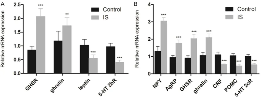
Maternal IS alters gene expression level in stomachs and hypothalamus of maternal mice
The mRNA expression levels of ghrelin pathway related genes including ghrelin, leptin, GHSR and 5-HT2bR in stomachs were measured using RT-PCR, as well as the expression levels of ghrelin, NPY, AgRP, GHSR, CRF, POMC and 5-HT2cR in hypothalamus of maternal mice on PND22. As shown in Figure 3A, the mRNA expression of ghrelin in the stomach, which is the main source of serum ghrelin, increased
significantly in the IS group compared with the
control group (P < 0.001). Besides, the expres-sion level of GHSR also showed an increase
with significant difference compared with that
in the control group (P < 0.01), while the mRNA expression levels of leptin and 5-HT2bR
decreased significantly (P < 0.001). The gene expression levels of major ghrelin pathway in the hypothalamus of maternal mouse were also analyzed, which was shown in Figure 3B. The expression levels of ghrelin, GHSR, AgRP
and NPY were significantly higher than that in
the control group (P < 0.001), while the
expres-sion levels of POMC, CRF and 5-HT2cR
decreased significantly (P < 0.001). These data suggest that the major targets in hypothalamus of IS may be ghrelin-producing cells and Leptin-producing cells.
Discussion
A series of studies were conducted in order to
explore the mechanism involves in the influ -ence of postpartum depression on the develop-ment and growth of the offspring [12-14]. Low body weight in faints due to the separation of mother and baby was found using maternal deprivation stress model. In the previous work, postpartum restraint stress model was devel-oped using ICR mice based on the maternal deprivation stress model [15]. A 3 h per day extra restraint stress was given to maternal mice for 3 weeks continually, resulting in the low body weight of offspring. Different to the growth disorder due to the limitation of
mater-nal food intake, the offspring exhibits signifi -cant obesity or emaciation after weaning [16]. Previous studies in the growth disorder caused by postpartum restraint stress demonstrated
[image:5.629.100.531.131.292.2]the significant drop of expression levels of insu -lin-like growth factor (IGF-1) and growth hor-mone (GH) in serum and hypophyse, respec-tively, in the restraint stress group after wean-to GAPDH in the swean-tomach of maternal mice showed a tendency wean-to increase in the IS group with significant difference (P < 0.05). C. The expression level of ghrelin measured using SDS-PAGE. *P < 0.05 compared with the control group (Control: n = 3; IS: n = 3).
ing. Further study indicated the mechanism involves in the inhibition of prolactin (PRL) was related to the low expression levels of tran-scription factors Pit-1 and PitX2 which regulate GH and PRL. However, the expression levels of IGF-1, GH, PRL, Pit-1 and PitX2 showed no
sig-nificant difference to the control group after
free diet for 5 weeks, suggesting the direct rela-tion between the growth disorder and the changed metabolism due to the decrease in the IGF-1. In view of the ‘matabolic program-ming’ caused by the maternal restraint stress during lactation period, the low nutrition condi-tion including low body weight and food intake might be related to the metabolism correlative
factors ghrelin, leptin, IL-6 and TNFα [17, 18].
Therefore, the mechanism involves in the growth disorder of offspring was explored through the low nutrition condition due to the restraint stress during the lactation period. It was reported that two systems, appetite pro-motion and suppression systems, exist in human body [19, 20]. Ghrelin is the only hor-mone with the appetite regulation function pro-duced in peripheral nerve, especially in stom-ach. Under stressed condition, the production of 5-HT increases and activates 5-HT2b recep-tors, leading to the inhibition of acylated ghrelin secreted by X/A-like cells in the gastric mucosa. The decreased acylated ghrelin in blood inhib-its the combination of ghrelin with GHSR, result-ing in the inhibition of the activation of NPY/
AgRP in hypothalamus, finally leading to the
down-regulation of appetite. While in the cen-tral nervous system, especially in hypothala-mus, CRF was activated by increased 5-HT, resulting in the combination of CRF and 5-HT2R, and then leading to the secretion of ghrelin in blood. At the same time, POMC and 5-HT2c receptor activated by 5-HT also inhibit the secretion of ghrelin. Besides, the acylated ghre-lin secreted by ghreghre-lin nerve could be inhibited by the activated 5-HT2cR in the hypothalamus, resulting in the loss of appetite [19, 20]. In a word, the appetite promotion system including NPY/AgRP pathway in hypothalamus and ghre-lin pathway in stomach are inhibited, while the appetite suppression system containing CRF and 5-HT2cR in hypothalamus and 5-HT2bR in stomach are enhanced under stressed condi-tion, resulting in the down-regulation of appe-tite. According to the results obtained from real time RT-PCR assay, the increased gene expres-sion level of ghrelin suggested the up
regula-tion of ghrelin. In the study, there was no signifi -cant difference in the food intake of maternal mice in the IS group, suggesting the increased ghrelin level in stomach which resulting in the activation of ghrelin pathway so as to up regu-late the appetite, which correspond to the gene expression level measured by real time RT-PCR assay. However, in view of the decreased IGF-1 level in the blood of maternal mice based on the previous study, indicating the malnutrition condition in maternal mice, and leading to the low body weight of faints.
The limitation existing in the study is the lack of ghrelin data in the blood. Further study will be conducted to analyze the levels of acylated and deacylated ghrelin in the blood in order to sup-ply reliable theory base for the study of post-partum stress clinically.
Conclusion
In conclusion, the low body weight of maternal mice due to the IS was related to the expres-sion of ghrelin in the stomach which involves in the brain-gut ghrelin pathway. On the other hand, the ghrelin pathway stimulated by the increased ghrelin in stomach up regulated the appetite in order to feed the offspring, resulting
in the decreased food intake without significant
difference. Our study revealed the potential relation and mechanism involve in the effect of IS on appetite and ghrelin pathway in maternal mice.
Acknowledgements
The National Natural Science Foundation of China (Grant No. 81473610).
Disclosure of conflict of interest
None.
Address correspondence to: Dr. Pengfei Gao, Depart- ment of TCM, Jinshan Hospital of Fudan University, Shanghai, China. Tel: 57039600; Fax: 86-21-67226910; E-mail: [email protected]
References
[2] Fumagalli F, Molteni R, Racagni G and Riva MA. Stress during development: Impact on neuro-plasticity and relevance to psychopathology. Prog Neurobiol 2007; 81: 197-217.
[3] Batten SV, Aslan M, Maciejewski PK and Mazure CM. Childhood maltreatment as a risk factor for adult cardiovascular disease and de-pression. Batten SV 2004; 65: 249-254. [4] Mirescu C, Peters JD and Gould E. Early life
ex-perience alters response of adult neurogene-sis to stress. Nat Neurosci 2004; 7: 841-846. [5] Downey G and Coyne JC. Children of depressed
parents: an integrative review. Psychol Bull 1990; 108: 50-76.
[6] O’Brien LM, Heycock EG, Hanna M, Jones PW and Cox JL. Postnatal depression and faltering growth: a community study. Pediatrics 2004; 113: 1242-1247.
[7] Paulson JF, Dauber S and Leiferman JA. Individual and combined effects of posttum depression in mothers and fathers on par-enting behavior. Pediatrics 2006; 118: 659-668.
[8] Wright CM, Parkinson Kn and Drewett RF. The influence of maternal socioeconomic and emo -tional factors on infant weight gain and weight faltering (failure to thrive): data from a pro-spective birth cohort. Arch Dis Child 2006; 91: 312-317.
[9] Kojima M, Date Y, Nakazato M, Matsuo H, Kangawa K. Ghrelin is a growth-hormone-re-leasing acylated peptide from stomach. Nature 1999; 402: 656-660.
[10] Date Y, Kojima M, Hosoda H, Sawaguchi A, Mondal MS, Suganuma T, Matsukura S, Kangawa K and Nakazato M. Ghrelin, a novel growth hormone-releasing acylated peptide, is synthesized in a distinct endocrine cell type in the gastrointestinal tracts of rats and humans. Endocrinology 2000; 141: 4255-4261. [11] Livak KJ and Schmittgen TD. Analysis of
rela-tive gene expression data using real-time quantitative PCR and the 2(-Delta Delta C(T)) Method. Methods 2001; 25: 402-408. [12] Hendrick V, Smith LM, Hwang S, Altshuler LL
and Haynes D. Weight gain in breastfed infants of mothers taking antidepressant medica-tions. J Clin Psychiatry 2003; 64: 410-412.
[13] Patel V, Rahman A, Jacob KS and Hughes M. Effect of maternal mental health on infant growth in low income countries: new evidence from South Asia. BMJ 2004; 328: 820-823. [14] Surkan PJ, Kawachi I, Ryan LM, Berkman LF,
Carvalho Vieira LM and Peterson KE. Maternal depressive symptoms, parenting self-efficacy, and child growth. Am J Public Health 2008; 98: 125-132.
[15] de Moura EG and Passos MC. Neonatal pro-gramming of body weight regulation and ener-getic metabolism. Biosci Rep 2005; 25: 251-269.
[16] Teixeira C, Passos M, Ramos C, Dutra S and Moura E. Leptin serum concentration, food in-take and body weight in rats whose mothers were exposed to malnutrition during lactation. J Nutr Biochem 2002; 13: 429.
[17] Nakata H, Watanabe K, Murakami Y, Gao P, Tsuiji K, Nishimura K, Plotnikoff GA, Kurihara N, Irie Y and Ishige A. Stress on a postpartum mother inhibits the secretion of growth hor-mone in the offspring and causes persistent growth impairment. Methods Find Exp Clin Pharmacol 2009; 31: 433-441.
[18] Gao P, Ishige A, Murakami Y, Nakata H, Oka JI, Munakata K, Yamamoto M, Nishimura K, Watanabe K. Maternal stress affects postnatal growth and the pituitary expression of prolac-tin in mouse offspring. J Neurosci Res 2011; 89: 329-340.
[19] Yakabi K, Sadakane C, Noguchi M, Ohno S, Ro S, Chinen K, Aoyama T, Sakurada T, Takabayashi H and Hattori T. Reduced ghrelin secretion in the hypothalamus of rats due to cisplatin-induced anorexia. Endocrinology 2010; 151: 3773-3782.