Original Article
Increased expression of CCRL2 is correlated with
clinical prognosis in human bladder cancer
Ruihai Xiao*, Yi Yu*, Qiufeng Pan, Shaochen Shen, Renrui Kuang
Department of Urology, The Second Affiliated Hospital of Nanchang University, Nanchang 330006, P.R. China.
*Co-first authors.
Received February 24, 2017; Accepted May 24, 2018; Epub January 15, 2019; Published January 30, 2019
Abstract: A large amount of evidence suggests that expression of chemokine (C-C motif) receptor-like 2 (CCRL2) is associated with tumorigenesis. However, the relationship of CCRL2 expression with bladder cancer remains un-clear. The aim of the present study was to investigate the clinical significance and biological role of CCRL2 in blad-der cancer. In the present study, CCRL2 mRNA levels in 10 pairs of bladblad-der cancer and adjacent noncancerous tissue samples were detected using real-time quantitative reverse transcription PCR (RT-PCR). In addition, immu-nohistochemistry was performed to detect the expression of CCRL2 in 100 clinical bladder cancer tissue samples. Furthermore, the correlations between CCRL2 expression and clinicopathological features of bladder cancer were analyzed. The results showed that expression levels of CCRL2 were significantly higher in bladder cancer tissues compared with those in adjacent normal tissues (p < 0.05). In addition, high expression of CCRL2 was significantly associated with differentiation (p = 0.006), tumor types (p = 0.038), and recurrence (p = 0.023). According to Ka-plan-Meier analysis, CCRL2 expression was closely correlated with overall survival (p = 0.005) of bladder cancer, but not with recurrence-free survival (p = 0.112). Moreover, Cox multivariate regression analyses revealed that CCRL2 expression was an independent predictor of overall survival (p = 0.018, hazard ratio = 2.697, confidence interval 1.161-6.264). The present study data suggest that expression of CCRL2 is closely associated with human bladder cancer, and this protein might be used as a new target and prognostic factor in human bladder cancer.
Keywords: CCRL2, bladder cancer, survival, predictor, prognostic factor
Introduction
Bladder cancer is one of the most common genitourinary tumors, and the incidence of this cancer has been increasing rapidly over the last few decades in China [1]. Among newly diagnosed cases of bladder cancer, approxi-mately 75% are non-muscle-invasive bladder cancer (NMIBC) cases and 25% are muscle-invasive bladder cancer (MIBC) cases [2]. The usual treatments for bladder cancer include surgical resection and chemotherapy. In recent decades, despite developments in surgical techniques, there has been no improvement in overall 10-year diseasespecific survival [3, 4]. Recurrence and progression are common char-acteristics of bladder cancer, and it is challeng-ing to accurately differentiate patients with a high risk of recurrence and progression from those with a low risk after initial adequate ther-apy [5]. This difficulty results from the lack of
current clinical and pathologic variables able to predict the progression and survival of patients with bladder cancer. Therefore, it is imperative to develop novel strategies to prolong survival and minimize morbidity. Ongoing research is focused on the identification of molecular bio-markers that can provide useful information regarding diagnosis, disease progression, and surveillance in bladder cancer patients. Un- fortunately, there remains a lack of prognostic biomarkers for predicting the course of bladder cancer. A bladder tumor biomarker with high sensitivity and specificity would be a useful adjunct tool for detection or follow-up in pa- tients with bladder cancer.
chemo-kines play an important role in cancer progres-sion (including tumor growth, adheprogres-sion, migra-tion, and metastasis of tumor cells), ang- iogenesis, lymphatic invasion by malignant tumors [7-12], and regulation of leukocyte traf-fic [13]. C-C motif chemokine receptor-like 2 (CCRL2) is a representative member of the che-mokine receptor family. It was first isolated from a mouse macrophage cell line following lipopolysaccharide (LPS) stimulation, and it binds to chemokines such as CCL2, CCL5, CCL7, CCL8, and CCL19 [14, 15]. CCRL2 expression at the mRNA level has been report-ed in murine macrophages [16], astrocytes, microglia, and glial cells stimulated with LPS [17, 18], as well as in mast cells [19]. A highly stable complex formed through the combina-tion of CCRL2 and pRb effectively inhibits the induction of transcription by pRb, thus inhibit-ing pRb blockage of cell cycle activation. In addition, CCRL2 can combine with SMAD2 and SMAD3 to enhance the transcription of trans-forming growth factor-beta (TGF-β), which plays an important role in the regulation of cell growth and differentiation [20]. Thus, CCRL2 can regu-late some key signaling pathways, such as those involved in cell proliferation and differen-tiation, that may have effects on tumorigenesis and tumor development [19, 21, 22].
Previous studies have reported that CCRL2 plays an important role in many human cancers such as breast cancer and gliomas [23, 24]. However, no study has yet identified the rele-vance of CCRL2 expression in bladder cancer. In the present study, we aimed to evaluate the expression levels of CCRL2 in both bladder cancer and adjacent noncancerous tissues using real-time quantitative reverse transcrip-tion polymerase chain reactranscrip-tion (RT-PCR), and to investigate the protein expression of CCRL2 in low and high-grade bladder cancer samples using immunohistochemistry. In addition, we evaluated the relationship between CCRL2 expression and clinicopathological features of bladder cancer to determine the prognostic value of this expression for postoperative sur-vival of patients with bladder cancer.
Patients and methods
A total of 100 consecutive patients with blad-der cancer were enrolled from October 2012 to October 2014 at the Department of Urology, Second Affiliated Hospital of NanChang University. Patients were enrolled if they met
the following criteria: first-time diagnosis of bladder transitional cell carcinoma by histopa-thology, no history of other malignant tumors, and no anti-cancer treatment of any form prior to surgery. Patients with incomplete follow-up data were excluded to render the study more robust.
All tumors were graded and staged by experi-enced pathologists in our department accord-ing to the 2009 World Health organization (WHO) grading system and the 7th edition of the tumor, node, metastasis (TNM) classifica-tion [25-27]. According to these criteria, among the 100 enrolled patients, 89 had primary NMIBC (pTa-pT1) tumors and 11 had MIBC (pT2-pT4) tumors (Table 1). The pathological grades were classified according to the WHO Cl- assification Criteria of Urothelial Carcinoma. Low-grade urothelial carcinomas included Grade I and Grade II (Grade I and Grade II are well and moderately differentiated tumors, respectively), and high-grade carcinomas were classified as Grade III (poorly differentiated tumors). Among the 100 patients, 47 were clas-sified as having high-grade urothelial carcino-ma, and 53 as having low-grade urothelial carcinoma.
In addition, 10 paired samples of fresh frozen tissue were used in the study, including tissues from bladder cancer (diagnosed as bladder cancer by histopathology) and adjacent non-cancerous tissues (examined to verify that they showed evidence of malignancy). All of the sam-ples were quickly frozen in liquid nitrogen and stored at -80°C until used. This study was approved by the ethics committee of the Se- cond Affiliated Hospital of NanChang University. Written informed consent was obtained from each participant.
Quantification of CCRL2 mRNA levels by real-time RT-PCR
system (Bio-Rad Laboratories, Inc., Hercules, CA, USA) as follows: 94°C for 3 min, 94°C for 30 s with 45 cycles, and 72°C for 30 s, and the levels of gene expression were detected follow-ing the manufacturer’s instructions. Each ex- periment was repeated 3 times. The house-keeping gene GAPDH was used as an internal control. The following primers were used: CC- RL2 specific primers (forward, 5AGGATCCCCA- CCATGGTCTACACCCGT T TCT TA A A AGG -3, reverse, 5AGGATCCCCACCATGGCCAATTACACG- CTG-3) and GAPDH specific primers (forward, 5-AGGATCCCCACCATGGCC-3, reverse, 5TTGTAG- TCCACTTCGGTGGAATG-3).
Immunohistochemistry
Briefly, 5-μm-thick sections were cut from par-affin-embedded bladder cancer samples and mounted onto poly-L-lysine-coated slides. After deparaffinizing and rehydrating, the sections were boiled in 10 μmol/L citrate buffer solution (pH 6.0) for 10 min, and 0.3% hydrogen perox-ide was applied for 30 min to block endogenous peroxidase activity. The slides were treated with 1% fish skin gelatin for 30 min at room temperature to block non-specific staining. Then, the slides were incubated overnight with primary rabbit anti-CCRL2 antibody (1:100; BioVision Inc., Milpitas, CA, USA). After washing with phosphate buffer saline (PBS), the slides were incubated with the prediluted secondary rabbit anti-CCRL2 antibody (1:100; BioVision Inc.). After rinsing, diaminobenzidine (SuoLaibao Technology Co., Ltd., Beijing, China) was used for visualization. Finally, the sections were counterstained with hematoxylin and mounted. The sections were observed and photographed under a light microscope (Olympus Corp., Tokyo, Japan). The proportion of stained cells and the extent of the staining were used as evaluation criteria. For each sample, the inten-sity of staining was graded into 4 levels: 0, neg-ative staining; 1, weak staining (light yellow); 2, moderate staining (yellow brown), and 3, strong staining (brown). The percentage of stained cells was scored as follows: 0, 5% of the cells or less; 1, 5% to 10% of cells; 2, 11% to 50% of cells; 3, 51% to 80% of cells; and 4, 80% or more positive cells. A final score was deter-mined by multiplying the percentage of positive cells by the intensity. The final score was defined as low expression for a score of 0-4 and high expression for scores of 6-12.
Outcomes
Follow-up was performed by cystoscopy and cytology every 3 months for the first 2 years and then every 6 months. The procedures were performed by 3 specialists in a blinded fashion. Recurrence-free survival (RFS) was defined as the time from initial surgery to the date of the first documented bladder cancer relapse, and overall survival (OS) was defined as the time from surgery to the time of death.
Statistical analysis
Values were expressed as mean ± standard error. Data evaluation was performed using SPSS v. 18.0 (SPSS Inc., Chicago, IL, USA). The quantification of CCRL2 by real-time PCR was assessed using the independent samples t test. The chi-square test and Fisher’s exact test were used to assess the relationship between CCRL2 and clinicopathological features in patients with bladder cancer. The relationship between the expression of CCRL2 and patho-logical features of bladder cancer was assessed using the Cox proportional hazards regression model. Kaplan-Meier analysis was used to esti-mate patient survival (OS and RFS), and differ-ences were compared using the log-rank test. The significance level in all tests was defined as p < 0.05.
Results
Patient characteristics
A total of 100 patients, with their respective surgical specimens, were eligible for the pres-ent study. Table 1 lists baseline patient charac-teristics. The study population included 84 (84%) men and 16 (16%) women. The mean age of men at diagnosis was 61.0 ± 12.9 years (range: 29-88), and that for women was 61.1 ± 12.4 years (range: 39-88). Among the patients, 89 had NMIBC, while 11 had MIBC. Using the 2009 WHO grading system and the 7th edition of the TNM classification, experienced patholo-gists in our department classified 47 tumors as high-grade urothelial carcinoma (47%) and 53 as low-grade urothelial carcinoma (53%). The mRNA expression of CCRL2 in bladder tissue samples and adjacent noncancerous tissues
Table 1. Relationships between CCRL2 expression and clinical and pathological features
Parameter Total NO CCRL2 expression P
High No. (%) Low No. (%)
Sex 0.572
Male 84 55 (65.5) 29 (34.5)
Female 16 9 (56.3) 7 (43.7)
Age (y) 0.765
≤45 13 9 (69.2) 4 (30.8)
>45 87 55 (63.2) 32 (36.8)
Differentiation 0.006*
Low Grade (Grade I/II) 53 27 (50.9) 26 (49.1) High Grade (Grade III) 47 37 (78.7) 10 (21.3)
Tumor types 0.038*
NMIBC 89 60 (67.4) 29 (32.6)
MIBC 11 4 (36.4) 7 (63.6)
Distant metastasis 0.489
- 90 59 (65.6) 31 (34.4)
+ 10 5 (50.0) 5 (50.0)
Recurrence 0.023*
- 54 29 (53.7) 25 (46.3)
+ 46 35 (76.1) 11 (23.9)
Statistical analyses were performed by the Cox regression analysis. *p < 0.05 was considered significant.
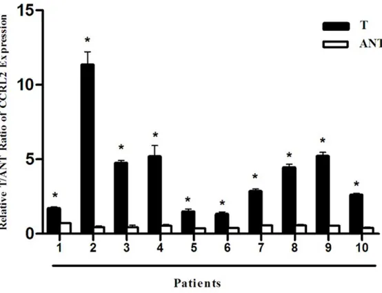
Figure 1. Elevations of CCRL2 mRNA expression in bladder cancer with real-time quantitative reverse transcription polymerase chain reaction (RT-PCR). RT-PCR analysis of CCRL2 expression in 10 paired bladder cancer tissue samples (T) and adjacent normal tissues (ANT). GAPDH was used as the loading control. *p < 0.05.
(ANT). As shown in Figure 1, the expression level of CCRL2 mRNA was significantly higher in bladder cancer tissue, com-pared with that in the matched adjacent noncancerous blad-der tissue (p < 0.05). Thus, the results of RT-PCR analysis demonstrated that CCRL2 mRNA expression in bladder cancer tissue was significantly higher than that in adjacent noncancerous tissue.
Protein expression in low- and high-grade urothelial carci-noma samples
In the present study, we per-formed immunohistochemis-try to examine CCRL2 expres-sion in 100 bladder cancer tissue sections, in order to investigate the potential roles of this protein in bladder can-cer. As shown in Figure 2, in high-grade urothelial carcino-ma samples, high expression of CCRL2 was observed in 37 out of 47 cases (78.7%), and low expression of CCRL2 was seen in 27 out of 47 cases (21.3%). In the low-grade uro-thelial carcinoma samples, high expression of CCRL2 was observed in only 27 out of 53 cases (50.9%), while low expression of CCRL2 was found in 26 out of 53 cases (49.1%). The difference bet- ween the two groups in the proportion of cases express-ing high levels of CCRL2 was statistically significant (p < 0.05). In addition, a close rela-tionship was observed bet- ween the expression of CCRL2 in bladder cancer and OS or RFS (Table 2). These results indicate that the expression of CCRL2 is increased on the mRNA in 10 paired samples of bladder cancer
[image:4.612.93.370.405.618.2]Correlation of CCRL2 expression with overall survival and recurrence-free survival in pa-tients with bladder cancer
The correlation between CCRL2 expression and clinical features of bladder cancer was determined using Kaplan-Meier analysis. Pa- tients were divided into a low-CCRL2 group and a high-CCRL2 group according to the expres-sion level of CCRL2. As shown in Table 1, CCRL2 expression was significantly associated with differentiation (p = 0.006), tumor type (p = 0.038), and recurrence (p = 0.023). However, there was no significant correlation between CCRL2 expression and other clinicopathologi-cal characteristics. Based on these results, we presumed that CCRL2 expression has a signifi-cant correlation with clinical prognosis. From
was confirmed as an independent predictor of RFS (hazard ratio [HR], 6.340; 95% confidence interval [CI], 1.333-30.131; p = 0.019) and OS (HR, 2.697; 95% CI, 1.161-6.264; p = 0.018) (shown in Table 2). All of these results indicate that CCRL2 might be an important prognostic mar- ker for bladder cancer patients.
Discussion
[image:5.612.90.375.70.400.2]Human bladder cancer is one of the most com-mon malignancies worldwide. Although there has been considerable improvement in surgical techniques and subsequent therapies [28-31], a reliable method to estimate risks and predict the progress of bladder cancer is still not avail-able. In addition, information regarding the bio-logical characteristics of bladder cancer is also
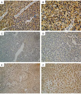
Figure 2.Representative immunostaining of CCRL2 in high-grade urothelial carcinoma samples (A, B), low-grade urothelial carcinoma samples (C, D), and isotype matched negative samples (E, F). High expression of CCRL2 (A, B) is observed in high-grade urothelial carcinoma samples; low expression of CCRL2 (C, D) is observed in low-grade urothelial carcinoma samples, and expression of CCRL2 is observed in isotype matched negative samples (A, C and E 10 × 20), (B, D and F 10 × 40).
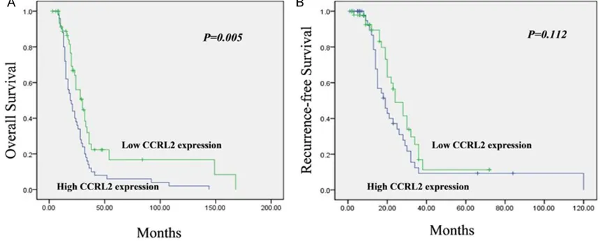
the outcomes of clinical fol-low-up and survival analysis, we also observed a close cor-relation between CCRL2 ex- pression and RFS or OS in bladder cancer patients. As shown in Figure 3A, a signifi-cant association between CCRL2 expression in the blad-der cancer tissues and OS was seen. The high-CCRL2 group had a longer OS than the low-CCRL2 group. The dif-ference between the two groups was found to be signifi-cant by the log-rank test (p = 0.005). Thus, the expression level of CCRL2 showed a sig-nificant correlation with OS in bladder cancer patients. How- ever, as shown in Figure 3B, although RFS in the low-CCRL2 group was shorter than that in the high-CCRL2 group, no significant differ-ence between the two groups was found using the log-rank test (p = 0.112), indicating that there was no significant correlation between CCRL2 expression and RFS.
insufficient [32]. Therefore, identification of novel molecular biomarkers that can serve as prognostic factors is necessary for early diag-nosis and targeted therapy for patients with bladder cancer. However, to date, there has been no research on the clinical significance and prognostic role of CCRL2 in patients with bladder cancer.
Tumor invasion and metastasis share many similarities with leukocyte trafficking, which is distinctly regulated by chemokines and their receptors [33]. Recently, large-scale clinical studies have demonstrated that chemokines play a pivotal role in cancer progression because they trigger numerous important cel-lular responses such as growth, adhesion, migration, and metastasis of tumor cells [34, 35]. Increasing attention has focused on the chemokine systems. One of the most recent additions to the atypical chemokine receptor family is CCRL2, which is also known as L-CCR (lipopolysaccharide inducible C-C chemokine
[image:6.612.87.524.98.167.2]receptor related gene), HCR (human chemokine receptor), and CRAM (chemokine receptor on activated macrophages) [16]. Recently, a num-ber of researchers have focused on the role of CCRL2 in the prognosis of human cancer. CCRL2 could thus become a new therapeutic target in treatment approaches to tumor inva-sion and dissemination. The present study is the first to report that CCRL2 functions as a prognostic biomarker in patients with bladder cancer. To investigate the role of CCRL2 in blad-der cancer, we analyzed the expression levels of CCRL2 mRNA using real-time quantitative RT-PCR, and found that bladder tissue showed significantly higher expression levels of CCRL2 mRNA compared with adjacent normal bladder tissues. Thus, we speculate that CCRL2 may play an important role in bladder cancer. Participants in this study included 47 patients with high-grade urothelial carcinoma and 53 patients with lowgrade urothelial carcinoma. To investigate the relationships between the Table 2. Multivariate Cox proportional hazard analysis for factors affecting overall survival and
recurrence-free survival of patients with bladder cancer
Prognostic variables OS P RFS P
Hazard ratio (95% CI) Hazard ratio (95% CI)
Age (>45 vs ≤45) 1.127 (0.294-4.237) 0.979 1.248 (0.430-3.619) 0.682 Differentiation (High Grade vs Low Grade) 1.021 (0.214-4.859) 0.869 1.133 (0.487-2.634) 0.768 CCRL2 (High vs Low) 6.340 (1.333-30.131) 0.019* 2.697 (1.161-6.264) 0.018* Statistical analyses were performed by the Cox regression analysis. *p < 0.05 was considered significant.
[image:6.612.89.519.205.378.2]expression of CCRL2 in bladder cancer sam-ples and clinicopathological features of bladder cancer, as well as prognosis, we performed both postoperative follow-up and CCRL2 immu-nohistochemistry. The results of statistical analysis revealed that CCRL2 expression has a close correlation with OS in bladder cancer patients. Patients with high expression of CCRL2 exhibited relatively shorter OS com-pared with patients with low CCRL2 expression. In addition, using multivariate Cox regression analysis, we found that expression of CCRL2 was associated with differentiation (p = 0.006), tumor type (p = 0.038), and recurrence (p = 0.023), indicating that CCRL2 may be involved in progression of bladder cancer. Tumor grade and other clinicopathological features are used to provide guidance for clinical treatment. The strong correlation of CCRL2 expression with these adverse tumor characteristics suggests that CCRL2 may have practical relevance in the prognostic evaluation of patients with bladder cancer. Overall, our research suggests that CCRL2 may play an important role in bladder carcinogenesis, and its expression may serve as a useful biomarker to predict the prognosis of patients with bladder cancer.
Recently, several studies have provided insights into the molecular mechanisms of CCRL2, and its target proteins and genes are gradually being identified in various cancers. A series of intriguing studies found that CCRL2 may play an essential role in the regulation of many can-cer-related proteins [23, 24], as well as a num-ber of key signaling pathways [20]. Campbell et al. reported that CCRL2 can combine with pRb to form a highly stable complex that can effec-tively inhibit the induction of transcription by pRb, thus inhibiting pRb blockage of cell cycle activation. In addition, CCRL2 can also com-bine with SMAD2 and SMAD3 to enhance the transcription of transforming growth factor-beta (TGF-β), which regulates cell growth and differentiation [20]. Akram et al. demonstrated that CCRL2 is a heptahelical transmembrane receptor, the expression of which has been found in almost all human hematopoietic cells [36]. Furthermore, CCRL2 is highly expressed during the dynamic phase of metastasis, such as during early liver colonization [37]. Another group of researchers reported that CCRL2 expression levels are elevated in human glioma
patient samples and cell lines. Overexpression of CCRL2 alone is sufficient to increase the migration and invasion of glioma tumor cells. The magnitude of increase in expression is pos-itively associated with increasing tumor grade, with the highest level observed in grade IV glio-blastoma [24]. In the present study, we observed that the expression level of CCRL2 mRNA in bladder cancer tissue was significant-ly higher than that found in the matched adja-cent noncancerous bladder tissue. Moreover, the proportion of high-grade urothelial carcino-ma samples (78.7%) demonstrating high expression of CCRL2 was higher than that observed among the low-grade urothelial carci-noma samples (50.9%). According to the previ-ous studies cited above, as well as our present results, it is clear that expression of CCRL2 is closely correlated with clinical prognosis in patients with bladder cancer. In addition, CCRL2 may play a role in modulating immune response, regulating cell growth, and promot-ing cell proliferation, migration, and angiogen-esis in the malignant progression of bladder cancer. Thus, CCRL2 might be an important prognostic marker in bladder cancer patients. However, the downstream target and signaling molecules of pathways mediating the effects of CCRL2 on the biological behavior of bladder cancer remain unclear and require further elucidation.
There are some limitations to the current study. First, the number of the patients enrolled in the study is insufficient. Second, the regulation mechanisms of CCRL2 in bladder tumor cells, including those of cell division, invasion, and apoptosis, remain unknown. In addition, we did not perform further research in cells and ani-mals to verify the differences between high and low expression of CCRL2 in bladder cancer cells in regard to invasion and metastasis. Thus, additional research is necessary to focus on these aspects.
expres-sion of CCRL2 was associated with shorter OS. The present study is the first to report that expression of CCRL2 may be associated with clinical outcomes in bladder cancer, and this protein may be a promising biomarker for early diagnosis and evaluation of prognosis in blad-der cancer.
Disclosure of conflict of interest
None.
Address correspondence to: Renrui Kuang, Depart- ment of Urology, The Second Affiliated Hospital of Nanchang University, No. 1 Minde Road, Nanchang 330006, Jiangxi Province, P.R. China. Tel: +86-791-86297662; Fax: +86-791-+86-791-86297662; E-mail: [email protected]
References
[1] Yang GL, Zhang LH, Bo JJ, Huo XJ, Chen HG, Cao M, Liu DM, Huang YR. Increased expres-sion of hmgb1 is associated with poor progno-sis in human bladder cancer. J Surg Oncol 2012; 106: 57-61.
[2] Zhu Z, Wang X, Shen Z, Lu Y, Zhong S, Xu C. Risk of bladder cancer in patients with diabe-tes mellitus: an updated meta-analysis of 36 observational studies. BMC Cancer 2013; 13: 310.
[3] Herr HW, Faulkner JR, Grossman HB, Natale RB, deVere White R, Sarosdy MF, Crawford ED. Surgical factors influence bladder cancer out-comes: a cooperative group report. J Clin Oncol 2004; 22: 2781-9.
[4] Herr HW, Dotan Z, Donat SM, Bajorin DF. Defin-ing optimal therapy for muscle invasive blad-der cancer. J Urol 2007; 177: 437-43.
[5] Lamm DL, Blumenstein BA, Crissman JD, Mon-tie JE, Gottesman JE, Lowe BA, Sarosdy MF, Bohl RD, Grossman HB, Beck TM, Leimert JT, Crawford ED. Maintenance bacillus calmette-guerin immunotherapy for recurrent Ta, T1 and carcinoma in situ transitional cell carcinoma of the bladder: a randomized southwest oncology group study. J Urol 2000; 163: 1124-1129. [6] Lazennec G, Richmond A. Chemokines and
chemokine receptors: new insights into can-cer-related inflammation. Trends Mol Med 2010; 16: 133-44.
[7] Müller A, Homey B, Soto H, Ge N, Catron D, Bu-chanan ME, Mcclanahan T, Murphy E, Yuan W, Wagner SN. Involvement of chemokine recep-tors in breast cancer metastasis. Nature 2001; 410: 50-56.
[8] Ghadjar P, Coupland SE, Na IK, Noutsias M, Letsch A, Stroux A, Bauer S, Buhr HJ, Thiel E, Scheibenbogen C. Chemokine receptor ccr6
expression level and liver metastases in co- lorectal cancer. J Clin Oncol 2006; 24: 1910-6. [9] Ghadjar P, Rubie C, Aebersold DM, Keilholz U.
The chemokine ccl20 and its receptor ccr6 in human malignancy with focus on colorectal cancer. Int J Cancer 2009; 125: 741-745. [10] Rubie C, Frick VO, Pfeil S, Wagner M, Kollmar
O, Kopp B, Graber S, Rau BM, Schilling MK. Correlation of il-8 with induction, progression and metastatic potential of colorectal cancer. World J Gastroenterol 2007; 13: 4996-5002. [11] Rubie C, Frick VO, Wagner M, Schuld J, Gräber
S, Brittner B, Bohle RM, Schilling MK. Elr+ cxc chemokine expression in benign and malig-nant colorectal conditions. BMC Cancer 2008; 8: 1-11.
[12] Rubie C. Cxc receptor-4 mrna silencing abro-gates cxcl12-induced migration of colorectal cancer cells. J Transl Med 2011; 9: 22. [13] Zlotnik A, Yoshie O. Chemokines: a new
classi-fication system and their role in immunity. Im-munity 2000; 12: 121–127.
[14] Leick M, Catusse JM, Nibbs RJ, Hartmann TN, Veelken H, Burger M. Ccl19 is a specific ligand of the constitutively recycling atypical human chemokine receptor cram-b. Immunology 2010; 129: 536–546.
[15] Biber K, Zuurman MW, Homan H, Boddeke HW. Expression of l-ccr in hek 293 cells reveals functional responses to ccl2, ccl5, ccl7, and ccl8. J Leukoc Biol 2003; 74: 243-251. [16] Shimada T, Matsumoto M, Tatsumi Y,
Kana-maru A, Akira S. A novel lipopolysaccharide in-ducible c-c chemokine receptor related gene in murine macrophages. FEBS Letters 1998; 425: 490-494.
[17] Brouwer N, Zuurman MW, Wei T, Ransohoff RM, Boddeke HW, Biber K. Induction of glial l-ccr mrna expression in spinal cord and brain in experimental autoimmune encephalomyelitis. Glia 2004; 46: 84-94.
[18] Zuurman MW, Heeroma J, Brouwer N, Bodde-ke HW, Biber K. Lps-induced expression of a novel chemokine receptor (l-ccr) in mouse glial cells in vitro and in vivo. Glia 2003; 41: 327– 336.
[19] Zabel BA, Nakae S, Zúñiga L, Kim JY, Ohyama T, Alt C, Pan J, Suto H, Soler D, Allen SJ. Mast cell-expressed orphan receptor ccrl2 binds chemerin and is required for optimal induction of ige-mediated passive cutaneous anaphylax-is. J Exp Med 2008; 205: 2207-2220.
[20] Campbell EL, Louis NA, Tomassetti SE, Canny GO, Arita M, Serhan CN, Colgan SP. Resolvin e1 promotes mucosal surface clearance of neutrophils: a new paradigm for inflammatory resolution. FASEB J 2007; 21: 3162-70. [21] Migeotte I, Franssen JD, Goriely S, Willems F,
expression of the putative human chemokine receptor hcr in leukocyte populations. Eur J Im-munol 2002; 32: 494-501.
[22] Samaras V, Piperi C, Korkolopoulou P, Zisakis A, Levidou G, Themistocleous MS, Boviatsis EI, Sakas DE, Lea RW, Kalofoutis A. Application of the elispot method for comparative analysis of interleukin (il)-6 and il-10 secretion in periph-eral blood of patients with astroglial tumors. Mol Cell Biochem 2007; 96: 343-351.
[23] Wang LP, Cao J, Zhang J, Wang BY, Hu XC, Shao ZM, Wang ZH, Ou ZL. The human chemokine receptor ccrl2 suppresses chemotaxis and in-vasion by blocking ccl2-induced phosphoryla-tion of p38 mapk in human breast cancer cells. Med Oncol 2015; 32: 1-9.
[24] Yin F, Xu Z, Wang Z, Yao H, Shen Z, Yu F, Tang Y, Fu D, Lin S, Lu G. Elevated chemokine cc-motif receptor-like 2 (ccrl2) promotes cell migration and invasion in glioblastoma. Biochem Bio-phys Res Commun 2012; 429: 168-172. [25] Greene FL. The american joint committee on
cancer: updating the strategies in cancer stag-ing. Bull Am Coll Surg 2002; 87: 13-15. [26] Oosterlinck W, Lobel B, Jakse G, Malmström
PU, Stöckle M, Sternberg C. Guidelines on bladder cancer. Eur Urol 2002; 41: 105-112. [27] Edge SB, Compton CC. The american joint
committee on cancer: the 7th edition of the ajcc cancer staging manual and the future of tnm. Ann Surg Oncol 2010; 17: 1471-1474. [28] Lei Y, Yan S, Lei MD, Na L, Han RF. Prognostic
significance of aurora-a expression in human bladder cancer. Acta Histochemica 2011; 113: 514-518.
[29] Naito S, Bilim V, Yuuki K, Ugolkov A, Motoyama T, Nagaoka A, Kato T, Tomita Y. Glycogen syn-thase kinase-3beta: A prognostic marker and a potential therapeutic target in human bladder cancer. Clin Cancer Res 2010; 16: 5124-5132.
[30] Wu CF, Ng KF, Chen CS, Chang PL, Chuang CK, Weng WH, Liao SK, Pang ST. Expression of parvin-|[beta]| is a prognostic factor for pa-tients with urothelial cell carcinoma of the up-per urinary tract. British Journal of Cancer 2010; 103: 852–860.
[31] Kwak C, Lee SE, Jeong IG, Ku JH. Adjuvant sys-temic chemotherapy in the treatment of pa-tients with invasive transitional cell carcinoma of the upper urinary tract. Urology 2006; 68: 53-57.
[32] Matsushita K, Cha EK, Matsumoto K, Baba S, Chromecki TF, Fajkovic H, Sun M, Karakiewicz PI, Scherr DS, Shariat SF. Immunohistochemi-cal biomarkers for bladder cancer prognosis. Int J Urol 2011; 18: 616-29
[33] Wang CL, Sun BS, Tang Y, Zhuang HQ, Cao WZ. Ccr1 knockdown suppresses human non-small cell lung cancer cell invasion. J Cancer Res Clin Oncol 2009; 135: 695-701.
[34] Horuk R. Chemokine receptor antagonists: overcoming developmental hurdles. Nat Rev Drug Discov 2009; 8: 23-33.
[35] Vandercappellen J, Van Damme J, Struyf S. The role of cxc chemokines and their receptors in cancer. Cancer Letters 2008; 267: 226-244. [36] Akram IG, Georges R, Hielscher T, Adwan H,
Berger MR. The chemokines CCR1 and CCRL2 have a role in colorectal cancer liver metasta-sis. Tumour Biol 2016; 37: 2461-71.