Antimicrobial Activity of Terminalia catappa
Extracts against Some Pathogenic Microbial
Strains
Abul Manzur1*, Arifuddin Raju1, Shahedur Rahman2
1
Department of Microbiology, North South University, Dhaka, Bangladesh; 2Department of Biotechnology and Genetic Engineering, Islamic University, Kushtia, Bangladesh.
Email: *[email protected]
Received September 4th, 2011; revised September 27th, 2011; accepted October 20th, 2011.
ABSTRACT
The methanol, acetone and N,N-dimethylformamide extracts of Terminalia catappa L. leaf were evaluated for antibac-terial and antifungal activity. Piperacillin and gentamicin were used as standards for antibacantibac-terial assay, while nystatin and flucanazole were used as standards for antifungal assay. 91 clinically important strains were used for the study which were both clinical isolates as well as identified strains. The antimicrobial activity of all the extracts was deter-mined by agar disc diffusion method. The antibacterial activity was more pronounced against bacteria than fungal strains. The Gram positive bacteria were more susceptible than Gram negative bacteria. The methanol extract showed best antibacterial activity. T. catappa leaf extracts showed better antibacterial activity than commercially used antibi-otics.
Keywords: Terminalia catappa, Antibacterial Activity, Antifungal Activity, Clinical Strains, Organic Solvents
1. Introduction
Traditional medicine has been practiced for many centu-ries in many parts of the world, including Bangladesh especially in rural areas due to availability and low cost. Nature has provided a source of medicinal agents for thousands of years and an impressive number of modern drugs have been isolated from natural sources, many based on their use in traditional medicine [1]. There has been an increasing incidence of multiple resistances in human pathogenic microorganisms, largely due to the indiscriminate use of commercial antimicrobial drugs commonly employed in the treatment of infectious dis-eases [2]. The development of bacterial resistance to presently available antibiotics has necessitated the search for new antibacterial agents. Numerous studies have been conducted with the extracts of various plants, screening antimicrobial activity as well as for the discovery of new antimicrobial compounds [3-6]. The efforts of scientists in establishing plants with promising antimicrobial prop-erty is yielding fruitful results as a number of plants with high antimicrobial property have been elucidated [7-13].
Terminalia catappa L. belongs to the family
Combre-taceae. T. catappa is used primarily as an ornamental,
shade, and salt-tolerant street tree, but the leaves provide food for the Tasar silkworm, and the seeds are edible like almonds with similar oils. On the Malay peninsular and through the Canary islands this tree is known as the
tropical almond. T. catappa has been claimed to have
therapeutic effects for liver related diseases [14]. In Java, it is attributed with cholagogue action. In India, it is used as cardiac stimulant. Its leaves are widely used as a folk medicine in Southeast Asia for the treatment of dermato-sis and hepatitis [15]. More and more pharmacological studies have reported that the extract of T. catappa leaves and fruits have anticancer, antioxidant, anti-HIV reverse transcriptase, anti-inflammatory, antidiabetic effects and hepatoprotective activities [16-19] but the effective com- ponents and related mechanisms remain unknown.
In the present work, antimicrobial activity of T. catappa leaf extracts were investigated against an array of clini-cally isolated as well as standard microbial cultures.
2. Material and Methods
2.1. Plant Material
The leaves of T. catappa were collected from the Boro
Bangla-desh, in January 2009 and identified by Dr. Masudur Rahman, Bangladesh National Herbarium, Dhaka.
2.2. Extraction
The leaves of T. catappa were air dried and then
pow-dered in a homogenizer and 10 g was used for different solvent extraction N,N-dimethylformamide DMF, ace-tone and methanol, the sample was extracted in solvent kept on a rotary shaker overnight, and then the filtrate was collected and centrifuged at 5000 rpm. The solvent was then evaporated to dryness under reduced pressure and the extracted compound left was used for the antim-icrobial assay. The percentage yield of N,N-dimethyl- formamide DMF, acetone and methanol extracts were 20.92, 4.96 and 14.48 respectively.
2.3. Microorganisms Studied
91 clinically important microbial strains which included 20 Gram positive, 55 Gram negative and 16 fungal strains were studied for the antimicrobial activity. These strains included both clinical isolates as well as identified strains. The identified strains were provided by Depart-ment of Microbiology, North South University and clini-
cal isolates were obtained from Lab Aid Diagnostic Laboratory, Dhaka, Bangladesh (Tables 1-5). The bacte-ria were grown in the nutrient broth and maintained on nutrient agar slants at 4˚C while fungal strains were grown in Sabouraud broth and maintained on MGYP slants yeast and potato dextrose agar slants mould at 4˚C.
2.4. Antimicrobial Assay
The Dimethylformamide extract TDE, acetone extract TAE and Methanol extract TME were dissolved in DMSO. The antimicrobial activity was evaluated at a concentration of 250 g/disc. Antimicrobial activity was performed by agar disc diffusion method [20,21]. The bacterial strains were grown in nutrient broth while fun-gal strains were grown in MGYP Malt glucose yeast peptone broth. Mueller Hinton Agar No. 2 was the media used to study the antibacterial susceptibility while Sa-broaud agar was used to study the antifungal susceptibil-ity test. The cultures were grown for 24 h, and the turbid-ity of the culture was maintained according to the 0.5
MacFarland standards. The inoculum’s size was 1 × 10⁸
[image:2.595.57.537.386.700.2]cells/ml. The media Mueller Hinton Agar No. 2 and MRS media and the test bacterial cultures were poured into
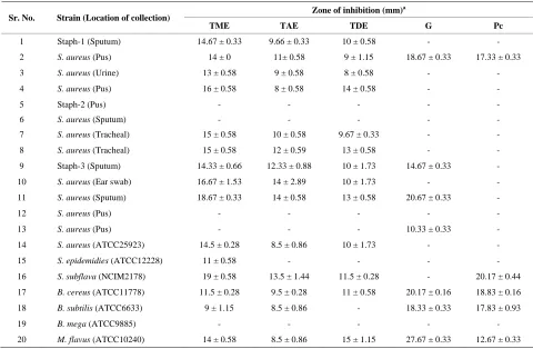
Table 1. Antibacterial activity of Terminalia catappa leaf extracts against some Gram positive bacteria.
Zone of inhibition (mm)a
Sr. No. Strain (Location of collection)
TME TAE TDE G Pc
1 Staph-1 (Sputum) 14.67 ± 0.33 9.66 ± 0.33 10 ± 0.58 - -
2 S. aureus (Pus) 14 ± 0 11± 0.58 9 ± 1.15 18.67 ± 0.33 17.33 ± 0.33
3 S. aureus (Urine) 13 ± 0.58 9 ± 0.58 8 ± 0.58 - -
4 S. aureus (Pus) 16 ± 0.58 8 ± 0.58 14 ± 0.58 - -
5 Staph-2 (Pus) - - -
6 S. aureus (Sputum) - - -
7 S. aureus (Tracheal) 15 ± 0.58 10 ± 0.58 9.67 ± 0.33 - -
8 S. aureus (Tracheal) 15 ± 0.58 12 ± 0.59 13 ± 0.58 - -
9 Staph-3 (Sputum) 14.33 ± 0.66 12.33 ± 0.88 10 ± 1.73 14.67 ± 0.33 -
10 S. aureus (Ear swab) 16.67 ± 1.53 14 ± 2.89 10 ± 1.73 - -
11 S. aureus (Sputum) 18.67 ± 0.33 14 ± 0.58 13 ± 0.58 20.67 ± 0.33 -
12 S. aureus (Pus) - - -
13 S. aureus (Pus) - - - 10.33 ± 0.33 -
14 S. aureus (ATCC25923) 14.5 ± 0.28 8.5 ± 0.86 10 ± 1.73 - -
15 S. epidemidies (ATCC12228) 11 ± 0.58 - - - -
16 S. subflava (NCIM2178) 19 ± 0.58 13.5 ± 1.44 11.5 ± 0.28 - 20.17 ± 0.44
17 B. cereus (ATCC11778) 11.5 ± 0.28 9.5 ± 0.28 11 ± 0.58 20.17 ± 0.16 18.83 ± 0.16
18 B. subtilis (ATCC6633) 9 ± 1.15 8.5 ± 0.86 - 18.33 ± 0.33 17.83 ± 0.93
19 B. mega (ATCC9885) - - -
20 M. flavus (ATCC10240) 14 ± 0.58 8.5 ± 0.86 15 ± 1.15 27.67 ± 0.33 12.67 ± 0.33
Table 2. Antibacterial activity of Terminalia catappa leaf extracts against some Pseudomonas species.
Zone of inhibition (mm)a
Sr. No. Strain (Location of collection)
TME TAE TDE G Pc
1 Ps. aeruginosa (ATCC27853) - - - 17 ± 1.15 12.33 ± 0.66
2 Ps. Aeruginosa (Sputum) - - - 16.67 ± 0.67 -
3 Ps. aeruginosa (Pus) - - - 19.67 ± 0.33 -
4 Ps. Fluorescence (Tracheal) 8.67 ± 0.33 - 12.67 ± 1.44 - -
5 Ps. fluorescence (Pus) 13.67 ± 3.18 8 ± 0.58 - - -
6 Ps. fluorescence (Urine) - - -
7 Ps. testosteroni (NCIM 5098) - - - 22.33 ± 0.66 -
8 Ps. pseudoalcaligenes (ATCC17440) 15.5 ± 0.28 12.5 ± 0.86 14.5 ± 028 19.33 ± 0.6 -
9 Pseudo-1 (Sputum) 11 ± 2.31 13 ± 0.58 11.67 ± 0.33 14 ± 0.58 -
10 Pseudo-2 (Pus) 13.67 ± 3.18 8 ± 0.58 - - -
11 Pseudo-3 (Urine) 14.67 ± 1.45 16 ± 0.58 14.67 ± 0.33 - -
12 Pseudo-4 (Pus) 14 ± 0.58 10.6 ± 2.34 9.33 ± 1.23 - -
13 Pseudo-5 (Tracheal) - - -
14 Pseudo-6 (Wound swab) - - -
15 Pseudo-7 (Pus) 16 ± 0.56 10 ± 0.58 12 ± 1.15 - -
16 Pseudo-8 (Tracheal secretion) 14 ± 1.15 9 ± 1.15 9 ± 1.15 - -
17 Pseudo-9 (Pus) 11.67 ± 0.88 9.33 ± 1.20 - - -
18 Pseudo-10 (Sputum) 17 ± 0.58 12 ± 0.33 13.67 ± 0.88 - -
19 Pseudo-11 (Sputum) 18.33 ± 0.33 16.33 ± 1.45 13 ± 0.58 20 ± 0.58 -
a
Values are Mean ± SEM, n = 3, zone includes disc diameter 7 mm, G: Gentamicin (10 µg/disc), Pc: Piperacillin (100 µg/disc), TME: Methanol extract, TAE: Acetone extract, TDE: N,N-dimethylformamide (DMF) extract, means no activity; Pseudo: Pseudomonas species.
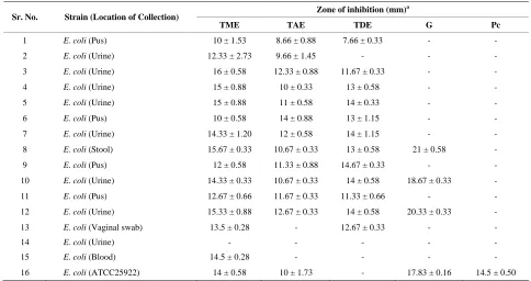
Table 3. Antibacterial activity of Terminalia catappa leaf extracts against some E. coli isolates.
Zone of inhibition (mm)a
Sr. No. Strain (Location of Collection)
TME TAE TDE G Pc
1 E. coli (Pus) 10 ± 1.53 8.66 ± 0.88 7.66 ± 0.33 - -
2 E. coli (Urine) 12.33 ± 2.73 9.66 ± 1.45 - - -
3 E. coli (Urine) 16 ± 0.58 12.33 ± 0.88 11.67 ± 0.33 - -
4 E. coli (Urine) 15 ± 0.88 10 ± 0.33 13 ± 0.58 - -
5 E. coli (Urine) 15 ± 0.88 11 ± 0.58 14 ± 0.33 - -
6 E. coli (Pus) 10 ± 0.58 14 ± 0.88 13 ± 1.15 - -
7 E. coli (Urine) 14.33 ± 1.20 12 ± 0.58 14 ± 1.15 - -
8 E. coli (Stool) 15.67 ± 0.33 10.67 ± 0.33 13 ± 0.58 21 ± 0.58 -
9 E. coli (Pus) 12 ± 0.58 11.33 ± 0.88 14.67 ± 0.33 - -
10 E. coli (Urine) 14.33 ± 0.33 10.67 ± 0.33 14 ± 0.58 18.67 ± 0.33 -
11 E. coli (Pus) 12.67 ± 0.66 11.67 ± 0.33 11.33 ± 0.66 - -
12 E. coli (Urine) 15.33 ± 0.88 12.67 ± 0.33 14 ± 0.58 20.33 ± 0.33 -
13 E. coli (Vaginal swab) 13.5 ± 0.28 - 12.67 ± 0.33 - -
14 E. coli (Urine) - - -
15 E. coli (Blood) 14.5 ± 0.28 - - - -
16 E. coli (ATCC25922) 14 ± 0.58 10 ± 1.73 - 17.83 ± 0.16 14.5 ± 0.50
a
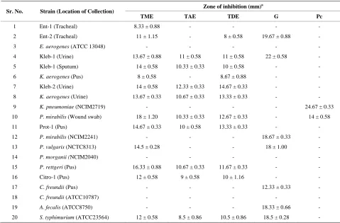
[image:3.595.55.539.442.698.2]Table 4. Antibacterial activity of Terminalia catappa leaf extracts against some Gram negative bacteria.
Zone of inhibition (mm)a
Sr. No. Strain (Location of Collection)
TME TAE TDE G Pc
1 Ent-1 (Tracheal) 8.33 ± 0.88 - - - -
2 Ent-2 (Tracheal) 11 ± 1.15 - 8 ± 0.58 19.67 ± 0.88 -
3 E. aerogenes (ATCC 13048) - - - - -
4 Kleb-1 (Urine) 13.67 ± 0.88 11 ± 0.58 11 ± 0.58 22 ± 0.58 -
5 Kleb-1 (Sputum) 14 ± 0.58 10.33 ± 0.33 10 ± 0.58 - -
6 K. aerogenes (Pus) 8 ± 0.58 - 8.67 ± 0.88 - -
7 Kleb-2 (Urine) 14 ± 0.58 12.33 ± 0.33 14.67 ± 0.33 - -
8 K. aerogenes (Urine) 13.67 ± 0.33 10.67 ± 0.33 13.33 ± 0.33 - -
9 K. pneumoniae (NCIM2719) - - - - 24.67 ± 0.33
10 P. mirabilis (Wound swab) 18 ± 1.20 10.33 ± 0.33 12.67 ± 0.33 - 14 ± 0.58
11 Prot-1 (Pus) 14.67 ± 0.33 10 ± 0.58 13.33 ± 0.33 - -
12 P. mirabilis (NCIM2241) - - - 18.67 ± 0.33 -
13 P. vulgaris (NCTC8313) 14.5 ± 0.28 - - 18 ± 1.00 -
14 P. morganii (NCIM2040) - - - - -
15 P. rettgeri (Pus) 16.33 ± 0.88 10.67 ± 0.33 11.67 ± 0.33 - -
16 Citro-1 (Pus) 12 ± 0.58 9 ± 0.58 10 ± 1.16 - -
17 C. freundii (Pus) - - - 12.33 ± 0.33 -
18 C. freundii (ATCC10787) - - -
19 A. fecalis (ATCC8750) - - - 18.33 ± 0.66 -
20 S. typhimurium (ATCC23564) 12 ± 0.58 8.5 ± 0.86 10.5 ± 0.86 18.5 ± 0.28 -
a
Values are Mean ± SEM, n = 3, zone includes disc diameter 7mm, G: Gentamicin (10 µg/disc), Pc: Piperacillin (100 µg/disc), TME: Methanol extract, TAE: Acetone extract, TDE: N,N-dimethylformamide (DMF) extract, means no activity; Ent: Enterobacter species; Kleb: Klebsiella species; Citro: Citrobacter species; Prot: Proteus species.
Petri dishes Hi-Media. The test strain 200 µl was inocu-lated into the media inoculums size 108 cells/ml when the temperature reached 40˚C - 42˚C. The test compound 20 µl was impregnated in to sterile discs 7 mm Hi-Media and was then allowed to dry. The disc was then intro-duced into medium with the bacteria. For each microbial strain negative controls were maintained where pure sol-vent DMSO was used instead of the extract since it does not possess any antimicrobial effect [22] and for positive control the standard antimicrobics Gentamicin 10 µg/disc and piperacillin 100 µg/disc for bacteria, nystatin 100 units/disc and flucanazole 10 µg/disc Himedia Labs for fungus were used for comparative studies. The plates were incubated overnight at 37˚C for bacterial strains and 42˚C for fungal strains. The experiment was performed under strict aseptic conditions. Microbial growth was de-termined by measuring the diameter of the zone of inhibi-tion. The experiment was performed in triplicates and the mean values of the result are shown in Tables 1-5.
3. Results and Discussion
Herbal medicine in developing countries is commonly
used for the traditional treatment of health problems [23]. In recent years multiple drug resistance in human patho-genic microorganisms have developed due to the indis-criminate use of commercial antimicrobial drugs com-monly used in the treatment of infectious diseases [24]. In addition to this problem, antibiotics are sometimes associated with adverse effects on host including hyper-sensitivity, immune suppression and allergic reactions [25]. Therefore there is a need to develop alternative an-timicrobial drugs for the treatment of infections obtained from various sources such as medicinal plants [26,27].
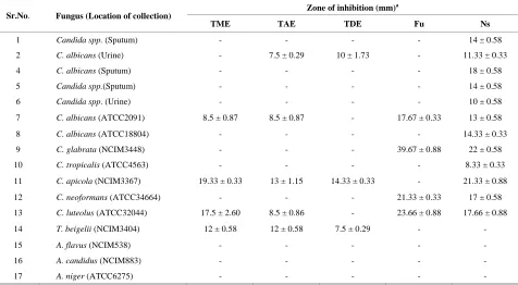
Table 5. Antifungal activity of Terminalia catappa leaf extracts.
Zone of inhibition (mm)a
Sr.No. Fungus (Location of collection)
TME TAE TDE Fu Ns
1 Candida spp. (Sputum) - - - - 14 ± 0.58
2 C. albicans (Urine) - 7.5 ± 0.29 10 ± 1.73 - 11.33 ± 0.33
4 C. albicans (Sputum) - - - - 18 ± 0.58
5 Candida spp.(Sputum) - - - - 14 ± 0.58
6 Candida spp. (Urine) - - - - 10 ± 0.58
7 C. albicans (ATCC2091) 8.5 ± 0.87 8.5 ± 0.87 - 17.67 ± 0.33 13 ± 0.58
8 C. albicans (ATCC18804) - - - - 14.33 ± 0.33
9 C. glabrata (NCIM3448) - - - 39.67 ± 0.88 22 ± 0.58
10 C. tropicalis (ATCC4563) - - - - 8.33 ± 0.33
11 C. apicola (NCIM3367) 19.33 ± 0.33 13 ± 1.15 14.33 ± 0.33 - 21.33 ± 0.88
12 C. neoformans (ATCC34664) - - - 21.33 ± 0.33 17 ± 0.58
13 C. luteolus (ATCC32044) 17.5 ± 2.60 8.5 ± 0.86 - 23.66 ± 0.88 17.66 ± 0.88
14 T. beigelii (NCIM3404) 12 ± 0.58 12 ± 0.58 7.5 ± 0.29 - -
15 A. flavus (NCIM538) - - -
16 A. candidus (NCIM883) - - -
17 A. niger (ATCC6275) - - -
a
Values are Mean ± SEM, n = 3, zone includes disc diameter 7mm, Ns: Nystatin (100 units/disc), Fu: Fluconazole (10 µg/disc) TME: Methanol extract, TAE: Acetone extract, TDE: N,N-dimethylformamide (DMF) extract, means no activity; Fu: Fluconazole, Ns: Nystatin.
All the three extracts of T. catappa TDE, TAE and
TME were active against 70% of the total Gram positive bacteria studied while only 63% of Gram negative bacte-ria were inhibited Table 1-4, on the other hand, the three
extracts of T. catappa were active against only 25% of
fungal strains Table 5. The best antibacterial activity was shown by the methanol extract. Similar results were also shown by Babayi et al., [29]. The Gram positive bacteria were more susceptible than Gram negative bacteria. This is in agreement with previous reports that plant extracts are more active against Gram positive bacteria than Gram negative bacteria [30-32]. These differences may be attributed to the fact that the cell wall in Gram posi-tive bacteria is of a single layer, whereas the Gram nega-tive cell wall is multilayered structure [33]. The most striking feature of the present findings is that many of the clinical isolates were resistant to the standard antimicro-bics used while the plant extracts showed moderate to good antibacterial activity. The need of the hour is to find new antimicrobics because the microorganisms are get-ting resistant to the exisget-ting antibiotics [34,35]. The per-sistent increase in multi drug resistant strains compels the search for more potent new antibiotics. Thus there is a need for a continuous search for new effective and af-fordable antimicrobial drugs. The results of present study signify the potentiality of T. catappa leaf as a source of therapeutic agents which may provide leads in the
ongo-ing search for antimicrobial botanicals.
REFERENCES
[1] G. M. Cragg and D. J. Newman, “Drugs from Nature: Past Achievements, Future Prospects,” In: M. M. Iwu and J. C. Wootton, Eds., Ethnomedicine and Drug Discovery, Elsevier Science, Amsterdam, 2002, pp. 23-37.
[2] A. A. Allero and A. J. Afolayan, “Antimicrobial Activity of Solanum tomentosum,” African Journal of Biotech-nology, Vol. 5, No. 4, 2006, pp. 369-372.
[3] B. Abu-Shanab, G. Adwan, D. Abu-Safiya, N. Jarrar and K. Adwan, “Antibacterial Activities of Some Plant Ex-tracts Utilized in Popular Medicine in Palestine,” Turkish Journal of Biology,Vol. 28, No. 2-4, 2004, pp. 99-102. [4] M. R. F. De Lima, J. De Souza Luna, A. F. Dos Santos, M.
C. C. De Andrade, A. E. G. Sant Ana, J. P. Genet, B. Marquez, L. Neuville and N. Moreau, “Antibacterial Acti- vity of Some Brazilian Medicinal Plants,” Journal of Eth-nopharmacology,Vol. 105, No. 1-2, 2005, pp. 137-147.
doi:10.1016/j.jep.2005.10.026
[5] J. Parekh, N. Karathia and S. Chanda, “Screening of Some Traditionally Used Medicinal Plants for Potential Antibacterial Activity,” Indian Journal of Pharmaceutical Science, Vol. 68, No. 6, 2006, pp. 832-834.
doi:10.4103/0250-474X.31031
No. 2, 2007, pp. 175-181.
[7] S. Dash, L. K. Nath, S. Bhise and B. Nihar, “Antioxidant and Antimicrobial Activities of Heracleum nepalense D Don Root,” Tropical Journal of Pharmaceutical Research, Vol. 4, No. 1, 2005, pp. 341-347.
[8] R. Nair, T. Kalariya and S. Chanda, “Antibacterial Activ-ity of Some Selected Indian Medicinal Flora,” Turkish Journal of Biology, Vol. 29, 2005, pp. 41-47.
[9] J. Parekh, R. Nair and S. Chanda, “Preliminary Screening of Some Folklore Plants from Western India for Potential Antimicrobial Activity,” Indian Journal of Pharmacology, Vol. 37, No. 6, 2005, pp. 408-409.
doi:10.4103/0253-7613.19085
[10] R. Nair and S. Chanda, “Antibacterial Activity of some Medicinal Plants against Some Medically Important Bac-terial Strains,” Indian Journal of Pharmacology, Vol. 38, No. 2, 2006, pp. 142-144. doi:10.4103/0253-7613.24625
[11] M. B. Tadhani and R. Subhash, “In Vitro Antimicrobial Activity of Stevia rebaudiana Bertoni Leaves,” Tropical Journal of Pharmaceutical Research, Vol. 5, No. 1, 2006, pp. 557-560.
[12] S. Mandal, M. D. Mandal and N. Pal, “Antibacterial Po-tential of Azadirechta indica Seed and Bacopa monniera Leaf Extracts against Multidrug Resistant Salmonella en-terica Serovar Typhi Isolates,” Archives of Medical Sci-ence, Vol. 3, No. 1, 2007, pp. 14-18.
[13] R. Nair and S. Chanda, “In-Vitro Antimicrobial Activity of Psidium guajava L. Leaf Extracts against Clinically Im-portant Pathogenic Microbial Strains,” Brazilian Journal of Microbiology, Vol. 38, No. 3, 2007, pp. 452-458.
doi:10.1590/S1517-83822007000300013
[14] N. Y. Chiu and K. H. Chang, “The Illustrated Medicinal Plants of Taiwan,” Vol. 1, SMC Publishing, Inc., Taipei, 1986, p. 129.
[15] C.C. Lin, Y. L. Chen, J. M. Lin and T. Ujiie, “Evaluation of the Antioxidant and Hepatoprotective Activity of Ter-minalia catappa,” American Journal of Chinese Medicine, Vol. 25, No. 2, 1997, pp. 153-161.
doi:10.1142/S0192415X97000172
[16] A. N. Nagappa, P. A. Thakurdesai, N. Venkat Rao and J. Singh, “Antidiabetic Activity of Terminalia catappa Linn Fruits,” Journal of Ethnopharmacology, Vol. 88, No. 1, 2003, pp. 45-50. doi:10.1016/S0378-8741(03)00208-3
[17] Y. M. Fan, L. Z. Xu, J. Gao, Y. Wang, X. H. Tang, X. N. Zhao and Z. X. Zhang, “Phytochemical and Anti-Inflam- matory Studies on Terminalia catappa,” Fitoterapia,Vol. 75, No. 3-4, 2004, pp. 253-260.
doi:10.1016/j.fitote.2003.11.007
[18] Y. F. Zhai, J. Yao, Y. M. Fan, L. Z. Xu, J. Gao and X. N. Zhao, “Inhibitory Effects of LR-98 on Proliferation of Hepatocarcinoma Cells,” Journal of Nanjing University of Natural Science, Vol. 37, No. 2, 2001, pp. 213-217. [19] L. Z. Xu, J. Gao, L. Zhu, M. Xu, S. Y. Lu, X. N. Zhao
and Z. X. Zhang, “Protective Effects of LR-98 on Hepa-totoxicity Induced by Carbon Tetrachloride and D-Galac- tosamine in Mice,” Journal of Nanjing University of Natural Science, Vol. 36, No. 2, 2000, pp. 197-201.
[20] A. W. Bauer, W. M. M. Kirby, J. C. Sherries and M. Truck, “Antibiotic Susceptibility Testing by a Standard-ized Single Disk Method,” American Journal of Clinical Pathology, Vol. 45, No. 4, 1966, pp. 426-493.
[21] J. Parekh and S. Chanda, “In Vitro Antimicrobial Activity of Trapa natans L. Fruit Rind Extracted in Different Sol-vents,” African Journal of Biotechnology, Vol. 6, No. 6, 2007, pp. 766-770.
[22] M. J. Pelczar, E. C. S. Chan and N. R. Krieg, “Microbiol-ogy Concepts and Applications,” McGraw-Hill Inc., New York, 1993, p. 967.
[23] M J. Martinez, J. Betancourt, N. Alanso-Gonzalea and A. Jauregui, “Screening of Some Cuban Medicinal Plants for Antimicrobial Activity,” Journal of Ethnopharmacology, Vol. 52, No. 3, 1996, pp. 171-174.
doi:10.1016/0378-8741(96)01405-5
[24] R. F. Service, “Antibiotics That Resist Resistance,” Sci-ence,Vol. 270, No. 5237, 1995, pp. 724-727.
doi:10.1126/science.270.5237.724
[25] I. Ahmad, Z. Mehmood and F. Mohammad, “Screening of Some Indian Medicinal Plants for Their Antimicrobial Properties,” Journal of Ethnopharmacology, Vol. 62, No. 2, 1998, pp. 183-193.
doi:10.1016/S0378-8741(98)00055-5
[26] G. A. Cordell, “Biodiversity and Drug Discovery a Sym- biotic Relationship,” Phytochemistry, Vol. 55, No. 66, 2000, pp. 463-480. doi:10.1016/S0031-9422(00)00230-2
[27] B. Dulger and A. Gonuz, “Antibacterial Activity of the Endemic Hypericum kazdaghensis,” Fitoterapia,Vol. 76, No. 2, 2005, pp. 237-239.
doi:10.1016/j.fitote.2004.12.010
[28] L. Stuart, “Infectious Diseases and Rise of Antibiotic Re- sistance,” Pharmaceutical News, Vol. 3, 1996, p. 21. [29] H. Babayi, I. Kolo, J. I. Okojun and U. J. J. Ijah, “The
Antimicrobial Activities of Methanolic Extracts of Euca-lyptus camaldulensis and Terminalia catappa against Some Pathogenic Microorganisms,” Biokemistri, Vol. 16, No. 2, 2004, pp. 106-111.
[30] J. E. Kelmanson, A. K. Jager and J. Van Staden, “Zulu Medicinal Plants with Antimicrobial Activity,” Journal of Ethnopharmacology, Vol. 69, No. 3, 2000, pp. 241-246.
doi:10.1016/S0378-8741(99)00147-6
[31] J. Parekh and S. Chanda, “Screening of Some India Me-dicinal Plants for Antibacterial Activity,” Indian Journal of Pharmaceutical Science, Vol. 68, No. 6, 2006, pp. 835-838. doi:10.4103/0250-474X.31032
[32] V. Prashanth Kumar, S. C. Neelam, H. Padh and M. Rajni, “Search for Antibacterial and Antifungal Agents from Selected Indian Medicinal Plants,” Journal of Ethnophar- macology,Vol. 107, No. 2, 2006, pp. 182-188.
doi:10.1016/j.jep.2006.03.013
[34] I. Bhattacharjee, A. Ghosh and G. Chandra, “Antimicro-bial Activity of the Essential Oil of Cestrum diurnum L. Solanales: Solanaceae,” African Journal of Biotechnology, Vol. 4, No. 4, 2005, pp. 371-374.