Original Article
Co-localization of galectin-3 and integrin β3 at
mouse maternal-fetal interface during
early embryo implantation
Lu Wang1, Wei Zhang1,2, Rui-Xiu Zhang1
1Department of Reproductive Endocrinology, Obstetrics and Gynecology Hospital, Fudan University, Shanghai,
China; 2Shanghai Key Laboratory of Female Reproductive Endocrine Related Disease, Shanghai, China
Received May 10, 2017; Accepted November 3, 2017; Epub December 15, 2017; Published December 30, 2017
Abstract: Galectin-3 (gal-3), a β-galactoside-binding protein, plays a role in the regulation of endometrial cell prolif-eration and adhesion and has been recognized as an important factor in endometrial receptivity. This study aimed to investigate the spatial expression pattern of gal-3 and integrin β3 in the uterus during embryo implantation and the co-localization of gal-3 with its ligands at the implantation and inter-implantation sites. We tested whether the embryo itself affects the expression and distribution of gal-3 in the uterus and we used uteri from a pseudo-preg-nant mouse model were used as a control. Real-time PCR and Western blotting analyses showed that mRNA and protein of gal-3 and integrin β3 expression in the implantation site were significantly higher than those in the inter-implantation site, and gal-3 expression was also strong in the pseudo-pregnant endometrium. Immunofluorescence data indicated that the expression of gal-3 and integrin β3 was enriched in the luminal epithelium, particularly at the implantation site. However, strong FN expression was observed in the stroma and blastocysts with a slight dif-ference between the two sites. Moreover, con-focal analysis revealed strong co-localization of gal-3 and integrin β3 while only sparse co-localization of gal-3 and FN in the luminal epithelium at the maternal-fetal interface. In conclu-sion, high expression and co-localization of gal-3 and integrin β3 is found at the maternal-fetal interface, suggest-ing that the interaction between gal-3 and integrin β3 at maternal-fetal interface may play an important role in the endometrial receptivity during early embryo implantation.
Keywords: Maternal-fetal interface, galectin-3, integrin β3, fibronectin, co-localization
Introduction
Embryo implantation is an intricate process whereby the developing embryo attaches and embeds in the endometrium, which is the first cross-talk between the embryo and receptive endometrium [1]. Prior to the implantation, the endometrium undergoes complex morphologi-cal, physiological and biochemical changes. Successful implantation is not only dependent on the invasive ability of the embryo, but also on the establishment of endometrial receptivity [2, 3]. Many cell adhesion molecules, such as integrins, are well known biomarkers of implan-tation. These adhesion molecules and other cytokines make the endometrium conducive to embryo implantation [3]. However, the cross-talk at the maternal-fetal interface has not been fully understood.
regulat-ed by 17β-estradiol, progesterone and human chorionic gonadotropin (hCG) [9, 10]. It has been demonstrated that endogenous gal-3 inhibits apoptosis but promotes proliferation of endometrial cells, while exogenous gal-3 plays the opposite role [10, 11], although their pre-cise mechanisms are still elusive.
Integrins, a family of heterodimeric cell surface glycoprotein receptors, are known as gal-3 binding ligands and are involved in the regula-tion of cell adhesion and trafficking [12]. It is well characterized that integrins play a pivotal role in cell adhesion, migration, organization of the cytoskeleton, and transduction of differen-tiation signals via interaction with the extracel-lular matrix, such as fibronectin (FN) and vi- tronectin [13]. In the endometrium, integrin expression is spatially and temporally regulated throughout the menstrual cycle and early preg-nancy [9, 14]. Among different integrins, integ-rin αVβ3 has been widely regarded as an epi-thelial marker that indicates opening of the implantation window, which directs the endo-metrium from a non-adhesive to an adhesive state [15]. Before embryo implantation, integ-rin αVβ3 relocates to the apical surface of tro-phoblast cells and is continuously expressed in the endometrial cells to facilitate endometrial cells proliferation and adhesive ability during peri-implantation [16, 24]. Therefore, we specu-late there may exist an association between gal-3 and integrin β3 at the maternal-fetal interface during early embryo implantation. In the present study, we investigated the expression pattern of gal-3 and integrin β3 at maternal-fetal interface and co-localization of gal-3 with its ligands, adhesive markers: integ-rin β3 and FN.
Materials and methods
Animals and sampling
Sexually mature Kunming (KM) mice (6-8 weeks old, weighing 23-25 g) were purchased from the Shanghai Laboratory Animal Center, Chi- nese Academy of Sciences. The mice were housed in a temperature- and humidity-con-trolled room under a 12/12 h light/dark cycle. This study had gotten the permission of re- search ethics committee at the Obstetrics and Gynecology Hospital, Fudan University.
Female mice were caged overnight with males and the presence of a vaginal plug was
consid-ered as day 1 of pregnancy. Under sterile condi-tions, sacrifice female mice on day 0, day 2, day 4, day 6 and day 8 after the vaginal plug observed (n = 20 in each group) and collect the uterus. To obtain endometrium at the receptive state, pregnant mice (n = 20) were sacrificed on day 5 at 8:00-11:00 a.m. after the injection of 0.1 ml of 1% Chicago blue dye (Sigma-Aldrich Inc., St. Louis, MO, USA) via tail vein. Implan- tation sites were determined by distinct blue bands. Under sterile conditions, the endome-trium from both the implantation site and inter-implantation site in each mouse was collected separately.
To induce the pseudo-pregnant model, female mice (n = 20) were mated with vasectomized males (n = 5). The mice were sacrificed on day 5 at 8:00-11:00 a.m. after vaginal plug presen-tation and endometrium samples were collect-ed under sterile condition.
In the control group, 20 female non-pregnant mice were sacrificed and endometrium sam-ples were collected.
For real-time PCR and Western blotting analy-ses, tissues from four groups were rapidly flash frozen and stored at -80°C, while the tissue for immunostaining were fixed in 4% parafor-maldehyde for 24 h at room temperature. The tissue from the peri-implantation models was embedded in paraffin blocks for immunohis-tichemistry while the other for immunofluro-scence was embedded in optical cutting tem-perature (OCT) medium after dehydration in 20% sucrose for 3-4 h and finally stored at -80°C. The study was performed with the per-mission of the Shanghai Scientific and Technical Committee (license No. 2012-36). All analyses and experiments were performed upon the approval of the research ethics committee at the Obstetrics and Gynecology Hospital, Fudan University.
Real-time PCR
four groups was determined by real-time PCR using SYBR Premix Ex Taq (Takara) with the Applied Biosystems 7900 system. Data were collected after each annealing step. β-actin was used as an endogenous control to normal-ize gal-3 mRNA expression in the four groups. Primer sequences and sizes of the amplified fragments were as follows: Gal-3: 5’-CAGG- AAAATGGCAGACAGCTT-3’ (sense), 5’-CCCATG- CACCCGGATATC-3’ (anti-sense); Integrin β3: 5’- GACGGATACTGGCAAAAACG-3’ (sense), 5’-CTC- AGGCTCTTCCACCACAT-3’ (anti-sense). β-actin: 5’-AGATTACTGCTCTGGCTCCT-3’ (sense), 5’-CAT- CTGCTGGAAGGTGGACA-3’ (anti-sense). Experi- ments were performed in triplicate for each sample.
Western blotting
The endometrial tissues were homogenized using cell lysis buffer and the protein concen-tration was determined using the BCA Protein Assay Kit (Beyotime, Jiangsu, China). Endo- metrial tissue proteins were separated by 8% sodium dodecyl sulfate-polyacrylamide gel electrophoresis (SDS-PAGE) gel and subse-quently transferred onto nitrocellulose mem-branes, then blocked in 5% nonfat milk for 1 h at room temperature. The membranes were incubated overnight at 4°C with primary bodies, including monoclonal mouse anti-mouse gal-3 (1:1000, Abcam, Cambridge, MA, USA), monoclonal rabbit anti mouse integrin β3 (1:500, Abcam, Cambridge, MA, USA) and β-actin (1:1000, Beyotime, Jiangsu, China) with gentle shaking, followed by incubation with horseradish peroxidase-conjugated secondary antibodies. Signals were detected using the enhanced chemiluminescence assay. The data were analyzed by Quantity One software. Each sample was analyzed in triplicate.
Immunhistochemistry staining
The tissue embedded paraffin blocks were cut into 4-µm sections and mounted on polylysine-coated slides, dewaxed and rehaydated. Then according to our previous publication [18], each endometrial slides were immunostained for gal-3 (1:500, ebioscience). The degree of slides staining was assigned in a blinded manner by two investigators with professional software (ImagePro Plus 6.0). For each sample, 10 ran-dom visual filed images at ×400 magnification were selected and scanned, and the average intensity of each sample was analyzed.
Immunofluorescence
The endometrial tissues embedded in OCT were cut into 4-µm sections and mounted on polylysine-coated slides. Tissue slides were washed in phosphate buffered saline (PBS) for 15 min, and blocked in 10% normal goat serum in PBS for 30 min, and then incubated with a mixture of the following primary antibodies: monoclonal rat anti-gal-3 antibody (1:200, Mi- llipore, Billerica, MA, USA) and monoclonal American hamster anti-integrin β3 (1:50, Millipore) or polyclonal rabbit anti-fibronectin (1:50, Abcam) overnight at 4°C. After several washes, the slides were incubated with a mix-ture of secondary antibodies, including Alexa Fluor 555-conjugated goat anti-rat IgG (1:500, Cell Signaling Technology, Danvers, MA, USA) and FITC-conjugated goat anti-American ham-ster IgG (1:100, eBioscience, San Diego, CA,USA) or Alexa Fluor 647-conjugated goat anti-rabbit IgG (1:200, Abcam) for 1 h. The slides were washed three times, mounted on glass microscope slides with Vectashield mounting medium with 4’,6-diamidino-2-phe-nylindole (DAPI) (Vector, Laboratories, Bur- lingame, CA, USA), and examined under a Leica TCS SP5 MP confocal microscope. Image analy-sis was performed using the software package provided by Image Pro Plus. Experiments were performed in duplicate and repeated at least three times.
Statistical analysis
All results are presented as the mean ± stan-dard error of mean (SEM). One-way ANOVA was performed and least significant difference was applied for post hoc test using Excel (Microsoft). A P < 0.05 was considered statistically signi- ficant.
Results
Gal-3 expression pattern during implantation period
The results indicated and validated that gal-3 expression began to increase after pregnancy reaching maximum on day 4 in glandular epi-thelium and day 6 in luminal epiepi-thelium (Figure 1).
Gal-3 and integrin β3 expression in the endo-metrium in different groups
Although the dynamic expression-pattern of gal-3 in the mouse endometrium during the peri-implantation period has been reported [18], the expression site of gal-3 in the
endo-are implantation markers as they exhibit increased expression in the endometrium dur-ing implantation [15, 17]. Moreover, we have demonstrated that integrin β3 is involved in the regulation of gal-3 mediating adhesion in endo-metrial cells in vitro [13]. Here we observed that gal-3 and integrin β3 were expressed mainly in the luminal epithelium in the four groups, but gal-3 and integrin β3 expression were not observed in the blastocysts (Figure 3). Moreover, the intensity of gal-3 and integrin β3 expression were the highest in the implantation site but very faint in the endometrium of non-Figure 1. Expression of gal-3 during implantation period, on day 0, day 2, day
4, day 6 and day 8. Control is from non-pregnant mouse at random day, and 3 samples were used for control. Data were analyzed using one-way ANOVA and least significant difference analysis. Experiments were performed in duplicate and repeated at least three times. Bar = 100 µm. (*P < 0.05, compared with day 0. G = gland; L = lumen of uterus; SC = stromal cells).
metrium is still unknown. In this study, we first compared the expression of gal-3 and integrin β3 mRNA and protein in the implantation site, inter-implantation site, pseudo-pre- gnant and non-pregnant endo-metrium. As shown in Figure 2A, gal-3 mRNA expression in the implantation site and in the pseudo-pregnant endo-metrium was significantly hig- her than that in the inter-implantation site, and the gal- 3 mRNA expression in the endometrium of non-pregnant mice was significantly lower than that in the other three groups. Moreover, the stron-gest gal-3 mRNA expression was observed at the implanta-tion site in the endometrium. The expression pattern of gal-3 protein was completely consistent with its mRNA ex- pression in the four groups (Figure 2B, 2C). As for in- tegrin β3 mRNA and protein expressions (Figure 2B, 2C), they were obviously the stron-gest at implantation site but no distinct difference was found between implantation site and pseudo-pregnant en- dometrium.
Co-localization of gal-3,
integ-rin β3 and FN at the implanta -tion site
[image:4.612.93.372.68.478.2]pregnant mice (Figure 3). Interestingly, FN expression was mainly found in the stroma and the blastocyst, and there was no obvious differ-ence in FN expression and localization among the four groups (Figure 3). Furthermore, we investigated the co-localization of gal-3 with
early embryo implantation (pregnant mouse on day 5) and the number of implanted embryos is substantially decreased in the mouse endome-trium after selective knock down of gal-3. These findings suggest that gal-3 not only facili-tates formation of the receptive endometrium Figure 2. Expression of gal-3 and integrin β3 in the endometrium from
preg-nant, pseudo-pregnant and non-pregnant mice using real-time PCR (A) and Western blotting (B, C). The endometrial tissues from pregnant mice were divided into two groups: implantation site and inter-implantation site. Each sample was analyzed in triplicate. Data were analyzed using one-way ANOVA and least significant difference analysis. (*P < 0.05, compared with control; #P < 0.05, compared with implantation site. IS: implantation site; inter-IS: inter-implantation site; pseudo: pseudo-pregnancy).
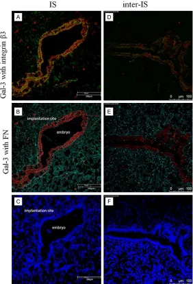
integrin β3 and FN at the implantation site. As shown in
Figures 3 and 4, gal-3 strongly colocalized with integrin β3 in the luminal epithelium at the maternal-fetal interface, espe-cially at the implantation site; however, co-localization of gal- 3 with FN was not frequently observed. These findings sug-gest that the co-expression of gal-3 and integrin β3 on the endometrium at the maternal-fetal interface facilitates en- dometrial receptivity for em- bryo attachment.
Discussion
but also promotes maternal-embryo cross-talk. However, this morphological evidence of spe-cific location and expression of gal-3 in the respective endometrium needs more support for our hypothesis that gal-3 involves in early
embryo attachment at maternal-fetal inter- face.
[image:6.612.99.520.70.594.2]signal transduction and gene expression [16]. In particular, integrin αVβ3 is an epithelial marker that indicates the opening of implanta-tion window, because it is specifically expressed in the luminal and glandular endometrial epi-thelium at the putative time of embryo implan-tation in humans [16, 22]. Although the precise mechanisms for how integrin αVβ3 functions during embryo implantation are largely un- known, it has been demonstrated that gal-3 can simultaneously change the affinity of integ-rins for extracellular matrix (ECM) proteins [23]. Moreover, our group demonstrated that exoge-nous gal-3 and integrin β1 induced endometrial
higher at the implantation site compared to the inter-implantation site. Compared with pseudo-pregnant endometrium, the expression of gal-3 in the endometrium from the implantation site was significantly higher; on the contrary, it sees no clear difference of integrin β3 expression between implantation site and pseudo-preg-nant endometrium. It indicates that embryo may be a stimulation for gal-3 spatial expres-sion, while stronger integrin β3 expression at implantation site is not induced by embryo. These results suggest that up-regulated expres-sion of gal-3 and its ligand integrin β3 at the implantation site may facilitate the establish-Figure 4. Representative images of co-localization of gal-3 (red) with integrin
β3 (green, A, D), or FN (turquoise, B, E) in the endometrium from the implan- tation site and inter-implantation site of pregnant uteri. Nuclei were counter-stained blue with DAPI (C, F). Experiments were performed in duplicate and repeated at least three times. Bar = 100 μm.
[image:7.612.94.376.73.487.2]cell apoptosis, but exogenous gal-3 and integrin β3 induced endometrial cell proliferation by partially competing with the apoptosis effect [24]. In terms of adhesion, the functional block of integrin β3 could result in reduced endometrial cell adhesion to FN in the presence of exogenous gal-3 [24, 25].
ment of endometrial receptivity during early implantation, and the presence of an embryo may have a positive effect on gal-3 but not integrin β3 expression in the endometrium. In the immunofluorescence images, we observed abundant co-localization of gal-3 with integrin β3 but not with FN in the uterine luminal epithe-lium, particularly at the implantation site of the maternal-fetal interface. These findings sug-gest that the interaction between gal-3 and integrin β3 is conducive for embryo attachment at the implantation site of the endometrium, while FN may not be a core partner for this pro-cess in mice. These results provide solid evi-dence that gal-3 with its ligand integrin β3 plays a role in the establishment of a receptive endometrium for embryo implantation. How- ever, the more factors inducing the expression of gal-3 in the endometrium require further elucidation.
In conclusion, we demonstrate that gal-3 and integrin β3 are highly expressed in the endo- metrium of pregnant mice, particularly at the implantation site, and gal-3 expression is highly co-localized with integrin β3 but not FN in the endometrium. Furthermore, high expression of gal-3 and integrin β3 may mediate embryo attachment at the maternal-fetal interface, although the precise mechanisms require fur-ther study.
Acknowledgements
This work was supported by grants from the Program of Shanghai Subject Chief Scientist (12XD1401200) and the Ph.D. Programs Foundation of Ministry of Education of China (20120071110074).
Disclosure of conflict of interest
None.
Address correspondence to: Dr. Wei Zhang, De- partment of Reproductive Endocrinology, Obstetrics and Gynecology Hospital, Fudan University, 413 Zhaozhou Road, Shanghai 200011, China. Tel: 86-21-63455050; Fax: 86-21-63455090; E-mail: [email protected]
References
[1] Rashid NA, Lalitkumar S, Lalitkumar PG, Gem-zell-Danielsson K. Endometrial receptivity and human embryo implantation. Am J Reprod Im-munol 2011; 66: 23-30.
[2] Donaghay M, Lessey BA. Uterine receptivity: alterations associated with benign gynecologi-cal disease. Semin Reprod Med 2007; 25: 461-75.
[3] Lessey BA. Adhesion molecules and implanta-tion. J Reprod Immunol 2002; 55: 101-12. [4] Yoo BC, Hong SH, Ku JL, Kim YH, Shin YK, Jang
SG, Kim IJ, Jeong SY, Park JG. Galectin-3 stabi-lizes heterogeneous nuclear ribonucleoprotein Q to maintain proliferation of human colon cancer cells. Cell Mol Life Sci 2009; 66: 350-64.
[5] Elola MT, Wolfenstein-Todel C, Troncoso MF, Vasta GR, Rabinovich GA. Galectins: matricel-lular glycan-binding proteins linking cell adhe-sion, migration, and survival. Cell Mol Life Sci 2007; 64: 1679-700.
[6] Dumic J, Dabelic S, Flögel M. Galectin-3: an open-ended story. Biochim Biophys Acta 2006; 1760: 616-35.
[7] Lee VH, Lee AB, Phillips EB, Roberts JK, Weit-lauf HM. Spatio-temporal pattern for expres-sion of galectin-3 in the murine utero-placental complex: evidence for differential regulation. Biol Reprod 1998; 58: 1277-82.
[8] DU GP, Zhang W, Wang L, Liu YK, Zhou JP. Iden-tification of differentially expressed genes in endometrium during the window of implanta-tion using suppression substractive hybridiza-tion. Zhonghua Fu Chan Ke Za Zhi 2007; 42: 187-91.
[9] Yang H, Taylor HS, Lei C, Cheng C, Zhang W. Hormonal regulation of galectin 3 in tropho-blasts and Its effects on endometrium. Reprod Sci 2011; 18: 1118-27.
[10] Yang H, Lei CX, Zhang W. Human chorionic go-nadotropin (hCG) regulation of galectin-3 ex-pression in endometrial epithelial cells and endometrial stromal cells. Acta Histochem 2013; 115: 3-7.
[11] Yang H, Lei C, Cheng C, Feng Y, Zhang W, Pe-tracco RG, Sak S. The antiapoptotic effect of galectin-3 in human endometrial cells under the regulation of estrogen and progesterone. Biol Reprod 2012; 23; 87: 39.
[12] Hynes RO. Integrins: versatility, modulation, and signaling in cell adhesion. Cell 1992; 69: 11-25.
[13] Cai L, Cao Y, Duan E. Role of αVβ3 integrin in embryo implantation in the mouse. Chinese Science Bulletin 2000; 45:2077-2113. [14] Bondza PK, Metz CN, Akoum A. Macrophage
migration inhibitory factor up-regulates alpha- (v)beta(3) integrin and vascular endothelial growth factor expression in endometrial ade-nocarcinoma cell line Ishikawa. J Reprod Im-munol 2008; 77: 142-51.
[16] Kaneko Y, Day ML, Murphy CR. Integrin β3 in rat blastocysts and epithelial cells is essential for implantation in vitro: studies with Ishikawa cells and small interfering RNA transfection. Hum Reprod 2011; 26: 1665-74.
[17] Du GP, Zhang W, Wang L, LIU YK, Zhou JP. Ex-pression of galectin-3 in human endometrium. Fudan Univ J Med Sci 2006; 2: 143-146. [18] Yang H, Lei CX, Zhang W. Expression of
galec-tin-3 in mouse endometrium and its effect dur-ing embryo implantation. Reprod Biomed On-line. 2012; 24, 116-22.
[19] Zhang S, Kong S, Lu J, Wang Q, Chen Y, Wang W, Wang B, Wang H. Deciphering the molecu-lar basis of uterine receptivity. Mol Reprod Dev 2013; 80: 8-21
[20] Knisley KA, Weitlauf HM. Compartmentalized reactivity of M3/38 (anti-Mac-2) and M3/84 (anti-Mac-3) in the uterus of pregnant mice. J Reprod Fertil 1993; 97: 521-7.
[21] Phillips B, Knisley K, Weitlauf KD, Dorsett J, Lee V, Weitlauf H. Differential expression of two beta-galactoside-binding lectins in the re-productive tracts of pregnant mice. Biol Re-prod 1996; 55: 548-58.
[22] Lessey BA, Castelbaum AJ, Buck CA, Lei Y, Yow-ell CW, Sun J. Further characterization of endo-metrial integrins during the menstral cycle and in pregnancy. Fertil Steril 1994; 62: 497-506. [23] Colin HR. Galectins as modulators of cell adhe-sion. Journal of Biochimie 2001; 83: 667-676. [24] Lei CX, Zhang W, Zhou JP, Liu YK. Interactions
between galectin-3 and integrin β3 in regulat-ing endometrial cell proliferation and adhe-sion. Human Reproduction 2009; 24: 2879-2889.