Volume-7 Issue-2
International Journal of Intellectual Advancements
and Research in Engineering Computations
MRI Brain Image Segmentation Using EM and FCM Algorithm
A.Predeep Raj
1, A.Ramkumar
2, V.Ravisankar
2, E. Padma
21
UG Students, Department of Computer Science and Engineering, Nandha Engineering
College (Autonomous), Erode, Tamilnadu, India.
2
Associate Professor, Department of Computer Science and Engineering, Nandha Engineering College
(Autonomous), Erode, Tamilnadu, India.
ABSTRACT
Automated segmentation and classification of tumors in different medical images are motivated by the necessity of high accuracy when dealing with human life. Hence, in this paper, we have presented a new methodology for segmentation and classification of brain using MRI brain images based on Framelet Transform. In the pre-processing stage given input images are segmented using Expectation Maximization (EM) and Fuzzy C Means (FCM) algorithm. Segmented images are given as input to the Framelet transform. The Framelet transform is used for feature extraction purpose. Tight wavelet frames also called as Framelet have advantageous features like shift invariant wavelet frame transforms and it may be helpful in recognize patterns in a redundant/repeated transforms. From that mean features are extracted and given as input to the next stage. Finally, Neural Network Classifier is used for classify the images whether it is normal or abnormal.
INTRODUCTION
Image segmentation refers to the process of partitioning a digital image into multiple regions. The goal of segmentation is to change the representation of an image to be more meaningful and easier to analyze. It is used in order to locate objects and boundaries in images. The result of image segmentation occurs as a set of regions that collectively covers the entire image. Therefore, medical image segmentation plays a significant role in clinical diagnosis [9]. It can be considered as a difficult problem because medical images commonly have poor contrasts, different types of noise, and missing or diffusive boundaries. The anatomy of the brain can be scanned by Magnetic Resonance Imaging (MRI) scan or computed tomography (CT) scan. The MRI scan is more comfortable than CT scan for diagnosis. It is not affect the human body because it does not use any radiation. It is based on the magnetic field and radio waves.
On the other hand, brain tumor is one of the leading causes of death among people. It is evidence that the chance of survival can be increased if the tumor is detected correctly at its early stage. In most cases, the physician gives the treatment for the strokes rather than the treatment for the tumor. Therefore, detection of the tumor is essential for the treatment. The lifetime of the person who affected by the brain tumor will increase if it is detected early. Thus, there is a need for an efficient medical image segmentation method with some preferred properties such as minimum user interaction, fast computation, accurate, and robust segmentation results.
LITERATURE REVIEW
This explains the existing approaches that are required to monitor the quality of image segmentation hybrid clustering algorithms to
segment an image to increasing accuracy and reliability for image segmentation.
Hybrid Clustering Techniques
Description
Medical image segmentation is considered as a hot research cluster. Each remaining object is clustered with the Medoid topic. Several researchers have suggested various methodoloor representative objects to which it is the most similar [14]. Kgies and algorithms for image segmentation. For example, Medoids method uses representative objects as reference. Bandhyopadhyay and Paul proposed a brain tumor segpoints rather than taking the mean value of the objects in each mentation method based on K-means clustering technique. cluster. The method consists of three steps: K-means algorithm based number of clusters to be partitioned among a set of n objects. Segmentation, local standard deviation guided grid based, the segmented images are highly dependent on the number of coarse grain localization, and local standard deviation guided segments or centers. They did not consider finding optimal grid based fine grain localization. The extraction of the number of brain segments to provide more accurate results, tumor region from the processed image requires the segmentation [12]. Islam and Ahmed proposed image segmentation section of the brain MRI images to two segments [11]. One segment unique based on K-means, K-Mediods, and Hierarchical 7 cluscontains the normal brain cells consisting of Grey Matter tering technologies. They made a comparison between these (GM), White Matter (WM), and the Cerebral Spinal Fluid three clustering techniques on natural images.
Approach Using NNC
Description
Techniques from statistical pattern recognition have, since the revival of neural networks, obtained a widespread use in digital image processing [11]. Initially, pattern recognition problems were often solved by linear and quadratic discriminants or the (non-parametric) k-nearest neighbour classi1er and the Parzen density estimator. In the mid-eighties, the PDP group
together with others, introduced the back-propagation learning algorithm for neural networks [15]. This algorithm for the 1rst time made it feasible to train a non-linear neural network equipped with layers of the so-called hidden nodes. Since then, neural networks with one or more hidden layers can, in theory, be trained to perform virtually any 10 regression or discrimination task. Moreover, no assumptions are made as with respect to the type of underlying (parametric) distribution of the input variables, which may be nominal, ordinal, real or any combination hereof. In their 1993 review article on image segmentation, Pal and Pal predicted that neural networks would become widely applied in image processing. This prediction turned out to be right. In this review article, we survey applications of neural networks developed to solve different problems in image processing.
ALGORITHMS IN PROPOSED
SYSTEM
There are some medical image segmentation systems which use K-means algorithm for detecting mass tumor in brain. The K-means algorithm is fast and simple to run on large datasets, but it suffers from incomplete detection of tumor, mainly if it is a malignant tumor [2]. On the other hand, other systems use Fuzzy C-means algorithm because it retains the more information of the original image to detect malignant tumor cells accurately compared to the K-means. These systems are sensitive to noise and outliers, and they take long execution time. In our proposed medical segmentation system, we get benefits from the last two algorithms [10]. The proposed medical image segmentation system consists of four stages: preprocessing, clustering, tumor extraction and contouring, and validation stages. The main idea of doing the integration is to reduce the number of iterations done by initializing the right cluster centers to Fuzzy C-means clustering techniques that, of course, minimizes execution time and give qualitative results
proposed system will be discussed in more detail in the subsequent sections.
EM algorithm
Estimation problems for 2-D random fields arise in contexts ranging from image processing to remote sensing. In some special cases most notably spatially stationary statistics over a rectangular grid with regularly spaced measurements very efficient algorithms based on the fast Fourier transform FFT can be used to obtain optimal estimates and error statistics. However, when one strays from these cases, the complexity of optimal solutions for many popular classes of stochastic models can become prohibitive with computational loads that do not scale well with problem size. One approach that overcomes many of these problems involves the use of multiscale stochastic models and associated multiscale estimation algorithms. These algorithms do not require stationarity, regularly shaped domains, or regularly spaced measurements. Moreover, these models have been shown to capture a rich class of random-field statistical behavior, making them useful for a number of important applications. We extend the applicability of this multiscale framework by marrying it with two other computationally powerful techniques, namely the expectation maximization EM algorithm and mean-field theory MFT. The result is a methodology that can be applied to a greatly 16 expanded range of applications, including texture segmentation, simultaneous classification and gain correction of magnetic resonance imaging MRI brain scans.
FCM Algorithm
To overcome the noise sensitiveness of conventional fuzzy c-means (FCM) clustering algorithm, a novel extended FCM algorithm for image segmentation is presented in this paper. The algorithm is developed by modifying the objective function of the standard FCM algorithm with a penalty term that takes into account the influence of the neighboring pixels on the centre pixels [3]. The penalty term acts as a regularizer in this algorithm, which is inspired from the neighborhood expectation maximization algorithm and is modified in order to satisfy the criterion of the FCM algorithm. The performance of our algorithm is discussed and compared to those of many derivatives of FCM algorithm. Experimental 18 results on segmentation of synthetic and real images demonstrate that the proposed algorithm is effective and robust. Image segmentation is an important and challenging problem and a necessary first step in image analysis as well as in high - level image interpretation and understanding such as robot vision, object recognition, and medical imaging [6]. The goal of image segmentation is to partition an image into a set of disjoint regions with uniform and homogeneous attributes such as intensity, colour, tone or texture, etc. Many different segmentation techniques have been developed. According to the image segmentation approaches can be divided into four categories: thresholding, clustering, edge detection and region extraction. In this paper, a clustering based method for image segmentation will be considered.
PROPOSED SYSTEM
There are some medical image segmentation systems which use K-means algorithm for detecting mass tumor in brain. The K-means algorithm is fast and simple to run on large datasets, but it suffers from incomplete detection of tumor, mainly if it is a malignant tumor. On the
other hand, other systems use Fuzzy C-means algorithm because it retains the more information of the original image to detect malignant tumor cells accurately compared to the K-means [5]. These systems are sensitive to noise and outliers, and they take long execution time.
Figure 2. The framework of the image segmentation system.
In our proposed medical segmentation system, we get benefits from the last two algorithms. The proposed medical image segmentation system consists of four stages: pretesting Brain Tumor Image EM Segmentation Training Brain Tumor Images Framelet based feature extraction EM Segmentation Database Normal Abnormal Neural Network Classifier Framelet based feature extraction 24 processing, clustering tumor extraction and contouring, and validation stages [4]. The main idea of doing the integration is to reduce the number of iterations done by initializing
the right cluster centers to Fuzzy C-means clustering techniques that, of course, minimizes execution time and give qualitative results. The results of our experiments clarified that our hybrid clustering method (KIFCM) can detect a tumor that cannot be detected by Fuzzy C means with less execution time.
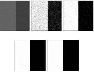
Thresholding the multiresolution map
These features assume that the asymmetry map of healthy brain contains a smaller value compared to the brain with tumor [1].
After the thresholding, in case of healthy brain, there is a smaller number of regions and also a smaller sum of their sizes. In most healthy cases, both numbers are close to zero. Examples of thresholding the asymmetry map by absolute values. 33 A comparison between the maximum asymmetry for multiresolution and each single resolution asymmetry map in brains with a tumor (green) and healthy brains (blue).
CONCLUSION AND FUTURE WORK
Image segmentation plays a significant role in medical image. In the field of medical diagnosis, an extensive diversity of imaging techniques is available presently, such as CT and MRI. MRI is the most effectively image model used for diagnostic image examination for brain tumor. The
MRI scan is more comfortable than CT scan for diagnosis. On the other hand, K-mean algorithm can detect a brain tumor faster than Fuzzy C-means, but Fuzzy C-means can predict tumor cells accurately. Original Fuzzy C-means algorithm fails to segment image corrupted by noise, outliers, and other imaging artifacts. Therefore, we developed a new approach that integrates the K means clustering algorithm with the Fuzzy C-means algorithm to detect brain tumor accurately and in minimal execution time.
In future work, the 3D evaluation of the brain tumor detection using 3D slicer will be carried out. As well as to increase the efficiency of the segmentation process, an intensity adjustment process will provide more challenging and may allow us to refine our segmentation techniques to the MRI brain tumor segmentation.
REFERENCES
[1]. Chowdhury, M.H.; Little, W.D,"Image thresholding techniques" IEEE Pacific Rim Conference on Communications, Computers, and Signal Processing.
[2]. Guillermo N. Abras and Virginia L. Ballarin,; "A Weighted K-means Algorithm applied to Brain Tissue Classification".
[3]. Keh-Shih Chuang, Hong-Long Tzeng, Sharon Chen, Jay Wu, TzongJer Chen, “Fuzzy c-means clustering with spatial information for image segmentation”,Computerized Medical Imaging and Graphics.
[4]. Matthew C. Clark “Segmenting MRI Volumes of the Brain With Knowledge- Based Clustering” MS Thesis, Department of Computer Science and Engineering, University of South Florida.
[5]. M. Masroor Ahmed, Dzulkifli Bin Mohamad,“Segmentation of Brain MR Images forTumor Extraction by Combining K-means Clustering and Perona-Malik Anisotropic.
[6]. Smita Pradhan, Dipti Patra, “Unsupervised Brain Magnetic Resonance Image Segmentation using HMRF- FCM framework”.
[7]. S.S. Mehta, C.R.Trivedi, N.S. Lingayat, “Identification and delineation of QRS Complexes in electrocardiogram using fuzzy C-means algorithm”, Journal of Theoretical and Applied Information Technology.
[8]. Engineering in Medicine and Biology Society (EMBS05), 2005, pp. 6411-6414. [9] Gopal,N.N. Karnan, M. ,―Diagnose brain tumor through MRI using image processing clustering algorithms such as Fuzzy C Means along with intelligent optimization techniques Computational Intelligence and Computing Research (ICCIC), 2010 IEEE International Conference, 28-29 ,2010, 1–4.
[9]. M. Schmidt, I. Levner, R. Greiner, et al. Segmenting brain tumors using alignment -based features[C]. Proceedings of the Fourth International Conference on Machine Learning and Applications, 2005, 6
[11]. Ahmed KHARRAT, Karim GASMI , Mohamed BEN MESSAOUD ,”A Hybrid Approach for Automatic Classification of Brain MRI Using Genetic Algorithm and Support Vector Machine ” Leonardo Journal of Sciences ISSN 1583-0233 17, 2010 ,71-82
[12]. V. Anitha and S. Murugavalli, “Brain tumour classification using two-tier classifier with adaptive segmentation technique,” IET Computer Vision, 10(1), 2016 , 9-17.
[13]. Sandabad Sara; Benba Achraf; Sayd Tahri Yassine; Hammouch Ahmed “New method of tumor extraction using a histogram study ”SAI Intelligent Systems Conference (IntelliSys), 10 -11. 2015 [14]. Nailah Afshan ,Shaima Qureshi,Syed Mujtiba Hussain,”Comparative study of tum ordetectional
gorithms”International Conference on Medical Imaging, m-Health and Emerging Communication Systems (MedCom) 2014