Gurpreet Kaur et al., J. Sci. Res. Phar. 2015, 4(1), 37-43
Journal of
Scientific
Research in
Pharmacy
Review Article
Available online through
ISSN: 2277-9469
www.jsrponl
ine.com
Fragment based Drug Discovery - Cancer Perspective
Gurpreet Kaur, Neelam VermaDepartment of Biotechnology, Punjabi University, Patiala-147002, Punjab, INDIA.
Received on: 13-02-2015; Revised and Accepted on: 10-03-2015
ABSTRACT
D
rug discovery is not a new process. Innovation in drug discovery field comes from its method of synthesis and generation of drugs. Since 1990, many new methods like sequencing, high throughput screening have played their role in research as well as in pharmaceutical industries. However due to less effective drugs and some other shortcoming now a days in-silico approach like fragment based drug design, modeling, pharmacophore study has emerged to overcome the lacking of traditional methods. Cancer is the worldwide crisis which has taken the attention of many researchers. The main focus of this review paper is to give brief information on the designing of anti-cancer drug using FBDD with assistance of few case studies.Key Words:Fragment based Drug designing, Cancer, Matrix metalloproteinase, Bcl-2, Aurora A &B, PKB/Akt, CDK-2, Hsp90, STAT3, hnRNP B1.
INTRODUCTION
C
ancer is one of the most lethal disease which affected 7.6 million population worldwide from 2008 – 2013, among them approximately 70% of cancer deaths were from low and middle-income nations. Although 30% cancer cases could be prevented but estimated mortality rate is still high[1, 2]. Cancer has signature of uncontrollable growth of cells followed by invasion in adjacent organs or tissues within body. So, the need of anti-cancer drug is a priority for both developed as well as developing nations. As the numbers of patients afflicted with cancer are increasing, the overall budget for research to synthesize anti-cancer drug is also upturning toward peak. From 2005- 2013 National Cancer Institute (NCI) budget reached $4.9 billion per year[3].For effectiveness the main focus of drug is to fit in the specific active site of target molecule whose suppression or activation further can control the progression of disease[4]. Although many anti-cancer drugs are available in the market but new medications are still needed due to deficient activity of present medicines to cure disease. Chemical synthesis is the traditional method of drug discovery which gets initiated by reaction of compounds named reactant to produce product[5]. This process is rather time-consuming and costly. Another method of drug designing is in-silico approach which is based on producing and screening of potent compound using computers. This approach has significantly reduced expensive lab time and cost[6]. Major roles of computational tools include virtual screening and de novo design; property modeling (in-silico ADME/T prediction); molecular design (determining protein-ligand binding and structure based drug design)[7].
This review will aim at describing the role of fragment based technology to discover new ligand/drugs and also their application in the field of cancer treatment.
Background:
The process of drug discovery starts with disease instead of treatment. History of drug development begins with traditional methods i.e. from isolation of medicinal plant substances or serendipity. As example, in 1928 a sudden encounter of Alexander Fleming with the moldPenicillium notatumsecreting antibacterial substance in plate culture ofStaphylococcussuggested the isolation
*Corresponding author:
Gurpreet Kaur
Department of Biotechnology,
Punjabi University, Patiala-147002, Punjab, INDIA. Contact No. - +919779074208, +919779006254.
*E-Mail: [email protected]; [email protected]
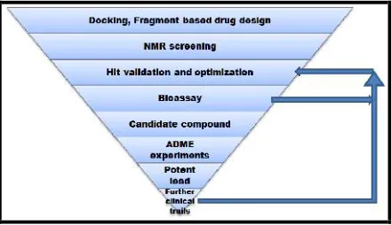
of antibiotic, Penicillin. With change in time, methods for drug synthesis also changed and recent advances of drug discovery have computers to solve their problems. Computers together with biology have overcome many disadvantages of traditional drug synthesis and have also saved laboratory time as well as funds. In-silico methods help in finding new medications considering the previous knowledge of specific target. Drugs are usually small organic compounds having the property to initiate or suppress a biological component, mainly protein, thus results in controlling the overexpressed biomolecule in diseased cells [8, 9]. The foremost requirement for drug designing is knowledge and structure of target molecule. It is believed that the chemical structure are some-how related with biological functions of compounds. This quantitative structure-activity relationship (QSAR) help in prediction study during lead discovery therefore prove to be the promising strategy for generating fragment based library of compounds[10]. For lead optimization virtual screening takes into account the structure of protein and small library of compounds. It filters the number of compounds from thousands to hundreds based on their pharmacophore (Fig. 1). Literature databases are available to seek information for protein or even genes. These databases contain full-length articles (PubMed, Sequence Retrieval System) and structural information (Protein Data Bank, PubChem, ZINC)[11].
*ADME-Absorption, Distribution, Metabolism, and Excretion
Fig. 1: Systematic flowchart for application of Fragment based drug designing
Lead identification by Fragment growth approach:
Gurpreet Kaur et al., J. Sci. Res. Phar. 2015, 4(1), 37-43
molecular mass<500 daltons, logP<5. It is recommended that an ideal fragment should follow these rules[10]. Recently commonly used method a macro version of 96- welled plate reaction systems, High Throughput Screening has the ability to observe diverse compounds at one time but because of its expensive nature alternative methods of drug designing like virtual high throughput screening (effort to dock compound libraries to a target) and prediction made on biological and physical properties of compound to fit in active site (like fragment based drug designing; FBDD) have taken place[12]. In short, purpose of computer aided drug designing through FBDD is to generate new compound by linkage which can fit in receptor active site in terms of affinity and potency.
NOTE:Often drug-likeness during lead optimization could not be maintained following the rule of five as in an attempt to improve affinity lipophilicity and molecular weight sometimes increases. Thus the rule was amended to Rule of Three which indicates the octanol-water partition coefficient (log P) ≤ 3, molecular mass ≤ 300 daltons, hydrogen bond donors≤3, hydrogen bond acceptors≤3, rotatable bonds≤3.
Fragment growth – evolving, linking, self-assembly, optimization:
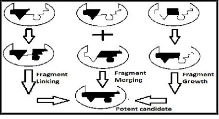
Significant feature of fragments are their low molecular weight (ranges between 100-250 Da) which requires very sensitive biophysical techniques for screening like X-ray crystallography, nuclear magnetic resonance spectroscopy (NMR) and surface plasmon resonance (SPR). Although fragments have low binding affinity, but this characteristic increases their chances to fit properly in the pocket of active site. FBDD method has been used widely in research laboratories to screen, generate, modified and also improve leads that have been further taken into clinical trials. Direct binding technique together with binding affinity information helps in evolving initial fragments. This support the building of complex molecule from simple compounds that interact with active site strongly thus leads to the tighter- binding molecules. Fragments present at adjacent vicinity can be linked chemically to increase the potency of compound. Requirements for fragment linking include identification of fragments close enough to be linked and a method for appropriate linkage to achieve the efficient results (Fig. 2). However it is assumed that for improved potency aggregate of free binding energy of all fragments should be equal to free energy of the molecule. This could be achieved if the loss of entropy on binding the active site is inconsiderable and involvement from the linker is negligible. One of the approaches to lead identification is the use of reactive fragments in presence of protein target. This is also known as fragment self-assembly because the protein selects its own fragment which could act as inhibitors or activators. Besides judging on binding affinity, fragment optimization cumulates the drug-likeness properties for a compound by modifying the properties of existing fragment[13].
Fig. 2: Illustration of Fragment evolution
Taking theory to Practical:
Main objective to discuss the case studies is to outline the application part of FBDD in selection of potent drug for preclinical trials. Some successful and some ongoing examples are illustrated.
Role of Biomarkers in Cancer Pathway:
Case 1 - Matrix Metalloproteinase: Restoration of extracellular matrix (ECM) is vital for development, incursion and proliferation of malignant tumors. Substantial aggregate of evidences suggested the capability of MMP to degrade ECM components thus significantly increasing cancerous condition. MMPs belong to structurally
correlated, zinc-dependent endopeptidases which jointly have the ability to distort the components of ECM. Main ingredient for MMP property is necessity of zinc in their active site to be vibrant[14]. MMPs inhibitors do not involve themselves in direct killing of cancer cells but as a substitute suppresses the processes like angiogenesis, invasion and cancer cells spreading. Till present, 21 human MMPs have been reported. Gelatinases have proposed to play crucial role in degrading the components of basement membranes thus helping in metastatic process. MMP-2 (Gelatinases A) is assumed to cause malignancies and tumor spread as evidences are reported that MMP-2 deficient mice express reduced angiogenesis and tumor spread while MMP-9 (Gelatinases B) showed diminished tumor formation and growth[15].
Case 2 - Bcl 2:Bcl-2 has been reported to cause various diseases as it belongs to the family responsible for impairment of several processes of immune systems such as apoptosis. Major reasons for dearth of cell death are over-expression of anti-apoptotic genes, and under-expression of pro-apoptotic genes. Moreover, studies suggested the correlation of Bcl-2 overexpression and up-regulation of VEGF expression showing an increase neoangiogenesis in human tumor xenograft[16].
Case 3 & Case 6 - Aurora A & B: Mitosis play a major role in development and prolongation of eukaryotic organisms[17]. One way to enhance the life span of patient is targeting the mitotic signaling pathway in diseased cells. Aurora kinases are reported to play crucial role in cell cycle regulation and mitosis. Aurora A is positioned at the centrosomes at prophase, and as mitosis proceeds, it is situated at the spindle poles through pro-metaphase and metaphase. In contrast, Aurora B, part of the chromosome passenger complex (CPC), is placed on the chromosome arms in prophase and at the centromeres during pro-metaphase and metaphase [18]. Aurora A (STK15, BTAK, Aurora-2) plays a fundamental role in centrosome maturation, segregation and organization of the mitotic bipolar spindle [19, 20]. Aurora B (STK12, Aurora-1) demands in mitosis is for chromosome condensation, alignment while metaphase and cytokinesis[21, 22]. Aurora A and B are up-regulated in numerous tumor cell lines. Uprooted expression of Aurora A in fibroblast cell lines direct toward centrosome amplification, genomic instability, and transformation in vitro as well as in vivo[23, 24]. Gullyet al.in 2012 reported that during mitosis at CPC Aurora B interact with p53 by examining cell cycle organizations, biochemical interaction techniques, and animal models. The hypothesis of Aurora kinases inhibition for treating cancer was introduced by testing Aurora A specific antisense oligonucleotides resulting in cell cycle arrest and introduction of cell death in pancreatic cell lines[25]. Moreover these inhibitors have also assist in better understanding of Aurora kinase performance in mitosis [26]. Furthermore, the working of inhibitors relies on inhibiting autophosphorylation of Aurora B and phosphorylation of histone H3 on Ser10 [27], but complete functioning of these small molecules are yet not known.
Case 4 - PKB/Akt: Stimulated Akt/protein kinase B transfers oncogenic indications leading to inhibition of apoptosis and cellular proliferation [28]. Interruption in oncogenesis pathway directs toward the overexpression of Akt thus causing tumor conditions. Pleckstrin homology (PH) domain binding product of phosphatidylinositol 3-kinase (PI3K) is the main reason for Akt activation through these signals. Identification of Akt was done as oncogene which was transduced by AKT8 acute transformation retrovirus as this virus had the capability to initiate malignancy in mink lung epithelial cell line CCL-retrovirus, particularly thymic lymphoma, in nude mice. Sequence analysis of the Akt viral oncogene and its cellular homology in early 1990 publicized that it encodes a serine-threonine protein kinase, possesing a carboxy-terminal kinase domain having similarity to that of PKC and PKA and an amino terminal PH domain[29].
Gurpreet Kaur et al., J. Sci. Res. Phar. 2015, 4(1), 37-43
Case 7 - Hsp90:Across all species Hsp90 is expressed at higher rate. Hsp90, a chaperone protein, actively participate in managing, stabilizing and functioning of proteins. Unfortunately, Hsp90 also works in stabilizing the tumor protein thus chosen as significant target for synthesis of anti-cancer drugs [31, 32]. In functioning of Hsp90 ATP binding domain, protein binding domain and dimerizing domain are responsible. The region near the N-terminus has high binding affinity for ATP while protein binding region is located at C-terminus. Hsp90 stabilizes most of the growth factors overexpressing in cancerous cells like signaling molecule P13K/Akt. It also stabilizes mutant protein, v-Src, fusion oncogene Bcr/Abl and transformed form of p53 usually observed throughout cell change process in cancer. Hsp90 also assist in angiogenesis and invasion process, metastasis, by promoting the MMPs. So, an inhibitor against Hsp90 could prove as a potent anti-cancer drug[33].
Case 8 - STAT3: STAT family members work in response to cytokines and growth factors. STAT members perform their role as transcription activators when phosphorylated by receptor-associated kinases then forming homo- or heterodimers further translocating to the cell nucleus. STAT plays a significant role in cell development and apoptosis. Essentially activation of STAT3 is allied with several human cancers and usually proposes poor prognosis [34-37]. It has anti-apoptotic and proliferative properties[34]. Huang et al. in 2011 studied the consequence of blockage of STAT3 signaling pathway on IL-6 persuading Epithelial-mesenchymal Transition (EMT) in human pancreatic cancer cells by using SW1990 cells. The results demonstrate the involvement of STAT3 signaling pathway in pancreatic cancer cell invasion and urge of EMT by IL-6 association with the triggering of STAT3 signaling pathway[38].
Case 9 - hnRNP B1: Heterogeneous nuclear ribonucleoprotein (hnRNP) belongs to family of protein that bound to RNA polymerase II transcript forming hnRNP particles. Several hnRNP genes have capability to form multiple isoforms through alternative splicing. Binding studies have revealed that sequence-specific essentials of DNA and RNA are associated with RNA recognition motif (RRM) also known as RNA binding motif usually situated at N-terminus. Evidences illustrate hnRNPs involvement at cellular level and suggest that it may control tumorigenesis. Carpenter et al. work with lung cancer cell line (A549) in 2006 and produced results that hnRNP members help in DNA repair and hnRNP B1 is a negative controller of DNA repair. Comparative study on hnRNP B1 with mRNA expression demonstrate that mRNA level decreases after leaving log phase in normal bronchial epithelial cell primary cultures while in tumor cell lines mRNA countenance remained steady regardless of growth phase. Furthermore high rate of microsatellite instability and heterozygosity loss were also allied with hnRNP B1 expression[39].
Case Studies - Discussion Of FBDD Approach In Lead Identification:
Case 1 – Target - Matrix Metalloproteinase (2001):
The Matrix Metalloproteinase (MMPs) belongs to zinc-dependent endopeptidases. Enzymes of this family play crucial role in maintenance and restoration of normal tissue. It is suggested that inappropriate functioning of MMPs are associated with tumor construction. Although the proper character of MMP is uncertain yet the consistent relation of MMP-2 and MMP-9 (gelatinases A and B) with tumor progression offers a new target for anti-tumor therapy. As evidence, MMP-2 and -9 deficient mice demonstrate suppression of tumor growth and metastasis without gross developing irregularities. Curtin et al in 2001 focused their effort on the detection of MMP inhibitor as anti-cancer agent. They found N -formylhydroxylamine(retrohydroxamate or ABT-770) as a potent MMP inhibitor. Pharmacokinetic analysis of ABT-770 specified low plasma clearance values at intravenous dose and high plasma concentration (of approximately 1.5µg/mL) at 3mg/kg at oral administration in rat, dog and monkey. In-vitro studies using B16 murine melanoma cells reported that dose significantly created 6.5 day delay to 1g tumor weight when 100mg/kg, bid, dose was given whose effect was resulted in 46% rise in life span. ABT-770 showed an additive effect when combined with conventional cytotoxic therapies, like ABT-770 plus paclitaxel, and as such no signs of overt toxicity were perceived. Preclinical studies were continued on ABT-770[40].
Further investigation with structure-activity relationship (SAR) accompanied with chemical synthesis of retrohydroxamates with various replacements for the biaryl ether and hydantoin groups testified that although ABT-770 has capability to selectively
inhibit MMP-2 but its activity against MMP-9 was moderate. So in order to improve potency of inhibitor a minor replacement was made in ABT-770 at ether linkage with sulfone group. This small change leads to the discovery of sulfone retrohydroxamates (ABT-518), which was taken to clinical trials[41].
Case 2 - Target - Bcl-2 (2005):
Inhibitor synthesis for Bcl-2 (B-cell lymphoma 2) opens a new treatment therapy to treat cancer as the overexpression of Bcl-2 family proteins contribute to tumor cell proliferation. The study used NMR- based screening, chemical synthesis and structure based drug designing to discover ABT-737, an inhibitor against apoptotic proteins, Bcl-2, Bcl-XL and Bcl-w, having improved magnitude with respect to affinity. Further studies disclose that compound activates the action of death signals particularly BH3 proteins, like Bid, Bim, instead of activating apoptotic system directly. These signals initiate apoptosis by prompting Bax and Bak oligomerization and leads to cytochrome c release. Activating BH3 proteins employ their pro-death function through binding anti-apoptotic Bcl-2 family members, thereby checking them from activating BH3 proteins. NMR based method was used to screen chemical library for small molecules having the capability to bind to the BH3-binding groove of Bcl-XL.40-fluorophenyl group and naphthalene derivative, 5,6,7,8-tetrahydro-naphthalen-1-ol, fit properly in hydrophobic pocket of BH3 region instead of carboxyl group of the 4-biphenylcarboxylic acid. Replacement of an acylsulphonamide for the biphenyl carboxyl group preserved the precise locating of the acidic proton that escapes steric intrusion by Phe 97. Further pharmacophore study to improve the efficiency of compound generated ABT-737 as potent candidate. This compound improved survival rates in various cancer cell lines. A mammalian two-hybrid system verified that ABT-737 interrupted with an intracellular Bcl-2 family protein–protein interaction by inhibiting the interaction of GAL 4–Bcl-XL and VP16– Bcl- XS by 44±1% and 55±2% at concentrations of 0.1 mM and 1.0 mM respectively. Combination of ABT-737 with chemotherapy and radiation reduces the median effective concentration (EC50) which further proved to be useful for treatment purpose. Moreover, during in-vivo experiments, ABT-737 caused the suppression of established SCLC tumour xenografts[42].
Case 3 - Target - Aurora A &B (2006):
Warner et al in 2006 focused their studies to detect a lead molecule which could act as an inhibitor of Aurora A and Aurora B. Approach used was de novo drug designing including modeling studies and FBDD. To reflectively resolve the aspect of homology model used for experiments root mean square deviation calculations were performed with Aurora A and a crystal structure with PDB-id 1MQ4 (Crystal Structure of Aurora-A Protein Kinase). Chemical fragment library was constructed using LUDI (de novo ligand design program). It included 70,000 two-dimensional structures and manually created 165 known serine/threonine and tyrosine kinases inhibitors. The lead compound (4-(6,7-dimethoxy-9H-1,3,9-triaza-fluoren-4-yl)-piperazine-1 carbothioic acid [4-(pyrimidin-2-ylsulfamoyl)-phenyl]-amide) was subjected to chemical synthesis followed by validation through cell based assays. Results suggested that compound showed an effective effort against target protein with an IC50 value of 0.094 µmol/L. Glide docking experiments helped in knowing the mode of action of compound. To experimentally confirm that the mode of action of compound is through competing with ATP in the active site various concentrations of ATP and lead compound in kinase reactions were prepared. The double-reciprocal plot of 1/v versus 1/[ATP] at constant concentrations of lead compound exhibited an intersecting arrangement reliable with competitive inhibition. Lead molecule subjected to secondary assays at 500nmol/L delivers certain evidences regarding its effective activity against Aurora A kinase (inhibition rate 71%) than other kinases. Dose dependent inhibition of G2-M arrest of the cell cycle (at concentrations >100µmol/L) and phosphohistone H3 levels (at concentrations >300µmol/L) were evaluated making the compound initial point for drug optimization and further improvement[43].
Case 4 - Target - PKB/Akt (2007):
Gurpreet Kaur et al., J. Sci. Res. Phar. 2015, 4(1), 37-43
tumor suppressor gene PTEN activates PKB. Fragment-based approach combined with virtual screening, X-ray crystallography and NMR were applied by Saxty et al in 2007 to detect novel hints as inhibitor for PKB. They docked approximately 300,000 available fragments. In-vitro, 8 fragments with potency range from 16µm-1mM were validated after pharmacophore studies. Further trials on xenograft model resulted in showing drug-likeness of 5-methyl-4-phenyl-1H-pyrazole. Structure based studies supported the fragment evolution by analyzing the improvement of hit to lead from 100 mM to 10nM respectively. Moreover with ligand efficiency maintained between 0.47-0.49 this lead was taken to clinical stage [44, 45].
Case 5 - Target - CDK-2 (2008):
Astex Therapeutics Ltd.,target in 2008 was to identify cyclin dependent kinase 2 (CDK2) inhibitors through fragment based drug discovery (FBDD). Being a family of serine/threonine protein kinases the major role of CDK2 is to regulate essentials of cell cycle progression. Cyclins expressions are strongly regulated during different stages of cell cycle which in-turn control the performance of CDKs. One of the key features of cancer is uncontrolled expression of cyclins levels throughout the cell cycle. So CDKs inhibition may prove as a treatment to cancer. In experiment, crystal of CDK was immersed in mixture of fragments of about 500 compounds to identify the hits bound to active site of receptor. For inhibitor designing process analysis of ATP binding site of CDK was performed and multiple low binding fragments were identified. Structural data and Ligand efficiency were the basis for optimization of potential fragments. In-vitro studies evaluated 2,6-dichlorophenyl derivative, N -(4-Piperidinyl)-4-(2,6-dichlorobenzoylamino)-1H-pyrazole-3-carboxamide (AT7519), as a potent inhibitor as it showed anti-proliferative effect in range of cell lines. When dosed at 9.1 mg/kg ip bid for 10 days 87% growth inhibition was shown by the compound on HCT116. Furthermore data from in-vivo antitumor activity in nude BALB/c mice bearing early stage A2780 human ovarian carcinoma xenografts of inhibiting the tumor growth to 86% at the 7.5 mg/kg dose level forms the foundation for its entry as preclinical development candidate[46].
Case 6 - Target - Aurora A& B (2009):
As target Aurora kinases from family of serine-threonine protein kinases, having two isoforms- Aurora A & B overexpressed in tumor cells was considered byAstex Therapeutics Ltd.in 2008. Fragments having molecular weight between 100-250 kDa were used. Through FBDD they optimized ligand efficiency of pyrazole-benzimidazole fragment, Pyrazol-4-yl Urea (AT9283). X-ray crystallographic structures helped in driving optimization of dual Aurora A/Aurora B inhibitors toward potency of approximately 3nM. Inhibited growth and proliferative rate of HCT116 cells proved this fact. This compound has shown its ability to inhibit a number of other kinases including JAK2, Flt3, and Abl (T315I) (IC50= 1-30 nM). In vivo studies are under progress for these inhibitors[47].
Case 7 - Target – Hsp90 (2007-2012):
Molecular chaperone named Heat shock protein 90 (Hsp90) after many scientific evidences are suggested to play significant role in tumor progression[48, 49]. An interference in Hsp90 functioning can cause disruption in various pathways of cancer growth. The chaperone cycle of Hsp90 comprises of output of ATP to ADP through an ATPase activity linked with the N-terminal domain of Hsp90[50]. Moreover studies demonstrate that inhibition at ATP binding site further downregulates the Hsp90 associated proteins which leads to suppression of antiproliferative activity[51]. Strategy followed by Astex Therapeutic Ltd. was the grouping of NMR and high throughput X-ray crystallography in order to fulfill the aim to make use of FBDD in designing Hsp90 inhibitor. In this study nearly 1600 compounds from our fragment library were screened in mixtures using ligand observed NMR. Although some fragments gave low ligand efficiency yet they recommended a number of design ideas for improving potency level through virtual screening and fragment screening approach. This approach leads to identification of clinical candidate. Pyrimidine series optimization showed an improved affinity by 40000-folds (250 µM to 6.3nM) by gaining active confirmation stabilization and hydrophobic contact for improvement with enzyme. Secondly, the phenolic series optimization process gave Resorcinol as lead compound with binding affinity 0.54 nM preparatory from the phenol fragment having affinity 790 μM[52]. Further preclinical studies were carried out using many fragments from which (2,4-Dihydroxy-5-
isopropylphenyl)-[5-(4-methylpiperazin-1-ylmethyl)-1,3-dihydroisoindol-2-yl]methanone (AT13387), proved to be potent preclinical candidate. Crucial factors for investigation like in-vivo target modulation, predicted human dose, selectivity/off-target effects, solubility, stability, formulation, and synthesis were successfully cleared by AT13387 as it binds in the ATPase site at N-terminus of Hsp90. It also proved its profile against a range of human tumor cell lines. Based on this data AT13387 has entered in clinical trials[53]. Recently it has been announced by the company that AT13387 is entering in Phase –II clinical trials for treatment of various cancers [54]. Astex Therapeutic Ltd.have also initiated in using this compound AT13387 for clinical trials phase I-II in prostate cancer[55].
Case 8 - Target - STAT3 (2005-2013):
STAT3 (Signal Transducers and Activators of Transcription 3) belongs to family of seven transcription factors (STATs 1, 2, 3, 4, 5a, 5b, and 6). They play significant role in transmitting signals from cell surface receptors to the nucleus, as well as signaling of various cytokines and growth factors that are involved in the fundamental cellular and biological processes leading to the immune response, angiogenesis, cell proliferation, differentiation, and apoptosis etc.[56-62]. Recently Chen et.al in 2013 used the cumulative effort of fragment based drug designing, chemical synthesis and pharmacology study to discover a compound named HJC0123 as a potent anti-cancer agent having the capability to inhibit STAT3 promoter activity, down-regulate phospho-STAT3, increase the expression of cleaved caspase-3, also in breast and pancreatic cancer cells, inhibit cell cycle progression and promote apoptosis with low micromolar to nanomolar IC50 values. Studies initiated with six fragments having characteristic of non-peptidic STAT3 inhibitors. To examine SAR study, in-vitro anticancer effect was evaluated using compounds bearing anti-cancerous property on human breast cancer cell lines MCF-7 (ER-positive) and MDAMB-231 (ER-negative and triple-negative), as well as on pancreatic cancer cell lines AsPC1 and Panc-1 through MTS assay. Notably, HJC0123 showed significant potency against all the tested cancer cells. According to previous reports STX-0119 with 2-phenylquinoline-4-carboxylic acid amide fragment (A1)[63, 64]and static with 1,1-dioxo-1H-1λ6- benzo[b]thiophen-6-yl fragment (B1) [65] were able to inhibit STAT3-SH2 domain. To investigate the pharmacological reasons AutoDock Vina docking approach was referred which proposed that the hydrophobic substituent at C2 of the quinoline or pyridine framework is important for targeting STAT3. Likewise, HJC0123 inhibited phosphorylated STAT3 at Tyr705. This compound during in-vivo studies considerably suppressed ER-negative breast cancer MDA-MB-231 xenograft tumor growth thus suggesting its potential as a successful and orally bioavailable drug lead candidate for human cancer therapy. Preclinical assessment with this favorable compound is under trials [66].
Case 9 - Heterogeneous Nuclear Ribonucleoprotein B1 (hnRNP B1):
Overexpression of hnRNP B1 has been noted at early stages of lung cancer. So this property makes it a strong target for synthesis of new anti-cancer drug against lung cancer. Our lab has been focusing on designing drug against hnRNP B1 (receptor) through computational approach. 64 compounds were docked in the active site of receptor which included lung cancer inhibitors like drugs already present in market, natural compound curcumin along with their analogue and other inhibitors of hnRNP B1. Free minimization results obtained on BioMed CaChe Software version 6.1.12.34 were taken for structure activity relationship studies where results suggested that Epigallocathechin gallate (EGCG) could be an effective candidate for hnRNP B1 inhibition. Our next approach was to modify EGCG structure for the purpose to improve binding affinity of the potent candidate. Pharmacophore studies revealed that improvement could be seen in docking score as well as in H-bond interactions between ligand and surrounding active sites amino acids. The binding affinity from parent compound to modified compound goes from -86.618 kcal/mol to -96.307 kcal/mol.
Gurpreet Kaur et al., J. Sci. Res. Phar. 2015, 4(1), 37-43
Table No. 1: Potent drug candidate modified from initial compound using Fragment Based Drug Designing
Target Initial Hit Lead Molecule Ref.
MMP-2 & 9
ABT-770
MMP-2 (IC50 ) 3.7 nM), it is
only moderately active against MMP-9 (IC50 ) 120 nM).
ABT-518
MMP-9 (IC50 ) 0.86 nM)
Unfortunately, this small change led to nearly complete loss of selectivity for inhibition of MMP-2 and -9 over MMP-1 (IC50 MMP-1/MMP-2 < 10-fold)
[41]
Bcl-2
Ki = 36 ±1.6 nM ABT-737; Ki ≤ 1 nM)
[42]
AURORA A & B
3 µmol/L
0.006 µmol/L
[43]
PKB/Akt
IC50 = 80µM ±38; LE = 0.47 IC50 = 31Nm ±14; LE = 0.49
[44, 45]
CDK-2
IC50 = 185µM; LE = 0.57 IC50 = 0.047 µM; LE = 0.42 HCT116 cells; IC50 = 0.082 µM
[46]
AURORA A& B
Aurora A = 0.91 µM Aurora A 52% I at 3.0 nM; Aurora B 58% I at 3.0 nM
[47]
Hsp-90
Hsp90; Kd(µM) = 0.00054 HCT116; IC50(µM) = 0.031
affinity forHsp90 (Kd= 0.5 nM)
[52, 53]
STAT3
IC50(µM)
Breast cancer ER-positive (MCF-7) = 0.1 Breast cancer ER-negative (MDA-MB-231) = 0.29 Pancreatic cancer (AsPC-1, Panc-1) = 1.25, 0.26 respectively
Gurpreet Kaur et al., J. Sci. Res. Phar. 2015, 4(1), 37-43
hnRNP B1
-86.618 kcal/mol; H-bonds- 1 to -96.307 kcal/mol; H-bonds- 3
Details will be published
in next paper/[67]
CONCLUSION
M
edicinal field require a lot of attention these days where disease are showing an epidemic effect. FBDD technique is effective; less time consuming and helpful for research as it identify the potent drug that can be synthesized. Although FBDD is new but its use for synthesis and screening of compounds, which may act as drugs, is attracting the interest of researchers worldwide.ACKNOWLEDGEMENT
W
e are thankful to University Grant Commission (UGC), India for their funds support.REFERENCES:
1. World Cancer Day 2013. < http://www.who.int/cancer/en/>
2. Siegel R, Naishadham D, Jemal A. Cancer Statistics, 2013.CA Cancer J. Clin.,2013; 63(1): 11-30.
3. The National Cancer Institute (NCI) fact sheet: Cancer
Research Funding
<http://www.cancer.gov/cancertopics/factsheet/NCI/resea rch-funding>
4. Feigenoff C. Explorations-Research Highlights from the University of Virginia: Precisely Targeted Drug Design (2002). < http://oscar.virginia.edu/explorations/> 5. Vogel AI, Tatchell AR, Furnis BS, Hannaford AJ, Smith PWG.
Vogel's Textbook of Practical Organic Chemistry, 5th Edition. Prentice Hall,1996. ISBN 0-582-46236-3.
6. Trisilowati, Mallet D G. In Silico Experimental Modeling of Cancer Treatment, ISRN Oncology,2012; 8.
7. Srinivasa VR, Srinivas K. Modern drug discovery process: An in silico approach, J. Bioinform. Seq. Anal.,2011; 2(5): 89-94.
8. Cohen NC. Guidebook on Molecular Modeling in Drug Design. Boston: Academic Press. ISBN 0-12-178245-X (1996).
9. Tollenaere JP. The role of structure-based ligand design and molecular modelling in drug discovery,Pharm. World Sci., 1996; 18(2): 56–62.
10. Leeson P. Drug discovery: Chemical beauty contest,Nature, 2012; 481: 455–456.
11. Pevsner J. Access to Sequence Data and Literature Information In Bioinformatics and Functional Genomics, Second edition, John Wiley & Sons, Inc., Hoboken, New Jersey (2009).
12. Howe D, Costanzo M, Fey P, Gojobori T, Hannick L, Hide W, Hill DP, Kania R, Schaeffer M, Pierre SS, Twigger S, White O, Rhee SY. Big data: The future of biocuration,Nature,2008; 455(7209): 47–50.
13. Rees DC, Congreve M, Murray CW, Carr R. Fragment-based lead discovery: leads by design,Drug Discov. Today, 2005; 10(14): 987-92.
14. Reunanen N, Kahari V. Madame Curie Bioscience Database. Matrix Metalloproteinases in Cancer Cell Invasion. Austin (TX): Landes Bioscience (2000) <http://www.ncbi.nlm.nih.gov/books/NBK6598/> 15. Rundhaug JE. Matrix Metalloproteinases, Angiogenesis, and
Cancer: Commentary re: A. C. Lockhart et al., Reduction of Wound Angiogenesis in Patients Treated with BMS-275291, a Broad Spectrum Matrix Metalloproteinase Inhibitor,Clin. Cancer Res.,2003; 9: 551-554.
16. Biroccio A, Candiloro A, Mottolese M, Sapora O, Albini A, Zupi G et al. Bcl-2 overexpression and hypoxia synergistically act to modulate vascular endothelial growth factor expression and in vivo angiogenesis in a breast carcinoma cell line,FASEB J.,2000; 14: 652-660.
17. Warner SL, Bashyam S, Vankayalapati H, Bearss DJ, Han H, Mahadevan D et al. Identification of a lead small-molecule inhibitor of the Aurora kinases using a structure-assisted, fragment-based approach,Mol. Cancer Ther.,2006; 5: 1764-1773.
18. Carmena M, Earnshaw, W.C. The cellular geography of aurora kinases,Nat. Rev. Mol. Cell Biol.,2003; 4: 842–854. 19. Warner S, Bearss DJ, Han H, Von Hoff DD. Targeting
Aurora-2 kinase in cancer.Mol. Cancer Ther.,2003; 2: 589–595. 20. Kimura M, Kotani S, Hattori T, Sumi N, Yoshioka T, Todokoro
K et al. Cell cycle-dependent expression and spindle pole localization of a novel human protein kinase, Aik, related to Aurora of Drosophila and yeast Ipl1,J. Biol. Chem.,1997; 272: 13766–13771.
21. Kimura M, Matsuda Y, Yoshioka T, Sumi N, Okano Y. Identification and characterization of STK12/Aik2: a human gene related to aurora of Drosophila and yeast IPL1, Cytogenet. Cell Genet.,1998; 82: 147–152.
22. Carmena M, Earnshaw WC. The cellular geography of aurora kinases,Nat. Rev. Mol. Cell Biol.,2003; 4: 842–854.
23. Bischoff, J.R., Anderson, L., Zhu, Y, Mossie K, Ng L, Souza B et al. A homologue of Drosophila aurora kinase is oncogenic and amplified in human colorectal cancers,EMBO J.,1998; 17: 3052–3065.
24. Zhou H, Kuang J, Zhong L, Kuo WL, Gray JW, Sahin A, et al. Tumour amplified kinase STK15/ BTAK induces centrosome amplification, aneuploidy and transformation, Nat. Genet., 1998; 20: 189–193.
25. Rojanala S, Han H, Munoz RM, Browne W, Nagle R, Von Hoff DD, et al. The mitotic serine threonine kinase, Aurora-2, is a potential target for drug development in human pancreatic cancer,Mol. Cancer Ther.,2004; 3: 451–457.
26. Yeung SC, Gully C, Lee MH. Aurora-B kinase inhibitors for cancer chemotherapy,Mini Rev. Med. Chem., 2008; 8(14): 1514-1525.
27. Lens SM, Voest EE, Medema RH. Shared and separate functions of polo-like kinases and aurora kinases in cancer, Nat. Rev. Cancer,2010; 10: 825–841.
28. Matsumoto J. Differential mechanisms of constitutive Akt/PKB activation and its influence on gene expression in pancreatic cancer cells, Jpn. J. Cancer Res.,2002; 93(12): 1317-1326.
29. Kim D, Dan HC, Park S, Yang L, Liu Q, Kaneko S, et al Akt/PKB signaling mechanisms in cancer and chemoresistance,Front. Biosci.,2005; 10: 975-987. 30. Shapiro GI. Cyclin-Dependent Kinase Pathways as Targets
for Cancer Treatment,J. Clin. Oncol.,2006; 24(11): 1770-1783.
31. Csermely P, Schnaider T, Soti C, Prohászka Z, Nardai G. The 90-kDa molecular chaperone family: structure, function, and clinical applications. A comprehensive review, Pharmacol. Ther.,1998; 79(2): 129–68.
32. Crevel G, Bates H, Huikeshoven H, Cotterill S. The Drosophila Dpit47 protein is a nuclear Hsp90 co-chaperone that interacts with DNA polymerase alpha, J. Cell. Sci. 2001; 114(Pt 11): 2015–25.
33. Calderwood SK, Khaleque MA, Sawyer DB, Ciocca DR. Heat shock proteins in cancer: chaperones of tumorigenesis, Trends Biochem. Sci.,2006; 31(3): 164–72.
34. Klampfer L. Signal transducers and activators of transcription (STATs): Novel targets of chemopreventive and chemotherapeutic drugs, Curr. Cancer Drug Targets, 2006; 6(2): 107–121.
35. Alvarez JV, Greulich H, Sellers WR, Meyerson M, Frank DA. Signal transducer and activator of transcription 3 is required for the oncogenic effects of non-small-cell lung cancer-associated mutations of the epidermal growth factor receptor,Cancer Res.,2006; 66(6): 3162–3168.
Gurpreet Kaur et al., J. Sci. Res. Phar. 2015, 4(1), 37-43
cell carcinoma cells in serum-free conditions,Mol. Cancer, 2006; 5(1): 15.
37. Kusaba T, Nakayama T, Yamazumi K, Yakata Y, Yoshizaki A, Inoue K, et al. Activation of STAT3 is a marker of poor prognosis in human colorectal cancer, Oncol. Rep.,2006; 15(6): 1445–1451.
38. Huang C, Yang G, Jiang T, Zhu G, Li H, Qiu Z. The effects and mechanisms of blockage of STAT3 signaling pathway on IL-6 inducing EMT in human pancreatic cancer cells in vitro, Neoplasma,2011; 58(5): 396-405.
39. Carpenter B, MacKay C, Alnabulsi A, MacKay M, Telfer C, Melvin WT et al. The roles of heterogeneous nuclear ribonucleoproteins in tumour development and progression,Biochimica et Biophysica Acta,2006; 1765: 85– 100.
40. Curtin ML, Florjancic AS, Heyman HR, Michaelides MR, Garland RB, Holms JH et al. Discovery and Characterization of the Potent, Selective and Orally Bioavailable MMP Inhibitor ABT-770,Bioorg. Med. Chem. Lett.,2011; 11: 1557-1560.
41. Wada CK, Holms JH, Curtin ML, Dai Y, Florjancic AS, Garland RB et al. Phenoxyphenyl Sulfone N-Formylhydroxylamines (Retrohydroxamates) as Potent, Selective, Orally Bioavailable Matrix Metalloproteinase Inhibitors, J. Med. Chem.,2002; 45: 219-232.
42. Oltersdorf T, Elmore SW, Shoemaker AR, Armstrong RC, Augeri DJ, Belli BA, et al. An inhibitor of Bcl-2 family proteins induces regression of solid tumours,Nature Letters,2005; 435: 677-681.
43. Warner SL, Bashyam S, Vankayalapati H, Bearss DJ, Han H, Mahadevan D, et al. Identification of a lead small-molecule inhibitor of the Aurora kinases using a structure-assisted, fragment-based approach,Mol. Cancer Ther.,2006; 5: 1764-1773.
44. Saxty G, Berdini BV, Blakemore W, Boyle RG, Carr RAE, Davies TG et al. Fragment-Based Approach to Orally Active PKB/AKT Inhibitors. < http://astx.com/content/resources/2007_Cam_Med_Chem_ G_Saxty_Poster.pdf >
45. Saxty G, Woodhead SJ, Berdini V, Davies TG, Verdonk ML, Wyatt PG, et al. Identification of Inhibitors of Protein Kinase B Using Fragment-Based Lead Discovery, J. Med. Chem., 2007; 50: 2293-2296.
46. Wyatt PG, Woodhead AJ, Berdini V, Boulstridge JA, Carr MG, Cross DM, et al. Identification of N-(4-Piperidinyl)-4-(2,6-dichlorobenzoylamino)-1H-pyrazole-3-carboxamide (AT7519), a Novel Cyclin Dependent Kinase Inhibitor Using Fragment-Based X-Ray Crystallography and Structure Based Drug Design,J Med Chem.,2008; 51(16): 4986-4999. 47. Howard S, Berdini V, Boulstridge JA, Carr MG, Cross DM,
Curry J, et al. Fragment-Based Discovery of the Pyrazol-4-yl Urea (AT9283), a Multitargeted Kinase Inhibitor with Potent Aurora Kinase Activity,J. Med. Chem.,2009; 52(2): 379-388. 48. Workman P, Burrows F, Neckers L, Rosen N. Drugging the
cancer chaperone HSP90: combinatorial therapeutic exploitation of oncogene addiction and tumor stress,Ann. N.Y. Acad. Sci.,2007; 1113: 202–216.
49. Whitesell L, Lindquist SL. HSP90 and the chaperoning of cancer,Nat. Rev. Cancer.,2005; 5: 761–772.
50. Pearl LH, Prodromou C. Structure and mechanism of the Hsp90 molecular chaperone machinery,Annu. Rev. Biochem., 2006; 75: 271–294.
51. Vilenchik M, Solit D, Basso A, Huezo H, Lucas B, He H, et al. Targeting wide-range oncogenic transformation via PU24FCl, a specific inhibitor of tumor Hsp90,Chem. Biol., 2004; 11: 787–797.
52. Murray CW, Carr MG, Callaghan O, Chessari G, Congreve M, Cowan S, et al. Fragment-Based Drug Discovery Applied to Hsp90. Discovery of Two Lead Series with High Ligand Efficiency,J. Med. Chem.,2010; 53: 5942–5955.
53. Woodhead AJ, Angove H, Carr MG, Chessari G, Congreve M, Coyle JE, et al. Discovery of (2,4-Dihydroxy-5- isopropylphenyl)-[5-(4-methylpiperazin-1-ylmethyl)-1,3-dihydroisoindol-2-yl]methanone (AT13387), a Novel Inhibitor of the Molecular Chaperone Hsp90 by Fragment Based Drug Design,J. Med. Chem.,2010; 53: 5956–5969. 54. Astex Theraputics. Astex scientists describe novel HSP90
inhibitors in two key publications in Journal of Medicinal Chemistry (2010) website [online], < http://investor.astx.com/releasedetail.cfm?ReleaseID=6639 70>
55. Astex Theraputics. Astex Pharmaceuticals Announces Initiation of HSP90 Inhibitor AT13387 Clinical Trial in Prostate Cancer Patients (2012) website [online], < http://investor.astx.com/releasedetail.cfm?ReleaseID=7057 92 >
56. Zhong Z, Wen Z, Darnell JE Jr. Stat3: a STAT family member activated by tyrosine phosphorylation in response to epidermal growth factor and interleukin-6,Science,1994; 264: 95-98.
57. Darnell JE. Jr. STATs and gene regulation,Science, 1997; 277: 1630-1635.
58. Bromberg JF, Wrzeszczynska MH, Devgan G, Zhao Y, Pestell RG, Albanese C, et al. STAT3 as an oncogene,Cell,1999; 98: 295-303.
59. Bromberg J, Darnell JE Jr. The role of STATs in transcriptional control and their impact on cellular function, Oncogene,2000; 19: 2468-2473.
60. Hirano T, Ishihara K, Hibi M. Roles of STAT3 in mediating the cell growth, differentiation and survival signals relayed through the IL-6 family of cytokine receptors, Oncogene, 2000; 19: 2548-2556.
61. Bowman T, Garcia R, Turkson J, Jove R. STATs in oncogenesis,Oncogene,2000; 19: 2474-2488.
62. Turkson J, Jove R. STAT proteins: novel molecular targets for cancer drug discovery,Oncogene,2000; 19: 6613-6626. 63. Matsuno K, Masuda Y, Uehara Y, Sato H, Muroya A,
Takahashi O, et al. Identification of a new series of STAT3 inhibitors by virtual screening,ACS Med. Chem. Lett.,2010; 1: 371-375.
64. Song H, Wang R, Wang S, Lin J. A low-molecular-weight compound discovered through virtual database screening inhibits Stat3 function in breast cancer cells,Proc. Natl. Acad. Sci.,2005; 102: 4700-4705.
65. Schust J, Sperl B, Hollis A, Mayer TU, Berg T. Stattic. A small-molecule inhibitor of STAT3 activation and dimerization, Chem. Biol.,2006; 13: 1235-1242.
66. Chen H, Yang Z, Ding C, Chu L, Zhang Y, Terry K, et al. Fragment-based drug design and identification of HJC0123, a novel orally bioavailable STAT3 inhibitor for cancer therapy,Eur. J. Med. Chem.,2013; 62: 498-507.
67. Verma N, Kaur G.In-silicodesigning of hnRNP B1 inhibitors against Lung Cancer: A Computational approach. Details will be mentioned in next paper.
How to cite this article:
Gurpreet Kaur, Neelam Verma: Fragment based Drug Discovery - Cancer Perspective, J. Sci. Res. Phar., 2015; 4(1): 37-43.
Conflict of interest:The authors have declared that no conflict of interest exists.