i
NOVEL FUNCTIONS OF ADRENOMEDULLIN, RECEPTOR ACTIVITY-MODIFYING PROTEIN 2, AND GPR182 IN CARDIOVASCULAR DISEASE AND CANCER
Daniel Oliver Kechele
A dissertation submitted to the faculty of the University of North Carolina at Chapel Hill in partial fulfillment of the requirements for the degree of Doctor of Philosophy in the
Department of Cell Biology and Physiology (Cell and Molecular Physiology)
Chapel Hill 2016
ii ©2016
iii
ABSTRACT
Daniel Oliver Kechele: Novel Functions of Adrenomedullin, Receptor Activity-Modifying Protein 2, and Gpr182 in Cardiovascular Disease and Cancer (Under the direction of Kathleen Caron)
The multifunctional circulating peptide, adrenomedullin (AM) signaling through its G protein-coupled receptor (GPCR) complex, comprised of the calcitonin receptor-like receptor (CLR) and receptor activity-modifying protein 2 (Ramp2), is required for lymphatic development and embryonic survival, as well as numerous functions in adult cardiovascular physiology. However these functions, especially during adulthood, have not been fully elucidated. Using both gain- and loss-of-function mouse models these studies aimed to further elucidate the roles of AM signaling during cardiovascular homeostasis and tumor progression. Endothelial restoration of Ramp2 is sufficient to rescue the embryonic lethality-associated with global loss of Ramp2. However, the developmental loss of Ramp2 in non-endothelial cells led to spontaneous hypotension, dilated cardiomyopathy, and multi-organ inflammation in adult survivors. Smooth muscle cell- or global-deletion of CLR did not recapitulate these phenotypes, indicating that Ramp2 is likely interacting with other GPCRs, including parathyroid hormone and glucagon receptors, to maintain cardiovascular homeostasis.
iv
While CLR/Ramp2 is the canonical AM receptor, it was previously thought that the AM receptor was the orphan GPCR, Gpr182, of which almost nothing is known. Using a knock-in reporter mouse model to map Gpr182 expression in vivo, Gpr182 was enriched in the active stem cells in the small intestine. Gpr182 was dispensable during development and homeostasis, but when challenged with irradiation injury or adenoma model, reduced
Gpr182 led to intestinal hyperproliferation during regeneration and enhanced adenoma
formation, respectively.
v
ACKNOWLEDGEMENTS
I would like to thank all members, past and present, of the Caron lab for intellectual discussions and technical assistance in all my projects. With regards to scientific data presented throughout this dissertation, I would like to thank the University of North Carolina Lineberger Animal Histopathology Core (NIH CA16086) and Kirk McNaughton and Ashley Ezzell of the University of North Carolina Histology Research Core for tissue embedding, sectioning, and staining expertise. I would also like to acknowledge Dr. Robert Bagnell and the University of North Carolina Microscopy Services Laboratory for confocal microscopy assistance. Regarding the data presented in Chapter II, I would like to thank the University of North Carolina Animal Models Core and the University of North Carolina Rodents Advanced Surgical Models Core, specifically Dr. Mauricio Rojas and Dr. Brian Cooley with echocardiography and intra-arterial blood pressure measurements, respectively. I would also like to thank Dr. Lin Xiao and Dr. Andrew Dudley (UNC-CH Cell Biology and Physiology) for endothelial isolation expertise, and Helen Willcockson, Dr. Samantha Hoopes, John Pawlak, and Dr. Wenjing Xu for technical assistance. For preliminary data presented in Chapter IV, I would like to thank Dr. James Dunleavey of the Dudley lab for providing melanoma cell line and tumor and flow cytometry expertise. For study presented in Chapter VI, I would like to thank all members of Dr. Kay Lund’s laboratory (UNC-CH Cell
vi
vii
TABLE OF CONTENTS
LIST OF TABLES ... x
LIST OF FIGURES ... xi
LIST OF ABBREVIATIONS ... xiii
PART I Chapter I: Adrenomedullin Function in Vascular Endothelial Cells: Insights from Genetic Mouse Models ... 1
Overview ... 1
Introduction ... 1
Development ... 6
Physiology and Pathology ...10
Summary and Future Directions ...16
Author Contributions...16
Tables ... 17
Figures ... 19
Chapter II: Endothelial restoration of receptor activity-modifying protein 2 is sufficeint to rescue lethality, but survivors develop dilated cardiomyopathy ...21
Overview ...21
Introduction ...22
Methods ...23
Results ...25
Discussion ...32
Perspectives ...35
viii
Author Contributions ...37
Tables ...38
Figures ...40
Supplemental Methods ...47
Supplemental Tables...54
Supplemental Figures ...58
Chapter III: Dosage of adrenomedullin correlates with tumor and lymph node lymphangiogenesis ...61
Overview ...61
Introduction ...62
Materials and Methods ...64
Results ...68
Discussion ...73
Author Contributions ...76
Figures ...77
Chapter IV: Conclusions and Future Directions ...85
Summary ...85
Current State of the Field – RAMPs, Cardiac Disease, and Tumor Lymphatics ...85
Future Directions ...92
Concluding Remarks ... 106
Figures ... 107
Part II Chapter V: The History of AM Receptors: The Forgotten GPCRs... 116
Overview ... 116
ix
CLR, Cxcr7 and Gpr182 in a RAMP World ... 120
Figure ... 122
Chapter VI: Gpr182 inhibits intestinal proliferation during regeneration and adenoma formation ... 123
Overview ... 123
Introduction ... 124
Methods ... 126
Results ... 131
Discussion ... 140
Author Contributions ... 142
Figures ... 143
Supplemental Tables... 152
Supplemental Figures ... 153
Chapter VII: Conclusions and Future Directions ... 157
Summary ... 157
Current State of the Field -- ISCs, regeneration, and carcinoma... 157
Future Directions ... 163
Concluding Remarks ... 176
Figures ... 177
x
LIST OF TABLES
Table 1-1 Gene Targeted Mouse Models for Studying Adrenomedullin Signaling ... 17 Table 1-2 Vascular Assays for Studying Adrenomedullin Function ... 18 Table 2-1 Endothelial restoration of Ramp2 can rescue
the global Ramp2-/- embryonic lethality ... 38 Table 2-2 Ramp2-/- Tg adults develop left ventricle dilatation
and declining heart function ... 39 Table 2-S1 Genotype and RT-PCR Gene Expression Primers and Probes ... 54 Table 2-S2 Relatively similar systemic circulating factors in
Ramp2+/+ ntg and Ramp2-/- Tg serum ... 55 Table 2-S3 Vascular smooth muscle cell conditional
Calcrl deletion does not lead to hypotension or
dilated cardiomyopathy phenotype in adult male mice ... 56 Table 2-S4 Global conditional Calcrl deletion does not lead to
xi
LIST OF FIGURES
Figure 1-1 Fold change in plasma adrenomedullin levels in a
variety of human conditions ... 19
Figure 1-2 Adrenomedullin signaling in development and vascular biology ... 20 Figure 2-1 Endothelial restoration of Ramp2 partially rescues embryonic
edema leading to prolonged survival of Ramp2-/-mice ... 40 Figure 2-2 Ramp2-/- Tg adults are hypotensive ... 41 Figure 2-3 Ramp2-/- Tg adults develop spontaneous dilated cardiomyopathy ... 42 Figure 2-4 Ramp2-/- Tg left ventricles hypertrophied with early
fibrotic and oxidative stress changes ... 43 Figure 2-5 Ramp2-/- Tg have vascular congestion and multi-organ
inflammation downstream of hypotension and dilated cardiomyopathy ... 44 Figure 2-6 Decreased signaling and expression of RAMP-associated
GPCRs in embryonic and adult Ramp2-/- Tg hearts ... 45 Figure 2-S1 Endothelial-specific overexpression of murine Ramp2 ... 58 Figure 2-S2 Generation of gene-targeted Ramp2
deficient mice with and without the Ramp2 Tg ... 59 Figure 2-S3 Generation and characterization of vascular
smooth muscle cell-specific Calcrl deficient mice ... 60 Figure 3-1 In vitro characterization of the LLC cell line ... 77
Figure 3-2 In vivo tumor analysis ... 78 Figure 3-3 Adm expression does not affect angiogenesis ... 79
Figure 3-4 Adm expression is necessary for lymphangiogenesis ... 80 Figure 3-5 Adm dosage does not affect macrophage or Vegf levels ... 81 Figure 3-6 Elevated Adm expression increases the diameter
of blood and lymphatic vessels ... 82 Figure 3-7 Tumor Adm expression influences sentinel lymph
xii
Figure 4-2 Ramp2-/- Tg dams can get pregnant but have reduced litter size ... 108 Figure 4-3 Ramp2-/- Tg spontaneous hypotension is sex-dependent ... 109 Figure 4-4 Systemic high AM leads to increased metastasis independent
of tumor growth or lymph(angiogenesis) ... 110 Figure 4-5 Increased tumor cell seeding in AdmHiHi mice
independent of primary mass ... 111 Figure 4-6 Chronic elevated systemic AM results in multi-organ inflammation ... 112 Figure 4-7 AM does not directly alter NK cell cytotoxicity ... 113 Figure 4-8 Effects of high systemic AM on tumor metastasis
through suppression of the cytotoxic immune system ... 115 Figure 5-1 Historical perspectives of AM pharmacology ... 122 Figure 6-1 Murine Gpr182 expression profile
during development and adulthood ... 143 Figure 6-2 Gpr182 is enriched in CBC intestinal stem cells ... 144 Figure 6-3 Gpr182 knockdown does not alter basal proliferation ... 145 Figure 6-4 Decreased Gpr182 leads to hyperproliferation during the regeneration phase after irradiation-induced injury ... 146 Figure 6-5 Reduced Gpr182 increased lethality and polyp
number in ApcMin/+mice ... 148 Figure 6-6 Mosaic Gpr182 expression corresponds to polyp
proliferation heterogeneity ... 149 Figure 6-7 Gpr182 knockdown leads to elevated Erk1/2 signaling upstream of the hyperproliferative intestinal crypt microenvironment ... 150 Figure 6-8 Low GPR182 expression in human carcinomas ... 151 Figure 6-S1 Additional murine Gpr182 expression during
xiii
LIST OF ABBREVIATIONS
GPCR (G protein-coupled receptor) RTK (receptor tyrosine kinase) AM (adrenomedulin protein)
Adm (adrenomedullin gene)
CLR (calcitonin receptor-like receptor protein)
Calcrl (calcitonin receptor-like receptor gene) RAMP (receptor activity modifying protein protein)
Ramp (receptor activity modifying proteingene)
PAM (peptidylglycine alpha-amidating monooxygenase) PAMP (proadrenomedullin N-terminal 20 amino acid peptide) CGRP (calcitonin gene-related peptide)
proADM (proadrenomedullin) Cdh5 (cadherin 5 or VE-cadherin) CTR (calcitonin receptor protein)
Calcr (calcitonin receptor gene)
Pthr1 (parathyroid hormone receptor 1) Gcgr (glucagon receptor)
GLP-1R (glucagon-like peptide 1 receptor) GLP-1 (glucagon-like peptide 1)
SCTR (secretin receptor)
VIPR1 (vasointesinal peptide receptor 1) CaSR (calcium-sensing receptor)
Gpr30 (G protein-coupled estrogen receptor)
xiv HIF-1 (hypoxia inducible factor-1)
Admr (L1/G10D/adrenomedullin receptor/Gpr182) Ackr3 (atypical chemokine receptor 3/Cxcr7) Gpr182 (G protein-coupled receptor 182)
Lgr5 (leucine-rich repeat-containing G protein-coupled receptor 5) -Gal (-galactosidase)
EGFP (enhanced green fluorescent protein) ApcMin (mutated adenoma polyposis coli) MAPK (mitogen-activated protein kinase)
Erk1/2 (p44/42 mitogen-activated protein kinase) VEGF (vascular endothelial growth factor) BMP (bone morphogenetic protein) IGF (insulin-like growth factor) FGF (fibroblast growth factor) EGF (epidermal growth factor) BEC (blood endothelial cell)
HUVEC (human umbilical vein endothelial cell) LEC (lymphatic endothelial cell)
LLC (Lewis Lung Carcinoma) LN (lymph node)
pNK Cell (peripheral Natural Killer Cell) vSMC (vascular smooth muscle cell) CBC (crypt base columnar)
ISCs (intestinal stem cells) KO (knockout)
xv ntg (non-transgenic)
OExp (overexpression) ISH (in situ hybridization) IHC (immunohistochemistry)
OPT (optical projection tomography) EdU (5-ethynyl-2’-deoxyuridine)
H&E (hematoxylin and eosin) DCM (dilated cardiomyopathy) IRR (irradiated)
4-HNE (4-hydroxynonenal) HW (heart weight)
BW (body weight) LV (left ventricle) RV (right ventricle) TL (tibia length)
BPM (beats per minute) d (diastole)
s (systole)
CO (cardiac output) EF (ejection fraction) FS (fractional shortening) IVS (interventricular septal)
LVID (left ventricle internal diameter) LVPW (left ventricle posterior wall)
xvi JLS (jugular lymph sac)
DA (dorsal aorta) VV (vitteline vein) PE (phenylephrine)
KOMP (Knockout Mouse Project) TCGA (The Cancer Genome Atlas) COAD (colon adenocarcinoma) KICH (kidney chromophobe) BRCA (breast invasive carcinoma) KIRC (kidney renal clear cell carcinoma) KIRP (kidney renal papillary cell carcinoma) LIHC (liver hepatocellular carcinoma) LUAD (lung adenocarcinoma)
LUSC (lung squamous cell carcinoma) PAAD (pancreatic adenocarcinoma) READ (rectum adenocarcinoma) STAD (stomach adenocarcinoma) THCA (thyroid carcinoma)
-MHC (-myosin heavy chain)
1
Chapter I. Introduction: Adrenomedullin Function in Vascular Endothelial
Cells: Insights from Genetic Mouse Models1
Overview
Adrenomedullin is a highly conserved peptide implicated in a variety of physiological processes ranging from pregnancy and embryonic development to tumor progression. This chapter highlights past and current studies that have contributed to our current appreciation of the important roles adrenomedullin plays in both normal and disease conditions. There is a particular emphasis on the functions of adrenomedullin in vascular endothelial cells and how experimental approaches in genetic mouse models have helped to drive the field forward.
Introduction
The Multifunctional Adrenomedullin Peptide
Adrenomedullin (gene=Adm; protein=AM) is a highly conserved multifunctional peptide that is implicated in a wide variety of physiological processes including angiogenesis and cardiovascular homeostasis.1 For over a decade, the association of ~2-fold elevations in plasma levels of AM peptide with a wide variety of cardiovascular disease conditions has prompted intense inquiry into understanding the functions and roles of AM in human disease
1 Reprinted with permission from: Natalie O. Karpinich, Samantha L. Hoopes, Daniel O.
Kechele, Patrica M. Lenhart, Kathleen M. Caron. Adrenomedullin function in vascular endothelial cells: insights form genetic mouse models. Current Hypertension Reviews.
2
(Figure 1-1). Moreover, the recent development of highly precise methods for the quantitation of proadrenomedullin (proADM) as a reliable surrogate of mature AM plasma levels,2 has paved the way for the introduction of AM as a clinically useful biomarker for the staging of adverse cardiovascular events, including myocardial infarction, sepsis and community acquired pneumonia.3-6 While it is clear that AM can elicit powerful effects on vascular smooth muscle cells and thus acutely modulate vascular tone, numerous studies in the past 5 years have elucidated essential functions of AM on vascular endothelial cells. In the following sections information is summarized pertaining to the multi-faceted role of AM in endothelial cells during development, how perturbations in AM signaling may lead to vascular pathologies, and recent discoveries regarding AM that have contributed in substantial ways to the broader field of vascular biology. Much of these discoveries have been unraveled through the use of sophisticated genetic animal models (Tables 1 and 1-2), and so special emphasis has been placed on describing the merits and shortcomings of these approaches and also highlighting current questions that are of predominant interest to the field today.
Adrenomedullin GPCR-Mediated Signaling in Endothelial Cells
3
GPCRs to particular ligands. For example, AM binds to the CLR receptor when CLR is associated with either RAMP2 or RAMP3. However, co-expression of CLR with RAMP1 changes the ligand specificity to another potent vasodilator called calcitonin gene-related peptide (CGRP), a related family member of the AM peptide. The ability of CLR to bind multiple ligands provides a unique mechanism by which the receptor can initiate a variety of signaling pathways. Since the AM receptor CLR and the 3 mammalian RAMPs are highly expressed in the vasculature, this cell signaling paradigm is being intensely investigated to determine how it can be exploited for the potential treatment of conditions such as pulmonary hypertension8, cardiovascular disorders,9 and the inhibition of cancer metastasis.10
The binding of AM to its receptor CLR results in a myriad of downstream effects including modulation of endothelial cell survival, proliferation, and vessel permeability. For example, AM-induced proliferation and migration of lymphatic endothelial cells is mediated in part by cAMP and downstream MEK/ERK pathways.11 Similar results were shown using cultured HUVECs. AM-mediated induction of HUVEC proliferation and migration through activation of PKA, PI3K, and focal adhesion kinase were observed and then further substantiated in whole animal studies.12, 13 AM induced the proliferation and migration of cultured human umbilical vein endothelial cells (HUVECs)12 and numerous studies have shown a direct role for AM in endothelial growth and survival.14-16
4
various functions of the blood brain barrier including increasing transendothelial electrical resistance, reducing fluid-phase endocytosis, and reducing permeability for sodium fluorescein which indicate that the cerebral endothelial cell junctions are tightened by AM.20 Also in an in vivo model, AM treatment reduced lung vascular permeability resulting from ventilator use in a mouse model where prolonged mechanical ventilation was administered.21 Overall, these data provide evidence for the role of AM as a junctional tightening factor to help regulate endothelial cell permeability.
Although AM functions to promote endothelial cell growth and proliferation in both the blood and lymphatic vasculatures, Fritz-Six et al. have shown that there is an enhanced effect of AM on lymphatic endothelial cells (LECs) as compared to blood endothelial cells (BECs).22 This biological distinction in AM function is based upon the finding that LECs are enriched in the expression of AM and its receptor components, Calcrl and Ramp2.22-24 This increase in Calcrl expression is mediated in part by induction of the transcriptional regulator of lymphatic specification, Prox1.22 It is therefore not surprising that loss of any component of the AM signaling axis (Adm, Calcrl, or Ramp2) results in embryonic lethality associated with profound lymphatic vascular defects.22 Furthermore, several in vitro and in vivo
5
lymphatic vessels—a dynamic that may ultimately help resolve the complex functions of AM
peptide in cardiovascular disease, tumor progression and inflammation.
6
Development
Endothelial Adrenomedullin Signaling is Essential for Embryonic Development
Work by multiple independent groups has established the importance of AM signaling during development. The use of gene targeted mouse models clearly indicates that functional AM signaling is essential for embryonic survival. The genetic ablation of
Adm,30-32 Calcrl,33 Ramp218, 22, 34 or the enzyme responsible for functional AM amidation,
peptidylglycine alpha-amidating monooxygenase (PAM)35 all result in midgestational lethality
associated with severe interstitial edema and cardiovascular defects. The conserved phenotypes between these knockout (KO) mice is compelling genetic evidence that the CLR/RAMP2 complex is the main receptor of AM during development, and also is the first in vivo confirmation that RAMP2 functionally interacts with CLR.22
Although the overt phenotypes of these KO mice are conserved, the physiological cause of edema and lethality is still debated. One possible hypothesis is that loss of AM signaling causes developmental cardiac abnormalities that lead to heart failure, thus resulting in edema and death that is similar to previously characterized KO mice with developmental heart failure.36-38 Supporting this line of thought, our lab showed that Adm-/-,
Calcrl-/-, and Ramp2-/- mice have smaller hearts due to decreased myocyte proliferation and
increased apoptosis. Additionally, they have increased left ventricle trabecularization, which leads to decreased ventricular volume.22, 30, 33 However, an alternative hypothesis is that vascular defects are responsible for the phenotypes, since Adm,30 Calcrl,33 and Ramp218
are abundantly expressed in the developing endothelium and vascular smooth muscle cells (vSMC). To help resolve between the two hypotheses, we generated an endothelial-specific
Calcrl-/- mouse using a Tie2 promoter to drive Cre recombinase expression which
recapitulated the phenotype observed in global KO mice,22 indicating that AM signaling in endothelial cells is essential for embryonic development, further supported by Ramp2
7
mediated excision also occurs in developing endocardial cells. Therefore, to definitively determine if cardiac abnormalities contribute to this phenotype the reverse experiment using
Cre lines specific to cardiac myocytes would be beneficial.
Although vascular defects are responsible for the edema in these KO mice, it remained unclear whether defects in the blood or lymphatic endothelium were the principle cause of the phenotypes. Given the role of AM in regulating vascular permeability, it seems reasonable that loss of AM signaling could lead to increased vascular permeability and a resulting buildup of interstitial fluid. In support of this idea, the KO mice have thinner aorta and carotid artery walls due to a decrease in vSMC proliferation,18, 30, 33 although the endothelium lining the aorta appeared to be normal.33 There are reported abnormalities in endothelial basement membranes and a down-regulation of junctional proteins in Adm-/- and
Ramp2-/- embryos that may lead to increased vascular permeability and hemorrhage,18, 31 but these phenotypes were observed in a small proportion of animals and not conserved in all studies. In addition, the severity of the edema and their survival beyond e10.5 does not resonate with other knockout mouse models with established vascular permeability defects.39-41 In contrast, the onset (Calcrl=e12.5, Adm=e13.5, Ramp2=e14.5) and severity of the phenotype closely resembles other genetic mouse models that delete genes essential for lymphatic development, including Prox1,42 Sox18,43 and Vegfc.44 To determine whether lymphatic vasculature defects may contribute to the edema observed in AM signaling KO animals, we performed a comprehensive study of AM signaling expression and function during lymphatic vascular development.22 It is most likely that a combination of both blood and lymphatic defects leads to the edema and lethality in the KO mice given the integrated physiology between the two vasculatures. However, more specialized genetic assays are required to resolve the relative contributions of each vasculature within these KO mice.45
8
developmental phenotypes have been reported in gain-of-function mouse models of AM signaling, either by vascular Adm overexpression46 or vSMC-specific Ramp2
overexpression,47 though these models displayed adult cardiovascular phenotypes. Given the essential nature of AM signaling within the endothelium, it would be interesting to over-express Calcrl or Ramp2 specifically in the endothelium, which to our knowledge, has not yet been reported (See Chapter II).
Adrenomedullin vs. Proadrenomedullin
9
Developmental Role of RAMP2 vs. RAMP3
While Ramp2-/- mice recapitulated the Adm-/- and Calcrl-/- phenotypes, it appears that RAMP3, another RAMP that associates with CLR and binds AM, is not essential for embryonic survival since Ramp3-/- mice develop normally to adulthood. There also appears to be no functional redundancy between RAMP2 and RAMP3 in development, since there is no transcriptional compensatory mechanism of either RAMP in response to loss of the other.18, 34 Although RAMP3 has been implicated in receptor trafficking,7, 53, 54 the functional role of the AM/CLR/RAMP3 signaling complex is not well understood in vivo.
New Developmental Insights of Adrenomedullin Pathway
A recent study by Nicoli et al. expanded our knowledge regarding the role of CLR during embryonic vascular development using a zebrafish model. By knocking down crlr
they showed drastic vascular defects due to decreased expression of vegf. While vegf
appears to be the critical mediator in the vascular development since overexpression of vegf
is able to rescue the crlr knockdown phenotype, it still appears that crlr is essential for appropriate levels of vegf. This study provides in vivo evidence that crlr is downstream of
10
Physiology and Pathophysiology
Adrenomedullin Signaling in Pregnancy
AM signaling is known to be a critical component for initiation and progression of normal pregnancy. By the third trimester of a normal pregnancy, plasma levels of AM increase 4- to 5-fold.60-65 AM is highly expressed in all vascular tissues which include the placenta and uterus.66, 67 Our previous studies in Adm+/- female mice expressing 50% less AM revealed that there is disrupted fertility, placentation, uterine receptivity, and fetal growth resulting from reduced AM expression.68 AM signaling components are also expressed in the trophoblast cells,69-74 which take on an endothelial-like function during the process of decidual maternal spiral artery remodeling during pregnancy. The trophoblast giant cells deriving from the trophectoderm invade and replace the vascular wall by inducing a loss of endothelial cells and smooth muscle cell coverage to allow for higher blood flow to the fetus through the spiral arteries. Failure of this remodeling process to occur is a hallmark feature of preeclampsia. Further research needs to be performed to determine the extent to which AM signaling affects trophoblast cells in the process of maternal spiral artery remodeling during pregnancy.
Adrenomedullin Signaling and Cardiovascular Biology
AM has been reported to be upregulated in various cardiovascular conditions1, 75, 76 and is a potent angiogenic factor as well as a cardioprotective factor.1 Plasma AM increases 2-fold in conditions such as essential hypertension, renal failure and congestive heart failure (Figure 1-1).77, 78 Previous studies with gene-targeted KO mice for Adm and
11
heart from hypertrophy and fibrosis during cardiovascular stress such as hypertension and cardiac hypertrophy, myocardial infarction, heart failure and atherosclerosis,80, 81 but the exact mechanisms of AM-mediated cardioprotection have not been fully elucidated. A comprehensive review of the cardioprotective function of AM during hypertension and heart failure has recently been provided by several groups.9, 82
Endothelial dysfunction is characterized by reduced endothelium-dependent vascular relaxation which is associated with most forms of cardiovascular disease. It is partially impacted by reduced nitric oxide and upregulation of adhesion molecules to result in a proinflammatory and prothrombotic state.83 Research also suggests that endothelial dysfunction may act as an early marker of atherosclerosis.84 One study indicated that Adm
12
dysfunction, but further research needs to be performed to investigate how AM directly impacts on the cardiac endothelial cells to regulate their function.
The Role of Adrenomedullin Signaling in Response to Injury, Vascular Dysfunction and
Wound Healing
Endothelial proliferation and angiogenesis are known to be impacted by AM signaling. In a hind-limb ischemia model, AM promotes endothelial cell proliferation and capillary formation and conversely, Adm+/- mice showed reduced blood flow and capillary development.56 Other whole animal studies using matrigel plugs to assess vascular growth demonstrated the role of AM in vascular regeneration because AM increased blood flow and capillary densities through PKA- and PI3K-dependent pathways.12, 13 AM also induced tube-formation of HUVECs cultured on matrigel.14 Another study pertaining to RAMP2 expression also revealed similar findings. An aortic ring assay and matrigel plug assay with adult Ramp2+/- mice revealed that with decreased RAMP2 expression there was reduced neovascularization in response to growth factor stimulation.18 Collectively, these studies indicate the importance of AM in endothelial cell proliferation and angiogenesis in adult mice.
AM signaling is known to impact the blood and lymphatic vasculature in other physiological processes and pathological conditions. In a pathological mouse model of subcortical vascular dementia (chronic cerebral hypoperfusion), AM was shown to promote arteriogenesis and angiogenesis as well as inhibit oxidative stress and preserve white matter in the brain.96 AM signaling can also induce anti-apoptotic and anti-inflammatory effects in response to injury. In the sinusoidal endothelial cells of the liver, AM helps to protects these cells from cold injury during the process of cold preservation for a liver transplant by decreasing endothelial cell apoptosis and inflammation.88 Conversely, in
13
the liver after cold injury88 further indicating that AM signaling helps to regulate apoptosis. Wound healing is an essential physiological process that requires angiogenesis and lymphangiogenesis for proper healing. Since AM is a known angiogenic factor and lymphangiogenic factor,22 it is not surprising that AM signaling is necessary in the wound healing process. In an ischemia/reperfusion mouse model of a pressure ulcer, AM administration reduced the wound area and accelerated angiogenesis as well as lymphangiogenesis.97 Also in a wounded HUVEC monolayer, AM promoted vascular regeneration via activation of endothelial Akt in a PKA- PI3K- dependent manner.12 Lymphedema is a hallmark condition of lymphatic dysfunction resulting in the swelling of one or more limbs due to accumulation of interstitial fluid. In Balb/C mice with tail lymphedema, AM treatment improved lymphedema and increased the number of lymphatic and blood vessels near the injury site.11 Taken together, these data indicate that AM is an essential component for proper endothelial cell function in both physiological and pathological states to regulate apoptosis, inflammation, and lymphangiogenesis as well as angiogenesis.
14
Adrenomedullin Expression in Tumor Progression
The AM peptide was initially isolated from a human adrenal tumor (pheochromocytoma) due to its platelet cAMP elevating activity.76 Since this discovery almost 20 years ago, investigation into the role of AM in tumors has greatly expanded. Early studies noticed elevated levels of AM in lung and brain tumors98, 99 and a comprehensive survey of human tumor cell lines from lung, breast, brain, ovary, colon, and prostate substantiated those reports.100 AM has been implicated in a variety of pro-tumor functions including acting as an autocrine growth factor,100-102 apoptosis survival factor,15 promoter of tumor cell motility and invasion,102-104 and molecular intermediate to enhance communication between tumor cells and immune cell infiltrates105. Furthermore, it has been suggested that the presence of AM in tumors may signify a more aggressive tumor phenotype due to correlation between Adm gene expression and histological tumor grade.102, 106
The mechanism(s) by which Adm gene expression is transcriptionally regulated in tumors remains unclear. It is likely that AM can be both an autocrine and paracrine factor107 by providing tumor cells a growth advantage in addition to acting on surrounding endothelial cells to promote proliferation and changes in vessel permeability to perhaps facilitate metastasis. Moreover, it has been suggested that hypoxia may play a role in AM production.8, 108 Tumors often develop hypoxic zones in areas where blood flow is inadequate to supply the necessary oxygen required for the growing tumor cells. As a result of this unfavorable state, hypoxia inducible factor-1 (HIF-1) is activated which in turn upregulates a number of genes to compensate for the reduced oxygen microenvironment. Interestingly, a HIF-1 dependent mechanism was found to increase the expression of Adm
15
responsive to hypoxic conditions and may provide a mechanism for elevated AM levels in a tumor setting.
Although the precise role of AM in tumor development and progression is still unresolved, significant progress has been made to better understand how AM affects not only a tumor cell, but also the endothelial cells in the surrounding microenvironment. Analysis of immunohistochemical staining of human ovarian cancer found that in addition to tumor cells, AM was also localized to the endothelial cells of the surrounding stroma.106 Furthermore, an in vitro co-culture system found that HUVECs became activated upon exposure to tumor cells and consequently increased transcriptional activity of Adm, among other factors.111 Since AM directly impacts endothelial cell proliferation and permeability, AM induced modulation of vessels may affect the spread of cancer cells to distant sites via blood or lymphatic vasculature, as shown in Chapter III. Research groups have been performing the in vivo studies necessary to confirm that AM promotes tumor progression through its known angiogenic properties. Several reports have shown that inhibition of AM action by neutralizing antibodies or AM antagonist AM22-52 have reduced the growth of tumor
xenografts in vivo by suppressing vascular development.56, 112, 113
16
the blood and lymphatic endothelium, a key question that remains to be answered is by what mechanisms do tumor cells disseminate into the blood and/or lymphatic vessels.
Summary and Future Directions
The use of genetic animal models in the field of AM research has produced significant contributions toward understanding the biology of this pleiotropic molecule, with a renewed appreciation for is critical regulation of endothelial cells function during development and vascular diseases. To date, AM has been implicated in lymphatic vascular development, in proper functioning of blood and lymphatic endothelial cells and in a variety of conditions such as pregnancy, cardiovascular disease, and tumor progression (Figure 1-2). Despite the strides that have been made, there is much more to learn regarding the mechanisms mediating AM function and regulation. With the generation of additional sophisticated molecular biology tools such as genetic mouse models, we are poised to refine our current knowledge as well as discover other novel roles for this peptide and signaling partners in normal and disease physiology.
Author Contributions
N.O.K., S.L.H, and D.O.K. all contributed equally to this manuscript. N.O.K., S.L.H., and D.O.K. drafted layout and edited manuscript. N.O.K wrote the Introduction, “Adrenomedullin Expression in Tumor Progression”, and the Summary and Future
17
Tables
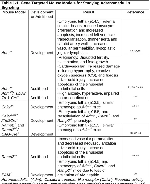
Table 1-1: Gene Targeted Mouse Models for Studying Adrenomedullin Signaling
Mouse Model Development or Adulthood
Result Reference
Adm-/- Development
-Embryonic lethal (e14.5), edema, smaller hearts, reduced myocyte proliferation and increased apoptosis, increased left ventricle trabecularization, thinner aorta and carotid artery walls, increased vascular permeability, hypoplastic
jugular lymph sac 22, 30-32
Adm+/- Adulthood
-Pregnancy: Disrupted fertility,
placentation, and fetal growth -Cardiovascular: Increased damage
including hypertrophy, reactive
oxygen species (ROS), and fibrosis -Liver cold injury: increased
apoptosis of the sinusoidal
endothelial cells 32, 68, 79, 88
Admfl/fl/Tubulin
Tα-1-Cre+
Adulthood
-High anxiety, hyperactive, impaired
motor coordination 114
Calcrl-/- Development
-Embryonic lethal (e13.5), similar
phenotype as Adm-/- mice 22, 33
Calcrl
LoxP/-/Tie2Cre+ Development
-Embryonic lethal (e16.5) and recapitulation of Adm-/-, Calcrl-/-, and
Ramp2-/- phenotype 22
Ramp2-/-and
Ramp2fl/fl/
CAG-Cre+ Development
-Embryonic lethal (e15.5), similar phenotype as Adm-/- mice
18, 22, 34
Ramp2+/- Adulthood
-Increased vascular permeability
and decreased neovascularization -Liver cold injury: increased
apoptosis of the sinusoidal
endothelial cells 18, 88
PAM-/- Development
-Embryonic lethal (e14.5) and phenocopy of Adm-/-, Calcrl-/-, and
Ramp2-/-mice due to loss of
amidation of AM peptide 35
18
Table 1-2: Vascular Assays for Studying Adrenomedullin Function
Assay Result Reference
Atherogenic Model
-Atherogenic diet and AM treatment in ApoE
-/-mice resulted in reduced formation of
atherosclerotic lesions 85
Tail
microlymphography
-AM injected mice showed reduced permeability of the dermal lymphatic
capillaries 25
Matrigel plug
-AM increased vascular regeneration -Ramp2+/- mice exhibited reduced
neovascularization 12, 13, 18
Aortic ring
-Ramp2+/-mice exhibited reduced
neovascularization in response to growth
factor stimulation 18
AngII/high-salt
-AM+/- mice exhibited increased reactive oxygen species (ROS), vascular fibrosis, and
intimal thickening 39
Prolonged mechanical ventilation
-AM treatment reduced lung vascular permeability resulting from ventilator use
21
Chronic cerebral hypoperfusion
-AM promoted arteriogenesis and
angiogenesis 96
Hind-limb ischemia
-AM promotes endothelial cell proliferation and
capillary formation -Adm+/- mice showed reduced blood flow and
capillary development 56
Wound healing (Pressure Ulcer
-Ischemia reperfusion model)
-AM reduced wound area and increased angiogenesis and lymphangiogenesis
97
Tail lymphedema
-AM improved lymphedema and increased
number of lymph and blood vessels 11 Tumor xenografts
-Blocking AM signaling results in reduced
19
Figures
Figure 1-1. Fold Change in Plasma Adrenomedullin Levels in a Variety of Human Conditions. Bars indicate average fold change in circulating AM levels in various disease categories or conditions based on published human clinical data. The dashed horizontal line at 2.33 represents the average fold increase in plasma AM levels across all conditions depicted. Number above each bar indicates the number of published observations
assessing plasma AM levels in each category. The clinical papers that were used for our analysis are listed according to the following broad categories: cancer,115-120
20
Figure 1-2. Adrenomedullin Signaling in Development and Vascular Biology.
(A) Loss of AM signaling causes embryonic lethality due to severe edema associated with impaired lymphatic vascular development. (B) In the adult, AM is an angiogenic,
21
Chapter II: Endothelial restoration of receptor activity-modifying protein 2 is sufficient
to rescue lethality, but survivors develop dilated cardiomyopathy2
Overview
Receptor activity-modifying proteins (RAMPs) serve as oligomeric modulators for numerous G protein-coupled receptors (GPCRs), yet elucidating the physiological relevance of these interactions remains complex. Receptor activity-modifying protein 2 (Ramp2) null mice are embryonic lethal, with cardiovascular developmental defects similar to those observed in mice null for canonical adrenomedullin/calcitonin receptor-like receptor (CLR) signaling. We aimed to genetically rescue the Ramp2-/- lethality in order to further delineate the spatiotemporal requirements for RAMP2 function during development and thereby enable the elucidation of an expanded repertoire of RAMP2 functions with Family B GPCRs in adult homeostasis. Endothelial-specific expression of Ramp2 under the VE-cadherin promoter resulted in the partial rescue of Ramp2-/- mice, demonstrating that endothelial expression of Ramp2 is necessary and sufficient for survival. The surviving Ramp2-/- Tg
animals lived to adulthood and developed spontaneous hypotension and dilated cardiomyopathy, which was not observed in adult mice lacking CLR. Yet, the hearts of
Ramp2-/- Tg animals displayed dysregulation of Family B GPCRs, including parathyroid
hormone and glucagon receptors as well as their downstream signaling pathways. These data suggest a functional requirement for RAMP2 in the modulation of additional GPCR
2
22
pathways in vivo, which is critical for sustained cardiovascular homeostasis. The cardiovascular importance of RAMP2 extends beyond the endothelium and canonical adrenomedullin/CLR signaling, in which future studies could elucidate novel and pharmacologically-tractable pathways for treating cardiovascular diseases.
Introduction
Receptor activity-modifying proteins (RAMPs) are single-pass transmembrane proteins that physically interact with numerous G protein-coupled receptors (GPCRs) to regulate receptor trafficking, ligand binding specificity and downstream G protein coupling and signaling. Biochemical and pharmacological studies have revealed functional RAMP interactions with many GPCRs including, calcitonin receptor-like receptor (CLR = protein,
Calcrl = gene), calcitonin receptor (CTR), parathyroid hormone receptor 1 and 2 (PTHR1 and PTHR2), glucagon receptor (GCGR), secretin receptor (SCTR), vasointestinal peptide receptor 1 and 2 (VIPR1 and VIPR2), calcium sensing receptor (CaSR), an estrogen receptor (GPR30), and likely additional RAMP-interacting GPCRs will be identified in the future.7, 191-193 Thus, in their capacity as endogenous oligomeric GPCR partners, RAMPs can exert powerful modulation on nearly every aspect of GPCR function and on a wide range of physiological processes. However, our understanding regarding the complexity and consequences of RAMP-GPCR interactions in vivo lags behind our current structural, biochemical and pharmacological knowledge and thus presents a limitation toward harnessing the potential pharmacological utility of this family of proteins.
23
cardiovascular defects in the heart, blood, and lymphatic vasculatures.18, 22, 34, 194, 196, 197 These phenotypes are essentially identical to those observed in mice lacking the genes that encode for CLR and its Ramp2-associated ligand, adrenomedullin (AM), indicating that RAMP2 is essential for canonical CLR/AM signaling during embryonic development. Yet, mice that are haploinsufficient for Ramp2 also exhibit an expanded constellation of endocrine-related phenotypes that are not observed in CLR and AM haploinsufficient mouse models.198 Therefore, these data imply that RAMP2 must exert functional modulation of other GPCRs, which is supported by its in vitro biochemical interaction with CTR, PTHR1/2, GCGR, and VIPR1/2.191 To date, ascribing a functional relevance for these other putative RAMP2-interacting GPCR pathways in adult physiological systems has been precluded by the embryonic lethality of Ramp2-/- mice.
Here, we have developed a novel Ramp2 transgenic mouse model which overexpresses Ramp2 specifically in the endothelium, with the hypothesis that restoration of RAMP2 in the endothelium will rescue the lethality-causing vascular defects associated with loss of CLR/AM signaling during embryogenesis. Indeed, endothelial RAMP2 is sufficient to rescue the embryonic lethality of many Ramp2-/- mice. Thus, the surviving mice, which express Ramp2 in the endothelium but lack Ramp2 in every other cell type, provided us with the opportunity to further elucidate the functional implications of the loss of Ramp2 on adult cardiovascular physiology and respective changes in other Ramp2-associated GPCRs.
Methods
Mouse Studies
Previously published SvEv-Ramp2+/- and Calcrlflox/flox mice were both fully backcrossed (>10 generations) onto a C57BL/6J genetic background for these studies.22, 34 A novel endothelial-specific Ramp2 transgenic mouse was generated and crossed to the
24
C57BL/6J genetic background. The Calcrlflox/flox mice were crossed with either the
SJL-Tg(Tagln-cre)1Her (SM22-Cre) or the inducible CAGG-CreERTM mouse to conditionally
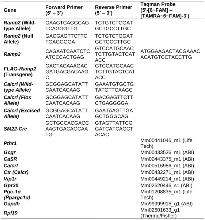
delete Calcrl specifically within developmental vascular smooth muscle cells (vSMC) or ubiquitously in adult mice, respectively. Adult CAGG-CreERTM females were administered with tamoxifen (TAM) as previously published.199 Genotyping and RT-PCR primers and probes are listed in Supplemental Table 2-S1. Females between 4-8 months of age were used in all Ramp2-associated studies, males 3-4 months of age for vSMC-specific Calcrl
studies, and females 16 month old for ubiquitous Calcrl studies. Mice were acclimated and conscious for both non-invasive tail cuff blood pressure and echocardiography analysis and anesthetized with isoflurane for intra-arterial blood pressure measurements. Organs weights and heart chamber dissections were normalized to either body weight or tibia length as previously described.200 Biological N of 3-10 mice per genotype was used for each experiment. Endothelial cells from adult mice were isolated with magnetic-associated cell sorting using CD31-specific antibodies as previously described.201 Cardiomyocytes from adult mice were isolated using collagenase digestion as previously described.202 All animal experiments were approved by the Institutional Animal Care and Use Committee of The University of North Carolina at Chapel Hill.
Statistical Analysis
Statistical analysis was determined with GraphPad 5.0 and data is represented as mean ± SEM. The unpaired student T-test was used to compare two groups, while one way ANOVA with Tukey’s Multiple Comparison test was used to compare ≥ 3 groups. Survival
data were compared by Mantel-Cox and Gehan-Breslow-Wilcoxon tests. Significant differences are represented as * p <0.05, ** p <0.01, ***p <0.001.
25
Results
Generation and Characterization of Endothelial-Specific Ramp2 Transgenic Animals
A diagram of the Cdh5-Ramp2 transgene (Tg) depicts the murine vascular cadherin 5 (Cdh5) promoter driving the expression of a flagged-tagged murine Ramp2 cDNA (Supplemental Figure 2-S1A), which was successfully integrated into the genome of two independent founder lines on the C57BL/6J genetic background. Expression of FLAG-Ramp2 protein was confirmed using both transgenic founder lines in a variety of adult tissues including the heart, kidney, lung and intestine (Supplemental Figure 2-S1B). Semi-quantitative RT-PCR revealed the presence of FLAG transcript within whole adult lung tissue and CD31+ endothelial-enriched cells in the Tg animals, but not wild-type endothelium (Supplemental Figure 2-S1C), which resulted in a modest increase in overall
Ramp2 gene expression levels within lung CD31+ endothelial cells of Tg animals compared
to wild-type animals (Supplemental Figure 2-S1D). Ramp2 expression was significantly higher in the left ventricles of Tg animals compared to wild-type—a finding which we
attribute to the endothelial-driven transgene expression, as compared to the low levels of endogenous Ramp2 expression in isolated cardiomyocytes (Supplemental Figure 2-S1E) and previously reported expression in vSMCs.92, 203 Tg(Cdh5-Ramp2) mice bred normally, with expected Mendelian and sex ratios at birth, and no obvious phenotypic defects.
Transgenic Endothelial Ramp2 Partially Rescues Embryonic Lethality of Ramp2-/- Mice
We sought to determine whether endothelial Ramp2 expression could rescue the previously reported Ramp2-/- embryonic lethality by interbreeding the hemizygous
Tg(Cdh5-Ramp2) animals with Ramp2+/- non-transgenic (ntg) animals, on an isogenic C57BL/6J
26
(Supplemental Figure 2-S2A). As expected, embryos lacking Ramp2 display non-hemorrhagic edema associated with arrested lymphangiogenesis22, yet the Ramp2-/- Tg
embryos showed significantly less edema than Ramp2-/- ntg littermates (Figure 2-1A-B). Unlike the small, hypoplastic jugular lymph sacs of Ramp2-/- ntg embryos, the jugular lymph sacs of the Ramp2-/- Tg were comparable in size to those of Ramp2+/+ littermates (Figure 2-1C-D), demonstrating that transgenic restoration of functional Ramp2 in the endothelium leads to improvement of the edematous phenotype.
In addition to defects in lymphatic development, loss of Ramp2 or AM/CLR signaling leads to small, disorganized hearts and thin vSMC walls.22, 30, 33 The aortic endothelium of viable embryos appeared intact with no signs of hemorrhagic leakage or endothelial dysfunction as previously reported in some Ramp2-/-embryos.18 However, the aortic vSMC layer was significantly thinner in Ramp2-/- ntg and Ramp2-/- Tg embryos (Figure 2-1E-F). Likewise, the hearts of e14.5 Ramp2-/- Tg animals were comparable in size to Ramp2-/- ntg
littermates, which were both significantly smaller than controls, despite detectable expression of Ramp2 in Ramp2-/- Tg hearts (Figure 2-1G-I). Consistent with previous reports, these data reflect that Ramp2 expression is required for normal vSMC and cardiac development and that endothelial Ramp2 restoration is not sufficient to rescue these defects, thus confirming non-endothelial roles of Ramp2 during cardiovascular development.
Unlike the Ramp2-/- ntg embryos, which uniformly die by e15.5, Ramp2-/- Tg embryos survive to term at near-Mendelian ratios, but a large number of these animals were found stillborn at postnatal day 1 (Supplemental Figure 2-S2B). Nevertheless, by postnatal day 7, 40% of the expected Mendelian ratio of Ramp2-/- Tg pups were viable, which represents a significant survival rescue when compared to the completely penetrant lethality of Ramp2 -/-ntg mice (Table 2-1). These surviving Ramp2-/- Tg mice were indistinguishable from
27
These data demonstrate that transgenic endothelial restoration of Ramp2 is able to blunt the endothelial and edematous phenotypes observed with global Ramp2 genetic deletion, leading to significantly improved survival.
Surviving Ramp2-/- Tg Adult Mice are Hypotensive
The surviving Ramp2-/- Tg mice, which express Ramp2 in the endothelium but lack
Ramp2 in all other cells, provided an opportunity to evaluate how Ramp2 loss-of-function during development affects adult cardiovascular homeostasis. Diastolic, systolic, and mean arterial blood pressures were significantly reduced in Ramp2-/- Tg compared to Ramp2+/+ ntg
and Tg female controls (Figure 2-2A). Importantly, there were no significant differences in basal blood pressure between Ramp2+/+ ntg and Ramp2+/+ Tg mice, indicating that the decreased blood pressure was not caused solely by the transgene. Moreover, the reduced basal blood pressure of Ramp2-/- Tg mice was observed without the exogenous administration of the canonical, hypotensive ligand AM, which was previously required to lower blood pressures in a vSMC-specific overexpression model of Ramp2.47, 204
Similar to the developmental defects in vSMC wall thickness of the Ramp2-/- Tg
embryos and other models lacking AM/CLR/Ramp2 function22, 30, 33, the vSMC walls were significantly thinner in Ramp2-/- Tg adult descending aortas compared to wild-type controls (Figure 2-2B). Intra-arterial blood pressure measurements further confirmed the hypotensive phenotype of adult Ramp2-/- Tg females (Figure 2-2C). To test vSMC responsiveness, Ramp2-/- Tg and controls were challenged with intravenous injections of the -adrenergic receptor agonist, phenylephrine, and the vasodilator, AM. The Ramp2-/- Tg
28
Ramp2-/- Tg Mice Develop Spontaneous Dilated Cardiomyopathy Phenotype
Considering the hypotension and the developmental defects observed in Ramp2-/- Tg
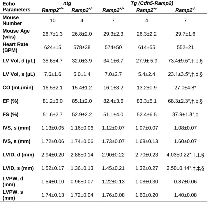
embryonic hearts, we next assessed how cardiac function and morphology were altered in adult Ramp2-/- Tg mice. Echocardiography on conscious mice revealed significantly dilated left ventricles during both diastole and systole, with significantly larger left ventricle volumes and left ventricle internal diameter dimensions in the Ramp2-/- Tg mice compared to
Ramp2+/+ and Ramp2+/-, with and without the transgene (Table 2-2). Representative M-mode echocardiograms from Ramp2+/+ ntg, Ramp2+/+ Tg, and Ramp2-/- Tg illustrate the ventricular dilation in Ramp2-/- Tg mice (Figure 2-3A), which resulted in a trending increase in calculated cardiac output in these animals (Table 2-2). Septum and left ventricle posterior wall dimensions of Ramp2-/- Tg mice were unchanged from controls, although there was a non-significant trend towards a thinner posterior wall. There was also a modest, but significant reduction in both ejection fraction and fractional shortening in the Ramp2-/- Tg
dilated hearts. These data indicate that loss of Ramp2 in non-endothelial cells leads to a spontaneous dilated cardiomyopathy (DCM)–like phenotype in adult mice, which at 6
months of age had not yet progressed to heart failure, as indicated by the elevated cardiac output and sufficient heart function.
Upon dissection, we observed that the hearts of Ramp2-/- Tg mice were grossly enlarged compared to wild-type and Ramp2-/- ntg mice (Figure 2-3B-C). The adult Tg mice had no differences in body weight (Ramp2+/+ ntg: 28.4 ± 1.4 g, Ramp2+/+ Tg: 28.4 ± 1.2 g,
29
ntg: 0.15 ± 0.01 mg/g, Ramp2+/+ Tg: 0.15 ± 0.01 mg/g, Ramp2-/- Tg: 0.21 ± 0.02* mg/g; *p<0.05) all exhibited significant enlargement in the Ramp2-/- Tg animals compared to all other genotypes. Similar significant trends were observed when the individual chamber weights were normalized to tibia length (LV:TL; Ramp2+/+ ntg: 5.4 ± 0.3 mg/mm, Ramp2+/+ Tg: 4.8 ± 0.2 mg/mm, Ramp2-/- Tg: 6.5 ± 0.2** mg/mm; **p<0.01).
Cross-sectional area of myocytes within the left ventricle revealed slight, but significant cardiomyocyte hypertrophy, with no changes in left ventricular capillary density, in
the Ramp2-/- Tg mice compared to controls (Figure 2-4A-D). Picrosirius red staining showed
no differences in perivascular fibrosis, but there was significantly increased interstitial fibrosis in the Ramp2-/- Tg hearts compared to hearts of Ramp2+/+ and Ramp2+/+ Tg mice (Figure 2-4E-H). There were elevated levels of the oxidative stress indicator, lipid peroxidase evidenced by 4-hydroxynonenal (4-HNE) staining in Ramp2-/- Tg hearts (Figure 2-4I-J).205 Together, the modest increases in hypertrophy, fibrosis, and oxidative stress, as well as modest decline in heart function, further support that 6 month old Ramp2-/- Tg mice exhibit a compensated, DCM-like phenotype.
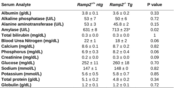
Adult Ramp2-/- Tg Mice Develop Multi-Organ Inflammation
30
hepatocellular or renal damage (Supplemental Table 2-S2). This supports that the end-organ inflammation in Ramp2-/- Tg mice is likely downstream of altered hemodynamics due to DCM and hypotension, rather than primarily due to loss-of-function of Ramp2 in end-organs.
Conditional Calcrl Deletion Does Not Lead to a Dilated Cardiomyopathy Phenotype
Since CLR and AM represent the most well-characterized, canonical pathway for RAMP2 modulation and the knockout mice for these genes recapitulate the Ramp2
-/-developmental phenotypes, it is reasonable to consider that disruption of this pathway, which has been demonstrated to be cardioprotective in both animal studies and in humans32,
77, 81, 209, may underlie the DCM and hypotensive phenotypes in Ramp2-/- Tg mice. To test
the functions of CLR in non-endothelial cells, we generated cardiac- and vSMC-specific
Calcrl null mice using the SM22-Cre mediated excision. The CalcrlloxP/loxP; SM22Cre+ mice
were born at expected Mendelian ratios and survived to adulthood (Supplemental Figure 2-S3A). Relative Calcrl expression was significantly reduced in aortic vSMCs and hearts of
CalcrlloxP/loxP; SM22-Cre+ compared to CalcrlloxP/+; SM22-Cre+ littermates (Supplemental Figure 2-S3B). Importantly, CalcrlloxP/loxP; SM22-Cre+ adult males were normotensive and had no basal changes in heart function, size, or morphology (Supplemental Table 2-S3 or Figure 2-S3C). Similarly, cardiac-specific Calcrl deletion using the αMHC-Cre+ transgenic line also failed to recapitulate the DCM phenotype, with animals surviving to adulthood with no basal cardiac dysfunction (Dackor R & Caron K.M., unpublished data, [2016]). These data demonstrate that cardiac- and vSMC-specific Calcrl expression is not required for embryonic development or adult cardiovascular maintenance.
We have previously shown that temporal, global deletion of Calcrl in adult
31
cardiac phenotypes and they had similar heart to body weight ratio as TAM-injected control mice (CalcrlloxP/loxP; CAGG-CreERTM: 4.43 ± 0.19 mg/gversus Calcrlflox/flox: 4.13 ± 0.14 mg/g, respectively). Furthermore, conscious echocardiography of CalcrlloxP/loxP; CAGG-CreERTM
female mice, even at 14 months of age, failed to reveal any significant changes in left ventricle internal diameter, function, or heart size compared to control mice (Supplemental Table 2-S4). Collectively, these data, generated from three independent series of conditional deletion approaches, indicate that the global-, cardiac- or vSMC-specific loss of
Calcrl does not recapitulate the DCM phenotype observed in the Ramp2-/- Tg animals. Therefore, this strongly suggests that the Ramp2-/- Tg phenotype is imparted by other RAMP2-associated GPCR pathways.
Ramp2-/- Tg hearts exhibit reduced signaling pathways and expression of numerous
RAMP-associated GPCRs
The genetic reduction or down-regulation of the transcriptional regulator CREB207, 210 and the crucial PPAR pathway transcription factor, Pgc-1α211-213, have both been shown to be involved in DCM pathogenesis. Thus as expected, the relative expression of Pgc-1α was significantly down-regulated in left ventricles and in an enriched cardiomyocytes fraction of
Ramp2-/- Tg hearts compared to controls (Figure 2-6A-B). There was also a significant reduction in phosphorylated CREB compared to total CREB and Gapdh in the Ramp2-/- Tg
32
encoding for other RAMP-associated GPCRs, like Calcr, Vipr1, and Gpr30, were unchanged (Figure 2-6D).
Similar gene expression changes in these Family B GPCR expression profiles were confirmed in isolated cardiomyocyte fractions (Figure 2-6E), further supporting the myocyte-specific genetic dysregulation. Calcrl expression was significantly increased in left ventricle, but not in the cardiomyocyte-enriched fraction, demonstrating that Calcrl upregulation is from a non-cardiomyocyte cell type. The expression of both Pthr1 and Gcgr were significantly decreased during development in the hearts of Ramp2-/- ntg and Ramp2-/- Tg
embryos (Figure 2-6F), supporting that these changes are specific to Ramp2 loss rather than secondary to the Cdh5-driven Ramp2 Tg or the DCM phenotype, as might be the case for CaSR. Serum analysis revealed no significant dysregulation of circulating calcium or glucose, thereby eliminating uncompensated Pthr1 or Gcgr systemic signaling as a cause for the cardiovascular phenotypes (Supplemental Table 2-S2). Collectively, these data demonstrate that genetic loss of Ramp2 in non-endothelial cells of the heart leads to down-regulation of RAMP2-associated GPCRs, Gcgr and Pthr1, as an underlying mechanistic basis for the decreased pCREB and Pgc-1α responsible for the pathogenesis of the DCM-like phenotype in Ramp2-/- Tg mice (Figure 2-6G).
Discussion
In this study, we generated an endothelial-specific Ramp2 Tg mouse model to attempt to rescue the embryonic lethality due to global loss of Ramp2. While no Ramp2 null mice survive to birth, we observed a significant number of Ramp2-/- Tg born and able to survive into adulthood. This result further confirms that endothelial Ramp2 is essential for embryonic survival and represents, to our knowledge, the first genetic rescue of the global
33
It remains unclear why a significant number of Ramp2-/- Tg pups survive to late-gestation, but are stillborn. Interestingly, it was recently shown that mice with endothelial excision of Ramp2 during development using a Cdh5-Cre died during late-gestation.206 Additionally, they report that approximately 5% of the endothelial Ramp2 knockouts live into adulthood and develop large hearts, hypotension, and multi-organ vasculitis. In this current study we found that 40% of Ramp2-/- Tg survived to adulthood, and also developed similar cardiovascular and inflammatory phenotypes. These two mouse models, as well as a previously published endothelial-specific deletion of Calcrl, demonstrate that adequate levels and timing of endothelial Ramp2/CLR/AM signaling are critical for embryonic survival.22
In addition, these studies identify potential sex-dependent mechanisms of Ramp2, or Cdh5, regulation and function, as evidenced by the substantially reduced numbers of male
Ramp2-/- Tg survivors. We have previously shown that adult Ramp2+/- females have endocrine phenotypes not present in Ramp2+/- males.198 In addition, Ramp3-/- males, but not females, displayed exacerbated cardiovascular phenotypes when challenged with hypertension.193, 196 Thus, while RAMP2 and RAMP3 interact with both similar and different GPCRs, our observations of sex-dependent phenotypes in these genetic animals will provide an area for interesting future investigations.
34
development and function, which likely contributes to the pathogenesis of hypotension and DCM in Ramp2-/- Tg survivors. Yet, the hypotension and DCM phenotypes occur despite maintenance of normal cardiac output. It is further possible that the developmentally-induced thin vSMC walls contribute to reduced vascular tone; however we demonstrated their ability to effectively respond to acute phenylephrine vasoconstriction. Therefore, additional studies that explore the entire repertoire of RAMP-mediated GPCRs will be required for full elucidation of the mechanistic basis for the hypotension in Ramp2-/- Tg
survivors.
Loss of Ramp2 in multiple non-endothelial cardiac cells, along with altered humoral signaling could lead to cardiac dysfunction and DCM pathogenesis. The genetic dysregulation in isolated cardiomyocytes suggests roles of Ramp2/GPCRs specifically in cardiomyocytes. Interestingly, a recently published study demonstrated that a cardiomyocyte-specific Ramp2 deletion led to a DCM-like phenotype due to mitochondrial dysfunction and irregular calcium handling, which the authors attribute to loss of CLR/AM signaling.222 Similarly, this study suggests that cardiomyocyte loss of Ramp2 in the Ramp2 -/-Tg mice is likely responsible for the DCM. Moreover, our data demonstrates that neither cardiomyocyte- or vSMC-specific loss of Calcrl during development or conditional Calcrl
deletion in adults recapitulate the DCM phenotype observed in the Ramp2-/- Tg and the aforementioned Ramp2flox/flox; MHC-MerCreMer mice.222 Therefore, collectively, these studies imply the involvement of other RAMP-associated GPCRs.
It is apparent that RAMPs interact with numerous GPCRs biochemically, although in vivo physiologic evidence of these interactions is limited. We found that lack of Ramp2 in non-endothelial cells leads to decreased expression of cardiac Pthr1 and Gcgr.
35
GCGR ligand selectivity between glucagon and glucagon-like peptide 1, which have opposing physiologic effects on glucose homeostasis and cardiovascular function.223 While GCGR has not been directly connected to DCM pathogenesis, glucagon can alter calcium signaling in myocytes and glucagon-like peptide 1 improves glucose uptake and survival of canines with DCM.214, 224 Likewise PTHR1 signaling is important in vitamin D and calcium homeostasis, which have both been associated with DCM.225, 226 Together, Ramp2 loss-of-function can not only alter both Pthr1 and Gcgr expression simultaneously, but may also alter their ability to reach plasma membrane, bind ligands, and signal. Additionally, we observed decreased signaling through CREB and downregulation of Pgc-1α, which both have been shown to lead to DCM.207, 210, 211 Both Pthr1 and Gcgr signal through CREB and murine deletion of Gcgr led to decreased phosphorylated CREB and Pgc-1a expression levels.215, 217 Thus, loss of Ramp2 likely has numerous mechanisms for the development and maintenance of cardiac functions both dependent and independent of canonical AM/CLR/Ramp2 signaling.
Perspectives
The in vivo interplay between GPCR/RAMP/Ligand is highly complex and is only