LGN-DEPENDENT MICROTUBULE REGULATION INFLUENCES ENDOTHELIAL CELL MIGRATION, ADHESION, AND SPROUT INTEGRITY
Catherine Elizabeth Wright
A dissertation submitted to the faculty at the University of North Carolina at Chapel Hill in partial fulfillment of the requirements for the degree of Doctor of Philosophy in the Department
of Biology.
Chapel Hill 2014
Approved by: Victoria Bautch
Jean Cook Andrew Dudley Bob Goldstein
ii © 2014
iii ABSTRACT
Catherine Elizabeth Wright: LGN-Dependent Microtubule Regulation Influences Endothelial Cell Migration, Adhesion, and Sprout Integrity
(Under the direction of Dr. Victoria Bautch)
Blood vessels form during organismal development and maintain integrity to provide
oxygen and nutrients to the tissues. Vessels are comprised of endothelial cells that coordinate their individual behaviors to generate functional sprouts. Endothelial cells undergo directional
migration, oriented divisions, and lumen formation through organization of the microtubule network. Microtubules are actively growing and shrinking polymers that direct the shape and movement of cells. Disruption of the microtubule network is detrimental for the cell. Here I
investigated the role of the mitotic polarity protein LGN in endothelial cells and sprouting angiogenesis. To study LGN in the vasculature, I utilized a three-dimensional model for
sprouting angiogenesis. Surprisingly, loss of LGN did not affect oriented division of endothelial cells within a sprout, but perturbed overall sprouting and branching. I utilized two-dimensional
assays to investigate the cause behind three-dimensional sprout defects in LGN KD endothelial cells. At the cellular level, LGN KD resulted in reduced endothelial cell migration and
dysregulated cell-cell adhesions. Endothelial cells with LGN knockdown displayed stabilized
microtubules at the growing plus-end. The data fits a model in which LGN promotes turnover of microtubules in endothelial cells, which in turn regulates migration, cell-cell adhesion, and
iv
v
ACKNOWLEDGMENTS
I want to thank the members of the Bautch lab past and present for being such a wonderful
group to work with. I thank Little John for being there at each stage of the grad school experience with me. I thank Jess and Diana for their amazing friendship. And I thank everyone else for being
so supportive of my ideas and research these past several years.
I want to thank my committee, who has been so supportive and helpful in shaping my project. Each member has expertise that influenced my project and I am grateful for that. I’d also
like to thank my advisor, Dr. Vicki Bautch. She taught me to think critically about everything and to not be afraid to challenge dogma. The amount of confidence I have now compared to when I
first started is astronomical and I credit that to Vicki’s advising.
My friends and family have been through everything with me. They are always willing to lend an ear if I need advice, whether scientific or personal. I especially cherish my non-science
vi
TABLE OF CONTENTS
LIST OF FIGURES ... viii
LIST OF TABLES ... ix
LIST OF ABBREVIATIONS ... x
CHAPTER I – General Introduction... 1
A. Mechanisms of Vascular Development ... 1
B. Cell Polarity and Division in Development ... 2
C. Regulation of Cell Division by LGN ... 3
I. LGN acts with MTs during Cell Division ... 4
D. LGN Structure and Function ... 5
E. Microtubules Regulate Cell Behavior... 6
I. Microtubule Morphology and Formation ... 6
II. Microtubule Organization in Cell Division ... 7
III. Microtubules Direct Cell Migration ... 7
F. Vascular Adhesions... 8
G. Summary ... 9
REFERENCES ... 12
CHAPTER II – MATERIALS AND METHODS ... 18
REFERENCES ... 23
CHAPTER III – RESULTS ... 24
vii
B. LGN SUPPORTS ANGIOGENIC SPROUT FORMATION ... 26
C. LGN IS REQUIRED FOR CELL MIGRATION AND DIRECTIONAL CHANGE ... 27
D. LGN KD HUVEC HAVE ENHANCED MT NUCLEATION ... 28
E. FOCAL ADHESIONS ARE ENHANCED IN LGN KD HUVEC ... 30
F. VE-CADHERIN LOCALIZATION IN LGN KD HUVEC SUGGESTS JUNCTIONAL INSTABILITY ... 31
G. VE-CADHERIN TRAFFICKING IS ENHANCED IN LGN KD HUVEC ... 32
H. OVERALL MEMBRANE ADHESION IMMUNOSTAINING IS DISRUPTED ... 33
I. DISCUSSION ... 33
REFERENCES ... 47
CHAPTER IV – DISCUSSION... 52
A. LGN IS REQUIRED FOR ENDOTHELIAL SPROUT MORPHOLOGY ... 52
B. LGN REGULATES MICROTUBULE TURNOVER ... 53
C. MODELING A MECHANISM FOR LGN ACTIVITY THROUGH A PREDICTED BINDING PARTNER, ZO-1 ... 55
D. AN ALTERNATE MODEL FOR AN LGN MECHANISM THROUGH A KNOWN BINDING PARTNER, 14-3-3 ... 56
E. EVIDENCE THAT LGN FUNCTIONS THROUGHOUT THE CELL CYCLE ... 56
F. ROLE OF LGN IN MAMMALIAN EPITHELIUM AND ENDOTHELIUM ... 58
G. SUMMARY ... 59
viii
LIST OF FIGURES
Figure 1.1 – Types of Cell Divisions during Development and the Vasculature ... 10
Figure 1.2 – Selective localization of LGN at the membrane directs spindle orientation ... 11
Figure 2.1 – Loss of LGN leads to loss of sprout integrity in sprouting angiogenesis ... 36
Figure 2.2 – Additional shRNA targeted to LGN show defects in angiogenic sprouts ... 37
Figure 2.3 – LGN KD HUVEC display reduced migratory capacity and directional change. ... 39
Figure 2.4 – Additional shRNA against LGN lead to reduced migration in HUVEC... 40
Figure 2.5 – Microtubule Dynamics are Disrupted in LGN KD HUVEC ... 41
Figure 2.6 – LGN KD HUVEC maintain some regulation of MT dynamics ... 43
Figure 2.7 – Focal Adhesions, Adherens Junctions and Membrane Markers are disrupted in LGN KD HUVEC ... 44
Figure 2.8 – VE-Cadherin localization is misregulated at Cell-Cell Borders in LGN KD HUVEC ... 46
Figure 3.1 – Proposed pathways for LGN in Endothelial cells ... 60
ix
LIST OF TABLES
x
LIST OF ABBREVIATIONS
AJs – Adherins junctions
aPKC – atypical protein kinase C
BSA – bovine serum albumin
DAPI – 4’,6-diamindino-2-phenylindole
Dlg – Discs Large
DMEM – Dulbecco’s modified eagle medium
EB1 – End binding protein 1
EBM-2/EGM-2 – Endothelial Basal/Growth Medium-2
EC – Endothelial cell
FA – Focal Adhesion
FBS – fetal bovine serum
GFP – Green Fluorescent Protein
Gαi – Guanine nucleotide binding-protein alpha subunit i
HEK293T – Human embryonic kidney 293 cells containing SV40 large T antigen
HUVEC – Human umbilical vein endothelial cells
Insc – Inscuteable
LGN – Leucine Glycine Asparagine
xi MT – Microtubule
MTOC – Microtubule Organizing Center
NHLF – Normal Human Lung Fibroblasts
NuMA – Nuclear mitotic apparatus protein
Par3 – partitioning defective 3
Par6 – partitioning defective 6
PBS – phosphate buffered saline
PECAM – platelet endothelial cell adhesion molecule
PFA – paraformaldehyde
PH3 – phospho-histone H3
s.e.m – standard error about the mean
tdTomato – tandem dimer tomato
TPR – tetratricopeptide repeats
VE-Cadherin – Vascular Endothelial Cadherin
VEGF – Vascular Endothelial Growth Factor
ZO-1 – Zonula Occludens protein 1
1
CHAPTER I – GENERAL INTRODUCTION
A. Mechanisms of Vascular Development
The vascular system is essential for providing oxygen and nutrients to the body1–3. The
vascular network is organized into a hierarchy of veins, arteries, and capillary vessels. These
vessels are formed when endothelial cells undergo cell migration, changes in adhesion, and
proliferation2,4,5. Endothelial cells migrate and coalesce during initial vessel formation to
generate a primitive network4,6,7. To expand the primitive network, endothelial cells generate
branches by sprouting from the parent vessel8. Endothelial adhesions are formed and maintained
through a cycle of externalization and recycling of adhesive proteins to promote vessel
integrity2,9. Vessel elongation occurs through migration and oriented cell divisions10. The
coordination of migration, adhesion, and cell division promotes healthy vasculature that supports
the organism. Understanding these processes and how their disruption contributes to disease
states is crucial11.
Growth factor signaling, particularity vascular endothelial growth factor (VEGF),
promotes overall vessel network formation and integrity2,6,12. Mice lacking VEGF ligand or
receptors die during embryonic development, due to failure of vessel patterning and
hemorrhaging13. Excessive VEGF activity disrupts downstream signaling pathways, inhibits
vascular sprouting, and leads to randomized division orientation10,14. While many
endothelial-specific pathways have been studied in relation to VEGF, it is unknown what is downstream of
VEGF in regulation of division orientation. Common pathways in division orientation have been
2
reported in endothelial cells15–17. Unpublished data suggests that endothelial cells can orient
spindles when some cell polarity proteins are genetically reduced, but further work needs to be
done to fully understand the exact mechanism (C. Lee and V. Bautch, unpublished).
B. Cell Polarity and Division in Development
Polarity influences cell behavior throughout development, including cell and tissue
migration, spatial identity, and oriented division18,19. Cell polarity is generated and maintained by
the asymmetric localization and activation of protein complexes20–22. One polarity complex is
Par3/Par6/aPKC, which facilitates the selective positioning of fate-determining and spindle
orientating factors, among other functions23,24. Par3/Par6/aPKC polarity is maintained through
mitosis to ensure proper spindle placement and establish daughter cell polarity25–27.
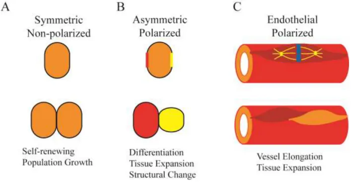
When a cell undergoes division, the cytoplasm is segregated just as the genetic material16.
The cytoplasm can be divided symmetrically or asymmetrically, which can dictate the identity,
function, and position of the daughter cells (Figure 1.1A,B)20,28. Asymmetric division is used to
structure tissues, differentiate cell types, and maintain stem cell progenitor populations. During
division, the microtubules organize into a spindle, which can be aligned specifically to ensure
daughter cells receive the proper material15. Spindle orientation can be established in response to
asymmetrically polarized factors at the cell membrane. Spindles in endothelial cells orient along
the long axis of the vessel, but the mechanism dictating this orientation is not known (Figure
1.1C)10. Endothelial cell shape or vessel polarity via flow could be contributing factors in spindle
alignment.
Endothelial cells are polarized on two axes: 1) proximal-distal as established by flow and
3
side faces the lumen and the basal side faces the external environment29. Proximal-distal polarity
is important in the migration of ECs and the generation of new vessels while apical-basal
polarity establishes lumens in new sprouts29–31. The Par3/Par6/aPKC polarity complex
establishes and supports lumen formation in the vasculature19,31. Loss of aPKC in the vasculature
leads to a delay in lumenization of the vessel, but no observed change in oriented division31.
Downstream of aPKC and cell polarity is LGN, a protein necessary in many cell types for
oriented cell division, which has not been characterized in endothelial cells32–34.
C. Regulation of Cell Division by LGN
LGN was first identified as a binding partner of Inscuteable (Insc) in Drosophila
melanogaster32. LGN complexes with Insc and Par3/Par6/aPKC to establish asymmetry in
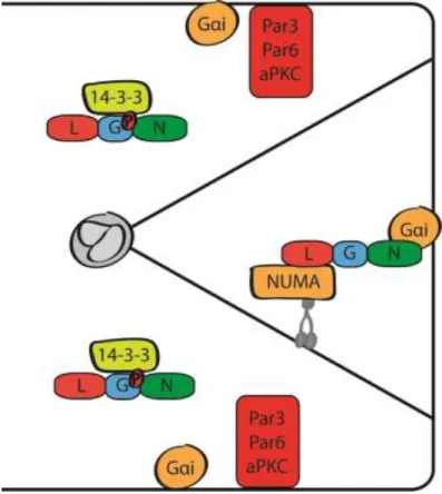
Drosophila melanogaster neuroblast cell division33,35,36. LGN acts in complex with G-alpha-i
and NUMA to direct and anchor the astral MTs (MTs extending from the spindle to the cell
membrane) (Figure 1.2). In mammalian epithelial cyst culture, Par3/Par6/aPKC excludes LGN
from acting on astral MTs26. LGN is phosphorylated by aPKC, preventing it from binding
G-alpha-i and NUMA. By limiting where the LGN complex forms, the astral MTs only anchor at
specified regions of the membrane26,36. In mammalian epithelium, the LGN complex is
distributed on both sides of the spindle poles, while in Drosophila melanogaster neuroblasts, the
complex is restricted to one daughter cell26,37,38. When LGN activity is disrupted (either through
depletion, truncation, or mislocalization), the spindles fail to properly orient26,37–39. Roles for
LGN are established in Drosophila melanogaster and mammalian epithelium, but its role in
4 I. LGN acts with MTs during Cell Division
At the start of mitosis, the nuclear envelope breaks down, releasing NUMA into the
cytoplasm40,41. NUMA traffics to the spindle poles in complex with dynein and dynactin42. From
there, NUMA is brought out to the membrane where it binds LGN and creates a bridge between
the membrane and the astral microtubules43. The bridge consists of NUMA bound to LGN,
which binds to G-alpha-i (in the GDP-bound state) at the membrane. Once the bridge has
formed, the minus-end directed motor dynein moves toward the spindle pole while in complex
with NUMA/LGN/G-alpha-i, generating the forces to pull the spindle poles apart37,42,44.
At the spindle poles, LGN is phosphorylated by Aurora A kinase, which recruits Discs
Large (Dlg)45. Discs large is normally present at the membrane during interphase and acts to
promote cellular junctions36,46. Dlg function changes during mitosis to help direct spindle
orientation by complexing with LGN and plus-end MT motor protein, kinesin-heavy-chain 73
(khc73)17,46,47. Kinesins align and shorten MTs during mitosis, generating additional force for
separating the spindle poles48.
These examples establish that LGN acts with the MT network to influence cell behavior
during mitosis. The current body of work on LGN has focused primarily on cell division, with
limited evidence showing that LGN was non-functional during interphase. However, some recent
studies challenge that model, and early studies that excluded LGN from interphase function no
longer fits with the current model for LGN. LGN is required for neutrophil polarization and
chemotaxis in mammalian immune response. Neutrophils rely on the actin cytoskeleton to
generate pseudopods in the direction of migration, facilitated by LGNl/G-alpha-i signaling77.
5
likely require LGN. The next section will discuss the early and current studies in context with
LGN protein structure.
D. LGN Structure and Function
LGN is a 74kDa protein consisting of 3 distinct structural domains: the TPR motif, the
Linker, and the GoLoco repeats. LGN conformation can be closed or open based on the
proximity of the TPR and the GoLoco repeats and dependent on the presence of binding
partners43,49,50. The Linker region does not contain any secondary protein folding, acting as a
hinge for LGN’s conformational changes. Each domain has documented interactions to facilitate
LGN function.
The tetratricopeptide repeats (TPR) are a series of alpha-helices that arrange in parallel to
form an amphipathic channel50,51. The channel creates a binding pocket that allows for
protein-protein interaction based on amino-acid charges. Charge-based interactions are flexible, and
allow for LGN to interact with multiple proteins via the TPR51. Due to the flexibility of the TPR
binding channel, it is unlikely that all possible binding partners of LGN are known. Several of
the known binding partners of the TPR, including NuMA and Insc, are involved in orienting the
mitotic spindle34,52. Expression of LGN-truncation mutants lacking the TPRs mimic
loss-of-function phenotypes in MDCK cysts26,37.
The GoLoco repeats are a series of highly specific binding pockets for G-alpha, which is
tethered to the membrane53. G-alpha is a member of the heterotrimeric G-protein signaling
cascade, but its LGN interactions are G-protein signaling-independent54. There are multiple
G-alpha isoforms, but G-G-alpha-i has the highest affinity for binding to LGN54 and can only bind to
6
of binding a single G-alpha55. LGN-mediated division orientation is abrogated when the GoLoco
domains cannot bind G-alpha-i37,56.
The TPR domain and GoLoco repeats are extensively characterized. Less attention has
been given to the flexible Linker region between the TPR and GoLoco domains. Previous studies
truncated LGN within the Linker region to study the function of the TPR or the GoLocos.
However, recent findings support the Linker having target sites for phosphorylation and
downstream function26,45. It is not known if LGN conformation influences kinase activity on the
Linker region. Linker-dependent function suggests that LGN truncations must be carefully
designed to eliminate any bias. There are limited studies of the Linker’s contribution to LGN
function, and it will be interesting to monitor the research published in the field in the future.
E. Microtubules Regulate Cell Behavior
I. Microtubule Morphology and Formation
Microtubules (MTs) are essential in cell migration, vesicle and protein trafficking, and
cell division. MTs are hollow, tube-shaped polymers of alpha and beta tubulin heterodimers.
MTs alternate between states of growth and catastrophe, a behavior termed dynamic instability57.
MT growth is characterized by the addition of heterodimers to the plus-end, located distally from
the centrosome. The centrosome is also referred to as the microtubule-organizing center
(MTOC). MT catastrophe occurs at the plus end, and involves sudden MT depolymerization,
7 II. Microtubule Organization in Cell Division
Microtubules are essential during cell division, as they establish the bipolar spindle and
anchor the spindles to the cell membrane to generate pulling forces that separate the
chromosomes. One of the two centrosomes migrates away from the other during prophase (early
mitosis), which positions it on the opposite side of the nucleus. This is done using overlapping
microtubules linked by motor proteins that push against each centriole until the pushing forces
equalize. The microtubules connecting the spindle poles to the chromosomes are the spindle
microtubules while the MTs that grow from the spindle toward the cortex are the astral
microtubules. Anchoring of the MTs to the cortex leads generates the forces necessary to
separate the spindle poles. Once the astral MTs are anchored, they begin to shorten, pulling the
spindle poles toward the cortex. Failure to shorten astral MTs leads to spindle instability,
however it was not determined what effect this had on completing division58. Once the
chromosomes separate fully, MTs concentrate between the separated chromosomes and generate
a contractile ring to initiate cytokinesis.
III. Microtubules Direct Cell Migration
Tthe MTOC position during migration follows the direction of movement. This allows
the majority of nucleating MTs to grow towards the direction of migration59. Additionally,
polarization of the MTOC provides cues to other organelles, leading to their re-positioning57,60,61.
The Golgi orients in response to MTOC polarity to traffic vesicles to the leading edge62. Cell
migration is promoted through directed trafficking of proteins (including Cdc42, WASP, and
Arp2/3) in Golgi-derived vesicles along microtubules62,63. These factors act to promote MT
8 F. Vascular Adhesions
Focal adhesions (FAs) are cytoskeletal complexes containing adhesion proteins (vinculin,
paxillin), integrins, and actin65. FAs are present at the leading edge of cells and at the ends of
actin stress fibers66. The formation, maturation, and degradation of FAs are necessary for cell
signaling, migration, and cell cycle progression65,67. FAs are capable of integrin cross-talk which
coordinates the actin cytoskeleton with the MT network68. During cell migration, cues from the
extracellular matrix trigger the integrin signaling network57,69. This leads to the production and
turnover of FAs at the leading edge67. These FAs provide a physical anchor for the cell to pull
the cell body during movement. Stabilization of FAs, through reduced MT dynamics, lead to
reduced cell migration and defects in cell division, as proper anchoring of the cell to its
environment is critical for establishing the pulling forces for cytokinesis64,70. FA size correlates
with stability, with larger FAs being more mature and stable71.
Adherens junctions (AJs) are cell-cell junction complexes that protect against barrier
disruption and leakiness in vessels72. In blood vessels, flow generates tension on the surface of
the ECs, which activates the formation and remodeling of AJs8,73. AJs are formed when adhesion
molecules, such as Vascular Endothelial Cadherin (VE-Cadherin, EC specific) are deposited at
the membrane5. VE-Cadherin acts by dimerizing its extracellular domain with the extracellular
domain of VE-Cadherin on adjacent cells9,74. The extracellular domains mediate homophilic
interactions between cells while the cytoplasmic domains generate scaffolding with catenins and
the actin cytoskeleton. Constant recycling of VE-cadherin at the membrane promotes strong
adhesions75. Endothelial cell adhesions have stereotypical VE-cadherin localization patterns, and
9 G. Summary
Endothelial cell behavior is dependent upon coordinated molecular and cytoskeletal
activity. Molecular components of cell polarity work in conjunction with the cytoskeleton to
regulate division, migration, and adhesion. LGN, a known regulator of cell division, interacts
with the microtubule network to orient cell division. There are multiple known binding partners
that are involved in cell division orientation, but these binding partners and others have
additional functions. This introduces the potential that LGN has a more diverse function that is
facilitated by the variety of interactions. This thesis proposes that LGN influences MT behavior
10
Figure 1.1 – Types of Cell Divisions during Development and the Vasculature
A. Cell divisions that are symmetrical have equal distribution of their contents. Symmetrical
divisions are utilized to maintain progenitor cell populations.
B. Asymmetric divisions have unequal distribution of cellular contents. Daughter cells can
assume different identities, sizes, or positions.
C. Endothelial cell divisions polarize along the proximal/distal sprout axis. Daughter cells
11
Figure 1.2 – Selective localization of LGN at the membrane directs spindle orientation
12 REFERENCES
1. Eichmann, A. & Simons, M. VEGF signaling inside vascular endothelial cells and
beyond. Curr. Opin. Cell Biol.24, 188–93 (2012).
2. Carmeliet, P. & Collen, D. Molecular basis of angiogenesis. Role of VEGF and
VE-cadherin. Ann. N. Y. Acad. Sci.902, 249–62; discussion 262–4 (2000).
3. Coultas, L., Chawengsaksophak, K. & Rossant, J. Endothelial cells and VEGF in vascular
development. Nature438, 937–45 (2005).
4. Lamalice, L., Le Boeuf, F. & Huot, J. Endothelial cell migration during angiogenesis.
Circ. Res.100, 782–94 (2007).
5. Dejana, E., Orsenigo, F. & Lampugnani, M. G. The role of adherens junctions and
VE-cadherin in the control of vascular permeability. J. Cell Sci.121, 2115–22 (2008).
6. Patel-Hett, S. & D’Amore, P. A. Signal transduction in vasculogenesis and developmental
angiogenesis. Int. J. Dev. Biol.55, 353–63 (2011).
7. Coffin, J. D. & Poole, T. J. Endothelial cell origin and migration in embryonic heart and
cranial blood vessel development. Anat. Rec.231, 383–95 (1991).
8. Semenza, G. L. Vasculogenesis, angiogenesis, and arteriogenesis: mechanisms of blood
vessel formation and remodeling. J. Cell. Biochem.102, 840–7 (2007).
9. Esser, S., Lampugnani, M., Corada, M., Dejana, E. & Risau, W. Vascular endothelial
growth factor induces VE-cadherin tyrosine phosphorylation in endothelial cells. J. Cell
Sci.111, 1853–1865 (1998).
10. Zeng, G. et al. Orientation of endothelial cell division is regulated by VEGF signaling
during blood vessel formation. Blood109, 1345–52 (2007).
11. Adams, R. H. & Alitalo, K. Molecular regulation of angiogenesis and lymphangiogenesis.
Nat. Rev. Mol. Cell Biol.8, 464–78 (2007).
12. Chappell, J. C., Wiley, D. M. & Bautch, V. L. How blood vessel networks are made and
measured. Cells. Tissues. Organs195, 94–107 (2012).
13. Ruhrberg, C. (Ed). (2008) VEGF in Development. Springer Science & Business Media,
2008)
14. Kearney, J. B., Kappas, N. C., Ellerstrom, C., DiPaola, F. W. & Bautch, V. L. The VEGF
receptor flt-1 (VEGFR-1) is a positive modulator of vascular sprout formation and
13
15. Siller, K. H. & Doe, C. Q. Spindle orientation during asymmetric cell division. Nat. Cell
Biol.11, 365–74 (2009).
16. Doe, C. Q. Asymmetric cell division and neurogenesis. Curr. Opin. Genet. Dev.6, 562–6
(1996).
17. Albertson, R. & Doe, C. Q. Dlg, Scrib and Lgl regulate neuroblast cell size and mitotic
spindle asymmetry. Nat. Cell Biol.5, 166–70 (2003).
18. Wodarz, A. Establishing cell polarity in development. Nat. Cell Biol.4, E39–44 (2002).
19. Zovein, A. C. et al. Beta1 integrin establishes endothelial cell polarity and arteriolar
lumen formation via a Par3-dependent mechanism. Dev. Cell18, 39–51 (2010).
20. Etemad-Moghadam, B., Guo, S. & Kemphues, K. J. Asymmetrically distributed PAR-3
protein contributes to cell polarity and spindle alignment in early C. elegans embryos. Cell
83, 743–52 (1995).
21. Patalano, S. et al. The aPKC-PAR-6-PAR-3 cell polarity complex localizes to the
centrosome attracting body, a macroscopic cortical structure responsible for asymmetric
divisions in the early ascidian embryo. J. Cell Sci.119, 1592–603 (2006).
22. Laprise, P. & Tepass, U. Novel insights into epithelial polarity proteins in Drosophila.
Trends Cell Biol.21, 401–8 (2011).
23. Joberty, G., Petersen, C., Gao, L. & Macara, I. G. The cell-polarity protein Par6 links Par3
and atypical protein kinase C to Cdc42. Nat. Cell Biol.2, 531–9 (2000).
24. McCaffrey, L. M. & Macara, I. G. The Par3/aPKC interaction is essential for end bud
remodeling and progenitor differentiation during mammary gland morphogenesis. Genes
Dev.23, 1450–60 (2009).
25. Izaki, T., Kamakura, S., Kohjima, M. & Sumimoto, H. Two forms of human
Inscuteable-related protein that links Par3 to the Pins homologues LGN and AGS3. Biochem. Biophys.
Res. Commun.341, 1001–6 (2006).
26. Hao, Y. et al. Par3 controls epithelial spindle orientation by aPKC-mediated
phosphorylation of apical Pins. Curr. Biol.20, 1809–18 (2010).
27. Prehoda, K. E. Polarization of Drosophila neuroblasts during asymmetric division. Cold
Spring Harb. Perspect. Biol.1, a001388 (2009).
28. Lechler, T. & Fuchs, E. Asymmetric cell divisions promote stratification and
14
29. Lee, C. Y. & Bautch, V. L. Ups and downs of guided vessel sprouting: the role of polarity.
Physiology (Bethesda).26, 326–33 (2011).
30. Jain, R. K. Molecular regulation of vessel maturation. Nat. Med.9, 685–93 (2003).
31. Pelton, J. C., Wright, C. E., Leitges, M. & Bautch, V. L. Multiple Endothelial Cells
Comprise the Tip of Developing Blood Vessels and Polarize to Promote Lumen
Formation. Development
32. Schaefer, M., Shevchenko, A. & Knoblich, J. A. A protein complex containing
Inscuteable and the Galpha-binding protein Pins orients asymmetric cell divisions in
Drosophila. Curr. Biol.10, 353–62 (2000).
33. Yu, F. et al. A mouse homologue of Drosophila pins can asymmetrically localize and
substitute for pins function in Drosophila neuroblasts. J. Cell Sci.116, 887–96 (2003).
34. Yu, F., Morin, X., Cai, Y., Yang, X. & Chia, W. Analysis of partner of inscuteable, a
novel player of Drosophila asymmetric divisions, reveals two distinct steps in inscuteable
apical localization. Cell100, 399–409 (2000).
35. Siller, K. H., Cabernard, C. & Doe, C. Q. The NuMA-related Mud protein binds Pins and
regulates spindle orientation in Drosophila neuroblasts. Nat. Cell Biol.8, 594–600 (2006).
36. Siegrist, S. E. & Doe, C. Q. Microtubule-induced Pins/Galphai cortical polarity in
Drosophila neuroblasts. Cell123, 1323–35 (2005).
37. Zheng, Z. et al. LGN regulates mitotic spindle orientation during epithelial
morphogenesis. J. Cell Biol.189, 275–88 (2010).
38. Lee, C.-Y., Robinson, K. J. & Doe, C. Q. Lgl, Pins and aPKC regulate neuroblast
self-renewal versus differentiation. Nature439, 594–8 (2006).
39. Williams, S. E., Beronja, S., Pasolli, H. A. & Fuchs, E. Asymmetric cell divisions promote
Notch-dependent epidermal differentiation. Nature470, 353–8 (2011).
40. Merdes, A., Heald, R., Samejima, K., Earnshaw, W. C. & Cleveland, D. W. Formation of
spindle poles by dynein/dynactin-dependent transport of NuMA. J. Cell Biol.149, 851–62
(2000).
41. Sun, Q.-Y. & Schatten, H. Regulation of dynamic events by microfilaments during oocyte
maturation and fertilization. Reproduction131, 193–205 (2006).
42. Kisurina-Evgenieva, O. et al. Multiple mechanisms regulate NuMA dynamics at spindle
15
43. Du, Q. & Macara, I. G. Mammalian Pins is a conformational switch that links NuMA to
heterotrimeric G proteins. Cell119, 503–16 (2004).
44. Du, Q., Taylor, L., Compton, D. A. & Macara, I. G. LGN blocks the ability of NuMA to
bind and stabilize microtubules. A mechanism for mitotic spindle assembly regulation.
Curr. Biol.12, 1928–33 (2002).
45. Johnston, C. A., Hirono, K., Prehoda, K. E. & Doe, C. Q. Identification of an
Aurora-A/PinsLINKER/Dlg spindle orientation pathway using induced cell polarity in S2 cells.
Cell138, 1150–63 (2009).
46. Unno, K., Hanada, T. & Chishti, A. H. Functional involvement of human discs large
tumor suppressor in cytokinesis. Exp. Cell Res.314, 3118–29 (2008).
47. Bellaïche, Y. et al. The Partner of Inscuteable/Discs-large complex is required to establish
planar polarity during asymmetric cell division in Drosophila. Cell106, 355–66 (2001).
48. Goshima, G., Nédélec, F. & Vale, R. D. Mechanisms for focusing mitotic spindle poles by
minus end-directed motor proteins. J. Cell Biol.171, 229–40 (2005).
49. Yuzawa, S., Kamakura, S., Iwakiri, Y., Hayase, J. & Sumimoto, H. Structural basis for
interaction between the conserved cell polarity proteins Inscuteable and Leu-Gly-Asn
repeat-enriched protein (LGN). Proc. Natl. Acad. Sci. U. S. A.108, 19210–5 (2011).
50. Zhu, J. et al. LGN/mInsc and LGN/NuMA complex structures suggest distinct functions
in asymmetric cell division for the Par3/mInsc/LGN and Gαi/LGN/NuMA pathways. Mol.
Cell43, 418–31 (2011).
51. D’Andrea, L. D. & Regan, L. TPR proteins: the versatile helix. Trends Biochem. Sci.28,
655–62 (2003).
52. Du, Q., Stukenberg, P. T. & Macara, I. G. A mammalian Partner of inscuteable binds
NuMA and regulates mitotic spindle organization. Nat. Cell Biol.3, 1069–75 (2001).
53. Siderovski, D. P., Diversé-Pierluissi, M. a & De Vries, L. The GoLoco motif: a Galphai/o
binding motif and potential guanine-nucleotide exchange factor. Trends Biochem. Sci.24,
340–1 (1999).
54. McCudden, C. R. et al. G alpha selectivity and inhibitor function of the multiple GoLoco
motif protein GPSM2/LGN. Biochim. Biophys. Acta1745, 254–64 (2005).
55. Nipper, R. W., Siller, K. H., Smith, N. R., Doe, C. Q. & Prehoda, K. E. Galphai generates
multiple Pins activation states to link cortical polarity and spindle orientation in
16
56. Willard, F. S. et al. A point mutation to Galphai selectively blocks GoLoco motif binding:
direct evidence for Galpha.GoLoco complexes in mitotic spindle dynamics. J. Biol. Chem.
283, 36698–710 (2008).
57. Watanabe, T., Noritake, J. & Kaibuchi, K. Regulation of microtubules in cell migration.
Trends Cell Biol.15, 76–83 (2005).
58. Rankin, K. E. & Wordeman, L. Long astral microtubules uncouple mitotic spindles from
the cytokinetic furrow. J. Cell Biol.190, 35–43 (2010).
59. Kaverina, I. & Straube, A. Regulation of cell migration by dynamic microtubules. Semin.
Cell Dev. Biol.22, 968–74 (2011).
60. Bergen, L. G., Kuriyama, R. & Borisy, G. G. Polarity of microtubules nucleated by
centrosomes and chromosomes of Chinese hamster ovary cells in vitro. J. Cell Biol.84,
151–9 (1980).
61. Wong, M. K. & Gotlieb, A. I. The reorganization of microfilaments, centrosomes, and
microtubules during in vitro small wound reendothelialization. J. Cell Biol.107, 1777–83
(1988).
62. Millarte, V. & Farhan, H. The Golgi in cell migration: regulation by signal transduction
and its implications for cancer cell metastasis. ScientificWorldJournal.2012, 498278
(2012).
63. Matas, O. B., Martínez-Menárguez, J. A. & Egea, G. Association of
Cdc42/N-WASP/Arp2/3 signaling pathway with Golgi membranes. Traffic5, 838–46 (2004).
64. Petrie, R. J., Doyle, A. D. & Yamada, K. M. Random versus directionally persistent cell
migration. Nat. Rev. Mol. Cell Biol.10, 538–49 (2009).
65. Wozniak, M. A., Modzelewska, K., Kwong, L. & Keely, P. J. Focal adhesion regulation of
cell behavior. Biochim. Biophys. Acta1692, 103–19 (2004).
66. Kaverina, I. Targeting, Capture, and Stabilization of Microtubules at Early Focal
Adhesions. J. Cell Biol.142, 181–190 (1998).
67. Ezratty, E. J., Partridge, M. A. & Gundersen, G. G. Microtubule-induced focal adhesion
disassembly is mediated by dynamin and focal adhesion kinase. Nat. Cell Biol.7, 581–90
(2005).
68. Worth, D. C. & Parsons, M. Adhesion dynamics: mechanisms and measurements. Int. J.
17
69. Webb, D. J., Parsons, J. T. & Horwitz, A. F. Adhesion assembly, disassembly and
turnover in migrating cells -- over and over and over again. Nat. Cell Biol.4, E97–100
(2002).
70. Kocgozlu, L. et al. The control of chromosome segregation during mitosis in epithelial
cells by substrate elasticity. Biomaterials33, 798–809 (2012).
71. Kwong, L., Wozniak, M. A., Collins, A. S., Wilson, S. D. & Keely, P. J. R-Ras promotes
focal adhesion formation through focal adhesion kinase and p130(Cas) by a novel
mechanism that differs from integrins. Mol. Cell. Biol.23, 933–49 (2003).
72. Niessen, C. M. Tight junctions/adherens junctions: basic structure and function. J. Invest.
Dermatol.127, 2525–32 (2007).
73. Udan, R. S., Vadakkan, T. J. & Dickinson, M. E. Dynamic responses of endothelial cells
to changes in blood flow during vascular remodeling of the mouse yolk sac. Development
140, 4041–50 (2013).
74. Carmeliet, P. et al. Targeted deficiency or cytosolic truncation of the VE-cadherin gene in
mice impairs VEGF-mediated endothelial survival and angiogenesis. Cell98, 147–57
(1999).
75. Yap, A. S., Crampton, M. S. & Hardin, J. Making and breaking contacts: the cellular
biology of cadherin regulation. Curr. Opin. Cell Biol.19, 508–14 (2007).
76. Bentley, K. et al. The role of differential VE-cadherin dynamics in cell rearrangement
during angiogenesis. Nat. Cell Biol.16, 309–21 (2014).
77. Ezan, J. et al. Primary cilium migration depends on G-protein signalling control of
18
CHAPTER II – MATERIALS AND METHODS
Cell Culture
Human Umbilical Vein Endothelial Cells (HUVEC) were obtained from Lonza, cultured
in EBM2 (Lonza) supplemented with EGM2 bullet kit (Lonza) and 1X Antibiotic-Antimycotic
(Gibco), and used between passages 2-6. For starvation conditions, OptiMEM (Gibco) was
supplemented with 0.5% fetal bovine serum (FBS, Gibco) and 1X Anti-Anti. HEK293T
(Clontech) and Normal Human Lung Fibroblasts (NHLF, Lonza) were cultured in DMEM with
10% FBS and 1% Anti-Anti and used between passages 4-12.
Lentiviral Constructs and Production
A tdTomato reporter was introduced into LGN KD and EV constructs1 at the GFP
reporter site. Lentivirus was produced by the UNC Lentiviral Core. Additional LGN targeting
constructs (TRCN0000011025, TRCN0000006469) were obtained from Thermo Scientific.
Targeting constructs were co-transfected with viral packaging plasmids pRSV Rev, pMDL RRE,
and pVSV-g (Addgene) into HEK293T cells, and viral supernatants were collected 48 hr post
transfection.
In Vitro Angiogenesis Assay
The sprouting angiogenesis assay was performed as described2. HUVEC were infected
19
analysis. 106 HUVEC were coated onto Cytodex microcarrier beads and allowed to settle
overnight, then suspended in 2mg/mL fibrinogen (Sigma, Fisher) plus 0.15 units/mL aprotinin
(Sigma) in PBS. Upon addition of 0.625 units/mL thrombin (Sigma), the fibrinogen clotted to
form a fibrin matrix. NHLF were plated on top of the fibrin, and media (EBM2 supplemented
with EGM2 bullet kit) was added and changed every 2 days.
Random Migration Assay
HUVEC were sparsely plated on coverslips treated with 1ug/mL fibronectin 4 hours prior
to imaging. Single cells expressing the viral reporter were selected, and images were acquired at
10 minute intervals over 12 hours. Cells that migrated out of frame or underwent mitosis were
excluded. The center of the nucleus followed, and migration coordinates were obtained using
Manual Tracking plug-in in FIJI and quantified in Excel.
Immunofluorescence
Cultured HUVEC were fixed in 4% PFA for 10 min followed by 10 min permeabilization
in PBS/0.5% Triton X-100. Sprouting HUVEC were fixed in 2% PFA for 20 min followed by 2
hours permeabilzation in PBS/0.5% Triton X-100. Samples were blocked in staining solution
(PBS/0.5% Triton X-100/1% BSA/1% Goat Serum/0.2% sodium azide) for 2 hours at RT or
overnight at 4oC. Primary antibodies (Table 1) in stain solution were incubated at 4oC overnight.
Samples were washed 3X 10 min and incubated in Alexa-fluor 305, 568, and 647 (Life
Technologies) (1:250 dilution for 1 hour at 37˚ in cultured HUVEC and 1:50 dilution overnight
20
incubated overnight, and Dapi and Draq5 (1:5000) were incubated 1 hour at RT. Conjugated
phosphohistone H3 488/555 (Cell Signaling, 1:100) was incubated overnight at 4oC.
Nocodazole Washout and MT Nucleation Assays
HUVEC expressing control or LGN KD were incubated in OptiMEM plus nocodazole
(5ug/ml in DMSO; Sigma) for 3 hr at 37°C. Cells were rinsed 2X in cold OptiMEM then
incubated in EBM2 at 37°C and fixed in 100% cold methanol at the following timepoints: 2 min
for microtubule nucleation and stained with alpha-tubulin-555 and 10 min for acetylated tubulin
and Alexa-567.
Focal Adhesion Analysis
HUVEC were treated with nocodazole and fixed in 2% PFA after incubating in EMB2 at
37°C for 20 min. Cells were stained with vinculin and Alexa 567 Images were captured at the
same zoom factor, 15 images per condition. Static properties of focal adhesions were analyzed
using FAAS (http://faas.bme.unc.edu/) with the following parameters: detection threshold 2,
minimum adhesion size 2 pixels, and minimum FAAI ratio 3. Output was processed in Excel.
PlusTip Tracking and Analysis
Cultured HUVEC were co-infected with control or LGN KD-tdTomato and EB1-GFP
and imaged as described3. Imaging utilized a PerkinElmer UltraView spinning disk confocal
ORCA-ER camera, Nikon 60× Plan Apo NA 1.4, and MetaMorph software. Briefly, images
21
plusTipTracker in MatLab4. The entire cell was analyzed with the region of interest outlining the
cell perimeter.
Adherens Junctions and EDTA Recovery Assay
Control or LGN KD HUVEC were plated at confluency and treated with 2.5 nM EDTA
for 2 hours prior to release (Goh et al., 2010). Cells were fixed and analyzed pre-, during, and 1
hour post-EDTA release. Cells were stained with VE-Cadherin, PECAM, or ICAM2 with or
without permeabilzation.
Imaging and Quantification
Cultured HUVEC were imaged on Leica DMI 6000B and Olympus LSM5 confocal
microscope. Sprouting HUVEC were imaged on Olympus LSM5. Live imaging of HUVEC was
performed on Olympus FV10 and Olympus VivaView. Images were processed in LSM Image
Browser and FIJI with Manual Tracking, Metamorph, and Chemotaxis plug-ins. Quantification
of cell detachment, sprout length, branchpoint frequency, transwell migration, and line scans
22
TABLE OF PRIMARY ANTIBODIES
Antibody Dilution Source
Alpha-tubulin-555 1:200 Cell Signaling
Phosphohistone H3-555 1:200 Cell Signaling
Phalloidin 1:50 Life Technologies
Dapi 1:5000 Invitrogen
Draq 1:5000 Invitrogen
Vinculin 1:200 Abcam
VE-Cadherin 1:200 Enzo
PECAM 1:200 Cell Signaling
ICAM2 1:200 Abcam
23 REFERENCES
1. Du, Q. & Macara, I. G. Mammalian Pins is a conformational switch that links NuMA to
heterotrimeric G proteins. Cell119, 503–16 (2004).
2. Nakatsu, M. N. & Hughes, C. C. W. An optimized three-dimensional in vitro model for
the analysis of angiogenesis. Methods Enzymol.443, 65–82 (2008).
3. Myers, K. A., Applegate, K. T., Danuser, G., Fischer, R. S. & Waterman, C. M. Distinct
ECM mechanosensing pathways regulate microtubule dynamics to control endothelial cell
branching morphogenesis. J. Cell Biol.192, 321–34 (2011).
4. Applegate, K. T. et al. plusTipTracker: Quantitative image analysis software for the
24
CHAPTER III – RESULTS1
A. INTRODUCTION
Understanding how endothelial cells (ECs) cooperate to form and maintain the
vasculature is crucial for disease prevention1. Disease states, such as cancerous tumors, utilize
the normal processes that promote the vessel network to grow and metastasize2. Formation of
vessel networks requires intricate coordination of endothelial cell migration, adhesion, and
polarization1,3. In response to angiogenic cues, EC assume a pro-migratory phenotype; altering
cell polarity and de-stabilizing cell-cell and cell-matrix junctions to facilitate vessel growth4,5.
However, to maintain the integrity of growing blood vessels, individual ECs must strike a
delicate balance between growth and vascular stability6.
The generation of a new sprout is controlled by molecular and morphological
mechanisms7,8. New sprouts form by re-orienting their polarity and activating pro-migratory
pathways9. Proximal/distal polarity promotes the formation and elongation of sprouts; it is also
critical in cell migration3. When ECs undergo mitosis, they divide along the proximal/distal axis,
which contributes to the lengthening of the sprout10. The formation of the lumen is promoted by
apical/basal polarization within the sprout11,12. Apical/basal polarization allows the EC to
distinguish and respond to luminal and extracellular matrix signals13.
1This chapter is adapted from a manuscript that was submitted in October 2014. I designed, performed, and
25
Various signaling pathways, including VEGF, provide environmental cues to ECs to
regulate the formation of branched networks1,14. The VEGF signaling pathway has been
extensively studied for its requirement in EC survival and morphogenesis4,7,10,15,16. EC sprouts
exposed to VEGF break down their adhesions and migrate toward the signal, proliferating and
lumenizing to generate a new sprout13,14,17. The VEGF signaling cascade is complex and
influences many aspects of EC behavior, making it difficult to dissect the exact mechanisms
behind each phenotype. Studying mechanisms that directly affect sprouting, migration, oriented
division, and adhesion in the context of endothelial cells will provide a more thorough
understanding of EC behavior. Because ECs alter their division orientation in response to VEGF,
we were interested in studying factors known to influence division in the context of developing
structures.
LGN, an adapter protein previously shown to function in mitotic orientation, has not been
previously studied in endothelial cells. LGN acts to anchor astral microtubules, which emanate
from the spindle pole towards the cortex18–22. LGN is necessary for asymmetric division in two
separate mouse epithelial tissues, the epidermis and the neuroepithelium, to maintain progenitor
cells and generate differentiated epithelial cells23,24. Without proper distinction between the two
cell populations, both epithelial tissues are unable to correctly form. LGN binds to G-alpha-i and
NuMA (or Discs Large) to create a bridge between the microtubules and the cortex21,25,26.
Microtubules are similarly regulated at the cortex during cell migration and adhesion dynamics27.
As in the epithelium, we predict that LGN promotes endothelial sprout formation and
maintenance through the microtubule network.
Here we present the first studies of LGN in angiogenic sprouting and EC behavior. We
26
mosaic for LGN knockdown displayed reduced sprouting and branching behavior and increased
occurrence of isolated ECs present in the matrix. We determined that two EC behaviors
important in sprout formation, migration and cell adhesion, were disrupted in a manner
consistent with reduced sprout maintenance. We challenged microtubules in LGN KD HUVEC
and concluded that LGN is required for proper MT dynamics, a molecular mechanism upstream
of migration and adhesion in ECs. We propose that LGN control EC behavior through the
microtubule network during interphase, upstream of migration and adhesion.
B. LGN SUPPORTS ANGIOGENIC SPROUT FORMATION
To explore the role of LGN during sprouting angiogenesis, we took a genetic knockdown
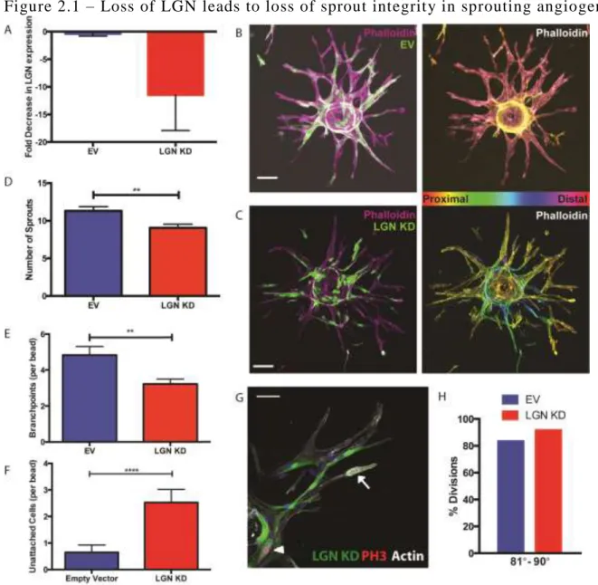
approach. We obtained a previously characterized LGN shRNA lentivirus20, and validated its
efficacy in Human Umbilical Vein EC (HUVEC). HUVEC infected with LGN KD virus showed
over a 10-fold decrease in LGN expression levels 72 hours post infection compared to control
empty vector virus (Fig 2.1A).
In order to examine LGN in endothelial sprouts, we utilized a sprouting angiogenesis
model that has been previously characterized28. Mosaic sprouts where quantified if at least half
of participating cells were LGN KD. By looking at mosaic sprouts, we were able to determine
whether LGN was globally required within a sprout for morphogenesis. We quantified the
number of sprouts that emerged from individual beads in each condition (Fig 2.1B-D). LGN KD
beads formed significantly fewer sprouts than control beads. Additionally, the sprouts that did
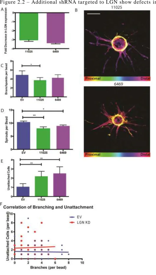
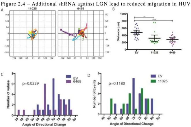
form in LGN KD beads displayed reduced branching (Fig 2.1E). To determine if the phenotype
we observed in the sprouting angiogenesis assay was due to loss of LGN, we obtained two
27
2.2A) and led to a reduction in sprouting and branching in the sprouting angiogenesis assay (Fig
2.2B-D). Taken together, these data suggests that LGN promotes the generation of new sprouts
and branches.
During the analysis of LGN KD and control beads, we observed that LGN KD cells were
more likely to be dissociated from a sprout (Fig 2.1F, 2.2E). We initially hypothesized that the
dissociated cells might represent branching EC that failed to remain connected to the parent
vessel. We compared the frequency of isolated cells against branching frequency within the same
bead and saw that there was no correlation between the parameters (Fig 2.2F). We concluded
that the isolated cells were not a result of failed branching attempts, but an independent effect of
LGN KD.
LGN directs spindle orientation during mitosis in epithelial tissue development23,24.
Therefore, we determined if the sprouting defects might be due to disrupted mitotic orientation.
However, in contrast to previous studies, we found that LGN was dispensable for orienting the
spindle (Fig 2.1G, H) in endothelial sprouts in our model. This suggests that LGN promotes
endothelial sprout formation through mechanisms other than spindle orientation.
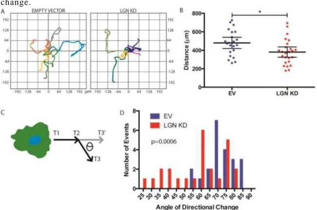
C. LGN IS REQUIRED FOR CELL MIGRATION AND DIRECTIONAL CHANGE
The process of endothelial sprouting involves multiple cellular events, including cell
migration to generate branches and networks1. We hypothesized that reduced sprouting in LGN
KD sprouts was a consequence of defective cell migration. To determine the effect of LGN KD
28
2.3A,B). LGN KD HUVEC traveled a significantly shorter distance than control cells over the
same time period (Fig 2.3B and Fig 2.4A,B), suggesting LGN facilitates cellular motility.
Reduced cell migration has many potential root causes, including defects in altering
directional migration, a cell behavior critical in the tip cell competition that guides growing
sprouts29. Therefore, we sought to determine if LGN had any influence on the ability of EC to
change direction during migration. To quantify directional changes, we generated vectors for
individual cells’ movement from one time-point to the next in live-imaging movies, then
calculated the magnitudes of angles between vectors (Fig 2.3C). Loss of LGN significantly
impaired the ability of HUVEC to make large (>30°) directional changes. Instead, almost 25% of
directional changes were of 30° or less (Fig 2.3D and Fig 2.4C,D). These data support a role for
LGN in re-orienting the cytoskeleton to initiate and alter migration.
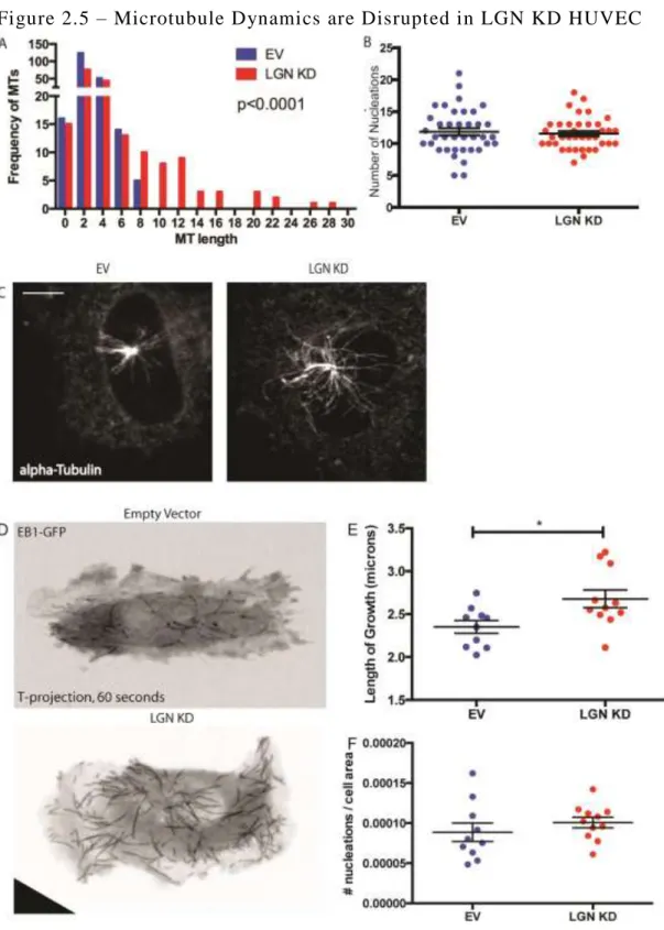
D. LGN KD HUVEC HAVE ENHANCED MT NUCLEATION
Cell migration requires dynamic remodeling of the cytoskeleton by the microtubule
organizing center (MTOC) and microtubules (MTs)27,30. Since our observations of LGN KD
HUVEC were consistent with cytoskeletal defects, we hypothesized that the MT network was
impaired in LGN KD HUVEC, contributing to the migration defects. Our lab has previously
shown that excess centrosomes can alter MT dynamics and EC migration. Therefore, we
quantified centrosome numbers in interphase LGN KD and control HUVEC, and found them to
be indistinguishable. (Fig 2.5A). We then tested the nucleation capacity of the centrosomes. We
quantified the length and number of nucleations in control and LGN KD HUVEC 1 minute after
nocodazole washout, a common method to destabilize MTs and then monitor their re-growth31.
29
compared to control HUVEC (Fig 2.5A-C). We hypothesize that LGN KD leads to more stable
MTs.
We interrogated steady-state MT dynamics through live-imaging of HUVEC infected
with an EB1-GFP lentiviral vector, which labels the plus-end of growing MTs32,33. Consistent
with our data from the nocodazole washout assay, we observed significantly longer comets and
no difference in nucleation rate in LGN KD HUVEC (Fig 2.5E-F). Combined, these data
reinforces our hypothesis that LGN regulates MT length.
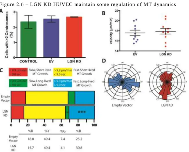
Microtubules in LGN KD cells are longer, which could be due to increased rate of MT
polymerization or elevated MT stabilization30,32. We quantified the velocity of EB1 comets in
both control and LGN KD HUVEC and we observed no global difference in comet velocity (Fig
2.6B). This suggests that the longer MTs are due to stabilization. When MTs are categorized by
their growth rate and lifetime, we observed a significantly larger population of fast, longer-lived
MTs in LGN KD (Fig 2.6C). This further supports a model in which LGN regulates MT
stability.
Our data suggest that LGN influences MT stabilization (Fig 2.5A, E, 2.6C). Nucleations
polarize toward the cell membrane and in the direction of migration27,34,35. Because of this, we
hypothesized that MT polarity would be skewed in LGN KD HUVEC. However, we failed to see
30
directional growth of MTs (Fig 2.6D). Overall, we conclude that LGN promotes the dynamic
instability necessary for MT growth and turnover.
E. FOCAL ADHESIONS ARE ENHANCED IN LGN KD HUVEC
In addition in influencing migration, the microtubule network regulates the formation,
maturation, and recycling of cellular adhesions30,36–38. Adhesions require signaling cross-talk
between MTs and the actin cytoskeleton. Focal adhesions (FAs) provide anchors to the
extracellular environment to promote cell movement, and require active MT polymerization and
catastrophe36,39. Based on our observation that LGN KD HUVEC have longer and more stable
MTs, we hypothesized that FA morphology was disrupted in LGN KD HUVEC. To investigate
FAs in LGN KD HUVEC, we pretreated the cells with nocodazole to halt FA turnover36. Once
the cells were released from nocodazole, FA turnover resumes. We quantified FA length in
control and LGN KD HUVEC, and observed that LGN KD cells had a higher frequency of long
focal adhesions (Fig 2.7A). LGN KD HUVEC not only have more long focal adhesions, but the
longest FAs in LGN KD cells are significantly longer than in control cells (Fig 2.7B). Longer
focal adhesions after washout suggests that turnover is reduced, which impairs cell migration. In
31
KD cells have longer FAs and reduced cell migration, it is likely a mechanism independent from
Ras activation, which relies on the actin cytoskeleton, not MT.
F. VE-CADHERIN LOCALIZATION IN LGN KD HUVEC SUGGESTS
JUNCTIONAL INSTABILITY
In addition to FAs, endothelial cells form adherens junctions (AJs) when in contact with
another cell40. The AJs are crucial in preventing leakiness and promoting structural integrity
during branching and sprouting5,41. VE-Cadherin promotes the formation of AJs and its
localization pattern can be used to extrapolate junctional stability42. VE-Cadherin contains an
extracellular domain that dimerizes with other VE-Cadherins on cells at the junction15.
VE-Cadherin gets recycled back into the cell, leading to separate populations of VE-VE-Cadherin43. We
immunostained confluent monolayers with VE-Cadherin to determine the integrity of junctions
in control and LGN KD HUVEC. We observed that LGN KD junctions had a higher
VE-Cadherin intensity and wider signal peak than control junctions, consistent with having a less
stable junction (Fig 2.8A-B)42.
To distinguish between internal and junctional VE-Cadherin, we repeated the
immunostaining without permeabilizing the cells, which will only label cell-surface
VE-Cadherin (Fig 2.8A-B). Under these conditions, LGN KD junctions had more VE-VE-Cadherin at
the junction (Fig 2.8C-D). Combined, this suggests that LGN KD HUVEC have more
32
could be caused by stabilized MTs, because there is evidence that VE-cadherin trafficking to and
from the cell membrane requires dynamic MTs44.
Our initial analyses of VE-Cadherin in LGN KD HUVEC focused on borders between
LGN KD positive and negative cells. We questioned how LGN KD cells contributed excess
VE-Cadherin at the AJ. To address this, we performed additional analyses of junctions between two
LGN KD cells. We observed an LGN-dependent increase in total VE-Cadherin present at the
junction, while only one LGN KD cell was necessary to observe higher intensity signals (Fig
2.8E-F).
G. VE-CADHERIN TRAFFICKING IS ENHANCED IN LGN KD HUVEC
We showed that VE-Cadherin has disrupted localization in steady-state LGN KD
monolayers (Fig 2.8). We sought to determine if the rate of junction formation was increased in
LGN KD monolayers. We investigated VE-Cadherin trafficking to the junctions through
modification of Ca2+ signaling45. By blocking Ca2+ signaling, VE-Cadherin is internalized and
junctions begin to break down. Once Ca2+ signaling resumes, VE-Cadherin re-localizes to the
junction. When HUVEC monolayers were treated with EDTA (a Ca2+ chelator) and released, we
quantified the rate of VE-Cadherin localization to the junctions. The rate of VE-Cadherin
re-localization in LGN KD junctions was increased two-fold compared to control junctions (Fig
33
dependent on proper MT turnover, suggesting that the VE-Cadherin localization in LGN KD
cells is downstream of the MT phenotype46.
H. OVERALL MEMBRANE ADHESION IMMUNOSTAINING IS DISRUPTED
We observed that VE-Cadherin was not properly localized in LGN KD AJs. However,
VE-Cadherin is not the only adhesion molecule involved in EC junctions. We sought to
determine if LGN KD effect was VE-Cadherin-specific. We immunostained for PECAM and
ICAM-2 and observed that LGN KD junctions had significantly different patterns than control
junctions (Fig 2.7G-L). Additionally, PECAM localization following EDTA treatment recovers
to pre-treatment levels in both control and LGN KD HUVEC (Fig 2.7C-D, F). These data
suggests that overall regulation of adhesion molecule trafficking is disrupted in LGN KD
HUVEC, but that VE-Cadherin and PECAM are affected differently. Because multiple adhesion
molecules are not properly localizing at LGN KD AJs, the effect that LGN has on junction
formation is likely more general and not directed at specific adhesion pathways.
I. DISCUSSION
In this study, we show that LGN influences endothelial cell functions that support blood
vessel formation. EC sprouts rely on effective cell adhesion and cell migration 3,47, which are
both impaired by MT stabilization 46,48, a feature observed in LGN KD HUVEC. This study is
the first to characterize LGN in endothelial cells, and the first to identify a requirement for LGN
in non-canonical functions. Additionally, this study shows that LGN has the potential to act
34
The role that LGN plays in anchoring astral microtubules during mitosis is extensively
detailed 18,19,49,50. Previous studies would predict that LGN is indispensable for establishing
spindle polarity in a tissue. Here, we show that EC sprouts do not require LGN to establish and
undergo oriented divisions. We predict that HUVEC rely on alternate mechanisms to promote
oriented divisions. Cell shape during interphase can dictate the formation, orientation, and
maintenance of a bipolar spindle 51–53. Endothelial cells have highly elongated cell shapes 54. We
predict that EC shape promotes oriented division in the sprouting angiogenesis model. The
sprouting angiogenesis model as a tool for observing division orientation led us to conclude that
LGN was not required for EC division orientation, but instead had novel influences on cell
behavior.
Although LGN was dispensable for division orientation in a sprout, we observed that loss
of LGN altered focal adhesion patterns and adherens junction protein localization, which are
important for maintaining sprout shape and integrity 15,4055. We predict that the elongated focal
adhesions and enhanced VE-cadherin localization are result from increased MT stability
observed in LGN KD HUVEC. Microtubules actively target focal adhesions to promote their
growth and disassembly 56, but stabilization of MTs produced excessive focal adhesion growth
48. Microtubules are also necessary in adhesion receptor recycling, which promotes stable
junctions in endothelial cells 46.
LGN associates with the astral MTs during mitosis through binding Discs Large 21,57.
Discs Large is recruited to the spindle poles and binds to phosphorylated LGN (pLGN) 57. This
binding promotes astral MT positioning and orientation in Drosophila S2 cells. The mammalian
homolog of Discs Large, ZO1, localizes to focal adhesions and promotes their life cycle 58,59, and
35
HUVEC. Another documented interaction that we might consider is pLGN/14-3-3 binding 50,
however this complex has no documented MT association. 14-3-3 has other functions, which
includes the stabilization of focal adhesions60. If LGN interacts with 14-3-3 in mammalian cells
during interphase, the binding of LGN and 14-3-3 would remove the complex from FAs and
promote FA disassembly. In LGN KD HUVEC, 14-3-3 would be maintained at the membrane
and continue stabilizing FAs, leading to reduced cell migration 35,53. While we predict that the
primary effect of LGN is on the MT network, we do not exclude the possibility that LGN may
participate directly in adhesion turnover.
We predict that LGN influences EC behavior through regulation of the MT network, thus
influencing downstream cell migration and adhesion. Previous studies have shown that LGN was
required for primary cilia migration61 during interphase. Another study implicated LGN in
pseudopod formation in neutrophils62. Our studies utilized established tools and assays to
directly characterize LGN function in endothelial cells, and further, we uncoupled LGN function
from mitosis in EC. With the evidence that LGN can act during interphase in angiogenic
36
Figure 2.1 – Loss of LGN leads to loss of sprout integrity in sprouting angiogenesis
37
38
39
Figure 2.3 – LGN KD HUVEC display reduced migratory capacity and directional change.
40
Figure 2.4 – Additional shRNA against LGN lead to reduced migration in HUVEC
41
Figure 2.5 – Microtubule Dynamics are Disrupted in LGN KD HUVEC
EB1-42
43
Figure 2.6 – LGN KD HUVEC maintain some regulation of MT dynamics
A. Quantification of excess centrosomes in control and LGN KD HUVEC. Bars show SEM.B.
Scatter plot showing average velocity of microtubule comet growth in control and LGN KD
HUVEC. Bars show mean and 95% CI.C. Distribution of MT plus ends based on lifetime length
and growth speed. **, p<0.01; ***, p<0.001D. Rose plot showing the distribution of MT plus
44
Figure 2.7 – Focal Adhesions, Adherens Junctions and Membrane Markers are disrupted in LGN KD HUVEC
A. Distribution graph of focal adhesion length in control and LGN KD HUVEC. Bars show
SEM. ***, p<0.001; *, p<0.05; **, p<0.01B. Scatter plot showing the longest 2% FAs in both
45
showing VE Cadherin, Dapi, and PECAM staining in confluent EV HUVEC before, during, and
after EDTA treatment. Scale bar is 50nm.D. Confocal images showing VE Cadherin, Dapi, and
PECAM staining in confluent LGN KD HUVEC before, during, and after EDTA treatment.
Scale bar is 50nm.E. Time course showing total VE Cadherin levels with EDTA treatment and
recovery. ****, p<0.0001F. Time course showing total PECAM levels with EDTA treatment
and recovery. **, p<0.01; ***, p<0.001G. Bar graph showing total PECAM signal in control
and LGN KD line scans. ****, p<0.0001H. Bar graph showing maximum signal intensity of
PECAM in control and LGN KD line scans.I. Confocal images showing PECAM
immunostaining in EV and LGN KD HUVEC monolayers. Scale bar is 50nm.J. Bar graph
showing total ICAM2 signal in control and LGN KD line scans.K. Bar graph showing
maximum signal intensity of ICAM2 in control and LGN KD line scans. ****, p<0.0001L.
46
Figure 2.8 – VE-Cadherin localization is misregulated at Cell -Cell Borders in LGN KD HUVEC
47 REFERENCES
1. Adams, R. H. & Alitalo, K. Molecular regulation of angiogenesis and lymphangiogenesis.
Nat. Rev. Mol. Cell Biol.8, 464–78 (2007).
2. Folkman, J. Angiogenesis in cancer, vascular, rheumatoid and other disease. Nat. Med.1,
27–31 (1995).
3. Lamalice, L., Le Boeuf, F. & Huot, J. Endothelial cell migration during angiogenesis.
Circ. Res.100, 782–94 (2007).
4. Carmeliet, P. & Collen, D. Molecular basis of angiogenesis. Role of VEGF and
VE-cadherin. Ann. N. Y. Acad. Sci.902, 249–62; discussion 262–4 (2000).
5. Dejana, E., Orsenigo, F. & Lampugnani, M. G. The role of adherens junctions and
VE-cadherin in the control of vascular permeability. J. Cell Sci.121, 2115–22 (2008).
6. Chappell, J. C., Wiley, D. M. & Bautch, V. L. How blood vessel networks are made and
measured. Cells. Tissues. Organs195, 94–107 (2012).
7. Kearney, J. B., Kappas, N. C., Ellerstrom, C., DiPaola, F. W. & Bautch, V. L. The VEGF
receptor flt-1 (VEGFR-1) is a positive modulator of vascular sprout formation and
branching morphogenesis. Blood103, 4527–35 (2004).
8. Yancopoulos, G. D. et al. Vascular-specific growth factors and blood vessel formation.
Nature407, 242–8 (2000).
9. Wodarz, A. Establishing cell polarity in development. Nat. Cell Biol.4, E39–44 (2002).
10. Zeng, G. et al. Orientation of endothelial cell division is regulated by VEGF signaling
during blood vessel formation. Blood109, 1345–52 (2007).
11. Zovein, A. C. et al. Beta1 integrin establishes endothelial cell polarity and arteriolar
lumen formation via a Par3-dependent mechanism. Dev. Cell18, 39–51 (2010).
12. Bayless, K. J. & Davis, G. E. The Cdc42 and Rac1 GTPases are required for capillary
lumen formation in three-dimensional extracellular matrices. J. Cell Sci.115, 1123–1136
(2002).
13. Patel-Hett, S. & D’Amore, P. A. Signal transduction in vasculogenesis and developmental
angiogenesis. Int. J. Dev. Biol.55, 353–63 (2011).