THE EFFECTS OF WHOLE BODY VIBRATION (WBV) AND LOCAL MUSCLE VIBRATION (LMV) ON PEAK TORQUE (PT) AND RATE OF TORQUE DEVELOPMENT
(RTD)
Dustin Lee
A senior honors thesis submitted to the faculty of the University of North Carolina at Chapel Hill in partial fulfillment of the requirements for the degree of Bachelor of the Arts in the Exercise
and Sport Science department in the College of Arts and Sciences.
Chapel Hill 2015
Approved by: J. Troy Blackburn Eric Ryan
ii © 2015 Dustin Lee
iii ABSTRACT
Dustin Lee: The Effects of Whole Body Vibration (WBV) and Local Muscle Vibration (LMV) on Peak Torque (PT) and Rate of Torque Development (RTD)
(Under the direction of J. Troy Blackburn)
Purpose: Patients with anterior cruciate ligament (ACL) reconstruction have quadriceps
dysfunction from arthrogenic muscle inhibition (AMI). AMI increases the risk of post-traumatic osteoarthritis, and current rehabilitation methods do not address AMI. Direct (local muscle vibration-LMV) and indirect (whole body vibration-WBV) vibratory stimuli improve muscle function and may be used for rehabilitation and performance enhancement. The purpose of this study was to compare the effects of WBV and LMV on quadriceps function.
Methods: Fifty-six healthy volunteers were randomized to one of three groups: WBV (n=19), LMV (n=19), or Control (n=18). All groups performed isometric squats while receiving their assigned intervention. Voluntary knee extensor peak torque (PT) and rate of torque development (RTD) were measured at baseline and again immediately, 10, and 20 minutes following
intervention (WBV, LMV, control) during maximal isometric knee extension on an isokinetic dynamometer. Dependent variables were evaluated using 3 (group) by 4 (time) repeated measures ANOVA.
iv
v
TABLE OF CONTENTS
LIST OF TABLES ... vii
LIST OF FIGURES ... viii
LIST OF ABBREVIATIONS... viiii
CHAPTER 1: INTRODUCTION ...1
CHAPTER 2: LITERATURE REVIEW ...7
INTRODUCTION ...7
ACL INJURY EPIDEMIOLOGY ...7
NEUROMUSCULAR ALTERATIONS FOLLOWING INJURY ...8
POST-TRAUMATIC OA ... 10
DEFICITS IN PT AND RTD FOLLOWING INJURY ... 13
VT ... 14
Effects on Muscle Function ... 17
WBV vs. LMV ... 18
CHAPTER 3: EXPERIMENTAL DESIGN AND METHODS ... 21
SUBJECTS... 21
EXPERIMENTAL DESIGN ... 21
PT and RTD ... 22
Intervention Procedures ... 23
Data Reduction ... 23
Data Analysis ... 24
CHAPTER 4: RESULTS ... 26
PT ... 26
RTD ... 27
CHAPTER 5: DISCUSSION ... 34
LIMITATIONS ... 39
vi
LIST OF TABLES
Table 1 - Subject demographic characteristics ... 28
Table 2 - One way ANOVA of baseline values ... 29
Table 3 - Paired samples test data for PT ... 30
vii
LIST OF FIGURES
viii
LIST OF ABBREVIATIONS
ACL Anterior cruciate ligament AMI Arthrogenic muscle inhibition LMV Local muscle vibration
OA Osteoarthritis
PT Peak torque
RTD Rate of torque development
VT Vibration therapy
1
CHAPTER 1: INTRODUCTION
The leading cause of physical disability in America is tibiofemoral osteoarthritis (OA), which affects approximately 29 million people and carries an annual cost of $165 billion (Dillon et al., 2006; Maetzel et al., 2004). Tibiofemoral OA is defined as a gradual reduction of articular cartilage within the knee joint (Lohmander et al., 2004). Interestingly, new evidence suggests that people who experience anterior cruciate ligament (ACL) and other knee joint injuries
possess a greater risk for developing post-traumatic OA (Genuario et al., 2012; Lohmander et al., 2004; Mather et al., 2013; Neuman et al., 2008; Oiestad et al., 2011; E. M. Roos, 2005; von Porat, Roos, & Roos, 2004), which can occur as early as 5 years after the injury (Neuman et al., 2009; Suomalainen et al., 2012). Specifically, one out of three Americans 65 and older are affected by OA (Lawrence et al., 2008), which carries an annual cost of $5,700 per patient (Maetzel et al., 2004). Moreover, nearly 250,000 ACL injuries occur each year in the United States (Griffin et al., 2006), and these injuries are primarily suffered by younger adults (H. Roos et al., 1995). Above all, these individuals are 3-5 times more likely to develop knee OA than those with non-injured knees, and OA is seen in nearly 13% of people who have experienced an ACL injury, within ten years or less of follow-up (Oiestad et al., 2011). The high costs
2
experiencing its long term effects, which include sedentary behavior and comorbidities such as cardiovascular disease (Philbin et al., 1996; Singh et al., 2002).
After individuals undergo an ACL reconstruction surgery, quadriceps weakness is commonly an accompanying disorder, which is caused by a complex neuromuscular
phenomenon known as arthrogenic muscle inhibition (AMI). AMI refers to a form of neural inhibition which restricts the quadriceps’ ability to be voluntarily activated (Palmieri-Smith &
Thomas, 2009a; Slemenda et al., 1997; Slemenda et al., 1998; Urbach et al., 2001). Once an individual suffers an ACL injury, mechanoreceptors from the injured joint send irregular afferent information to the central nervous system, which reduces excitability of the alpha motorneuron pool that controls the quadriceps (Palmieri-Smith & Thomas, 2009b). Pain, joint effusion, damage to joint mechanoreceptors, and abnormal joint translations may all be contributing factors to the changes in afferent input (Palmieri et al., 2004; Palmieri et al., 2005). People suffering from AMI have reported difficulties in completing many activities of daily living such as balance, sitting, stair climbing, and walking (Perry et al., 2007; Winters & Rudolph, 2014). Additionally, abnormal quadriceps activation patterns can lead to functional deficits at the knee joint that contribute to kinematic and kinetic changes during gait, which may lead to OA (M. D. Lewek, Rudolph, & Snyder-Mackler, 2004; Mündermann, Dyrby, & Andriacchi, 2005;
Shelburne, Torry, & Pandy, 2006). During the early stance phase of gait, healthy quadriceps act as a shock absorber by dispersing the energy from the ground reaction force by eccentrically controlling knee flexion (Knoll, Kocsis, & Kiss, 2004). Conversely, quadriceps that are weakened by AMI are unable to adequately absorb the forces experienced during gait. Due to inefficient energy absorption, the unhindered forces are transferred to the knee’s articular
3
Interestingly, patients with ACL reconstruction have lesser internal knee extensor moments and knee flexion angles during gait, which increases the impulsive load on the knee by decreasing the time interval over which force is absorbed (M. Lewek et al., 2002).
4
Current ACL rehabilitation programs that aim to increase quadriceps strength through exercise are usually ineffective at restoring knee extensor strength because they do not address the primary muscle activation issues associated with AMI (Hopkins & Ingersoll, 2000). For example, strengthening a muscle suffering from AMI is difficult since it cannot be effectively overloaded to stimulate hypertrophy without near maximal voluntary activation. Therefore, increasing the excitability of a muscle before exercise may be an effective mechanism for
increasing muscle function (Hurley, Jones, & Newham, 1994). Consequently, new rehabilitation methods need to be implemented that treat the deficits associated with AMI. Vibration therapy (VT) is a progressing rehabilitation modality that increases muscle strength, power, PT and physical function (Abercromby et al., 2007b; Bosco et al., 1999; Bosco, Cardinale, & Tsarpela, 1999; Luo et al., 2009; Moran, McNamara, & Luo, 2007; Ronnestad, 2009; Samuelson, Jorfeldt, & Ahlborg, 1989). When VT is applied, the muscle experiences a series of rapid lengthening and shortening contractions which triggers a tonic vibratory reflex (TVR). This reflexive contraction increases the excitatory input to the alpha motorneuron pool which may override AMI (D. J. Cochrane et al., 2010; Eklund & Hagbarth, 1966; Rittweger, 2010). This neuromuscular enhancement caused by vibratory stimuli supports the possibility of improving the efficacy of traditional rehabilitation methods, designed to improve muscle function, by increasing the excitability of a muscle with VT before the initiation of strength training.
5
Conversely, LMV also improves muscle function (Bosco et al., 1999; Moran et al., 2007),and it may counter the limitations of WBV. Specifically, LMV is easily portable and costs substantially less than WBV (~$250), which suggests that it could be a cost-effective alternative to WBV. LMV provides a similar stimulus as WBV, but its effects may differ, as they are administered differently. LMV may deliver a more efficient vibration stimulus as it is applied directly over the muscle being stimulated. Conversely, the stimulus generated by WBV is damped as it passes through the lower extremity to the knee. For example, the transmissibility of WBV is dependent on the stance of the individual receiving the stimulus (i.e. standing erect vs. knees bent), as differences in muscle activation attenuate the stimulus differently (Rubin et al., 2003). Conversely, WBV may provide additional benefits by stimulating multiple sensory receptors in other areas of the lower extremity, such as cutaneous receptors in the feet and mechanoreceptors in other joints. WBV and LMV produce comparable increases in muscle activation and strength in individuals following experimental knee joint effusion (Blackburn et al., 2014), but it remains unclear how long these effects last, which would be necessary information if VT were to be used as a rehabilitation modality. Furthermore, no studies have compared the effects of WBV and LMV on quadriceps strength and RTD in healthy individuals without knee pathologies or determined how long the effects last.
6
by WBV, more people suffering from quadriceps weakness associated with AMI could receive treatment that may delay the initiation and progression of OA due to LMV being a portable, low-cost alternative to WBV. Therefore, the purpose of this study was to compare the effects of WBV and LMV on measures of quadriceps function and determine their duration. The specific aims were as follows:
1) To determine the effects of WBV and LMV on quadriceps strength and RTD during a maximal isometric contraction in healthy adults. I hypothesized that WBV and LMV would increase quadriceps strength and RTD, and that these changes would be
greater than those observed in a control group receiving no treatment.
2) To compare the effects of WBV and LMV on quadriceps strength and RTD during a maximal isometric contraction in healthy adults. I hypothesized that WBV and LMV would increase quadriceps strength and RTD with similar magnitudes.
3) To determine the duration of the effect of WBV and LMV on quadriceps strength and RTD during a maximal isometric contraction in healthy adults. I hypothesized that the effect of WBV and LMV on quadriceps strength and RTD would last at least 10
7
CHAPTER 2: LITERATURE REVIEW
INTRODUCTION
The purpose of this literature review was to analyze relevant studies and identify their areas of limited understanding. Primarily, the muscular function deficits that people experience after suffering an ACL injury were addressed (i.e. quadriceps weakness). Additionally, the muscular shortfalls associated with post traumatic knee injury were highlighted to explain a potential pathway from ACL injury to OA through characteristic alterations in gait kinematics and kinetics. Moreover, the relationship between neuromuscular deficiencies and the failure to complete activities of daily living were identified. This review also explained the insufficiencies regarding current rehabilitation methods and recognized VT as a potential approach to increase muscular function after ACL injury. Lastly, the efficacies of LMV and WBV were compared to determine if LMV could be implemented as a possible cost-effective and portable alternative to WBV.
ACL INJURY EPIDEMIOLOGY
8
can increase the odds of acquiring tibiofemoral OA (Lohmander et al., 2004; Neuman et al., 2009; E. M. Roos, 2005; von Porat et al., 2004). Tibiofemoral OA is defined as a gradual breakdown of articular cartilage within a joint (Oiestad et al., 2011). Interestingly, patients with an ACL injury are 3-5 more times likely to develop OA than people with non-injured knees (Lohmander et al., 2004; Neuman et al., 2009), and OA development can occur as early as 5 years after an ACL injury (Ajuied et al., 2014). In fact, within ten years or less, OA is present in nearly 13% of all knees with no associated meniscal injury, and up to 48% of knees with an associated meniscal injury (Oiestad et al., 2011). Moreover, ACL injuries are most commonly suffered by younger adults, and the annual costs associated with OA are approximately $5,700 per patient (Maetzel et al., 2004). The high expenses associated with OA and the increased probability of a younger population acquiring OA contributes to an increased healthcare burden, as these younger individuals live with the costly effects of OA for a substantial portion of their lives.
NEUROMUSCULAR ALTERATIONS FOLLOWING INJURY
After an ACL injury, quadriceps weakness is often an accompanying disorder that is commonly considered a result of disuse atrophy (Palmieri-Smith & Thomas, 2009a). This may be one factor involved in the development of quadriceps weakness, but this hypothesis remains questionable because near instantaneous reductions in strength have been demonstrated post-injury and quadriceps weakness often persists after arduous rehabilitation programs (Hopkins & Ingersoll, 2000; Hurley et al., 1994). AMI, the failure to completely voluntarily activate a
9
units in muscles surrounding an injured joint (Palmieri-Smith, Thomas, & Wojtys, 2008). This neural response is thought to be a protective mechanism initiated to prevent pain and
unnecessary motion in the injured joint (Palmieri-Smith & Thomas, 2009a). AMI is nearly a ubiquitous disorder, and it is commonly measured using central activation ratio (CAR), which estimates the percentage of motor units that can be contracted voluntarily. Various studies using CAR to measure quadriceps activation after an ACL injury have reported deficits in nearly all patients with magnitudes ranging from 8% to 45% (Hart et al., 2010). Additionally, these insufficiencies often persist several years after the injury and extensive rehabilitation, potentially leading to joint degeneration. (Hopkins & Ingersoll, 2000). Interestingly, a current meta-analysis conducted by Pietrosimone et al. (2011) emphasized that quadriceps muscle activation deficits exist in persons with tibiofemoral OA at comparable rates with magnitudes of approximately 20%. Similar quadriceps muscle activation patterns between people with ACL injuries and individuals with OA supports the hypothesis that quadriceps weakness could be a precursor to OA (Palmieri-Smith & Thomas, 2009a).
The ACL houses mechanoreceptors which are responsible for sending afferent sensory signals to the central nervous system (CNS) to provide proprioception information regarding joint translations, position, and loading (Andriacchi & Mündermann, 2006). Mechanoreceptors also initiate defensive reflex mechanisms that help protect and stabilize an injured joint
10
contributing factors to the changes in afferent input (Palmieri-Smith & Thomas, 2009a; Rice & McNair, 2010).
POST-TRAUMATIC OA
OA refers to the gradual breakdown of articular cartilage within a joint, and is
traditionally diagnosed via its symptoms and physical examination. However, its severity and progression are commonly assessed with the Kelgren-Lawrence scale, which uses radiographic data to determine the quantity of osteophytes and the magnitude of joint space narrowing within a patient’s knee (grade 1 – doubtful narrowing of joint space and possible osteophytic lipping,
grade 2 – definite osteophytes and definite narrowing of joint space, grade 3 – moderate multiple osteophytes and definite narrowing of joint space, grade 4 – large osteophytes and marked narrowing of joint space) (Ajuied et al., 2014; Oiestad et al., 2011). OA is the primary cause of physical disability in America, affecting nearly 33% of adults older than 65 (Lawrence et al., 2008), and knee OA is the post prevalent form of OA, which affects nearly 29 million people (Dillon et al., 2006), and carries an annual cost of $165 billion (Maetzel et al., 2004). Knee OA is classified as idiopathic when it’s a result of a non-specific cause, but when it occurs after a knee injury, such as a ruptured ACL, it’s categorized as post-traumatic. After patients suffer an
11
After an individual suffers an ACL injury, quadriceps weakness is usually an
accompanying complication which can be partly associated with AMI, and is present in nearly all patients who are ACL deficient and approximately 80% of patients who have undergone ACL reconstruction (Hart et al., 2010). Healthy quadriceps help maintain normal knee ambulation and protect the knee joint by attenuating and evenly distributing the ground reaction forces imposed during gait (Andriacchi & Mündermann, 2006; Palmieri-Smith et al., 2008; Urbach et al., 2001). Deficits in quadriceps strength can lead to joint laxity and biomechanical alterations, which can influence articular cartilage breakdown by increasing the magnitude of force delivered to the knee and increasing the rate at which it is delivered(Andriacchi & Mündermann, 2006; Palmieri-Smith & Thomas, 2009a).
Articular cartilage is a smooth, lubricating, connective tissue that, when under normal conditions, can efficiently attenuate the cyclic loading patterns experienced in joint articulations (Radin et al., 1984). Additionally, articular cartilage is viscoelastic, which means it can
momentarily deform when subjected to a stress and return to its natural state once the stress is removed (Andriacchi & Dyrby, 2005; Chaudhari et al., 2008; Radin et al., 1984). For example, articular cartilage’s viscoelastic properties allow it to efficiently attenuate forces as long as it is afforded adequate time to recover and regain its original state between the applications of forces. Therefore, articular cartilage’s force absorbing abilities are time dependent, and it is capable of
12
alterations caused by changes in knee joint loading patterns that people experience after an ACL injury induce an irreversible form of articular cartilage breakdown and the initiation of OA.
When the quadriceps loses its ability to be fully activated, its force generating capability declines, which can alter its response to the external demands experienced during gait (Palmieri-Smith & Thomas, 2009a; Slemenda et al., 1997). Changes in force production and altered joint loads at the knee can induce kinematic and kinetic transformations, which can lead to articular cartilage breakdown and the initiation and progression of OA. For example, during the early stance phase of gait, the quadriceps act as a shock absorber by absorbing and dispersing the energy from the ground reaction force by eccentrically controlling knee flexion (Knoll et al., 2004; M. Lewek et al., 2002; M. D. Lewek et al., 2004; M. D. Lewek, Rudolph, & Snyder‐ Mackler, 2004). Concerning this, Jefferson et al. (1990) studied the role of quadriceps in controlling impulsive forces during gait, which compared knee flexion angles and ground
reaction forces before and after individuals experienced artificially induced quadriceps paralysis. After quadriceps paralysis, decreased knee flexion angles and impulsive loading characteristics were reported. Prior to the heel strike phase of gait, healthy quadriceps smoothly decelerate the lower limb and reduce the ground reaction force by softening the impact of the foot with the ground. Conversely, weak quadriceps interrupts this mechanism, which can interfere with the lower limb’s energy absorption capabilities. Interestingly, people with ACL injuries have
reduced internal knee extensor moments and knee flexion angles during gait, which increases the impulsive load characteristics on the knee by decreasing the time interval through which force is absorbed (Glass et al., 2013; Winters & Rudolph, 2014).
13
exposed to high stress (Andriacchi & Mündermann, 2006). This suggests that joints have adaption capabilities that mitigate stress on areas that experience chronic loading by increasing the amount of cartilage at specific locations (Andriacchi & Mündermann, 2006). However, ACL injury can cause knee joint laxity and changes in gait kinematics that alter tibiofemoral contact areas, potentially introducing areas of articular cartilage to forces to which they are
unaccustomed (Hopkins & Ingersoll, 2000; Palmieri-Smith et al., 2008). For example, after an ACL injury, joint surfaces that are composed of relatively thin areas of articular cartilage, not conditioned to frequent stress, and less efficient at absorbing forces may be loaded to a greater extent. Articular cartilage breakdown is initiated when thinner regions of cartilage that are less accustomed to frequent stress experience more frequent loading of greater magnitude before they have time to adapt. Therefore, changes in knee joint contact areas and increases in pressure on unaccustomed areas of articular cartilage may initiate OA by narrowing joint space and damaging articular cartilage.
DEFICITS IN PT AND RTD FOLLOWING INJURY
14
and slowed progression of OA (Palmieri-Smith et al., 2008). Therefore, quadriceps that are capable of producing forces needed to complete functional tasks and protect the knee from external forces are more proficient at maintaining a healthy joint than quadriceps that are
hindered by decreased strength. However, Sharma et al. (2000) reported that greater quadriceps PT was associated with OA progression in patients who have been diagnosed with OA and have lax knees. This finding suggests that other indices of muscle function may be involved in controlling biomechanical alterations and the progression of OA. Nearly all individuals with knee OA have altered quadriceps activation patterns associated with AMI (Hart et al., 2010), and the failure to adapt to knee joint laxity may be an indication of a decrease in the rate of force delivery by the quadriceps, caused by a deficiency in motor neuron recruitment (i.e. AMI). RTD provides an indication of how quickly an individual can develop maximal force (Winters & Rudolph, 2014), and rapid RTD has been shown to compensate for decreased quadriceps strength by delivering counter forces quickly in response to external demands. Interestingly, Staehli et al. (2010) demonstrated that quadriceps RTD is more closely related to activities of daily living that quadriceps PT. Therefore, the functional and biomechanical associations to RTD illustrate its importance to knee joint function. Lastly, the kinetic and kinematic alterations caused by quadriceps weakness that have been demonstrated to initiate joint degeneration could be mitigated by rehabilitation programs aimed at increasing both PT and RTD.
VT
15
rehabilitation professionals, focused on improving strength, commonly prescribe exercises with the fundamental purpose of inducing muscular hypertrophy (Hellebrandt, 1958). Muscular hypertrophy is a multidimensional process that is defined as an increase in muscle mass and cross-sectional area, which can be accomplished via the overload principle (Robergs & Roberts, 1997; Russell, Motlagh, & Ashley, 2000). In brief, the overload principle is a muscular
adaptation that occurs in response to exercise stimuli that force muscle fibers to experience tensile forces near their maximum capacity (Hellebrandt, 1958). Subsequently, the
accompanying musculature is forced adapt through hypertrophy, leading to an increase in the size of the individual muscle fibers and their consequent force production capabilities (Jones & Lees, 2003; Pearson et al., 2000; Russell et al., 2000). However, individuals suffering from neuromuscular deficits may not have the capacity to effectively overload all their muscle fibers and experience muscular hypertrophy and its associated strength gains, as activation scarcities limit muscle’s ability to efficiently respond to training stimuli (Hopkins & Ingersoll, 2000). Specifically, if a motor unit is not activated, its associated muscle fibers cannot be overloaded, and no adaptation can occur without overload (McNicol et al., 2009; Russell et al., 2000). Furthermore, neuromuscular deficiencies not only restrict individuals’ ability to acquire strength gains, it places them at risk to experience muscle atrophy and its debilitating effects (Hurley et al., 1994).
16
potential neural enhancement capabilities may validate it as a possible rehabilitation modality that could be used in combination with traditional strength training programs aimed at increasing muscle function.
Vibration provides a mechanical oscillation of force, acceleration, and displacement over time. VT is defined as a forced oscillation during which energy is transferred from an actuator (vibration device) to a resonator (human body) (Bazett-Jones, Finch, & Dugan, 2008). The transfer of energy causes reactive forces within the body, which can cause beneficial
neuromuscular responses, but it also carries the potential to harm tissues within the body. VT inflicts a force on the body that is proportional to its mass, and it causes the body to accelerate through sinusoidal oscillations in which the affected muscles and tendons act like springs by storing and releasing mechanical energy (Rittweger, 2010). An accumulation of mechanical energy within the body can damage muscles through increases in internal forces when the
frequency of the actuator matches the natural frequency of the resonator, which can be controlled by the body’s stiffness and mass (D. J. Cochrane et al., 2010; D. J. Cochrane, 2011). Therefore,
this damaging resonance effect can be overcome by muscles damping the signal through modifications in body position and changes in muscle stiffness. For example, the transmissibility of ground based vibrations is dependent on posture and the associated
17
When VT is applied, the energy transfer causes the muscle to go through a series of rapid lengthening and shortening contractions which triggers a tonic vibratory reflex (TVR) (Burke et al., 1976; Eklund & Hagbarth, 1966). The vibration causes muscles and their associated muscle spindles to lengthen and they become more easily excitable. The reflexive contraction
experienced by the muscles increases the excitatory input to the alpha motorneuron pool which can increase a muscle’s force generating capability by increasing the activation of its motor units (Rittweger, 2010). There are also additional mechanisms that can explain the neuromuscular increases associated with VT such as muscle tuning, motor unit synchronization, central motor command, and intramuscular coordination (D. J. Cochrane et al., 2010; D. Cochrane, 2011). Additionally, muscle enhancements can be caused by escalations in muscle temperature and blood flow, similar to a warm-up. Lastly, VT also inflicts a training-effect, which is caused by a muscle’s contractual response to increases in gravitational forces caused by vibration (D. J.
Cochrane, 2011; D. Cochrane, 2011).
Effects on Muscle Function
18
various enhancements in muscle function associated with VT may be a result of neuromuscular improvements (D. Cochrane, 2011). Regardless of these positive findings, there are also studies that show ambivalent results in one repetition maximum (Lau, Yip, & Pang, 2012; Pamukoff, Ryan, & Blackburn, 2014; Segal et al., 2013), and RTD (Pamukoff et al., 2014). Additionally, others have demonstrated negative effects on muscle strength (De Ruiter et al., 2003; Erskine et al., 2007a; Herda et al., 2009). For example, Erskine et al. (2007a) found a 9% decrease in knee extensor strength following 10 by 1 minute isometric half squat exercises while being exposed to WBV. Interestingly, the aforementioned VT studies were administered via different application methods and with dissimilar treatment parameters (i.e. duration, amplitude and frequency). Therefore, the lack of treatment guidelines existing for VT may be an explanation for the inconsistencies between these findings and the basis for the heterogeneous stimulation
parameters employed (D. J. Cochrane et al., 2010). The method by which VT is received may also change its potential benefits because changes in posture and muscle stiffness alter the transmissibility of vibration (Rubin et al., 2003). For example, Pamukoff et al. (2014) compared the effects of LMV on PT at 30 Hz and at 60 Hz and showed increases in PT at 30 Hz but no effect at 60 Hz, which enforces the idea that a muscle’s mechanical response varies with frequency.
WBV vs. LMV
The majority of VT studies have focused on the muscular improvements associated with WBV. However, WBV platforms have limited portability and they carry cost restrictions (~$12,000) (Blackburn et al., 2014). LMV also improves muscle function (Blackburn et al., 2014; Erskine et al., 2007a; Luo et al., 2009; Pamukoff et al., 2014) and may be a more
19
the efficacy of LMV, which is applied directly to the muscle-tendon unit, Iodice et al. (2011) revealed that leg extensor muscle strength increased following acute exposure to LMV.
However, other investigators have reported equivocal outcomes after LMV (Moran et al., 2007). Although WBV and LMV produce similar stimuli, the efficacy of the neuromuscular
enhancements that they provide may be altered by their modes of application due to different muscle damping characteristics.
By changing the delivery method of the VT (i.e. standing on platform or local application), the body’s musculature absorbs the associated energy differently, which may change the neuromuscular response (Abercromby et al., 2007a). Additionally, the characteristics of vibration stimuli are dependent on the physical properties of the material through which they travel. Specifically, changes in posture alter the frequency received by muscles through inherent alterations in muscle stiffness associated with variations in stance (Abercromby et al., 2007b; Rubin et al., 2003). Consequently, deviations in vibration frequency can affect the magnitude of a muscle’s TVR, which is directly associated with the extent of excitation increases in the alpha motorneuron pool that control the quadriceps (Rittweger, 2010).
20
21
CHAPTER 3: EXPERIMENTAL DESIGN AND METHODS
SUBJECTS
Sixty healthy individuals (30 males and 30 females) were recruited from the student and employee populations at the University of North Carolina at Chapel Hill and from the
surrounding area. Subjects were included if they were between the ages of 18 and 30 years and recreationally active, defined as participation in physical activity for at least 30 minutes 3 times per week. Subjects were excluded for any history of musculoskeletal injury within 6 months prior to testing, lower extremity surgery, neurological disorder, cardiovascular disease, hypertension, diabetes mellitus, concussion or head injury, stroke, epilepsy, peripheral
neuropathy, migraine headaches, cranial neural surgery, cancer in the brain or thigh musculature, cardiac pacemaker, implanted foreign metal object, or diagnosed psychiatric disorder. Inclusion and exclusion criteria were confirmed via self-report. Each subject was required to read and sign an informed consent form prior to data collection.
EXPERIMENTAL DESIGN
22
This investigation utilized a randomized controlled trial experimental design. Subjects were randomized to 1 of 3 groups following pre-test assessments and received either WBV, LMV, or Control (no vibration) treatment (n = 20 per group). Although subjects completed 3 testing visits as part of the larger ongoing study, data for this study was collected from a single visit to the Neuromuscular Research Laboratory at the University of North Carolina at Chapel Hill which lasted approximately 1 hour. Each subject completed a baseline evaluation of knee extensor PT and RTD during a 5-second maximal knee extension, received one of the
aforementioned interventions, and completed follow-up testing immediately, 10 minutes, and 20 minutes following the intervention.
ASSESSMENTS
PT and RTD
Subjects first underwent a brief 5-minute aerobic warm-up on a stationary cycle ergometer followed by baseline tests of isometric knee extensor PT and RTD using a
23
initiate knee extension, they were instructed to “kick as hard and as fast as possible” and
received verbal encouragement for each trial to ensure maximal effort. Sixty seconds of rest was given between assessments.
Intervention Procedures
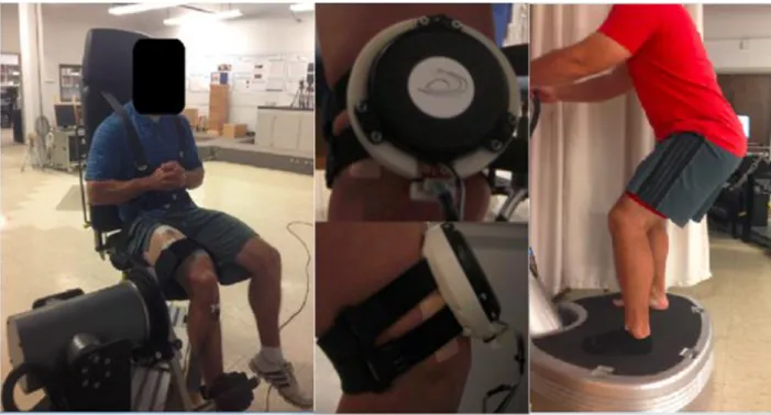
Following baseline testing, subjects were randomized to LMV, WBV, or Control (no vibration) groups. The LMV group received 6 exposures of 60 seconds of vibration with 2 minutes of rest between each exposure while standing in approximately 40° of knee flexion. A custom-made LMV device was positioned on the quadriceps tendon (Figure 1, Center). Subjects randomized to the WBV group stood on a vibrating platform that delivered a similar stimulus (Figure 1, Right). The LMV and WBV stimuli were delivered at 2g of acceleration at a frequency of 30Hz. The control group did not receive vibration, but assumed the same knee flexion position in 6x60 second intervals with 2 minutes of rest between exposures. All subjects stood on the WBV device during their respected interventions, but it was only activated for the subjects assigned to the WBV group. These parameters were similar to a previous study in our laboratory that represented comparable effects between LMV and WBV on voluntary muscle activation (Blackburn et al., 2014). Directly after the intervention, subjects repeated the
aforementioned assessments of PT and RTD during a maximal isometric contraction, and again 10 minutes and 20 minutes following the cessation of the intervention.
Data Reduction
24
successive 20ms intervals and divided by time to represent RTD. The peak RTD value was identified and normalized to body mass for statistical analysis. The mean PT and RTD across trials was calculated at each time point and used for all subsequent analyses.
Data Analysis
Data was inspected for normality using the Shapiro-Wilk test and homogeneity of
variance using Levene’s test to confirm the assumptions for analysis of variance (ANOVA). All
25
26
CHAPTER 4: RESULTS
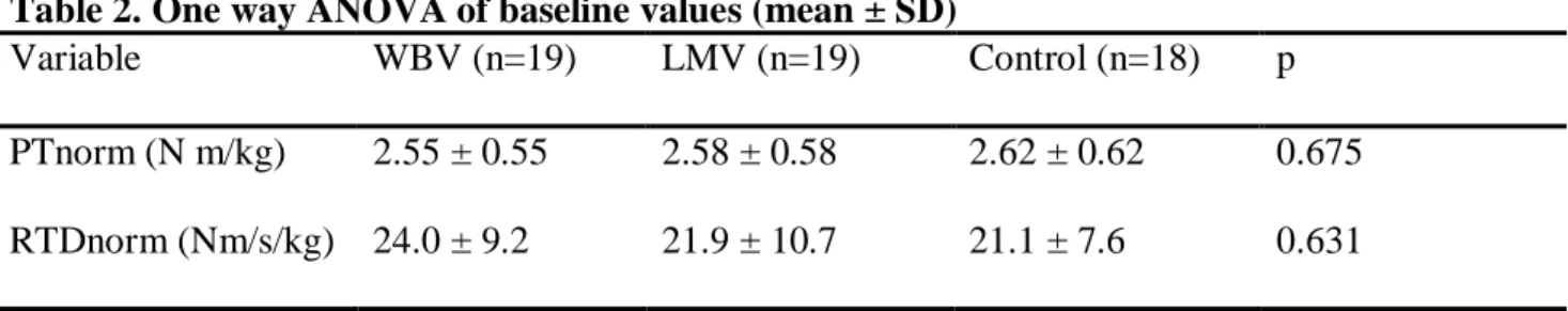
Box plots were utilized to identify outliers, defined as values more than 1.5 times the interquartile range. Four subjects’ data were identified as outliers and excluded from statistical analysis for subsequent evaluation. Demographic data for subjects retained in the final analyses are detailed in Table 1.
PT
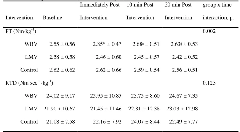
PT did not differ between groups at baseline (p = 0.675; Table 2). Additionally, the PT data violated the assumption of sphericity. As such, the Greenhouse-Geisser correction was used to assess the group by time interaction. The group by time interaction was significant (F2,53 =
4.26, p = 0.002). Post hoc testing (Table 3) revealed a significant increase for the WBV group (+0.29 Nm/kg, 95%CI: 0.18 – 0.42, p < 0.001) from baseline to immediately following
treatment. However, due to overly conservative post hoc analyses, there was not a significant change in the WBV group at 10 or 20 minutes following intervention. However, there was a statistical trend that approached significance 10 minutes (+0.13 Nm/kg, 95%CI: 0.05 – 0.21, p = 0.007) and 20 minutes (+0.08 Nm/kg, 95%CI: 0.01 - 0.15, p = 0.028) after WBV. There were no significant differences in either the LMV or control groups anytime following treatment.
27 RTD
RTD did not differ between groups at baseline (p = 0.631; Table 2). The group by time interaction was not significant for RTD (F2,53 = 1.71, p = 0.123). The effects of the vibratory
28
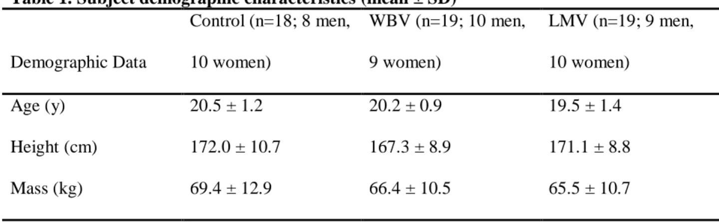
Table 1. Subject demographic characteristics (mean ± SD)
Demographic Data
Control (n=18; 8 men, 10 women)
WBV (n=19; 10 men, 9 women)
LMV (n=19; 9 men, 10 women)
Age (y) 20.5 ± 1.2 20.2 ± 0.9 19.5 ± 1.4
Height (cm) 172.0 ± 10.7 167.3 ± 8.9 171.1 ± 8.8
29
Table 2. One way ANOVA of baseline values (mean ± SD)
Variable WBV (n=19) LMV (n=19) Control (n=18) p
30 Table 3. Paired Samples Test Data for PT (WBV)
Comparison Mean Difference (%) 95% confidence interval Sig. (1-tailed)
Lower Upper
Baseline vs. Post * Baseline vs. 10 min Post ǂ Baseline vs. 20 min Post ǂ
0.299 0.128 0.081
0.18 0.05 0.01
0.42 0.21 0.15
< .001 .0065 .028
31
Table 4. Descriptive statistics for the vibratory interventions (mean ± SD)
Intervention Baseline
Immediately Post Intervention
10 min Post Intervention
20 min Post Intervention
group x time interaction, p:
PT (Nm·kg-1) 0.002
WBV 2.55 ± 0.56 2.85* ± 0.47 2.68ǂ ± 0.51 2.63ǂ ± 0.53 LMV 2.58 ± 0.58 2.46 ± 0.60 2.45 ± 0.57 2.42 ± 0.52 Control 2.62 ± 0.62 2.62 ± 0.66 2.59 ± 0.54 2.56 ± 0.51
RTD (Nm·sec-1·kg-1) 0.123
WBV 24.02 ± 9.17 25.95 ± 10.85 23.75 ± 8.60 24.67 ± 7.35 LMV 21.90 ± 10.67 21.45 ± 11.46 22.31 ± 12.38 23.03 ± 12.98 Control 21.08 ± 7.58 22.16 ± 7.92 24.07 ± 8.44 22.49 ± 7.77
32 Figure 2. Normalized PT.
1.00 1.50 2.00 2.50 3.00 3.50
Pre Post 10 min Post 20 min Post
P
T
(N
m
/k
g)
PT
WBV
LMV
33 Figure 3. Normalized RTD.
5 10 15 20 25 30 35 40
Pre Post 10 min Post 20 min Post
R
TD
(N
m
/s
ec
/k
g)
RTD
WBV
LMV
34
CHAPTER 5: DISCUSSION
The primary findings of this study were that WBV significantly increased quadriceps PT in healthy individuals immediately following treatment. Additionally, our results suggested that quadriceps PT remained elevated 10 and 20 minutes after WBV relative to baseline values. Though not statistically significant due to overly conservative post hoc analyses these outcomes displayed a statistical trend that approached significance. Moreover, there were no substantial improvements in PT following LMV at any time point. With respect to RTD, there were no significant differences in WBV or LMV anytime following intervention.
35
corticospinal excitability and intracortical processes (K. N. Mileva, Bowtell, & Kossev, 2009; Siggelkow et al., 1999). Accordingly, prospective research is needed to gain knowledge of the mechanism behind the muscle enhancements associated with WBV.
Another important finding was the presence of a trend approaching significance that quadriceps PT was still enhanced 10 and 20 minutes after WBV. Previous studies have reported increases in muscle force 5 minutes (Bazett-Jones et al., 2008) and 8 minutes (McBride et al., 2010) after vibration treatment. Moreover, many researchers credit the prolonged enhancements in muscle function succeeding vibration to elevated muscle temperature, synchronization,
muscular coordination and proprioceptor response (Adams et al., 2009; Cardinale & Bosco, 2003; D. J. Cochrane et al., 2008). However, our findings are in contrast to those of Erskine et al. (2007b) who found no increase in knee extensor strength following WBV. The discrepancy in results could be due do heterogeneous stimulation parameters between the two studies (2g vs. 5g), as skeletal muscle is a specialized tissue that modifies its overall functional capacity in response to different stimuli (Erskine et al., 2007b). In particular, the neurological adaptations elicited from vibration are directly related to the characteristics of the load imposed by the stimulus (D. J. Cochrane, 2011; Rittweger, 2010). Therefore, the 2 g acceleration delivered in this study as opposed to 5 g in the aforementioned investigation may be more appropriate to elicit positive neuromuscular adaptations in the quadriceps. Consequently, vibratory parameters such as amplitude, frequency, duration, and acceleration should be investigated further in efforts to improve future vibratory protocols.
36
the ANOVA model had a power of 0.34 (ES = 0.40), suggesting that we may have been underpowered to detect differences in PT. Accordingly, exploratory post hoc power analyses revealed that 78 subjects per group would be necessary to achieve a power of 0.80. The lack of difference between groups may have been a result of the heterogeneous nature of our sample. Specifically, the sample utilized in this study consisted of individuals with diverse physical activity statuses, ranging from minimal physical activity to DI athletes. Consequently, the large variability in PT discovered within groups caused their respective standard deviations to overlap, producing insignificant statistical differences between the groups.
LMV did not influence PT at any point following application. Our hypothesis that PT would increase after LMV was based on several findings that demonstrated improvements in muscular function following LMV (Bongiovanni & Hagbarth, 1990; Couto et al., 2013; Iodice et al., 2011; Mischi & Cardinale, 2009; Ribot-Ciscar, Butler, & Thomas, 2003). For example, Iodice et al. (2011) revealed that leg extensor muscle strength and jump performance increased following acute and prolonged exposure to LMV. Moreover, LMV’s ability to elicit
neuromuscular improvements along with its portability capabilities and cost effectiveness made it an appealing modality to investigate in effort to find an alternative to large and expensive WBV platforms. However, other studies have suggested a decline in force output following LMV (Kouzaki, Shinohara, & Fukunaga, 2000; Mottram et al., 2006), which could be associated with the parameters of stimulation. Accordingly, the inconsistencies in our PT results between WBV and LMV could be attributable to differences in the somatosensory receptors that are targeted by the WBV and LMV. For example, WBV stimulates receptors in skin and
37
effects are likely isolated to receptors surrounding the muscle-tendon unit to which it’s applied, potentially limiting its effectiveness. However, Blackburn et al. (2014) demonstrated significant improvements in quadriceps function following LMV in individuals with artificially induced quadriceps inhibition similar to individuals with knee pathologies. As such, this form of vibratory stimulus may be effective for enhancing quadriceps function in individuals with pathological knees, and prospective investigations are necessary to elucidate its effects since WBV platforms can be cost prohibitive and provide limited portability.
We did not observe a significant change in RTD subsequent to WBV or LMV at any point following treatment. RTD is influenced by neural and mechanical factors, and improved RTD is derived from increases in neural drive (i.e. firing frequency and motor unit activation) (Aagaard et al., 2002). Hence, our hypothesis that RTD would increase after VT was based on findings of improved alpha motorneuron excitability via the muscle spindle system (Cardinale & Bosco, 2003; Ritzmann et al., 2013) which could cause increases in firing frequency and motor unit activation (Burke & Gandevia, 1995; Eklund & Hagbarth, 1966). In opposition to our findings, Tihanyi et al. (2007) discovered a 19% increase in RTD in patients with stroke during a maximal isometric contraction following WBV. However, our subjects were young,
38
processing techniques. In our study, change in torque (final-initial) was calculated over successive 20ms intervals and divided by time to represent RTD. Conversely, Tihanyi et al. (2007) discovered a significant change in RTD by calculating RTD as the tangent (dM/dt) fitted to the steepest part of the torque-time curves. Consequently, future studies are needed to determine the most effective methodological approach to calculate RTD.
Quadriceps PT and RTD are important considerations for athletic performance and injury prevention, and key determinants for the elderly’s capacity to complete activities of daily living
(Bohannon & Andrews, 1990; Perry et al., 2007; Thompson et al., 2013; Winters & Rudolph, 2014). Moreover, muscle strength plays a significant role in balance and ambulation, and even healthy elderly persons complete activities of daily living such as stepping and walking at levels near their maximal voluntary joint torques (Hortobagyi et al., 2003; Kim & Eng, 2003). Pohl et al. (2002) reported that RTD is a better predictor of gait speed in stroke patients than PT. Moreover, rapid RTD has been shown to compensate for decreased quadriceps strength by delivering counter forces quickly in response to external demands (Nadeau et al., 1999; Winters & Rudolph, 2014). Unfortunately, individuals with knee OA and ACL injury often have deficits in PT and RTD originating from inhibited voluntary quadriceps activation (Hart et al., 2010; Pietrosimone et al., 2011), which have been shown to reduce the effectiveness of rehabilitation (Hopkins & Ingersoll, 2000; Hurley et al., 1994). Consequently, the prolonged effects of WBV on muscle function and its potential for producing local and peripheral neuromuscular
39 LIMITATIONS
There are inherent limitations associated with our study that should be considered when interpreting the results. First, our findings may reveal a ceiling effect due to the presence of healthy, young, and recreationally active subjects. Accordingly, a study conducted by Blackburn et al. (2014) that compared the effects of WBV and LMV on individuals with artificially induced quadriceps arthrogenic inhibition discovered greater benefits from vibration in individuals with more severe quadriceps inhibition. Additionally, other studies have reported reductions in pain and inflammation and increases in proprioception following VT (Aaboe et al., 2009; Kitay et al., 2009; Simão et al., 2012). Therefore, the positive effects of vibration stimuli may be greater in pathological and elderly populations who possess neuromuscular deficits. Secondly, our study did not investigate the mechanisms by which WBV enhances maximal voluntary quadriceps strength, which could be a result of enhanced excitation of alpha motorneurons via the muscle spindle system (Burke & Gandevia, 1995; Eklund & Hagbarth, 1966) or may be credited to alterations in corticospinal excitability and intracortical processes (K. N. Mileva et al., 2009; Siggelkow et al., 1999). Lastly, we did not investigate the source of increased quadriceps PT following WBV. Specifically, we did not measure the associated agonist and antagonist muscle co-activation patterns, which means the positive net force discovered from knee extension may have originated from increased agonist activity, reductions in antagonist activity, or a
combination of both. Accordingly, Tihanyi et al. (2007) studied EMGrms during eccentric
40 CONCLUSIONS
41
REFERENCES
1. Aaboe, J., Henriksen, M., Christensen, R., Bliddal, H., & Lund, H. (2009). Effect of whole body vibration exercise on muscle strength and proprioception in females with knee
osteoarthritis. The Knee, 16(4), 256-261.
2. Aagaard, P., Simonsen, E. B., Andersen, J. L., Magnusson, P., & Dyhre-Poulsen, P. (2002). Increased rate of force development and neural drive of human skeletal muscle following resistance training. Journal of Applied Physiology (Bethesda, Md.: 1985), 93(4), 1318-1326. doi:10.1152/japplphysiol.00283.2002 [doi]
3. Abercromby, A. F., Amonette, W. E., Layne, C. S., McFarlin, B. K., Hinman, M. R., &
Paloski, W. H. (2007a). Variation in neuromuscular responses during acute whole-body vibration exercise. Medicine and Science in Sports and Exercise, 39(9), 1642.
4. Abercromby, A. F., Amonette, W. E., Layne, C. S., McFarlin, B. K., Hinman, M. R., & Paloski, W. H. (2007b). Vibration exposure and biodynamic responses during whole-body vibration training. Medicine and Science in Sports and Exercise, 39(10), 1794.
5. Adams, J. B., Edwards, D., Serravite, D. H., Bedient, A. M., Huntsman, E., Jacobs, K. A., . . . Signorile, J. F. (2009). Optimal frequency, displacement, duration, and recovery patterns to maximize power output following acute whole-body vibration. Journal of Strength and Conditioning Research / National Strength & Conditioning Association, 23(1), 237-245. doi:10.1519/JSC.0b013e3181876830 [doi]
6. Ajuied, A., Wong, F., Smith, C., Norris, M., Earnshaw, P., Back, D., & Davies, A. (2014). Anterior cruciate ligament injury and radiologic progression of knee osteoarthritis: A systematic review and meta-analysis. The American Journal of Sports Medicine, 42(9), 2242-2252.
doi:10.1177/0363546513508376 [doi]
7. Andriacchi, T. P., & Dyrby, C. O. (2005). Interactions between kinematics and loading during walking for the normal and ACL deficient knee. Journal of Biomechanics, 38(2), 293-298. 8. Andriacchi, T. P., & Mündermann, A. (2006). The role of ambulatory mechanics in the initiation and progression of knee osteoarthritis. Current Opinion in Rheumatology, 18(5), 514-518.
9. Bazett-Jones, D. M., Finch, H. W., & Dugan, E. L. (2008). Comparing the effects of various whole-body vibration accelerations on counter-movement jump performance. Journal of Sports Science & Medicine, 7(1), 144.
42
11. Bohannon, R. W., & Andrews, A. W. (1990). Correlation of knee extensor muscle torque and spasticity with gait speed in patients with stroke. Archives of Physical Medicine and
Rehabilitation, 71(5), 330-333.
12. Bongiovanni, L., & Hagbarth, K. (1990). Tonic vibration reflexes elicited during fatigue from maximal voluntary contractions in man. The Journal of Physiology, 423(1), 1-14. 13. Bosco, C., Colli, R., Introini, E., Cardinale, M., Tsarpela, O., Madella, A., . . . Viru, A. (1999). Adaptive respsonses of human skeletal muscle to vibration exposure. CLINICAL
PHYSIOLOGY-OXFORD-, 19, 183-187.
14. Bosco, C., Cardinale, M., & Tsarpela, O. (1999). Influence of vibration on mechanical power and electromyogram activity in human arm flexor muscles. European Journal of Applied
Physiology and Occupational Physiology, 79(4), 306-311.
15. Burke, D., & Gandevia, S. (1995). The human muscle spindle and its fusimotor control. Neural control of movement (pp. 19-25) Springer.
16. Burke, D., Hagbarth, K. E., Lofstedt, L., & Wallin, B. G. (1976). The responses of human muscle spindle endings to vibration of non-contracting muscles. The Journal of Physiology, 261(3), 673-693.
17. Cardinale, M., & Bosco, C. (2003). The use of vibration as an exercise intervention. Exercise and Sport Sciences Reviews, 31(1), 3-7.
18. Chaudhari, A. M., Briant, P. L., Bevill, S. L., Koo, S., & Andriacchi, T. P. (2008). Knee kinematics, cartilage morphology, and osteoarthritis after ACL injury. Medicine and Science in Sports and Exercise, 40(2), 215-222. doi:10.1249/mss.0b013e31815cbb0e [doi]
19. Cochrane, D. J. (2011). The potential neural mechanisms of acute indirect vibration. Journal of Sports Science & Medicine, 10(1), 19.
20. Cochrane, D. J., Stannard, S. R., Firth, E. C., & Rittweger, J. (2010). Acute whole-body vibration elicits post-activation potentiation. European Journal of Applied Physiology, 108(2), 311-319.
21. Cochrane, D. J., Stannard, S. R., Sargeant, A. J., & Rittweger, J. (2008). The rate of muscle temperature increase during acute whole-body vibration exercise. European Journal of Applied Physiology, 103(4), 441-448.
22. Cochrane, D. (2011). Vibration exercise: The potential benefits. International Journal of Sports Medicine, 32(2), 75.
23. Couto, B. P., Silva, H. R., Filho, A. G., da Silveira Neves, S. R., Ramos, M. G.,
43
vibration. International Journal of Sports Medicine, 34(9), 814-819. doi:10.1055/s-0032-1331198 [doi]
24. De Ruiter, C., Van Der Linden, R., Van der Zijden, M., Hollander, A., & De Haan, A. (2003). Short-term effects of whole-body vibration on maximal voluntary isometric knee
extensor force and rate of force rise. European Journal of Applied Physiology, 88(4-5), 472-475. 25. Dillon, C. F., Rasch, E. K., Gu, Q., & Hirsch, R. (2006). Prevalence of knee osteoarthritis in the united states: Arthritis data from the third national health and nutrition examination survey 1991-94. The Journal of Rheumatology, 33(11), 2271-2279. doi:06/13/1011 [pii]
26. Eklund, G., & Hagbarth, K. (1966). Normal variability of tonic vibration reflexes in man. Experimental Neurology, 16(1), 80-92.
27. Erskine, J., Smillie, I., Leiper, J., Ball, D., & Cardinale, M. (2007a). Neuromuscular and hormonal responses to a single session of whole body vibration exercise in healthy young men. Clinical Physiology and Functional Imaging, 27(4), 242-248.
28. Erskine, J., Smillie, I., Leiper, J., Ball, D., & Cardinale, M. (2007b). Neuromuscular and hormonal responses to a single session of whole body vibration exercise in healthy young men. Clinical Physiology and Functional Imaging, 27(4), 242-248.
29. Genuario, J. W., Faucett, S. C., Boublik, M., & Schlegel, T. F. (2012). A cost-effectiveness analysis comparing 3 anterior cruciate ligament graft types: Bone-patellar tendon-bone autograft, hamstring autograft, and allograft. The American Journal of Sports Medicine, 40(2), 307-314. doi:10.1177/0363546511426088 [doi]
30. Glass, N., Torner, J., Frey Law, L., Wang, K., Yang, T., Nevitt, M., . . . Segal, N. (2013). The relationship between quadriceps muscle weakness and worsening of knee pain in the MOST cohort: A 5-year longitudinal study. Osteoarthritis and Cartilage, 21(9), 1154-1159.
31. Griffin, L. Y., Albohm, M. J., Arendt, E. A., Bahr, R., Beynnon, B. D., Demaio, M., . . . Yu, B. (2006). Understanding and preventing noncontact anterior cruciate ligament injuries: A review of the hunt valley II meeting, january 2005. The American Journal of Sports Medicine, 34(9), 1512-1532. doi:34/9/1512 [pii]
32. Hart, J. M., Pietrosimone, B., Hertel, J., & Ingersoll, C. D. (2010). Quadriceps activation following knee injuries: A systematic review. Journal of Athletic Training, 45(1), 87-97. doi:10.4085/1062-6050-45.1.87 [doi]
33. Hellebrandt, F. A. (1958). Application of the overload principle to muscle training in man. American Journal of Physical Medicine, 37(5), 278-283.
44
35. Hopkins, J. T., & Ingersoll, C. D. (2000). Arthrogenic muscle inhibition: A limiting factor in joint rehabilitation. Journal of Sport Rehabilitation, 9(2), 135-159.
36. Hortobagyi, T., Mizelle, C., Beam, S., & DeVita, P. (2003). Old adults perform activities of daily living near their maximal capabilities. The Journals of Gerontology.Series A, Biological Sciences and Medical Sciences, 58(5), M453-60.
37. Hurley, M. V., Jones, D. W., & Newham, D. J. (1994). Arthrogenic quadriceps inhibition and rehabilitation of patients with extensive traumatic knee injuries. Clinical Science (London, England : 1979), 86(3), 305-310.
38. Iodice, P., Bellomo, R. G., Gialluca, G., Fanò, G., & Saggini, R. (2011). Acute and cumulative effects of focused high-frequency vibrations on the endocrine system and muscle strength. European Journal of Applied Physiology, 111(6), 897-904.
39. Issurin, V., & Tenenbaum, G. (1999). Acute and residual effects of vibratory stimulation on explosive strength in elite and amateur athletes. Journal of Sports Sciences, 17(3), 177-182. 40. Jacobs, P. L., & Burns, P. (2009). Acute enhancement of lower-extremity dynamic strength and flexibility with whole-body vibration. Journal of Strength and Conditioning Research / National Strength & Conditioning Association, 23(1), 51-57.
doi:10.1519/JSC.0b013e3181839f19 [doi]
41. Jefferson, R. J., Collins, J. J., Whittle, M. W., Radin, E. L., & O'Connor, J. J. (1990). The role of the quadriceps in controlling impulsive forces around heel strike. Proceedings of the Institution of Mechanical Engineers.Part H, Journal of Engineering in Medicine, 204(1), 21-28. 42. Jones, P., & Lees, A. (2003). A biomechanical analysis of the acute effects of complex training using lower limb exercises. Journal of Strength and Conditioning Research, 17(4), 694-700.
43. Kim, C. M., & Eng, J. J. (2003). The relationship of lower-extremity muscle torque to locomotor performance in people with stroke. Physical Therapy, 83(1), 49-57.
44. Kitay, G., Koren, M., Helfet, D., Parides, M., & Markenson, J. (2009). Efficacy of combined local mechanical vibrations, continuous passive motion and thermotherapy in the management of osteoarthritis of the knee. Osteoarthritis and Cartilage, 17(10), 1269-1274.
45. Knoll, Z., Kocsis, L., & Kiss, R. M. (2004). Gait patterns before and after anterior cruciate ligament reconstruction. Knee Surgery, Sports Traumatology, Arthroscopy, 12(1), 7-14. 46. Kouzaki, M., Shinohara, M., & Fukunaga, T. (2000). Decrease in maximal voluntary
45
47. Lau, R. W., Yip, S. P., & Pang, M. Y. (2012). Whole-body vibration has no effect on neuromotor function and falls in chronic stroke. Medicine and Science in Sports and Exercise, 44(8), 1409-1418. doi:10.1249/MSS.0b013e31824e4f8c [doi]
48. Lawrence, R. C., Felson, D. T., Helmick, C. G., Arnold, L. M., Choi, H., Deyo, R. A., . . . Hunder, G. G. (2008). Estimates of the prevalence of arthritis and other rheumatic conditions in the united states: Part II. Arthritis & Rheumatism, 58(1), 26-35.
49. Lewek, M. D., Rudolph, K. S., & Snyder‐Mackler, L. (2004). Quadriceps femoris muscle weakness and activation failure in patients with symptomatic knee osteoarthritis. Journal of Orthopaedic Research, 22(1), 110-115.
50. Lewek, M. D., Rudolph, K. S., & Snyder-Mackler, L. (2004). Control of frontal plane knee laxity during gait in patients with medial compartment knee osteoarthritis. Osteoarthritis and Cartilage, 12(9), 745-751.
51. Lewek, M., Rudolph, K., Axe, M., & Snyder-Mackler, L. (2002). The effect of insufficient quadriceps strength on gait after anterior cruciate ligament reconstruction. Clinical
Biomechanics, 17(1), 56-63.
52. Lohmander, L., Östenberg, A., Englund, M., & Roos, H. (2004). High prevalence of knee osteoarthritis, pain, and functional limitations in female soccer players twelve years after anterior cruciate ligament injury. Arthritis & Rheumatism, 50(10), 3145-3152.
53. Luo, J., Clarke, M., McNamara, B., & Moran, K. (2009). Influence of resistance load on neuromuscular response to vibration training. Journal of Strength and Conditioning Research / National Strength & Conditioning Association, 23(2), 420-426.
doi:10.1519/JSC.0b013e318194241c [doi]
54. Maetzel, A., Li, L. C., Pencharz, J., Tomlinson, G., Bombardier, C., & Community
Hypertension and Arthritis Project Study Team. (2004). The economic burden associated with osteoarthritis, rheumatoid arthritis, and hypertension: A comparative study. Annals of the Rheumatic Diseases, 63(4), 395-401.
55. Mather, R. C., Koenig, L., Kocher, M. S., Dall, T. M., Gallo, P., Scott, D. J., . . . Spindler, K. P. (2013). Societal and economic impact of anterior cruciate ligament tears. The Journal of Bone & Joint Surgery, 95(19), 1751-1759.
56. McBride, J. M., Nuzzo, J. L., Dayne, A. M., Israetel, M. A., Nieman, D. C., & Triplett, N. T. (2010). Effect of an acute bout of whole body vibration exercise on muscle force output and motor neuron excitability. Journal of Strength and Conditioning Research / National Strength & Conditioning Association, 24(1), 184-189. doi:10.1519/JSC.0b013e31819b79cf [doi]
46
58. Mileva, K., Naleem, A. A., Biswas, S. K., Marwood, S., & Bowtell, J. L. (2006). Acute effects of a vibration-like stimulus during knee extension exercise. Medicine and Science in Sports and Exercise, 38(7), 1317.
59. Mileva, K. N., Bowtell, J. L., & Kossev, A. R. (2009). Effects of low‐frequency whole‐body vibration on motor‐evoked potentials in healthy men. Experimental Physiology, 94(1), 103-116. 60. Mischi, M., & Cardinale, M. (2009). The effects of a 28-hz vibration on arm muscle activity during isometric exercise. Medicine Science in Sports Exercise, 41(3), 645.
61. Moran, K., McNamara, B., & Luo, J. (2007). Effect of vibration training in maximal effort (70% 1RM) dynamic bicep curls. Medicine and Science in Sports and Exercise, 39(3), 526-533. doi:10.1249/mss.0b013e31802d11a7 [doi]
62. Mottram, C. J., Maluf, K. S., Stephenson, J. L., Anderson, M. K., & Enoka, R. M. (2006). Prolonged vibration of the biceps brachii tendon reduces time to failure when maintaining arm position with a submaximal load. Journal of Neurophysiology, 95(2), 1185-1193.
doi:00807.2005 [pii]
63. Mündermann, A., Dyrby, C. O., & Andriacchi, T. P. (2005). Secondary gait changes in patients with medial compartment knee osteoarthritis: Increased load at the ankle, knee, and hip during walking. Arthritis & Rheumatism, 52(9), 2835-2844.
64. Nadeau, S., Arsenault, A. B., Gravel, D., & Bourbonnais, D. (1999). Analysis of the clinical factors determining natural and maximal gait speeds in adults with A Stroke1. American Journal of Physical Medicine & Rehabilitation, 78(2), 123-130.
65. Neuman, P., Kostogiannis, I., Fridén, T., Roos, H., Dahlberg, L., & Englund, M. (2009). Patellofemoral osteoarthritis 15 years after anterior cruciate ligament injury–a prospective cohort study. Osteoarthritis and Cartilage, 17(3), 284-290.
66. Neuman, P., Englund, M., Kostogiannis, I., Friden, T., Roos, H., & Dahlberg, L. E. (2008). Prevalence of tibiofemoral osteoarthritis 15 years after nonoperative treatment of anterior cruciate ligament injury: A prospective cohort study. The American Journal of Sports Medicine, 36(9), 1717-1725. doi:10.1177/0363546508316770 [doi]
67. Oiestad, B. E., Holm, I., Engebretsen, L., & Risberg, M. A. (2011). The association between radiographic knee osteoarthritis and knee symptoms, function and quality of life 10-15 years after anterior cruciate ligament reconstruction. British Journal of Sports Medicine, 45(7), 583-588. doi:10.1136/bjsm.2010.073130 [doi]
47
69. Palmieri, R. M., Weltman, A., Edwards, J. E., Tom, J. A., Saliba, E. N., Mistry, D. J., & Ingersoll, C. D. (2005). Pre-synaptic modulation of quadriceps arthrogenic muscle inhibition. Knee Surgery, Sports Traumatology, Arthroscopy, 13(5), 370-376.
70. Palmieri-Smith, R. M., Thomas, A. C., & Wojtys, E. M. (2008). Maximizing quadriceps strength after ACL reconstruction. Clinics in Sports Medicine, 27(3), 405-424.
71. Palmieri-Smith, R. M., & Thomas, A. C. (2009a). A neuromuscular mechanism of
posttraumatic osteoarthritis associated with ACL injury. Exercise and Sport Sciences Reviews, 37(3), 147-153. doi:10.1097/JES.0b013e3181aa6669 [doi]
72. Palmieri-Smith, R. M., & Thomas, A. C. (2009b). A neuromuscular mechanism of
posttraumatic osteoarthritis associated with ACL injury. Exercise and Sport Sciences Reviews, 37(3), 147-153. doi:10.1097/JES.0b013e3181aa6669 [doi]
73. Pamukoff, D. N., Ryan, E. D., & Blackburn, J. T. (2014). The acute effects of local muscle vibration frequency on peak torque, rate of torque development, and EMG activity. Journal of Electromyography and Kinesiology,
74. Pearson, D., Faigenbaum, A., Conley, M., & Kraemer, W. J. (2000). The national strength and conditioning association's basic guidelines for the resistance training of athletes. Strength & Conditioning Journal, 22(4), 14.
75. Perry, M. C., Carville, S. F., Smith, I. C. H., Rutherford, O. M., & Newham, D. J. (2007). Strength, power output and symmetry of leg muscles: Effect of age and history of falling. European Journal of Applied Physiology, 100(5), 553-561.
76. Philbin, E. F., Ries, M. D., Groff, G. D., Sheesley, K. A., French, T. S., & Pearson, T. A. (1996). Osteoarthritis as a determinant of an adverse coronary heart disease risk profile. Journal of Cardiovascular Risk, 3(6), 529-533.
77. Pietrosimone, B. G., Hertel, J., Ingersoll, C. D., Hart, J. M., & Saliba, S. A. (2011). Voluntary quadriceps activation deficits in patients with tibiofemoral osteoarthritis: A meta-analysis. PM&R, 3(2), 153-162.
78. Pohl, P. S., Duncan, P., Perera, S., Long, J., Liu, W., Zhou, J., & Kautz, S. A. (2002). Rate of isometric knee extension strength development and walking speed after stroke. Journal of
Rehabilitation Research and Development, 39(6), 651-658.
79. Pollock, R. D., Woledge, R. C., Mills, K. R., Martin, F. C., & Newham, D. J. (2010). Muscle activity and acceleration during whole body vibration: Effect of frequency and amplitude.
Clinical Biomechanics, 25(8), 840-846.
80. Radin, E. L., Martin, R. B., Burr, D. B., Caterson, B., Boyd, R. D., & Goodwin, C. (1984). Effects of mechanical loading on the tissues of the rabbit knee. Journal of Orthopaedic
48
81. Ribot-Ciscar, E., Butler, J. E., & Thomas, C. K. (2003). Facilitation of triceps brachii muscle contraction by tendon vibration after chronic cervical spinal cord injury. Journal of Applied Physiology (Bethesda, Md.: 1985), 94(6), 2358-2367. doi:10.1152/japplphysiol.00894.2002 [doi] 82. Rice, D. A., & McNair, P. J. (2010). Quadriceps arthrogenic muscle inhibition: Neural
mechanisms and treatment perspectives. Paper presented at the Seminars in Arthritis and Rheumatism, , 40(3) 250-266.
83. Rittweger, J. (2010). Vibration as an exercise modality: How it may work, and what its potential might be. European Journal of Applied Physiology, 108(5), 877-904.
84. Ritzmann, R., Kramer, A., Gollhofer, A., & Taube, W. (2013). The effect of whole body vibration on the h‐reflex, the stretch reflex, and the short‐latency response during hopping. Scandinavian Journal of Medicine & Science in Sports, 23(3), 331-339.
85. Robergs, R., & Roberts, S. (1997). Exercise physiology: Exercise, performance and clinical applications. boston, USA, ed. WCB McGraw-hill, mosby-year book.
86. Roelants, M., Delecluse, C., & Verschueren, S. M. (2004). Whole‐Body‐Vibration training increases Knee‐Extension strength and speed of movement in older women. Journal of the American Geriatrics Society, 52(6), 901-908.
87. Ronnestad, B. R. (2009). Acute effects of various whole-body vibration frequencies on lower-body power in trained and untrained subjects. Journal of Strength and Conditioning Research / National Strength & Conditioning Association, 23(4), 1309-1315.
doi:10.1519/JSC.0b013e318199d720 [doi]
88. Roos, H., Adalberth, T., Dahlberg, L., & Lohmander, L. S. (1995). Osteoarthritis of the knee after injury to the anterior cruciate ligament or meniscus: The influence of time and age.
Osteoarthritis and Cartilage, 3(4), 261-267.
89. Roos, E. M. (2005). Joint injury causes knee osteoarthritis in young adults. Current Opinion in Rheumatology, 17(2), 195-200. doi:00002281-200503000-00016 [pii]
90. Rubin, C., Pope, M., Fritton, J. C., Magnusson, M., Hansson, T., & McLeod, K. (2003). Transmissibility of 15-hertz to 35-hertz vibrations to the human hip and lumbar spine:
Determining the physiologic feasibility of delivering low-level anabolic mechanical stimuli to skeletal regions at greatest risk of fracture because of osteoporosis. Spine, 28(23), 2621-2627. doi:10.1097/01.BRS.0000102682.61791.C9 [doi]
91. Russell, B., Motlagh, D., & Ashley, W. W. (2000). Form follows function: How muscle shape is regulated by work. Journal of Applied Physiology (Bethesda, Md.: 1985), 88(3), 1127-1132.