Copyright © 2002, American Society for Microbiology. All Rights Reserved.
mecA
Gene Is Widely Disseminated in
Staphylococcus aureus
Population
C. L. C. Wielders,* A. C. Fluit, S. Brisse, J. Verhoef, and F. J. Schmitz
Eijkman-Winkler Institute for Microbiology, Infectious Diseases and Inflammation, University Medical Centre, 3584 CX Utrecht, The Netherlands
Received 11 March 2002/Returned for modification 28 May 2002/Accepted 4 August 2002
Methicillin-resistantStaphylococcus aureus(MRSA) is one of the most important causes of hospital
infec-tions worldwide. High-level resistance to methicillin is caused by themecAgene, which encodes an alternative
penicillin-binding protein, PBP 2a. To determine the clonal relationships between methicillin-susceptible S.
aureus(MSSA) and MRSA, we typed 1,069 S. aureusisolates (493 MSSA isolates and 576 MRSA isolates),
collected mainly in North American and European hospitals between the 1960s and the year 2000, using
pulsed-field gel electrophoresis and ribotyping. Of 10 widespreadS. aureuslineages recognized, 8 had
corre-spondingmecA-positive strains. Multiresistant MRSA strains are found in hospitals worldwide, while
unre-lated and more susceptible strains represent less than 1% of the MRSA population. This supports the
hypothesis that horizontal transfer plays an important role in the dissemination of themecAgene in theS.
aureuspopulation.
Staphylococcus aureus strains resistant to methicillin and many other antibiotics are major causes of nosocomial infec-tions worldwide (8). Resistance to methicillin is determined by themecAgene, which encodes the low-affinity penicillin-bind-ing protein PBP 2A (3). ThemecAgene is part of a 21- to 60-kb staphylococcal chromosome cassettemec(SCCmec), a mobile genetic element that may also contain genetic structures such as Tn554, pUB110, and pT181 which encode resistance to non--lactam antibiotics (15). Two hypotheses have been raised to explain the evolutionary origin of methicillin-resistant
S. aureus(MRSA) strains. The single clone hypothesis, based on early analyses of the restriction fragment length polymor-phisms obtained for MRSA isolates collected worldwide by using probes formecAand Tn554, suggests thatmecAentered the S. aureuspopulation on one occasion and resulted in the formation of a single MRSA clone that has since spread around the world (15, 16). The second hypothesis, based on the detection ofmecAin diverseS. aureusmultilocus enzyme elec-trophoresis types, proposes that MRSA strains evolved a num-ber of times by means of the horizontal transfer ofmecAinto phylogenetically distinct methicillin-susceptible S. aureus
(MSSA) precursor strains (17). By using DNA microarray technology,mecAhas been detected in at least five divergent lineages, implying that horizontalmecAtransfer has played a fundamental role in the evolution of MRSA (9). The transfer ofmecAfromS. epidermidistoS. aureuswas recently witnessed in vivo, suggesting thatmecAmay transfer more frequently to MSSA (21).
MRSA is also emerging in the community, particularly in the United States, where 28% of community-acquired S. aureus
strains may be resistant to methicillin (4, 5, 8, 11). The prev-alence of MRSA in the community is predicted to increase
substantially due to the dissemination of a successful SCCmec
type by horizontal transfer (5, 13). We present molecular typ-ing data that support the theory of frequentmecAgene trans-fer into resident lineages ofS. aureus, with the resulting for-mation of numerous MRSA clones. The population framework that we established can be exchanged between laboratories by use of automated riboprinting.
MATERIALS AND METHODS
Bacterial isolates.The clonal relationships and antimicrobial susceptibilities of 1,069S. aureusisolates, including 576mecA-positive (mecA⫹) isolates (which are,
by definition, MRSA) and 493 mecA-negative (mecA⫺) isolates (which are
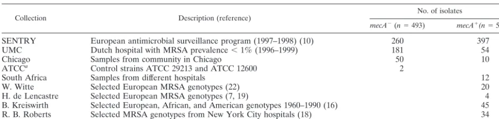
MSSA) were determined. These isolates were selected from different sources in order to study isolates from different temporal and geographic backgrounds. The origins of the isolates were as follows (Table 1): 397 MRSA and 260 MSSA isolates had been collected between April 1997 and December 1998 in 20 uni-versity hospitals in 12 European countries as part of the SENTRY Antimicrobial Surveillance Program (10). They included isolates from Athens, Greece (8
mecA⫺isolates, 19 mecA⫹isolates); Du¨sseldorf (10mecA⫺, 9mecA⫹) and
Freiburg (14mecA⫺, 2mecA⫹), Germany; Lausanne, Switzerland (56mecA⫺, 1
mecA⫹); Linz, Austria (7mecA⫺, 7mecA⫹); Paris (group 1) (10mecA⫺, 21
mecA⫹), Paris (group 2) (7mecA⫺, 33mecA⫹), Lille (7mecA⫺, 22mecA⫹), and
Lyon (7mecA⫺, 14mecA⫹), France; Coimbra, Portugal (16mecA⫺, 70mecA⫹);
Warsaw (7mecA⫺, 17mecA⫹) and Krakow (5mecA⫺, 5 mecA⫹), Poland;
Madrid (14mecA⫺, 2mecA⫹), Seville (18mecA⫺, 29mecA⫹), and Barcelona (7
mecA⫺, 6mecA⫹), Spain; Rome (10mecA⫺, 32mecA⫹) and Genoa (9mecA⫺,
34mecA⫹), Italy; Brussels, Belgium (10mecA⫺, 31mecA⫹); London, United
Kingdom (26mecA⫺, 36mecA⫹); and Istanbul, Turkey (12mecA⫺, 7mecA⫹).
An additional 181 MSSA and 54 MRSA isolates had been collected between 1996 and 1999 at the University Medical Center (UMC), Utrecht, The Nether-lands, where patients and staff coming from foreign hospitals are screened for MRSA carriage. These MRSA isolates, detected during 12 MRSA outbreak episodes, had evaded the hospital’s search-and-destroy procedure and could not be linked epidemiologically to any foreign hospitals. One hundred three more MRSA isolates were selected to represent the genetic diversity of the MRSA collections of Kreiswirth et al. (16), Roberts et al. (18), de Lencastre et al. (7, 19), and Witte et al. (22). They included the earliest isolates collected from Europe and Africa during the 1960s, North American isolates collected from the 1970s to the 1990s, and European reference strains like the Iberian clone (7), the Brazilian clone (7), the North German clone(22), the South German clone (22), the Berlin clone (22), the Hannover clone (22), the Portuguese clone (7), the Pediatric clone (19), EMRSA 15 (14), and EMRSA 16 (14). Another 12 MRSA isolates that had been collected in South Africa during 1998 were studied. For comparison, MSSA strains ATCC 29213 and ATCC 12600 were included.
Fi-* Corresponding author. Mailing address: Eijkman-Winkler Insti-tute for Microbiology, Infectious Diseases and Inflammation, Univer-sity Medical Centre, Room G04.614, Heidelberglaan 100, 3584 CX Utrecht, The Netherlands. Phone: 00 31 30 2506534. Fax: 00 31 30 2541770. E-mail: [email protected].
3970
on May 15, 2020 by guest
http://jcm.asm.org/
nally, 10 MRSA and 50 MSSA isolates that had been taken from colonized patients who had no clinical signs ofS. aureusinfection within 2 h after admission to the Cook County Hospital (Chicago, Ill.) were included.
The isolates were identified asS. aureusby routine microbiological methods. Only one isolate per patient was included.
Susceptibility testing.Susceptibility to oxacillin, erythromycin, clindamycin, rifampin, chloramphenicol, ciprofloxacin, gentamicin, and tetracycline was de-termined by the broth microdilution method defined by the National Committee for Clinical Laboratory Standards. Isolates were considered multiresistant when they displayed a decreased susceptibility to at least four of the eight antimicrobial agents tested.
Detection and amplification ofmecAby PCR.PCR was used to detectmecA
DNA in methicillin-resistant isolates. A few colonies were picked from blood agar; suspended in 200l of a lysis buffer containing 10 mM Tris-HCl buffer (pH 8.0), 50 mM NaCl, lysostaphin (100g/ml), achromopeptidase (100g/ml), and RNase (100g/ml); incubated at 30°C for 45 min; boiled for 5 min; and then diluted by the addition of 400l of TE (10 mM Tris-HCl [pH 8.0], 1 mM EDTA). For the PCR, 1l of lysate was added as a template to 24l of a reaction mixture containing 10 mM Tris-HCl (pH 9.0), 1.5 mM MgCl2, 50 mM KCl, 0.1% Triton
X-100, 0.2 mM each deoxynucleoside triphosphate, and 0.75 U of Supertaq DNA polymerase (HT Biotechnology, Cambridge, United Kingdom).MecADNA was amplified with the primers 5⬘-GTT GTA GTT GTC GGG TTT GG-3⬘ and 5⬘CTT CCA CAT ACC ATC TTC TTT AAC-3⬘(20M). These primers were designed on the basis of themecAsequence (GenBank accession no. X52593) with Primer software (Educational Software, State Line, Pa.). To test the quality of the template used for PCR, we added primers for the amplification of 16S ribosomal DNA (5⬘-AGG CCC GGG AAC GTA TTC AC-3⬘and 5⬘-GAG GAA GGT GGG GAT GAC GT-3⬘) (20M) to each PCR mixture and monitored the amplification of 16S ribosomal DNA. Samples were subjected to 30 cycles con-sisting of 1 min at 95°C, 1 min at an annealing temperature ramped from 65 to 55°C during the first 10 cycles, and 1 min at 72°C in a thermocycler. The PCR product was visualized on a 1.5% agarose gel by using ethidium bromide and a UV transilluminator.
Pulsed-field gel electrophoresis (PFGE) analysis.Genomic DNA was digested withSmaI and resolved with the CHEF-DRII system (Bio-Rad Laboratories, Hercules, Calif.), as described by the manufacturer.
Ribotyping.Ribotypes were determined with an automated riboprinter system (Qualicon, Wilmington, Del.) andEcoRI, as described by the manufacturer.
Analysis of restriction patterns.The restriction patterns were compared by calculating a similarity index by using the unweighted pair group method with arithmetic averages cluster algorithm and the Dice coefficient provided by the Bionumerics software (Applied Mathematics, Kortrijk, Belgium).
RESULTS
Clonal relationships among S. aureus isolates. When the
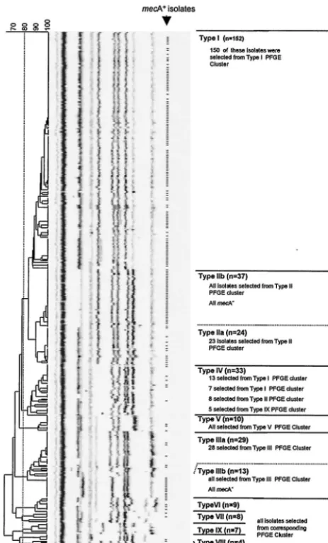
PFGE patterns of 576 MRSA isolates and 493 MSSA isolates were compared in a dendrogram, 10 major clusters of various sizes were discerned. To confirm the clonal relatedness of the isolates within these clusters, 330 isolates were selected for ribotyping, which covered the chromosomal diversity of the isolates (Fig. 1). Compared to the diversity of the PFGE pat-terns, the riboprints were much more conserved during
evolu-tion. Ten clusters were distinguished at the 80% similarity level, and these 10 clusters defined clonal lineages called S. aureustypes I to X (Fig. 2). A very good correlation between PFGE typing and ribotyping was observed: except for the type IV isolates, 99% of the isolates typed by both methods clus-tered in corresponding branches of both the riboprint and the PFGE dendrograms. The PFGE patterns obtained for the type IV isolates were found not only in a cluster of their own but also in the PFGE clusters of other types as well (Fig. 1). Furthermore, both type II and type III isolates could be di-vided into two subtypes (subtypes a and b) that formed sub-clusters in the ribotyping dendrogram and that had different susceptibility patterns.
The more successful S. aureus types consisted of isolates which displayed numerous band-shift variations on widely dis-seminated and frequently isolated dominant PFGE patterns (Fig. 1). Pandemic clones of isolates yielding identical PFGE patterns were present in many hospitals on both continents. Of the type I (40% of all samples), type II (20%), and type III (12%) lineages, indistinguishable isolates were present in nearly all hospitals studied. Type IV (8%), type VI (8%), type IX (7%), and type VIII (2%) clones were also detected in Europe and North America, although less frequently. Isolates of the remaining lineages (type V [2%], type VII [0.5%], and type X [0.5%]) were referred only from European countries.
Dissemination ofmecAin the differentS. aureuslineages.All
isolates was assessed for the presence ofmecAby PCR.MecA
was detected in representatives of all but the type IX and X lineages. Some pandemic mecA⫺ MSSA clones came with mecA⫹MRSA counterparts that shared the identical ribotype, while their PFGE patterns differed by a single band shift due to insertion of a fragment containingmecA(Fig. 3). However, we found no mecA⫺counterparts among type IIb and type IIIb isolates, which, with no exception, all containedmecA.
[image:2.603.43.547.80.200.2]More than 60% of themecA⫹isolates belonged to the type I lineage. These isolates predominated in North America, Af-rica, and Europe. The type I MRSA isolates included all MRSA isolates from the 1960s, the Brazilian clone (7), the North German clone (22), the Hannover clone (22), the Ibe-rian clone (7), and the Portuguese clone (7). One-quarter of themecA⫹isolates belonged to the type II lineage. Isolates of the type II lineage were dominant among the North American isolates from the 1980s and were later isolated in Europe and Africa. Type IIa MRSA isolates (14% of themecA⫹isolates) included the Pediatric clone (19). Type IIb MRSA isolates TABLE 1. Origins of isolates
Collection Description (reference) No. of isolates
mecA⫺(n⫽493) mecA⫹(n⫽576)
SENTRY European antimicrobial surveillance program (1997–1998) (10) 260 397
UMC Dutch hospital with MRSA prevalence⬍1% (1996–1999) 181 54
Chicago Samples from community in Chicago 50 10
ATCCa Control strains ATCC 29213 and ATCC 12600 2
South Africa Samples from different hospitals 12
W. Witte Selected European MRSA genotypes (22) 20
H. de Lencastre Selected European MRSA genotypes (7, 19) 4
B. Kreiswirth Selected European, African, and American genotypes 1960–1990 (16) 45
R. B. Roberts Selected MRSA genotypes from New York City hospitals (18) 34
aATCC, American Type Culture Collection.
VOL. 40, 2002 mecADISSEMINATION IN S. AUREUS POPULATION 3971
on May 15, 2020 by guest
http://jcm.asm.org/
FIG. 1. PFGE patterns from European and North American MSSA (n⫽493) and MRSA (n⫽576) isolates form 10 clusters. Isolates containingmecAare indicated by hyphens. In order to confirm the clonal relatedness of the isolates within the 10 clusters, a total of 330 MSSA and MRSA isolates representing the various PFGE types encountered were selected for ribotyping (indicated by hyphens). The positions of the type IV isolates in the PFGE dendrogram are indicated by black lines.
3972
on May 15, 2020 by guest
(11% of mecA⫹ isolates) included the South German clone (22).
Type III MRSA isolates were found among isolates recov-ered since the 1980s in Africa, Europe, and North America. Type IIIb MRSA isolates (6% of mecA⫹ isolates) included EMRSA 16 (14). In contrast, type IIIa, type IV (the Berlin clone [22]), type V (EMRSA 15 [14]), type VI, type VII, and type VIII MRSA strains, which appeared during the 1990s, were isolated only sporadically (⬍1% ofmecA⫹isolates). In-terestingly, these sporadic MRSA types were relatively abun-dant among the different UMC genotypes (33% type IIa, type IV, and type VI isolates), which are epidemiologically unlinked to the hospital epidemic MRSA isolates, and among the
mecA⫹isolates from the community in Chicago (20% type IV isolates).
Multiresistance and dissemination of MRSA in Europe.To
correlate the resistance profiles of MRSA strains with their current dissemination, the susceptibilities of the recent Euro-pean S. aureusisolates were compared. The mecA gene was present in all isolates resistant to four or more antibiotics. Moreover, this multiresistance was displayed by the most prev-alent and geographically widespread MRSA types (types I, IIa, IIb, and IIIb), which together represented 99% of themecA⫹ population in Europe. Of the European isolates that appeared to be susceptible to methicillin in the phenotypic test, 10% nevertheless containedmecA, and some of these were multire-sistant. In contrast, 5% of the phenotypically methicillin-resis-tant isolates did not carrymecAand displayed low-level resis-tance (⬎8g/ml).
[image:4.603.48.280.71.455.2]Most of the type I MRSA isolates, representing 68% of the recent European MRSA population and being present in 17 of 20 hospitals participating in the SENTRY program, were
FIG. 2. Ten clusters were obtained by ribotyping of 330 isolates, which represented the variability of PFGE types encountered. These clonalS. aureuslineages were called types I to X. There was an excel-lent correlation between PFGE and ribotyping. Except for the type IV isolates, 99% of the isolates typed by both methods were found in the corresponding PFGE and ribotype clusters. Isolates containingmecA
[image:4.603.307.528.285.676.2]are indicated by hyphens. Automated riboprinting allows standardized exchange of data between laboratories.
FIG. 3. PFGE patterns ofmecA⫹andmecA⫺counterparts (upper
panels) that differ by a single band shift (arrowhead) due to the inser-tion of a fragment that hybridizes with amecAprobe (lower panels). The type II and type VI MSSA PFGE patterns are for control strains ATCC 29213 and ATCC 12600, respectively.
VOL. 40, 2002 mecADISSEMINATION IN S. AUREUS POPULATION 3973
on May 15, 2020 by guest
http://jcm.asm.org/
resistant to erythromycin (97%), gentamicin (98%), and clin-damycin (89%) and showed decreased susceptibility to cipro-floxacin (98%), tetracycline (98%), and rifampin (98%). Al-though most of the type IIb MRSA isolates, which represented 16% of the recent European isolates and which were found in 5 of 20 hospitals participating in the SENTRY program, were resistant to erythromycin (98%), clindamycin (88%), cipro-floxacin (98%), and gentamicin (100%), they remained suscep-tible to tetracycline (98%) and rifampin (100%). Type IIIb MRSA isolates, which represented 8% of the recent European MRSA isolate population and which were found in 2 of 21 hospitals participating in the SENTRY program, were resis-tant to erythromycin (100%), ciprofloxacin (100%), and clin-damycin (84%) but remained susceptible to rifampin (100%), tetracycline (100%), and gentamicin (84%). Also, the type IIa MRSA isolates, which represented 7% of the MRSA isolate population and which were found in 6 of 20 hospitals par-ticipating in the SENTRY program, were mostly resistant to erythromycin (75%), clindamycin (69%), and ciprofloxacin (90%) but remained susceptible to rifampin (100%), tetracy-cline (100%), and gentamicin (100%). In contrast, type IIIa, type IV, type V, type VI, type VII, and type VIII MRSA iso-lates, which were isolated only sporadically (⬍1% of recent EuropeanmecA⫹isolates), mostly remained susceptible to all but the-lactam antibiotics (clindamycin, 100%; tetracycline, 100%; gentamicin, 100%; rifampin, 100%; erythromycin, 60%; ciprofloxacin, 60%).
DISCUSSION
ThemecAgene, which lies in the SCCmecresistance island (13), is carried by 95% of the isolates that display a phenotype of methicillin resistance and was detected in all multiresistant
S. aureusisolates. This study aimed to examine the dissemina-tion ofmecAin theS. aureus population. Two methods were applied to determine the clonal relationships betweenmecA⫹ MRSA andmecA⫺MSSA isolates collected between 1960 and 2000 from over 50 locations in the Western world. The overall chromosomal organizations of the isolates were first compared by using SmaI-generated PFGE patterns, which provide a relatively quickly evolving genotypic marker. Because there is little evolutionary pressure to conserve the SmaI restriction sites per se, this technique has high discriminatory power and highlights some of the differences between the strains. Ribo-typing was then used to combine evolutionarily closely related PFGE types into clonal lineages. The complete ribotyping pro-cedure has been automated and is coupled to a database man-agement system, allowing electronic data exchange between different centers. The genes that encode ribosomal DNA are more conserved during evolution and provide a relatively slowly evolving marker. Therefore, automated ribotyping re-sults in fewer ribotypes compared to the diversity of PFGE types. Combination of PFGE and ribotyping allowed grouping of all geographically widespread isolates in distinct clonally related lineages except for some of the type IV isolates, which may be ancestrally related to many types.
S. aureusisolates of 10 different lineages, called types I to X, were present in the hospitals studied, and 8 of these have acquiredmecA. Our data show the worldwide dissemination of both successful MSSA strains and successful MRSA strains.
From the major lineages, both pandemic MSSA and MRSA clones yielding identical PFGE patterns were collected in many European and North American hospitals and the com-munity. Several pandemicmecA⫺MSSA clones havemecA⫹ counterparts that share identical ribotypes, while their PFGE patterns differ by a single band shift due to acquisition of the element containingmecA. The worldwide appearance of spe-cific MRSA clones has been shown before (7), but the exis-tence of widespread MSSA counterparts was not described before in detail. In line with this observation, a comparison of MRSA and MSSA isolates isolated in the United Kingdom and Denmark in the early 1960s suggests that contemporary MSSA isolates served as an early recipient of themecAgene in Eu-rope (6).
The dissemination of particular MRSA lineages is corre-lated with their resistance profiles. The majority of the multi-resistant type I MRSA isolates, predominant on both conti-nents, lacked susceptibility to tetracycline, erythromycin, clindamycin, gentamicin, ciprofloxacin, and rifampin. A second multiresistant lineage (type IIb), found in North America and several European countries, was susceptible to tetracycline and rifampin. Smaller pandemics were caused by isolates suscepti-ble to gentamicin, tetracycline, and rifampin (type IIa and type IIIb). Although antibiotic selection pressure by itself provides a reasonable explanation for the widespread dissemination of such multiresistant strains, additional factors, e.g., modifica-tions in expression of virulence factors and binding capacities, may add to their high prevalence. In contrast, the sporadically isolatedmecA⫹MRSA types, type IIIa and types IV to VIII (1% of recent European isolates), generally remained suscep-tible to all except the -lactam antibiotics. Sporadic MRSA types were overrepresented in the samples from the commu-nity in Chicago (20%) and UMC (30%), where the search-and-destroy procedure prevents epidemic MRSA strains from entering the hospital, keeping the prevalence of MRSA isolates below 1%. In both the community and the hospital, sporadic MRSA types, which are not descendants of the epi-demic hospital clones, may be formed de novo by the horizon-tal transfer ofmecA to resident MSSA lineages, as was wit-nessed recently in vivo when the gene was passed from
S. epidermidistoS. aureusduring antibiotic treatment (21). Several investigators (2, 9, 17, 21) have questioned the hy-pothesis that the gene enteredS. aureuson only one occasion. The data presented here show the repeated horizontal transfer ofmecA to at least eight residentS. aureuslineages and the spread of more resistant clones favored most by antibiotic selection pressure. When DNA microarray technology was used to characterize the genetic diversity of 11 MRSA isolates,
mecA was detected in at least five highly divergent chromo-somal genetic groups (9). In addition, multilocus enzyme trophoresis data showed that MRSA isolates comprise 15 elec-trophoretic types that form six clusters or lineages, indicating that multiple MRSA lineages arose by horizontal transfer toS. aureus(17). Analysis of themecIandmecR1region, which lies 5⬘ of mecA, revealed that older isolates lacked part of this region, whereas more recent isolates showed an organization at this position similar to that in coagulase-negative staphylo-cocci (CoNS) (2, 12), and it has been proposed that CoNS serve as donors for the transfer of themecAgene toS. aureus
(1, 20, 21). In this context, it is noteworthy that 70 to 75% of all
on May 15, 2020 by guest
http://jcm.asm.org/
CoNS worldwide are now resistant to methicillin (8), thus representing a huge potential reservoir of resistance. The mechanism of transfer, however, remains unclear. There is evidence thatmecAresides within a mobile genetic element, SSCmec, that encodes recombinases for its excision from and integration into the staphylococcal chromosome (15). This el-ement may also contain other genetic elel-ements, like Tn554, pUB110, and pT181, which encode resistance to non--lactam antibiotics, causing multiresistance (15). Thus, while new MRSA strains will continue to emerge by the horizontal trans-fer of themecAgene, those strains disseminated widely possess additional resistance traits and are favored most by antibiotic selection pressure.
ACKNOWLEDGMENTS
We acknowledge B. Kreiswirth, R. B. Roberts, H. de Lencastre, and W. Witte, who collected many of the isolates. In addition, we thank Adrienne Box, Mirjam Klootwijk, Karlijn Kusters, Stefan de Vaal, and Roland Geisel for proficient technical support and Max Heck for advice on PFGE.
REFERENCES
1. Archer, G. L., and D. M. Niemeyer.1994. Origin and evolution of DNA associated with resistance to methicillin in staphylococci. Trends Microbiol.
2:343–347.
2. Archer, G. L., D. M. Niemeyer, J. A. Thanassi, and M. J. Pucci.1994. Dissemination among staphylococci of DNA sequences associated with methicillin resistance. Antimicrob. Agents Chemother.38:447–454. 3. Beck, W. D., B. Berger-Bachi, and F. H. Kayser.1986. Additional DNA in
methicillin-resistantStaphylococcus aureusand molecular cloning ofmec -specific DNA. J. Bacteriol.165:373–378.
4. Centers for Disease Control and Prevention.1999. Four pediatric deaths from community acquired methicillin-resistantS. aureus—Minnesota and North Dakota, 1997–1999. Morb. Mortal. Wkly. Rep.48:707–710. 5. Chambers, H. F.2001. The changing epidemiology of Staphylococcus
au-reus? Emerg. Infect. Dis.7:178–182.
6. Crisostomo, M. I., H. Westh, A. Tomasz, M. Chung, D. C. Oliveira, and H. de Lencastre.2001. The evolution of methicillin resistance in Staphylococcus aureus: similarity of genetic backgrounds in historically early methicillin-susceptible and -resistant isolates and contemporary epidemic clones. Proc. Natl. Acad. Sci. USA98:9865–9870.
7. de Sousa, M. A., I. S. Sanches, M. L. Ferro, M. J. Vaz, Z. Saraiva, T. Tendeiro, J. Serra, and H. de Lencastre.1998. Intercontinental spread of a multidrug-resistant methicillin-resistantStaphylococcus aureusclone. J. Clin. Microbiol.36:2590–2596.
8. Diekema, D. J., M. A. Pfaller, F. J. Schmitz, J. Smayevsky, J. Bell, R. N. Jones, and M. Beach.2001. Survey of infections due to Staphylococcus species: frequency of occurrence and antimicrobial susceptibility of isolates
collected in the United States, Canada, Latin America, Europe, and the Western Pacific region for the SENTRY antimicrobial surveillance program, 1997–1999. Clin. Infect Dis.32(Suppl. 2):S114–S132.
9. Fitzgerald, J. R., D. E. Sturdevant, S. M. Mackie, S. R. Gill, and J. M. Musser.2001. Evolutionary genomics of Staphylococcus aureus: insights into the origin of methicillin-resistant strains and the toxic shock syndrome epi-demic. Proc. Natl. Acad. Sci. USA98:8821–8826.
10. Fluit, A. C., C. L. Wielders, J. Verhoef, and F. J. Schmitz.2001. Epidemi-ology and susceptibility of 3,051Staphylococcus aureusisolates from 25 university hospitals participating in the European SENTRY Study. J. Clin. Microbiol.39:3727–3732.
11. Herold, B. C., L. C. Immergluck, M. C. Maranan, D. S. Lauderdale, R. E. Gaskin, S. Boyle-Vavra, C. D. Leitch, and R. S. Daum.1998. Community-acquired methicillin-resistant Staphylococcus aureus in children with no identified predisposing risk. JAMA279:593–598.
12. Hiramatsu, K.1995. Molecular evolution of MRSA. Microbiol. Immunol.
39:531–543.
13. Hiramatsu, K., L. Cui, M. Kuroda, and T. Ito.2001. The emergence and evolution of methicillin-resistant Staphylococcus aureus. Trends Microbiol.
9:486–493.
14. Hookey, J. V., J. F. Richardson, and B. D. Cookson.1998. Molecular typing ofStaphylococcus aureusbased on PCR-restriction fragment length polymor-phism and DNA sequence analysis of the coagulase gene. J. Clin. Microbiol.
36:1083–1089.
15. Ito, T., and K. Hiramatsu.1998. Acquisition of methicillin resistance and progression of multiantibiotic resistance in methicillin-resistant Staphylococ-cus aureus. Yonsei Med. J.39:526–533.
16. Kreiswirth, B., J. Kornblum, R. D. Arbeit, W. Eisner, J. N. Maslow, A. McGeer, D. E. Low, and R. P. Novick.1993. Evidence for a clonal origin of methicillin resistance in Staphylococcus aureus. Science259:227–230. 17. Musser, J. M., and V. Kapur.1992. Clonal analysis of methicillin-resistant
Staphylococcus aureusstrains from intercontinental sources: association of themecgene with divergent phylogenetic lineages implies dissemination by horizontal transfer and recombination. J. Clin. Microbiol.30:2058–2063. 18. Roberts, R. B., A. de Lencastre, W. Eisner, E. P. Severina, B. Shopsin, B. N.
Kreiswirth, A. Tomasz, et al.1998. Molecular epidemiology of methicillin-resistant Staphylococcus aureus in 12 New York hospitals. J. Infect. Dis.
178:164–171.
19. Sa-Leao, R., I. Santos Sanches, D. Dias, I. Peres, R. M. Barros, and H. de Lencastre.1999. Detection of an archaic clone ofStaphylococcus aureuswith low-level resistance to methicillin in a pediatric hospital in Portugal and in international samples: relics of a formerly widely disseminated strain? J. Clin. Microbiol.37:1913–1920.
20. Tesch, W., A. Strassle, B. Berger-Bachi, D. O’Hara, P. Reynolds, and F. H. Kayser.1988. Cloning and expression of methicillin resistance from Staph-ylococcus epidermidisinStaphylococcus carnosus. Antimicrob. Agents Che-mother.32:1494–1499.
21. Wielders, C. L., M. R. Vriens, S. Brisse, L. A. de Graaf-Miltenburg, A. Troelstra, A. Fleer, F. J. Schmitz, J. Verhoef, and A. C. Fluit.2001. Evidence for in-vivo transfer of mecA DNA between strains of Staphylococcus aureus. Lancet357:1674–1675.
22. Witte, W., M. Enright, F. J. Schmitz, C. Cuny, C. Braulke, and D. Heuck.
2001. Characteristics of a new epidemic MRSA in Germany ancestral to United Kingdom EMRSA 15. Int. J. Med. Microbiol.290:677–682.
VOL. 40, 2002 mecADISSEMINATION IN S. AUREUS POPULATION 3975