Desorption Ionization–Time of Flight Mass Spectrometry System for
Identification of Gram-Positive Aerobic Bacteria
Jenna Rychert,aCarey-Ann D. Burnham,bMaureen Bythrow,dOmai B. Garner,eChristine C. Ginocchio,fRebecca Jennemann,c Michael A. Lewinski,eRyhana Manji,dA. Brian Mochon,eGary W. Procop,gSandra S. Richter,gLinda Sercia,gLars F. Westblade,b* Mary Jane Ferraro,aJohn A. Brandaa
Department of Pathology, Massachusetts General Hospital and Harvard Medical School, Boston, Massachusetts, USAa; Department of Pathology & Immunology, Washington University School of Medicine, St. Louis, Missouri, USAb; Barnes Jewish Hospital, St. Louis, Missouri, USAc; Department of Pathology and Laboratory Medicine, North Shore-LIJ Health System Laboratories, Lake Success, New York, USAd; Department of Pathology and Laboratory Medicine, David Geffen School of Medicine, University of California, Los Angeles, Los Angeles, California, USAe; Department of Pathology and Laboratory Medicine, Hofstra North Shore-LIJ School of Medicine, Hempstead, New York, USAf; Department of Clinical Pathology, Cleveland Clinic, Cleveland, Ohio, USAg
Matrix-assisted laser desorption ionization–time of flight mass spectrometry (MALDI-TOF) is gaining momentum as a tool for
bacterial identification in the clinical microbiology laboratory. Compared with conventional methods, this technology can more
readily and conveniently identify a wide range of organisms. Here, we report the findings from a multicenter study to evaluate
the Vitek MS v2.0 system (bioMérieux, Inc.) for the identification of aerobic Gram-positive bacteria. A total of 1,146 unique
isolates, representing 13 genera and 42 species, were analyzed, and results were compared to those obtained by nucleic acid
sequence-based identification as the reference method. For 1,063 of 1,146 isolates (92.8%), the Vitek MS provided a single
identification that was accurate to the species level. For an additional 31 isolates (2.7%), multiple possible identifications
were provided, all correct at the genus level. Mixed-genus or single-choice incorrect identifications were provided for 18
isolates (1.6%). Although no identification was obtained for 33 isolates (2.9%), there was no specific bacterial species for
which the Vitek MS consistently failed to provide identification. In a subset of 463 isolates representing commonly
en-countered important pathogens, 95% were accurately identified to the species level and there were no misidentifications.
Also, in all but one instance, the Vitek MS correctly differentiated
Streptococcus pneumoniae
from other viridans group
streptococci. The findings demonstrate that the Vitek MS system is highly accurate for the identification of Gram-positive
aerobic bacteria in the clinical laboratory setting.
I
n the clinical microbiology laboratory, bacteria are typically
identified using phenotypic and biochemical methods. These
methods are generally reliable but have several drawbacks. In
par-ticular, they can be time-consuming, sometimes taking several
days before a definitive identification can be reached.
Further-more, many are subjectively interpreted, rendering them
suscep-tible to misinterpretation and requiring substantial training and
experience on the part of the technologist to establish and
main-tain competency. In the case of some Gram-positive bacteria, such
as the coagulase-negative staphylococci, the viridans group
strep-tococci, and some enterococci, distinguishing one species from
another using these methods can be unreliable or overly
compli-cated (
1
). Thus, many laboratories do not attempt to distinguish
among them, or do so only selectively.
Matrix-assisted laser desorption–ionization time of flight mass
spectrometry (MALDI-TOF MS) has the potential to address
these drawbacks. Proteomic analysis of bacteria by mass
spec-trometry can be performed as soon as colonies are isolated, often
in primary culture, and the analysis itself takes just a few minutes
(
2
–
6
). Thus, on average, routine bacterial identifications can be
obtained more than 30 hours earlier than identification by
con-ventional methods (
7
). MALDI-TOF MS also provides equal or
superior accuracy compared with conventional phenotypic
meth-ods (
8
–
10
) and does not require subjective interpretation. Finally,
it can readily differentiate bacterial species that have similar
phe-notypic characteristics (
11
–
19
).
There are only a few commercially available MALDI-TOF MS
systems used for identification of bacteria. Numerous studies have
described the performance of the two most common, the Bruker
Biotyper and the bioMérieux Vitek MS RUO, primarily compared
to phenotypic testing as the reference method (
3
,
4
,
6
,
8
,
9
,
13
–
23
).
There are currently no published reports available on the
perfor-mance of the Vitek MS IVD system, with its new method of
spec-tral analysis and updated database (version 2.0). Here, we report
the findings of a large multicenter evaluation of the Vitek MS v2.0
system for the identification of Gram-positive aerobic bacteria in
which DNA sequence-based identification served as the reference
method.
Received12 March 2013Returned for modification1 April 2013
Accepted30 April 2013
Published ahead of print8 May 2013
Address correspondence to Jenna Rychert, [email protected].
* Present address: Lars F. Westblade, Department of Pathology and Laboratory Medicine, Hofstra North Shore-LIJ School of Medicine, Hempstead, New York, USA.
Supplemental material for this article may be found athttp://dx.doi.org/10.1128 /JCM.00682-13.
Copyright © 2013, American Society for Microbiology. All Rights Reserved. doi:10.1128/JCM.00682-13
on May 16, 2020 by guest
http://jcm.asm.org/
MATERIALS AND METHODS
Study sites.The performance of the Vitek MS v2.0 system (bioMérieux, Inc., Durham, NC) was assessed at five sites within the United States (Boston, MA; St. Louis, MO; Los Angeles, CA; Lake Success, NY; and Cleveland, OH). Prior to initiation of the study, operators at each site were trained in target slide preparation, instrument use, and result review. Each operator was required to demonstrate proficiency by successfully analyz-ing a masked panel of organisms consistanalyz-ing of 10 isolates representanalyz-ing common aerobic and anaerobic Gram-positive and Gram-negative bac-teria and yeasts. This study was approved by the human subject commit-tees at the respective sites, when deemed necessary by their internal review boards.
Bacterial isolates.Fresh isolates obtained in the course of routine clinical work during the study period were collected and tested at each of the study sites. Each site was responsible for analyzing a minimum of 10 isolates per species for a predetermined set of frequently encountered organisms (Enterococcus faecalis,Enterococcus faecium,Staphylococcus au-reus, Staphylococcus epidermidis, Streptococcus agalactiae, Streptococcus pneumoniae, andStreptococcus pyogenes) and six isolates per species for a predetermined set of less frequently encountered organisms (including Enterococcus avium,Enterococcus casseliflavus,Enterococcus durans, En-terococcus gallinarum,Gardnerella vaginalis,Listeria monocytogenes, Mi-crococcus luteus/lylae,Staphylococcus capitis,Staphylococcus haemolyticus, Staphylococcus hominissubsp.hominis,Staphylococcus lugdunensis, Staph-ylococcus saprophyticus,Staphylococcus simulans,Staphylococcus warneri, Streptococcus anginosus,Streptococcus constellatus,Streptococcus dysgalac-tiaesubsp.equisimilis,Streptococcus mitis/Streptococcus oralis, and Strep-tococcus sanguinis). In the event that a rarer organism, not included in the list above, was tested, it was included in the study. If a site was unable to obtain the minimum number of preselected species, additional testing of frozen isolates obtained from their culture collections or provided by the study sponsor was permitted. In all cases, cultures were incubated under standard conditions for a minimum of 18 h at 35 to 37°C in ambient air or with CO2enrichment. Each isolate was analyzed on the Vitek MS system within 72 h of visible growth either from the primary culture or from a subculture. Subculturing was performed when colonies on the primary culture plate were insufficiently isolated or for convenience to allow batching. All subculturing was performed using tryptic soy agar with 5% sheep blood (Remel, Lenexa, KS). More than 95% of the isolates were tested after growth on this medium. Of the remaining isolates, 53 were tested after growth on brucella agar supplemented with 5% horse blood (Remel, Lenexa, KS), two after growth on choc-olate agar (Remel, Lenexa, KS), and one after growth on human blood-Tween bilayer (BD, Franklin Lakes, NJ). Frozen (archived) isolates were serially subcultured twice on tryptic soy agar with 5% sheep blood prior to analysis. Duplicate isolates of the same species from the same patient were not included in the study.
Sample preparation for MALDI-TOF MS analysis.Isolated bacterial colonies were applied to a single well of a disposable, barcode-labeled target slide (Vitek MS-DS; bioMérieux, Inc.) using a 1.0-l loop, overlaid with 1.0l of a saturated solution of alpha-cyano-4-hydroxycinnamic acid matrix in 50% acetonitrile and 2.5% trifluoroacetic acid (Vitek MS-CHCA; bioMérieux, Inc.), and air dried. The same bacterial culture was also used to set up a slide for Gram stain and prepare the organism for shipment to a centralized laboratory for reference testing.
For instrument calibration, an Escherichia coli reference strain (ATCC 8739) was transferred to designated wells on the target slide using the procedure described above. For quality control purposes, positive and negative controls were analyzed on each day of testing. The positive control consisted of at least one of four quality control organisms, includingStaphylococcus aureus(ATCC 29213),Klebsiella oxytoca(ATCC 13182),Pseudomonas aeruginosa(ATCC 10145), and Enterobacter aerogenes(ATCC 13048). The negative control was ma-trix alone.
Organism identification using the Vitek MS v2.0 system.The Vitek MS v2.0 system includes an OEM (original equipment manufacturer)-labeled Shimadzu AXIMA Assurance mass spectrometer linked to a reference database, referred to as Knowledge Base. During target in-terrogation, mass spectra within a range of 2,000 to 20,000 Da are recorded in linear positive mode at a laser frequency of 50 Hz. For each interrogation, laser shots at different positions within the target well produce up to 100 mass profiles that are summed into a single, raw mass spectrum. The spectrum is then processed by baseline correction, denoising, and peak detection to identify well-defined peaks. The list of these significant peaks is subjected to a proprietary process called “mass binning” (see the supplemental material). The processed (binned) data are used to query Knowledge Base to determine the unknown’s taxonomic identity. These results are then provided in the form of a single species-level (and sometimes subspecies-level) iden-tification, a split (low discrimination) identification with up to four species-level alternatives displayed, or no identification. For the pres-ent study, when a report of “no idpres-entification” was provided, or when analysis of an isolate yielded absent or poor-quality spectra, the organ-ism was reanalyzed a single time on a new target slide. Isolates yielding split identifications were not reanalyzed. Additionally, isolates that yielded a reference identification that is not included in the database were excluded from analysis.
Reference method for bacterial identification.All study isolates were sent to a centralized laboratory (MIDI Labs, Inc., Newark, DE) for nucleic acid sequence-based identification. Sequencing of a 527-bp region within the 16S rRNA gene was performed using universal 16S primers at posi-tions 0005F and 0531R (MicroSeq system; Applied Biosystems, Carlsbad, CA). The resulting sequence was run through the MicroSeq, GenBank, and BiBi databases to determine the identity. In the event of a discrepancy or low-discrimination result, or when no match was obtained using this method, supplemental sequencing of a different gene target (sodA or rpoB) and/or phenotypic testing was performed by bioMérieux, Inc. (Durham, NC).
Analysis.The Vitek MS result was considered accurate to the species level if a single identification was given and it matched that obtained by the reference method. The Vitek MS result was considered correct to the genus level if multiple alternative identifications, all from the same genus, were reported and this matched the genus obtained by the reference method. The Vitek MS result was considered incorrect if a single identification was given that did not match (at some taxonomic level) the result obtained with the reference method, when multiple identifications of different genera were reported, or when multiple identifications of the same genera were reported but did match the genus of the reference method.
RESULTS
Overall results.
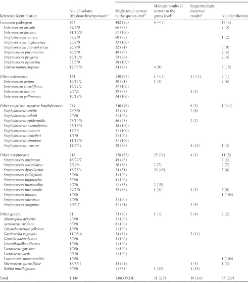
A total of 1,146 unique, aerobic, Gram-positive
isolates was analyzed, representing 16 genera and 42 species. As
shown in
Table 1
, for 1,063 isolates (92.8%), the Vitek MS
pro-vided a single identification that matched at the species level the
identification obtained by the reference method. An additional 31
isolates (2.7%) were correctly identified to the genus level,
mean-ing that the Vitek MS result included a split identification with
multiple alternative species all from the same genus, and the genus
in all cases matched that obtained by the reference method (
Tables
1
and
2
). For all but one of these isolates, the correct species was
listed among the alternate identifications provided (
Table 2
). A
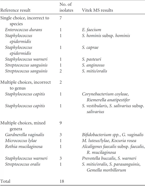
total of 18 isolates were not accurately identified (1.6%) (
Tables 1
and
3
). This included seven isolates for which a single
identifica-tion was provided that was accurate to the genus level but not the
species level, two isolates for which multiple identifications were
provided, all of which were incorrect to the genus level, and nine
isolates where multiple different genera were reported. In the later
on May 16, 2020 by guest
http://jcm.asm.org/
TABLE 1Accuracy of organism identification using the Vitek MS v2.0 system
Reference identification
No. of isolates (fresh/archive/sponsor)a
No. (%) with Vitek MS result
Single result correct to the species levelb
Multiple results all correct to the genus levelc
Single/multiple incorrect
resultsd No identification
Common pathogens 463 442 (95) 4 (⬍1) 17 (4)
Enterococcus faecalis 62/6/0 66 (97) 2 (3)
Enterococcus faecium 41/16/0 57 (100)
Staphylococcus aureus 59/2/0 60 (98) 1 (2)
Staphylococcus lugdunensis 25/8/0 33 (100)
Staphylococcus saprophyticus 26/9/0 32 (91) 3 (9)
Streptococcus pneumoniae 45/6/0 49 (96) 2 (4)
Streptococcus pyogenes 45/10/0 53 (96) 2 (4)
Streptococcus agalactiae 53/5/0 58 (100)
Listeria monocytogenes 12/33/0 34 (76) 4 (9) 7 (15)
Other enterococci 134 130 (97) 1 (⬍1) 1 (⬍1) 2 (1)
Enterococcus avium 16/15/2 30 (91) 1 (3) 2 (6)
Enterococcus casseliflavus 13/22/2 37 (100)
Enterococcus durans 2/7/21 29 (97) 1 (3)
Enterococcus gallinarum 10/19/5 34 (100)
Other coagulase-negative Staphylococci 249 240 (96) 8 (3) 1 (⬍1)
Staphylococcus capitis 26/8/0 32 (94) 2 (6)
Staphylococcus cohnii 2/0/0 2 (100)
Staphylococcus epidermidis 78/10/0 86 (98) 2 (2)
Staphylococcus haemolyticus 23/15/0 38 (100)
Staphylococcus hominis 17/3/1 21 (100)
Staphylococcus schleiferi 1/1/0 2 (100)
Staphylococcus simulans 11/14/6 31 (100)
Staphylococcus warneri 14/7/12 28 (85) 4 (12) 1 (3)
Other streptococci 218 178 (82) 25 (11) 4 (2) 11 (5)
Streptococcus anginosus 18/22/7 45 (96) 2 (4)
Streptococcus constellatus 7/19/4 26 (86) 2 (7) 2 (7)
Streptococcus dysgalactiae 18/25/4 24 (51) 20 (43) 3 (6)
Streptococcus gallolyticus 3/0/0 3 (100)
Streptococcus infantarius 5/0/0 4 (100)
Streptococcus intermedius 6/7/0 11 (85) 2 (15)
Streptococcus mitis/oralis 29/7/0 32 (86) 1 (3) 1 (3) 3 (8)
Streptococcus mutans 1/0/0 1 (100)
Streptococcus salivarius 2/0/0 2 (100)
Streptococcus sanguinis 9/8/17 31 (91) 3 (9)
Other genera 81 73 (90) 1 (1) 5 (6) 2 (2)
Abiotrophia defective 2/0/0 2 (100)
Aerococcus viridans 6/0/0 6 (100)
Corynebacterium jeikeium 1/0/0 1 (100)
Gardnerella vaginalis 11/0/16 24 (89) 3 (11)
Gemella haemolysans 3/0/0 3 (100)
Granulicatella adiacens 1/0/0 1 (100)
Lactococcus garvieae 1/0/0 1 (100)
Lactococcus lactis 0/1/0 1 (100)
Leuconostoc mesenteroides 1/0/0 1 (100)
Micrococcus luteus/lylae 16/8/11 33 (94) 1 (3) 1 (3)
Rothia mucilaginosa 3/0/0 1 (33) 1 (33) 1 (33)
Total 1,146 1,063 (92.8) 31 (2.7) 18 (1.6) 33 (2.9)
a
Fresh isolates were those obtained during the normal clinical workflow; archived isolates were obtained from frozen stocks at one of the trial sites; sponsor isolates were obtained from the sponsor’s frozen stocks. Sponsor-derived isolates were not used in the development of Knowledge Base.
b
The result was a single identification that matched the reference method to the species level.
cThe result was multiple possible identifications, all within the same genus, which matched (at the genus level) the identification obtained by the reference method.
d
The result was either a single identification that did not match the identification obtained by the reference method or multiple possible identifications that included more than one genus.
on May 16, 2020 by guest
http://jcm.asm.org/
cases, the correct species-level identification was included among
the alternatives. Finally, no identification was obtained for 33
iso-lates, all of which were represented in the database (2.9%) (
Table
1
). There was no specific bacterial species for which the Vitek MS
consistently failed.
Commonly encountered important pathogens.
Given the
breadth of species analyzed in this study, some of which are rarely
encountered in the clinical microbiology laboratory, we
consid-ered the performance of the Vitek MS system for the identification
of a subset of commonly encountered and clinically important
Gram-positive bacteria (
Table 1
). This set of organisms was
cho-sen prior to data analysis on the basis of being pathogenic and
commonly isolated from clinical specimens. Among the 463
iso-lates included in this group, 95% were accurately identified. The
level of accuracy was similar to that for all isolates combined (
P
⫽
0.0557, Fisher exact test). Although 17 isolates (4%) within this
subset were not identified, there were no organisms within this
subgroup that were misidentified.
Listeria monocytogenes
was the
only organism within this group that was correctly identified to
the species level less than 90% of the time (
Table 1
). Seven of the 45
L. monocytogenes
isolates (15%) were not identified, and an
addi-tional four (9%) were correctly identified to the genus but not the
species level (listed in
Table 2
).
Other enterococci, staphylococci, and streptococci.
Among
the Gram-positive bacteria, the enterococci, staphylococci, and
streptococci are the most commonly encountered in the clinical
laboratory. Aside from the organisms included above,
identifica-tion of these bacteria to the species level may not be feasible using
conventional methods due to the poor specificity and technical
challenges of these methods. With this in mind, we determined
the performance of the Vitek MS in accurately identifying these
groups of organisms (
Table 1
). Of the 134 enterococci, 130
(97%) were correctly identified to the species level. Similarly,
96% of the coagulase-negative staphylococci were correctly
identified to the species level (240 of 249). The accuracy was
lower for the streptococci, with 82% being identified to the
species level (178 of 218). In particular, only 51% of the
S.
dysgalactiae
isolates were correctly identified to the species
level (24 of 47). The remaining were either not identified or
correctly identified only to the genus level. Importantly, the
Vitek MS correctly distinguished
S. pneumoniae
from other
S.
mitis
group streptococci in all but one case. In this case, the
reference method identified an isolate as
S. mitis
, but the Vitek
MS reported a split identification between
S. mitis/oralis
and
S.
pneumoniae
. These data indicate that the Vitek MS can readily
provide species-level identifications for this challenging group
of organisms.
[image:4.585.40.545.78.224.2]Other genera.
The remaining isolates included in the study
represent an additional 10 genera. As shown in
Table 1
, the
ma-jority of these isolates were correctly identified to either the species
level (90%) or the genus level (1%). Six percent of the isolates in
this group were misidentified, including three
G. vaginalis
, one
M.
luteus/lylae
, and one
Rothia mucilaginosa
isolate. No identification
TABLE 2Split identifications reported by the Vitek MS v2.0 system that were accurate to the genus level
Reference identification
No. of
isolates Vitek MS identification
Listeria monocytogenes 3 L. ivanovii,L. monocytogenes
Listeria monocytogenes 1 L. ivanovii,L. monocytogenes,L. welshimeri
Enterococcus avium 1 E. raffinosus,E. avium
Streptococcus constellatus 2 S. anginosus,S. constellatus
Streptococcus dysgalactiaesubsp.equisimilis 1 S. equisubsp.zooepidemicus,S. equisubsp.equi
Streptococcus dysgalactiaesubsp.equisimilis 19 S. pyogenes,S. dysgalactiaesubsp.equisimilis,S. dysgalactiaesubsp. dysgalactiae
Streptococcus intermedius 2 S. constellatus,S. intermedius
Streptococcus mitis 1 S. mitis/oralis,S. pneumoniae
Rothia mucilaginosa 1 R. dentocariosa,R. mucilaginosa
Total 31
TABLE 3Inaccurate results reported by the Vitek MS v2.0 system
Reference result
No. of
isolates Vitek MS results
Single choice, incorrect to species
7
Enterococcus durans 1 E. faecium Staphylococcus
epidermidis
1 S. hominissubsp.hominis
Staphylococcus epidermidis
1 S. caprae
Staphylococcus warneri 1 S. pasteuri Streptococcus sanguinis 1 S. anginosus Streptococcus sanguinis 2 S. mitis/oralis
Multiple choices, incorrect to genus
2
Staphylococcus capitis 1 Corynebacterium coyleae, Riemerella anatipestifer Staphylococcus capitis 1 S. vestibularis,S. salivariussubsp.
salivarius
Multiple choices, mixed genera
9
Gardnerella vaginalis 3 Bifidobacterium spp.,G. vaginalis Micrococcus lylae 1 M. luteus/lylae,Kocuria rosea Rothia mucilaginosa 1 Alcaligenes faecalissubsp.faecalis,
R. mucilaginosa
Staphylococcus warneri 3 Prevotella buccalis,S. warneri Streptococcus oralis 1 S. mitis/oralis,S. parasanguinis,
Gemella morbillorum
Total 18
on May 16, 2020 by guest
http://jcm.asm.org/
[image:4.585.40.286.408.725.2]was obtained for 2% of these isolates, including one isolate each of
Leuconostoc mesenteroides
and
M. luteus/lylae
.
DISCUSSION
The purpose of this study was to evaluate the new Vitek MS v2.0
MALDI-TOF mass spectrometry system for the identification of
Gram-positive aerobic bacteria in the clinical laboratory setting.
Compared to the reference method, the Vitek MS system provided
an accurate, species-level identification for more than 92% of the
isolates tested. This level of accuracy is similar to that obtained
using modern biochemical testing platforms (
24
–
28
) and other
MALDI-TOF MS systems (
4
,
6
,
8
,
12
,
21
,
23
,
29
,
30
–
33
). The
advantage of this and other MALDI-TOF MS systems over
con-ventional methods is the potential improvement in turnaround
time and reduction in reagent and labor costs (
5
,
7
,
23
,
34
). This is
especially true for the less common species of
Enterococcus
,
Staph-ylococcus
, and
Streptococcus
, which can be difficult to identify at
the species level using conventional methods. Given the similar
level of accuracy among the commercially available MALDI-TOF
MS systems, factors including cost and ease of use will likely
influ-ence the choice of system.
Among the major commercial MALDI-TOF platforms, the
in-strument, sample preparation, and basic approach to signal
acqui-sition are similar, but the approach to database building and
anal-ysis differs significantly (
35
). One approach uses a database of
reference mass spectra and a pattern matching algorithm (
36
). In
this approach, a “mass fingerprint” or peak list with
mass-to-charge values and intensities is generated from an unknown
sam-ple. The mass fingerprint is then compared to a database of
refer-ence mass fingerprints, the mass signals and signal intensity (peak
height) are compared, and the closest matches are identified (
35
–
37
). In contrast, the Vitek MS v2.0 uses a “bin matrix” approach to
determine the identity of an unknown organism in comparison to
the reference database (see the supplemental material). This
anal-ysis assigns added weight to species-specific peaks (
35
), a method
that has been shown to aid in differentiating between very similar
mass spectra derived from closely related species or subspecies
(
36
). Additional studies are required to determine whether this
difference in spectral analysis leads to improvements in the
iden-tification of bacterial species encountered in the clinical
microbi-ology laboratory.
The Vitek MS v2.0 was able to reliably distinguish between
S.
pneumoniae
and other
S. mitis
group species. This appears to be an
advantage of this system in comparison to other MALDI-TOF MS
systems, which have been reported to misidentify
S. mitis
group
species (especially
S. mitis
,
S. oralis
, and
S. pseudopneumoniae
) as
S. pneumoniae
(
2
,
8
,
21
,
23
,
38
–
42
). It was previously suggested
that this difficulty may stem from the extremely close relationship
of these species (
35
). However, it has been demonstrated that the
mass spectra of these organisms do contain characteristic peaks
that clearly distinguish them (
43
), and in a head-to-head
compar-ison, an earlier version of the Vitek MS system was better able to
discriminate between these organisms than another commercial
MALDI-TOF MS platform (
6
). Follow up studies are necessary to
corroborate these findings.
This study has several strengths. First, it was a large multicenter
investigation with good geographic representation using
primar-ily freshly obtained clinical isolates. Second, the study relied upon
nucleic acid sequence-based identification as the reference
method, which is especially unusual for a study of this size, due to
the cost associated with sequencing. Finally, very strict rules were
applied when isolates were tested. In particular, a single deposit
rather than multiple deposits of each isolate was spotted on the
target plate, and protein extraction was not performed. This is a
more stringent approach than has been adopted in other studies
(
6
,
11
,
44
). Further, repeated testing was not allowed when split
identifications were obtained. In this regard, the study findings
may underestimate the system’s true capabilities, as it is possible to
obtain a single, species-level identification by repeating the
anal-ysis after initially obtaining multiple identifications. Although the
breadth of organisms tested in this study was extensive, one
limi-tation of the study was that there were several species for which
only a small number of isolates were analyzed, and for some of
these it was necessary to supplement the freshly collected isolates
with archived isolates.
In summary, the Vitek MS v2.0 is an accurate and
user-friendly system for identifying Gram-positive aerobic bacteria
in the clinical laboratory. For some clinical microbiology
lab-oratories, adoption of this technology is likely to improve
ser-vice by providing species-level identification for the
coagulase-negative staphylococci and the less common enterococci and
streptococci. It should be noted, however, that this technology
does not supersede the expertise of a trained microbiologist;
Gram staining and colony morphology are still a valuable part
of bacterial identification and can be used to identify errors
that may be overlooked by relying solely on the identification
result provided by the Vitek MS.
ACKNOWLEDGMENTS
This study was performed as part of an FDA trial of the Vitek MS v2.0 system and was funded by the device manufacturer (bioMérieux, Inc., Durham, NC). M.J.F., J.A.B., and J.R. have received research funding from bioMérieux and Becton, Dickinson. C.-A.D.B has received research funding from bioMérieux, Accler8, Cepheid, and T2 Biosystems. C.C.G. has received research funding and consultant fees from bioMérieux and Becton Dickenson. G.W.P. has received research funding from bioMérieux, Bruker, CDC, and Luminex. S.S.R. has received research funding from bioMérieux, Nanosphere, and Forest Laboratories.
We thank Marc van Nuenen and Maud Arsac for assistance in prepar-ing the supplemental information.
REFERENCES
1.Ruoff K.2011. General approaches to identification of aerobic gram-positive cocci, p 304 –307.InVersalovic J, Carroll KC, Funke G, Jor-gensen JH, Landry ML, Warnock DW (ed), Manual of clinical micro-biology, 10th ed, vol 1. ASM Press, Washington DC.
2.Neville SA, Lecordier A, Ziochos H, Chater MJ, Gosbell IB, Maley MW, van Hal SJ.2011. Utility of matrix-assisted laser desorption ionization-time of flight mass spectrometry following introduction for routine labo-ratory bacterial identification. J. Clin. Microbiol.49:2980 –2984. 3.Seng P, Drancourt M, Gouriet F, La Scola B, Fournier PE, Rolain JM,
Raoult D.2009. Ongoing revolution in bacteriology: routine identifica-tion of bacteria by matrix-assisted laser desorpidentifica-tion ionizaidentifica-tion time-of-flight mass spectrometry. Clin. Infect. Dis.49:543–551.
4.Eigner U, Holfelder M, Oberdorfer K, Betz-Wild U, Bertsch D, Fahr AM.2009. Performance of a matrix-assisted laser desorption ionization-time-of-flight mass spectrometry system for the identification of bacterial isolates in the clinical routine laboratory. Clin. Lab.55:289 –296. 5.El-Bouri K, Johnston S, Rees E, Thomas I, Bome-Mannathoko N, Jones
C, Reid M, Ben-Ismaeil B, Davies AR, Harris LG, Mack D. 2012. Comparison of bacterial identification by MALDI-TOF mass spectrome-try and conventional diagnostic microbiology methods: agreement, speed and cost implications. Br. J. Biomed. Sci.69:47–55.
6.Martiny D, Busson L, Wybo I, El Haj RA, Dediste A, Vandenberg O.
2012. Comparison of the Microflex LT and Vitek MS systems for routine
on May 16, 2020 by guest
http://jcm.asm.org/
identification of bacteria by matrix-assisted laser desorption ionization-time of flight mass spectrometry. J. Clin. Microbiol.50:1313–1325. 7.Tan KE, Ellis BC, Lee R, Stamper PD, Zhang SX, Carroll KC.2012.
Prospective evaluation of a matrix-assisted laser desorption ionization-time of flight mass spectrometry system in a hospital clinical microbiology laboratory for identification of bacteria and yeasts: a bench-by-bench study for assessing the impact on time to identification and cost-effectiveness. J. Clin. Microbiol.50:3301–3308.
8.van Veen SQ, Claas EC, Kuijper EJ.2010. High-throughput identifica-tion of bacteria and yeast by matrix-assisted laser desorpidentifica-tion ionizaidentifica-tion- ionization-time of flight mass spectrometry in conventional medical microbiology laboratories. J. Clin. Microbiol.48:900 –907.
9.Bessede E, Angla-Gre M, Delagarde Y, Sep Hieng S, Menard A, Me-graud F.2011. Matrix-assisted laser-desorption/ionization Biotyper: ex-perience in the routine of a University hospital. Clin. Microbiol. Infect.
17:533–538.
10. Benagli C, Rossi V, Dolina M, Tonolla M, Petrini O.2011. Matrix-assisted laser desorption ionization-time of flight mass spectrometry for the identification of clinically relevant bacteria. PLoS One6:e16424. doi: 10.1371/journal.pone.0016424.
11. Bizzini A, Jaton K, Romo D, Bille J, Prod’hom G, Greub G. 2011. Matrix-assisted laser desorption ionization-time of flight mass spectrom-etry as an alternative to 16S rRNA gene sequencing for identification of difficult-to-identify bacterial strains. J. Clin. Microbiol.49:693– 696. 12. Dupont C, Sivadon-Tardy V, Bille E, Dauphin B, Beretti JL, Alvarez AS,
Degand N, Ferroni A, Rottman M, Herrmann JL, Nassif X, Ronco E, Carbonnelle E.2010. Identification of clinical coagulase-negative staph-ylococci, isolated in microbiology laboratories, by matrix-assisted laser desorption/ionization-time of flight mass spectrometry and two auto-mated systems. Clin. Microbiol. Infect.16:998 –1004.
13. Marko DC, Saffert RT, Cunningham SA, Hyman J, Walsh J, Arbefeville S, Howard W, Pruessner J, Safwat N, Cockerill FR, Bossler AD, Patel R, Richter SS.2012. Evaluation of the Bruker Biotyper and Vitek MS matrix-assisted laser desorption ionization-time of flight mass spectrometry sys-tems for identification of nonfermenting gram-negative bacilli isolated from cultures from cystic fibrosis patients. J. Clin. Microbiol.50:2034 – 2039.
14. Justesen US, Holm A, Knudsen E, Andersen LB, Jensen TG, Kemp M, Skov MN, Gahrn-Hansen B, Moller JK.2011. Species identification of clinical isolates of anaerobic bacteria: a comparison of two matrix-assisted laser desorption ionization-time of flight mass spectrometry systems. J. Clin. Microbiol.49:4314 – 4318.
15. Mellmann A, Cloud J, Maier T, Keckevoet U, Ramminger I, Iwen P, Dunn J, Hall G, Wilson D, Lasala P, Kostrzewa M, Harmsen D.2008. Evaluation of matrix-assisted laser desorption ionization-time-of-flight mass spectrometry in comparison to 16S rRNA gene sequencing for spe-cies identification of nonfermenting bacteria. J. Clin. Microbiol.46:1946 – 1954.
16. Fedorko DP, Drake SK, Stock F, Murray PR.2012. Identification of clinical isolates of anaerobic bacteria using matrix-assisted laser desorp-tion ionizadesorp-tion-time of flight mass spectrometry. Eur. J. Clin. Microbiol. Infect. Dis.31:2257–2262.
17. Verroken A, Janssens M, Berhin C, Bogaerts P, Huang TD, Wauters G, Glupczynski Y.2010. Evaluation of matrix-assisted laser desorption ion-ization-time of flight mass spectrometry for identification of nocardia species. J. Clin. Microbiol.48:4015– 4021.
18. Jamal WY, Shahin M, Rotimi VO.2013. Comparison of two matrix-assisted laser desorption ionization-time of flight (MALDI-TOF) mass spectrometry methods and API 20AN for identification of clinically rele-vant anaerobic bacteria. J. Med. Microbiol.62:540 –544.
19. Nagy E, Maier T, Urban E, Terhes G, Kostrzewa M. 2009. Species identification of clinical isolates of Bacteroides by matrix-assisted laser-desorption/ionization time-of-flight mass spectrometry. Clin. Microbiol. Infect.15:796 – 802.
20. Bizzini A, Greub G.2010. Matrix-assisted laser desorption ionization time-of-flight mass spectrometry, a revolution in clinical microbial iden-tification. Clin. Microbiol. Infect.16:1614 –1619.
21. McElvania Tekippe E, Shuey S, Winkler DW, Butler MA, Burnham CA.
2013. Optimizing identification of clinically relevant Gram-positive or-ganisms using the Bruker Biotyper MALDI-TOF MS system. J. Clin. Mi-crobiol.51:1421–1427.
22. Dubois D, Grare M, Prere MF, Segonds C, Marty N, Oswald E.2012. Performances of the Vitek MS matrix-assisted laser desorption
ionization-time of flight mass spectrometry system for rapid identification of bacteria in routine clinical microbiology. J. Clin. Microbiol.50:2568 –2576. 23. Cherkaoui A, Hibbs J, Emonet S, Tangomo M, Girard M, Francois P,
Schrenzel J.2010. Comparison of two matrix-assisted laser desorption ionization-time of flight mass spectrometry methods with conventional phenotypic identification for routine identification of bacteria to the spe-cies level. J. Clin. Microbiol.48:1169 –1175.
24. Funke G, Funke-Kissling P.2005. Performance of the new VITEK 2 GP card for identification of medically relevant gram-positive cocci in a rou-tine clinical laboratory. J. Clin. Microbiol.43:84 – 88.
25. Chatzigeorgiou KS, Sergentanis TN, Tsiodras S, Hamodrakas SJ, Bagos PG.2011. Phoenix 100 versus Vitek 2 in the identification of gram-positive and gram-negative bacteria: a comprehensive meta-analysis. J. Clin. Microbiol.49:3284 –3291.
26. Donay JL, Mathieu D, Fernandes P, Pregermain C, Bruel P, Wargnier A, Casin I, Weill FX, Lagrange PH, Herrmann JL.2004. Evaluation of the automated phoenix system for potential routine use in the clinical microbiology laboratory. J. Clin. Microbiol.42:1542–1546.
27. McGregor A, Schio F, Beaton S, Boulton V, Perman M, Gilbert G.1995. The MicroScan WalkAway diagnostic microbiology system—an evalua-tion. Pathology27:172–176.
28. Stefaniuk E, Baraniak A, Gniadkowski M, Hryniewicz W.2003. Evalu-ation of the BD Phoenix automated identificEvalu-ation and susceptibility test-ing system in clinical microbiology laboratory practice. Eur. J. Clin. Mi-crobiol. Infect. Dis.22:479 – 485.
29. Bizzini A, Durussel C, Bille J, Greub G, Prod’hom G.2010. Performance of matrix-assisted laser desorption ionization-time of flight mass spec-trometry for identification of bacterial strains routinely isolated in a clin-ical microbiology laboratory. J. Clin. Microbiol.48:1549 –1554. 30. Harris LG, El-Bouri K, Johnston S, Rees E, Frommelt L, Siemssen N,
Christner M, Davies AP, Rohde H, Mack D.2010. Rapid identification of staphylococci from prosthetic joint infections using MALDI-TOF mass-spectrometry. Int. J. Artif. Organs33:568 –574.
31. Carpaij N, Willems RJ, Bonten MJ, Fluit AC.2011. Comparison of the identification of coagulase-negative staphylococci by matrix-assisted laser desorption ionization time-of-flight mass spectrometry and tuf sequenc-ing. Eur. J. Clin. Microbiol. Infect. Dis.30:1169 –1172.
32. Spanu T, De Carolis E, Fiori B, Sanguinetti M, D’Inzeo T, Fadda G, Posteraro B.2011. Evaluation of matrix-assisted laser desorption ioniza-tion-time-of-flight mass spectrometry in comparison to rpoB gene se-quencing for species identification of bloodstream infection staphylococ-cal isolates. Clin. Microbiol. Infect.17:44 – 49.
33. Fang H, Ohlsson AK, Ullberg M, Ozenci V.2012. Evaluation of species-specific PCR, Bruker MS, VITEK MS and the VITEK 2 system for the identification of clinical Enterococcus isolates. Eur. J. Clin. Microbiol. Infect. Dis.31:3073–3077.
34. Gaillot O, Blondiaux N, Loiez C, Wallet F, Lemaitre N, Herwegh S, Courcol RJ.2011. Cost-effectiveness of switch to matrix-assisted laser desorption ionization-time of flight mass spectrometry for routine bacte-rial identification. J. Clin. Microbiol.49:4412.
35. Welker M.2011. Proteomics for routine identification of microorgan-isms. Proteomics11:3143–3153.
36. Sauer S, Freiwald A, Maier T, Kube M, Reinhardt R, Kostrzewa M, Geider K.2008. Classification and identification of bacteria by mass spec-trometry and computational analysis. PLoS One3:e2843. doi:10.1371 /journal.pone.0002843.
37. Welker M, Moore ER.2011. Applications of whole-cell matrix-assisted laser-desorption/ionization time-of-flight mass spectrometry in system-atic microbiology. Syst. Appl. Microbiol.34:2–11.
38. Wessels E, Schelfaut JJ, Bernards AT, Claas EC.2012. Evaluation of several biochemical and molecular techniques for identification of Streptococcus pneumoniae and Streptococcus pseudopneumoniae and their detection in respiratory samples. J. Clin. Microbiol.50:1171– 1177.
39. Stevenson LG, Drake SK, Murray PR. 2010. Rapid identification of bacteria in positive blood culture broths by matrix-assisted laser desorp-tion ionizadesorp-tion-time of flight mass spectrometry. J. Clin. Microbiol.48:
444 – 447.
40. Ikryannikova LN, Lapin KN, Malakhova MV, Filimonova AV, Ilina EN, Dubovickaya VA, Sidorenko SV, Govorun VM.2011. Misidentification of alpha-hemolytic streptococci by routine tests in clinical practice. Infect. Genet. Evol.11:1709 –1715.
41. Ferroni A, Suarez S, Beretti JL, Dauphin B, Bille E, Meyer J, Bougnoux
on May 16, 2020 by guest
http://jcm.asm.org/
ME, Alanio A, Berche P, Nassif X. 2010. Real-time identification of bacteria and Candida species in positive blood culture broths by matrix-assisted laser desorption ionization-time of flight mass spectrometry. J. Clin. Microbiol.48:1542–1548.
42. Davies AP, Reid M, Hadfield SJ, Johnston S, Mikhail J, Harris LG, Jenkinson HF, Berry N, Lewis AM, El-Bouri K, Mack D.2012. Identi-fication of clinical isolates of alpha-hemolytic streptococci by 16S rRNA gene sequencing, matrix-assisted laser desorption ionization-time of flight mass spectrometry using MALDI Biotyper, and conventional phenotypic methods: a comparison. J. Clin. Microbiol.50:4087– 4090.
43. Werno AM, Christner M, Anderson TP, Murdoch DR.2012. Differen-tiation of Streptococcus pneumoniae from nonpneumococcal strepto-cocci of the Streptococcus mitis group by matrix-assisted laser desorption ionization-time of flight mass spectrometry. J. Clin. Microbiol.50:2863– 2867.
44. McElvania Tekippe E, Shuey S, Winkler DW, Butler MA, Burnham CA.
2013. Optimizing identification of clinically relevant gram-positive organ-isms by use of the Bruker Biotyper matrix-assisted laser desorption ion-ization-time of flight mass spectrometry system. J. Clin. Microbiol.51:
1421–1427.