5-Methyl-1
H
-indole-3-carbaldehyde
Sharifah Shafiqah Ismail, Hamid Khaledi* and Hapipah Mohd Ali
Department of Chemistry, University of Malaya, 50603 Kuala Lumpur, Malaysia Correspondence e-mail: [email protected]
Received 4 August 2012; accepted 7 August 2012
Key indicators: single-crystal X-ray study;T= 296 K; mean(C–C) = 0.004 A˚;
Rfactor = 0.038;wRfactor = 0.107; data-to-parameter ratio = 10.2.
The title molecule, C10H9NO, is almost planar with an r.m.s.
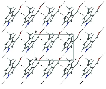
deviation for all non-H atoms of 0.0115 A˚ . In the crystal, molecules are connected through N—H O hydrogen bonds into chains running along [021]. The chains are further connectedviaC—H interactions, forming layers in thebc plane.
Related literature
For the structure of 1H-indole-3-carbaldehyde, see: Ng (2007) and for the structure of 6-bromo-1H-indole-3-carbaldehyde, see: Johnsonet al.(2009).
Experimental
Crystal data
C10H9NO
Mr= 159.18 Orthorhombic,Pca21 a= 16.9456 (19) A˚
b= 5.7029 (6) A˚ c= 8.6333 (9) A˚ V= 834.31 (15) A˚3
Z= 4
= 0.08 mm 0.470.150.05 mm
Data collection
Bruker APEXII CCD diffractometer
Absorption correction: multi-scan (SADABS; Sheldrick, 1996) Tmin= 0.962,Tmax= 0.996
5499 measured reflections 1147 independent reflections 717 reflections withI> 2(I) Rint= 0.039
Refinement
R[F2> 2(F2)] = 0.038
wR(F2) = 0.107
S= 0.98 1147 reflections 113 parameters 1 restraint
H atoms treated by a mixture of independent and constrained refinement
max= 0.12 e A˚3
min=0.14 e A˚
[image:1.610.311.566.297.338.2]3
Table 1
Hydrogen-bond geometry (A˚ ,).
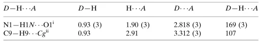
Cgis the centroid of the N1/C1/C2/C3/C8 ring.
D—H A D—H H A D A D—H A
N1—H1N O1i
0.93 (3) 1.90 (3) 2.818 (3) 169 (3) C9—H9 Cgii
0.93 2.91 3.312 (3) 107
Symmetry codes: (i)xþ1 2;y1;z
1 2; (ii)xþ
1 2;y;zþ
1 2.
Data collection:APEX2(Bruker, 2007); cell refinement:SAINT
(Bruker, 2007); data reduction:SAINT; program(s) used to solve structure:SHELXS97(Sheldrick, 2008); program(s) used to refine structure: SHELXL97 (Sheldrick, 2008); molecular graphics: X-SEED (Barbour, 2001); software used to prepare material for publication:SHELXL97andpublCIF(Westrip, 2010).
We thank the University of Malaya for funding this study (UMRG grant No. RG 066/12BIO).
Supplementary data and figures for this paper are available from the IUCr electronic archives (Reference: ZL2498).
References
Barbour, L. J. (2001).J. Supramol. Chem.1, 189–191.
Bruker (2007).APEX2andSAINT. Bruker AXS Inc., Madison, Wisconsin, USA.
Johnson, J. E., Canseco, D. C., Dolliver, D. D., Schetz, J. A. & Fronczek, F. R. (2009).J. Chem. Crystallogr.39, 329–336.
Ng, S. W. (2007).Acta Cryst.E63, o2732.
Sheldrick, G. M. (1996).SADABS. University of Go¨ttingen, Germany. Sheldrick, G. M. (2008).Acta Cryst.A64, 112–122.
Westrip, S. P. (2010).J. Appl. Cryst.43, 920–925. Structure Reports
Online
supplementary materials
Acta Cryst. (2012). E68, o2691 [doi:10.1107/S1600536812034873]
5-Methyl-1
H
-indole-3-carbaldehyde
Sharifah Shafiqah Ismail, Hamid Khaledi and Hapipah Mohd Ali
Comment
The structure of the title compound is isomorphous with that of 1H-indole-3-carbaldehyde (Ng, 2007). The planar
molecules are connected via N—H···O hydrogen bonds (Table 1) into chains in the [021] direction. The chains are further
linked through C—H···π interactions (Table 1) to form layers in the bc plane. The structure of 6-bromo-1H
-indole-3-carbaldehyde (Johnson et al., 2009) exhibits similar N—H···O bonded chains, however, further supramolecular
aggregation by Br-involved interactions is observed.
Experimental
The title crystals were obtained by slow evaporation of an ethanolic solution of the commercially available
5-methyl-indole-3-carboxaldehyde at room temperature.
Refinement
The C-bound hydrogen atoms were located in calculated positions and refined in a riding mode with C—H distances of
0.93 (Csp2) and 0.96 (Cmethyl) Å. The N-bound H atom was found in a difference Fourier map and refined freely. For all
hydrogen atoms, Uiso were set to 1.2–1.5Ueq(carrier atom). In the absence of significant anomalous scattering effects
Friedel pairs were merged.
Computing details
Data collection: APEX2 (Bruker, 2007); cell refinement: SAINT (Bruker, 2007); data reduction: SAINT (Bruker, 2007);
program(s) used to solve structure: SHELXS97 (Sheldrick, 2008); program(s) used to refine structure: SHELXL97
(Sheldrick, 2008); molecular graphics: X-SEED (Barbour, 2001); software used to prepare material for publication:
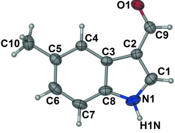
Figure 1
Molecular structure of the title compound showing thermal ellipsoids at the 30% prbability level. Hydrogen spheres are
Figure 2
Crystal packing view looking down the a axis, thus showing the two-dimensional-supramolecular structure formed by N
—H···O and C—H···π interactions (dashed lines).
5-Methyl-1H-indole-3-carbaldehyde
Crystal data
C10H9NO Mr = 159.18
Orthorhombic, Pca21 Hall symbol: P 2c -2ac a = 16.9456 (19) Å b = 5.7029 (6) Å c = 8.6333 (9) Å V = 834.31 (15) Å3 Z = 4
F(000) = 336 Dx = 1.267 Mg m−3
Mo Kα radiation, λ = 0.71073 Å Cell parameters from 1172 reflections θ = 2.4–22.1°
µ = 0.08 mm−1 T = 296 K Lath, yellow
0.47 × 0.15 × 0.05 mm
Data collection
Bruker APEXII CCD diffractometer
Radiation source: fine-focus sealed tube Graphite monochromator
φ and ω scans
Absorption correction: multi-scan (SADABS; Sheldrick, 1996)
5499 measured reflections 1147 independent reflections 717 reflections with I > 2σ(I) Rint = 0.039
θmax = 28.8°, θmin = 2.4° h = −22→13
Refinement on F2 Least-squares matrix: full R[F2 > 2σ(F2)] = 0.038 wR(F2) = 0.107 S = 0.98 1147 reflections 113 parameters 1 restraint
Primary atom site location: structure-invariant direct methods
Secondary atom site location: difference Fourier map
Hydrogen site location: inferred from neighbouring sites
H atoms treated by a mixture of independent and constrained refinement
w = 1/[σ2(F
o2) + (0.0596P)2] where P = (Fo2 + 2Fc2)/3 (Δ/σ)max < 0.001
Δρmax = 0.12 e Å−3 Δρmin = −0.14 e Å−3
Special details
Geometry. All e.s.d.'s (except the e.s.d. in the dihedral angle between two l.s. planes) are estimated using the full covariance matrix. The cell e.s.d.'s are taken into account individually in the estimation of e.s.d.'s in distances, angles and torsion angles; correlations between e.s.d.'s in cell parameters are only used when they are defined by crystal symmetry. An approximate (isotropic) treatment of cell e.s.d.'s is used for estimating e.s.d.'s involving l.s. planes.
Refinement. Refinement of F2 against ALL reflections. The weighted R-factor wR and goodness of fit S are based on F2, conventional R-factors R are based on F, with F set to zero for negative F2. The threshold expression of F2 > σ(F2) is used only for calculating R-factors(gt) etc. and is not relevant to the choice of reflections for refinement. R-factors based on F2 are statistically about twice as large as those based on F, and R- factors based on ALL data will be even larger.
Fractional atomic coordinates and isotropic or equivalent isotropic displacement parameters (Å2)
x y z Uiso*/Ueq
O1 0.27204 (11) 1.0610 (3) 0.6573 (2) 0.0742 (6)
N1 0.29282 (16) 0.4126 (4) 0.3481 (3) 0.0760 (7)
H1N 0.2766 (15) 0.285 (5) 0.290 (5) 0.091*
C1 0.2435 (2) 0.5303 (4) 0.4384 (3) 0.0717 (8)
H1 0.1913 0.4879 0.4563 0.086*
C2 0.27969 (14) 0.7233 (4) 0.5018 (3) 0.0587 (6)
C3 0.35916 (14) 0.7227 (4) 0.4414 (3) 0.0535 (6)
C4 0.42432 (15) 0.8710 (4) 0.4561 (3) 0.0554 (6)
H4 0.4212 1.0036 0.5185 0.066*
C5 0.49316 (16) 0.8208 (4) 0.3782 (3) 0.0638 (7)
C6 0.49673 (18) 0.6225 (5) 0.2839 (4) 0.0785 (8)
H6 0.5434 0.5904 0.2311 0.094*
C7 0.4341 (2) 0.4736 (5) 0.2661 (3) 0.0766 (8)
H7 0.4377 0.3422 0.2026 0.092*
C8 0.36502 (18) 0.5241 (4) 0.3453 (3) 0.0626 (7)
C9 0.24192 (16) 0.8851 (4) 0.6026 (3) 0.0640 (6)
H9 0.1897 0.8544 0.6292 0.077*
C10 0.56347 (18) 0.9773 (6) 0.3966 (4) 0.0860 (10)
H10A 0.5654 1.0866 0.3121 0.129*
H10B 0.6107 0.8842 0.3969 0.129*
Atomic displacement parameters (Å2)
U11 U22 U33 U12 U13 U23
O1 0.0843 (15) 0.0640 (9) 0.0743 (12) 0.0106 (9) 0.0086 (10) −0.0101 (9) N1 0.109 (2) 0.0565 (10) 0.0622 (13) −0.0129 (12) −0.0146 (15) −0.0053 (11) C1 0.0820 (19) 0.0660 (13) 0.0671 (17) −0.0143 (15) −0.0109 (17) 0.0081 (14) C2 0.0722 (18) 0.0533 (11) 0.0508 (12) −0.0012 (11) −0.0030 (12) 0.0033 (10) C3 0.0668 (17) 0.0499 (10) 0.0440 (11) 0.0056 (10) −0.0070 (11) −0.0013 (10) C4 0.0639 (16) 0.0528 (10) 0.0495 (12) 0.0026 (11) −0.0052 (12) −0.0003 (10) C5 0.0640 (18) 0.0699 (14) 0.0575 (14) 0.0141 (12) −0.0022 (13) 0.0073 (13) C6 0.081 (2) 0.0869 (18) 0.0678 (16) 0.0295 (15) 0.0074 (16) 0.0032 (15) C7 0.105 (2) 0.0657 (14) 0.0590 (16) 0.0237 (16) 0.0008 (17) −0.0133 (12) C8 0.089 (2) 0.0488 (10) 0.0502 (13) 0.0051 (12) −0.0112 (14) −0.0021 (11) C9 0.0662 (17) 0.0683 (13) 0.0574 (14) 0.0083 (14) 0.0013 (13) 0.0124 (13) C10 0.068 (2) 0.102 (2) 0.088 (2) 0.0008 (17) 0.0032 (16) 0.0136 (17)
Geometric parameters (Å, º)
O1—C9 1.221 (3) C4—H4 0.9300
N1—C1 1.326 (4) C5—C6 1.395 (4)
N1—C8 1.379 (4) C5—C10 1.497 (4)
N1—H1N 0.93 (3) C6—C7 1.368 (4)
C1—C2 1.374 (3) C6—H6 0.9300
C1—H1 0.9300 C7—C8 1.385 (4)
C2—C9 1.421 (3) C7—H7 0.9300
C2—C3 1.444 (3) C9—H9 0.9300
C3—C4 1.397 (3) C10—H10A 0.9600
C3—C8 1.408 (3) C10—H10B 0.9600
C4—C5 1.377 (3) C10—H10C 0.9600
C1—N1—C8 109.7 (2) C7—C6—C5 122.3 (3)
C1—N1—H1N 121.9 (18) C7—C6—H6 118.8
C8—N1—H1N 128.1 (18) C5—C6—H6 118.8
N1—C1—C2 111.0 (3) C6—C7—C8 118.1 (2)
N1—C1—H1 124.5 C6—C7—H7 121.0
C2—C1—H1 124.5 C8—C7—H7 121.0
C1—C2—C9 124.3 (3) N1—C8—C7 131.4 (2)
C1—C2—C3 105.7 (2) N1—C8—C3 107.3 (2)
C9—C2—C3 130.0 (2) C7—C8—C3 121.2 (3)
C4—C3—C8 119.0 (2) O1—C9—C2 125.6 (3)
C4—C3—C2 134.7 (2) O1—C9—H9 117.2
C8—C3—C2 106.3 (2) C2—C9—H9 117.2
C5—C4—C3 120.0 (2) C5—C10—H10A 109.5
C5—C4—H4 120.0 C5—C10—H10B 109.5
C3—C4—H4 120.0 H10A—C10—H10B 109.5
C4—C5—C6 119.4 (3) C5—C10—H10C 109.5
Cg is the centroid of the N1/C1/C2/C3/C8 ring.
D—H···A D—H H···A D···A D—H···A
N1—H1N···O1i 0.93 (3) 1.90 (3) 2.818 (3) 169 (3)
C9—H9···Cgii 0.93 2.91 3.312 (3) 107