Bcr/Abl expression stimulates integrin function
in hematopoietic cell lines.
G Bazzoni, … , J D Griffin, M E Hemler
J Clin Invest.
1996;
98(2)
:521-528.
https://doi.org/10.1172/JCI118820
.
Cell adhesion to the extracellular matrix is largely mediated by adhesion molecules of the
integrin family and is often diminished upon oncogenic transformation. However, we show
here that the chronic myelogenous leukemia oncogene Bcr/Abl has positive effects on
VLA-4 and VLA-5 integrin function. The presence of Bcr/Abl in the GM-CSF- or IL-3-dependent
hematopoietic cell lines MO7e, 32D, and BaF/3 enhanced cell binding to both soluble and
immobilized fibronectin. The effect was due to enhanced function of the VLA-5 integrin
fibronectin receptor and not to increased surface expression. In parallel, Bcr/Abl stimulated
cell adhesion to the VLA-4 integrin ligand VCAM-1. Stimulation of VLA-5 function directly
correlated with induction of Bcr/Abl tyrosine kinase activity in a temperature-sensitive kinase
mutant. Thus, Bcr/Abl stimulates integrin-dependent cell adhesion, by a mechanism
involving increased ligand binding, with the tyrosine kinase activity of Bcr/Abl likely playing
a key role. Consistent with these results, hematopoietic precursor cells from chronic
myelogenous leukemia patients also showed increased adhesion to fibronectin.
Research Article
Find the latest version:
J. Clin. Invest.
© The American Society for Clinical Investigation, Inc. 0021-9738/96/07/0521/08 $2.00
Volume 98, Number 2, July 1996, 521–528
Bcr/Abl Expression Stimulates Integrin Function in Hematopoietic Cell Lines
Gianfranco Bazzoni,* Nadia Carlesso,‡ James D. Griffin,‡ and Martin E. Hemler*
*Division of Tumor Virology, and ‡Laboratory of Hematologic Malignancies, Dana-Farber Cancer Institute, Harvard Medical School,
Boston, Massachusetts 02115
Abstract
Cell adhesion to the extracellular matrix is largely mediated by adhesion molecules of the integrin family and is often di-minished upon oncogenic transformation. However, we show here that the chronic myelogenous leukemia oncogene Bcr/ Abl has positive effects on VLA-4 and VLA-5 integrin func-tion. The presence of Bcr/Abl in the GM-CSF– or IL-3–depen-dent hematopoietic cell lines MO7e, 32D, and BaF/3 enhanced cell binding to both soluble and immobilized fibronectin. The effect was due to enhanced function of the VLA-5 inte-grin fibronectin receptor and not to increased surface ex-pression. In parallel, Bcr/Abl stimulated cell adhesion to the VLA-4 integrin ligand VCAM-1. Stimulation of VLA-5 function directly correlated with induction of Bcr/Abl ty-rosine kinase activity in a temperature-sensitive kinase mu-tant. Thus, Bcr/Abl stimulates integrin-dependent cell ad-hesion, by a mechanism involving increased ligand binding, with the tyrosine kinase activity of Bcr/Abl likely playing a key role. Consistent with these results, hematopoietic pre-cursor cells from chronic myelogenous leukemia patients also showed increased adhesion to fibronectin. (J. Clin. In-vest. 1996. 98:521–528.) Key words: integrin • Bcr/Abl • cell adhesion • chronic myelogenous leukemia • fibronectin
Introduction
Integrins are heterodimeric cell surface molecules that medi-ate cell adhesion and link extracellular ligands with cytoskeletal proteins (1, 2), and also may affect cell homeostasis by influ-encing the rate of cell proliferation (3) and cell death (4). Inte-grin function can be regulated, without changes in surface levels, such as during platelet aggregation (5), lymphocyte ac-tivation (6, 7), and keratinocyte differentiation (8). This pro-cess, known as “inside-out” signaling (9), may involve integrin clustering, modulation of ligand binding affinity, and associa-tion with the cytoskeleton (9).
Abnormal adhesive properties of neoplastic cells are often due to alteration of integrin function (1). Altered integrin func-tion may also occur in chronic myelogenous leukemia (CML)1
cells. CML is a myeloproliferative disorder associated with ex-pression of the Bcr/Abl oncogene, a hybrid gene created by the Philadelphia chromosome translocation (10), juxtaposing the BCR and c-ABL genes (located on chromosomes 22 and 9, respectively) to generate the fusion protein p210 Bcr/Abl. Compared with c-abl, the Bcr/Abl gene product has a higher tyrosine kinase activity that is essential for transformation (11). Compared with normal hematopoietic progenitors, pro-genitors from CML show decreased colony formation after ad-hesion to bone marrow stroma and/or fibronectin (12, 13). Thus it was suggested that Bcr/Abl might downregulate inte-grin-mediated adhesive activity. However, the specific effect of Bcr/Abl expression on integrin function has not been directly evaluated. In this study we have determined directly the ef-fects of p210 Bcr/Abl on both integrin-mediated adhesion to immobilized ligand and integrin binding of soluble ligand.
Methods
Cell lines and cell cultures.The human cell line MO7e (obtained from Steve Clark, Genetics Institute, Cambridge, MA) was derived from a patient with megakaryocytic leukemia and does not constitu-tively express Bcr/Abl. Although immortal, the MO7e cell line is completely dependent on GM-CSF or IL-3 for proliferation and via-bility. MO7e cells were cultured in RPMI-1640 (Mediatech, Washing-ton, DC) supplemented with 10% FBS and 1 ng/ml human GM-CSF (Genetics Institute). The murine myeloid progenitor cell line 32Dcl3 (32D) was obtained from Dr. J. Greenberger (University of Pitts-burgh, PittsPitts-burgh, PA). Bcr/Abl makes the 32D cell line leukemic in syngeneic mice where it is rapidly lethal (14). The murine BaF/3 pro-genitor cell line (from Dr. A. D’Andrea, Dana-Farber Cancer Insti-tute) has characteristics of immature B cells. Both 32D and BaF/3 cells depend on IL-3 for growth and viability. Both lines were cul-tured in RPMI-1640 supplemented with 10% FBS, and 10% WEHI-3B conditioned medium as a source of murine IL-3.
The p210 Bcr/Abl expression vector, pGDp210Bcr/Abl, was obtained from Dr. G. Daley and Dr. D. Baltimore (Massachusetts Institute of Technology, Cambridge, MA). The p210 Bcr/Abl temper-ature-sensitive mutant pGDp210Bcr/Abl-TS1 was generated by in-troducing the two point mutations Arg457→His and Tyr469→His in the
tyrosine kinase domain of Abl, as described (15), and cloned into the expression vector pGD. Sublines expressing pGDp210Bcr/Abl or pGDp210Bcr/Abl-TS1 were generated by transfection of the corre-sponding plasmids into MO7e, 32D, and BaF/3 cell lines by electropo-ration, using a Gene Pulser (Bio-Rad, Richmond, CA) and selected with G418 as described (15). Cell lines transfected with pGDp210Bcr/ Abl became factor independent and were cultured in RPMI-1640 sup-plemented with 10% FBS. 32D cells transfected with pGDp210Bcr/ Abl-TS1 were not factor independent when cultured at the nonper-missive temperature (398C), and proliferation required the addition of 10% WEHI-3B conditioned medium.
Adhesion assays. Cell adhesion to fibronectin was carried out as previously described (16). Briefly, soluble human fibronectin (GIBCO-BRL, Gaithersburg, MD) was coated onto 96-well microtiter plates (Flow Laboratories, Inc., McLean, VA) and incubated overnight at 48C. Then 0.1% heat-denatured BSA was added for 45 min to block nonspecific adhesion. After culturing in the presence or absence of IL-3 or GM-CSF for 16 h, cells were labeled by incubation with the The first two authors contributed equally to this study.
Address correspondence to Martin E. Hemler, Ph.D., Dana-Far-ber Cancer Institute, Room M-613, 44 Binney Street, Boston, MA 02115. Phone: 617-632-3410; FAX: 617-632-2662; E-mail: Martin_ Hem-ler@ DFCI.HARVARD.EDU
Received for publication 18 December 1995 and accepted in re-vised form 13 May 1996.
fluorescent dye BCECF-AM (Molecular Probes, Inc., Eugene, OR) for 30 min at 378C and then washed twice before resuspending in RPMI-1640 containing 0.1% heat-inactivated BSA. After evaluating cell viability by trypan blue exclusion, cells (5 3 104/well) were added
to each well in triplicate and incubated for 30 min at 378C. Plates were then washed three times with RPMI to remove unbound cells. Cells remaining attached to the plates were analyzed using a fluorescence analyzer (Cytofluor 2300; Millipore Corp., Bedford, MA). After sub-traction of background cell binding to BSA-coated wells, values for cells bound/mm2 were calculated as described (17).
For adhesion of 32D cells transfected with pGDp210Bcr/Abl-TS1, cytokine-deprived cells were grown at either nonpermissive (398C) or permissive (338C) temperatures for 16 h, and then adhesion was carried out at either 33 or 398C. For VCAM-1 adhesion assays, the purified VCAM-1-mouse C kappa fusion protein (2 mg/ml) was added (for 30 min at 378C) to 96-well plates that had been precoated overnight with a rat antibody to mouse C kappa. For negative control experiments, wells were precoated, but VCAM-1 was omitted.
For the comparison of normal and CML progenitor cells, hep-arinized bone marrow samples were obtained, after informed con-sent, from two patients with newly diagnosed CML in chronic phase, and two healthy volunteers. Low density mononuclear cells were iso-lated using standard Ficoll centrifugation. CD341 progenitor cells were obtained with an immunomagnetic isolation system (Dynal Co., Oslo, Norway) according to the manufacturer’s instructions. CD341
cells were cultured for 16 h in serum-free medium (AIM V; Gibco Laboratories, Grand Island, NY) in the absence of added IL-3, and then adhesion to fibronectin was evaluated as described above. The presence of p210 Bcr/Abl in cells from CML patients was confirmed by RT-PCR.
Ligand binding assay. Cells were washed once with TBS (Tris-buffered saline: 24 mM Tris-HCl, pH 7.4, 137 mM NaCl, 2.7 mM KCl), and preincubated at room temperature for 5 min with TBS con-taining 5% BSA and 0.02% sodium azide (washing buffer) and then, where indicated, with either GRGDSP peptide, fibronectin, or MnCl2. Aliquots of 105 cells were then incubated for 45 min on ice
with primary antibodies (ascites at a final dilution of 1:100, or purified mAbs at a final concentration of 1 mg/ml). Cells were washed three times with washing buffer and incubated for 30 min on ice with FITC-conjugated goat anti–mouse (GIBCO-BRL) or anti–rat IgG (Sigma Immunochemicals, St. Louis, MO). Cells were washed three times and analyzed using a FACScan machine (Becton Dickinson, Oxnard, CA).
Antibodies. The following mAbs against human integrins were used: mouse anti-a1, TS2/7 (18); anti-a2, A2-2E10 (19); anti-a3,
A3-IIF5 (20); anti-a4, B5G10 (21); anti-a6, A6-ELE (22); anti-b 1, TS2/16
(18) and A-1A5 (23); rat anti-a5, mAb 16 (24); rat anti-b
1, mAb 13
(24) and 9EG7 (25). Other mAbs were anti–mouse a4, MFR5; and a5,
R1-2 (Pharmingen, San Diego, CA) and antimycoplasma control an-tibody J2A2 (26).
Immunoblotting. Cells (108 cells/ml) were lysed in a solution (1%
NP-40, 150 nM NaCl, 20 mM Tris, pH 7.4, and 10% glycerol) contain-ing 1 mM PMSF, 5 mM aprotinin, and 1 mM Na3VO4. Lysates were
adjusted to contain equal amounts of total protein, using the
Brad-ford assay (Bio-Rad, Hercules, CA), and were boiled for 5 min in an equal volume of SDS sample buffer. Proteins were separated by 7.5% SDS-PAGE and electrophoretically transferred to nitrocellulose (Schleicher & Schuell, Keene, NH). Immunoreactive bands were de-tected with an mAb against c-Abl (gift of Dr. Naomi Rosenberg, Tufts University, Boston, MA) and visualized with alkaline phos-phatase staining reagents.
Results
[image:3.612.57.386.55.184.2]Expression of p210 Bcr/Abl increases cell adhesion to fibronec-tin. The hematopoietic cell lines MO7e, 32D, and BaF/3 were stably transfected with Bcr/Abl cDNA and the expression of p210 Bcr/Abl in transfected cells was confirmed by immuno-blotting (Fig. 1). Then, adhesion to fibronectin was examined using MO7e, a GM-CSF–dependent human megakaryocytic leukemia cell line (27). Since GM-CSF (as well as IL-3) has been reported to stimulate cell adhesion (28), both untrans-Figure 1. Expression of p210 Bcr/Abl on he-matopoietic cell lines. Lysates from untrans-fected (lanes a, c, and e) and Bcr/Abl-trans-fected (lanes b, d, and f) MO7e, 32D, and BaF/3 cells were fractionated on a 7.5% SDS-polyacrylamide gel and immunoblotted with an anti-Abl antibody. Notably, c-abl expres-sion was decreased in Bcr/Abl-transfected 32D and BaF/3 cells (lanes d and f). Also, de-graded fragments of Bcr/Abl are present, par-ticularly in lanes d and f.
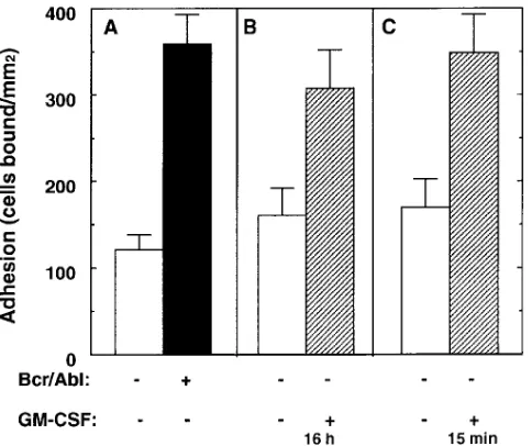
[image:3.612.316.558.428.631.2]fected and Bcr/Abl-transfected MO7e cells were deprived of GM-CSF for 16 h before the adhesion assay. In this condition, Bcr/Abl transfectants showed up to threefold higher adhesion than untransfected cells (Fig. 2 A). In a parallel experiment, adhesion of untransfected cells was induced up to a level com-parable with the Bcr/Abl transfectants, by preincubation with GM-CSF for 16 h (Fig. 2 B). Also, short-term treatment with GM-CSF (15 min) induced comparable adhesion (Fig. 2 C), despite growth factor deprivation for 16 h. Thus, factor depri-vation for 16 h had no obvious effect on adhesive potential. Adhesion of MO7e cells was mediated by the integrin VLA-5 (a5b
1), since it was completely inhibited by either anti-a5 mAb
16 or anti-b1 mAb 13 (not shown).
The effect of p210 Bcr/Abl expression was not due to up-regulation of VLA-5 expression, since comparable levels of VLA-5 were expressed at the surface of both parental and transfected MO7e cells, as determined by flow cytometry (Fig. 3). Furthermore, there were no changes in the expression of other b1 integrins. On both cell types we detected high levels
of VLA-4 and -5, low levels of VLA-2 and -6, and negligible amounts of VLA-1 and -3.
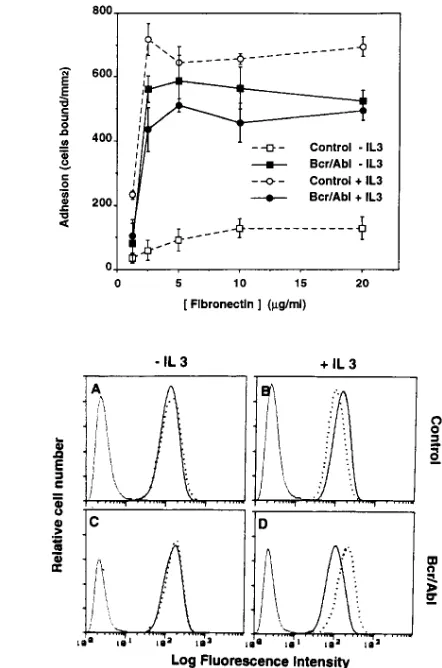
We next analyzed the effect of Bcr/Abl expression on the functions of two nonleukemic, IL-3–dependent, murine cell lines, 32D and BaF/3, derived from normal bone marrow cul-tures (29, 30). In the absence of IL-3, adhesion of 32D cells was very low even at the highest concentrations of fibronectin. In comparison, adhesion of the Bcr/Abl transfectants was greatly elevated at all levels of fibronectin tested. Culture for 16 h in IL-3 strongly increased adhesion of parental 32D cells, but had no further effect on the already adhesive Bcr/Abl-transfected cells (Fig. 4 A). No differences in cell viability were observed between parental and transfected cells either in presence or in absence of IL-3 (not shown). Brief incubation (15 min at 378C) of factor-starved 32D cells with IL-3 had effects (not shown) similar to preincubation with IL-3 for 16 h. Bcr/Abl expression similarly increased the adhesion to fibronectin (5 mg/ml) of IL-3–deprived BaF/3 cells from 595690 up to 829648 cells bound/mm2 in a representative experiment.
Adhesion of 32D cells to fibronectin was inhibited by the anti–mouse-a5 mAb MFR-5 (not shown). The VLA-5
expres-sion levels were similar in untransfected and Bcr/Abl-trans-fected cells, both in the absence or presence of IL-3 (Fig. 4 B). Adhesion was maximal within 30 min of exposure to immobi-lized fibronectin, but declined after 2 h and even further after 6 h (Fig. 5). Regardless of whether 32D cells expressed Bcr/ Abl, or were stimulated by IL-3, a similar adhesion time course was observed (Fig. 5).
Besides VLA-5, 32D cells express the VLA-4 (a4b 1)
inte-grin (Fig. 4 B) and this integrin mediated 32D cell adhesion to the VLA-4 ligand VCAM-1 (376625 cells bound/mm2).
Adhe-sion was increased both in the Bcr/Abl transfectants (in ab-sence of IL-3) and in untransfected cells that had been pre-treated with IL-3 (up to 669631 and 654616 cells bound/mm2,
respectively). Bcr/Abl transfection or IL-3 treatment had mini-mal effects on VLA-4 expression (Fig. 4 B).
Activation of p210 Bcr/Abl kinase activity stimulates adhe-sion of 32D cells to fibronectin. To analyze further the mecha-Figure 3. p210 Bcr/Abl does not affect b1 integrin expression levels.
[image:4.612.57.315.56.220.2]Untransfected (white bars) and p210-Bcr/Abl-transfected (black bars) MO7e cells cultured in the absence of GM-CSF were incubated with mAbs directed against different integrin subunits. Analysis by flow cytometry yielded mean fluorescence intensity values as indi-cated at the right of each bar.
[image:4.612.333.554.58.392.2]nism of Bcr/Abl-dependent stimulation of integrin function, we examined cell adhesion in 32D cells transfected with a tem-perature-sensitive form of p210 Bcr/Abl kinase (15). At the nonpermissive temperature (398C) kinase activity is minimal, and in the absence of IL-3, only low to moderate adhesion to fibronectin was seen in two different clones (Fig. 6, A and B). However, adhesion was markedly upregulated at the 338C per-missive temperature when the kinase was active. In the
pres-ence of IL-3, induction of Bcr-Abl kinase activity had less of an effect, because adhesion of both clones already showed nearly maximal stimulation (Fig. 6, A and B). In a control ex-periment, adhesion to fibronectin of 32D cells (either untrans-fected or transuntrans-fected with wild-type p210 Bcr/Abl) was not sig-nificantly different at either 39 or 338C (Fig. 6 C). Therefore, the differences in adhesion detected with the temperature-sen-sitive p210 Bcr/Abl mutants are not due to a nonspecific tem-perature effect on cell adhesion. Stimulation of adhesion cor-related with induction of Bcr/Abl kinase activity (Fig. 7). Switching from 39 to 338C for 6 h increased cell adhesion to fi-bronectin and induced in parallel p210 Bcr/Abl autophosphor-ylation. Only low levels of adhesion and phosphorylation were detected in cells kept at 398C throughout the assay.
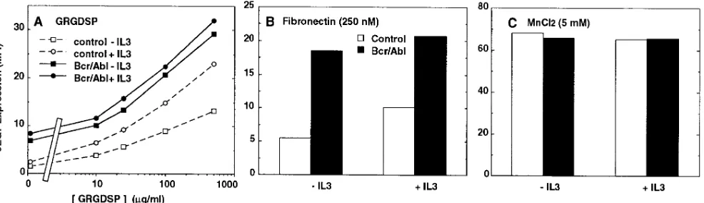
Expression of p210 Bcr/Abl stimulates binding of soluble fi-bronectin. Regulation of integrin-mediated adhesion may oc-cur by mechanisms that involve alterations in ligand binding affinity (e.g., reference 31). To determine whether Bcr/Abl and growth factors directly modulate integrin binding to fi-bronectin, we exploited the ability of the mAb 9EG7 to detect a conformational change of the b1 integrin subunit induced
upon ligand binding (32). Both untransfected and Bcr/Abl-transfected 32D cells were incubated with the ligand mimetic peptide GRGDSP and analyzed by flow cytometry for expres-sion of the 9EG7 epitope. In the absence of IL-3, very low lev-els of the epitope were induced as a result of peptide binding to untransfected cells. However, 9EG7 expression was induced nearly threefold on the Bcr/Abl transfectants. IL-3 stimulated 9EG7 expression on nontransfected cells, but had minimal ef-fect on the Bcr/Abl transef-fectants which appeared to be already maximally activated (Fig. 8 A). Bcr/Abl expression also greatly stimulated the binding of 250 nM soluble fibronectin (Fig. 8
[image:5.612.65.299.58.273.2]B). Also in Fig. 8 B, as seen in Fig. 8 A, the addition of IL-3 could stimulate untransfected control cells, but Bcr/Abl-trans-fected cells already showed near maximal fibronectin binding, as measured by 9EG7 epitope induction.
Figure 5. Time-dependent adhesion of 32D cells to fibronectin. Un-transfected (open symbols) and p210 Bcr/Abl-transfected (closed symbols) 32D cells were cultured for 16 h either in absence (squares) or presence (circles) of IL-3 before the adhesion assay. Adhesion to fibronectin (5 mg/ml) was evaluated at the time points indicated. Re-sults are mean6SD from triplicate determinations.
[image:5.612.57.561.499.682.2]The low expression of the epitope on untransfected cells was not due to an intrinsic defect of the integrin molecule. In-deed, when cells were treated with 5 mM manganese instead of soluble ligand, the epitope could be induced at comparable levels in both parental and transfected cells, either in the pres-ence or abspres-ence of IL-3 (Fig. 8 C). Like integrin ligands, man-ganese was previously found to induce the 9EG71
conforma-tion (32).
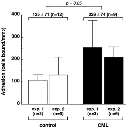
Adhesion of CML progenitor cells to fibronectin.We also analyzed the adhesive properties of CD341 hematopoietic
pre-cursors obtained from CML patients compared with those from normal individuals. In two separate experiments, the CD341 bone marrow precursors from CML patients showed
elevated adhesion to fibronectin (Fig. 9). The cumulative re-sults show that adhesion of CML precursors is elevated by nearly twofold. These results represent 12 adhesion assay data points for normal progenitors, and 9 data points for CML pro-genitors.
Discussion
To understand the molecular mechanisms responsible for the altered adhesiveness of hematopoietic progenitors in CML
(12, 13), we evaluated Bcr/Abl effects on integrin function in several model hematopoietic cell lines. We observed that: (a) p210 Bcr/Abl expression strongly enhances adhesion (in a 30-min assay) of hematopoietic cells to fibronectin and VCAM-1 mediated by the integrins VLA-5 and VLA-4; (b) the proad-hesive effects of Bcr/Abl closely mimic the proadproad-hesive effects of GM-CSF and IL-3; (c) the Bcr/Abl effect is at least partially due to enhanced ligand binding activity; (d) the tyrosine kinase activity of Bcr/Abl is necessary for its effect on cell adhesion; and (e) Bcr/Abl effects in cell lines correlate with increased ad-hesion seen in CML progenitor cells.
Bcr/Abl stimulates integrin-mediated cell adhesion.The stim-ulatory effects of Bcr/Abl on cell adhesion were verified using three different cell lines transfected with the Bcr/Abl onco-gene. In the human megakaryocytic leukemia cell line MO7e, in the murine myeloid progenitor line 32D, and in the murine lymphoid progenitor line BaF/3, the effects of Bcr/Abl on ad-hesion to fibronectin were remarkably similar. For each of these lines, the untransfected parental cell provides an ideal control, thus allowing definitive conclusions regarding the ef-fects of Bcr/Abl expression.
In contrast, primary CML cells are heterogeneous with re-spect to lineage and stage of differentiation, and it is difficult to define an ideal normal cell population for comparison. In addi-tion, it is difficult to control for possible cytokine secretion by stromal cells, macrophages, T lymphocytes, or even the leuke-mic cells themselves in these heterologous cultures. Nonethe-less, we did find that adhesion of CML progenitor cells was el-evated, compared with normal progenitor cells. We presume that this elevated adhesion is largely due to the presence of Bcr/Abl in the CML cells.
[image:6.612.56.298.59.219.2]Notably, integrin expression was unchanged in the Bcr/Abl transfectants. Also, integrin expression was unchanged in CML progenitors as compared with normal progenitors, as shown elsewhere (13). In addition, integrins from Bcr/Abl transfectants had normal electrophoretic mobility, suggestive of unaltered integrin maturation (not shown). These results in-dicate that Bcr/Abl expression causes an alteration of inside-out signaling that regulates integrin function. The Bcr/Abl-dependent stimulation of adhesion was a particularly surprising observa-tion, since inhibition of cell adhesion was predicted (see be-low). Also, impaired rather than enhanced cell adhesion is Figure 7. Induction of
p210 Bcr/Abl kinase ac-tivity correlates with stimulation of cell adhe-sion. 32D cells trans-fected with tempera-ture-sensitive p210 Bcr/ Abl kinase (clone 1) were incubated at 398C before switching to 338C for the indicated times. Cells were then tested for adhesion to 5
mg/ml fibronectin (top panel). In parallel,
32P-labeled cell lysates
were immunoprecipi-tated with an anti-Abl antibody and separated by SDS-PAGE to test autophosphorylation of Bcr/Abl (bottom).
[image:6.612.63.556.547.689.2]more commonly associated with oncogene expression or anti-oncogene suppression (33–35).
In one study, adhesion of CML bone marrow progenitors to purified fibronectin was suggested to be reduced compared with the adhesion of progenitors from normal donors (13). However, adhesion was evaluated indirectly by enumerating the number of colonies initiated by adherent cells after 3 and 5 wk in short- and long-term bone marrow cultures. At these time points, the number of colonies might not reflect differ-ences in the initial adhesion, but rather in the subsequent ex-pansion of the initiator cells (36, 37).
Parallel effects of Bcr/Abl and cytokines.Previously it was found that Bcr/Abl expression triggers the growth (38) and survival (39) of hematopoietic precursor cells and cell lines, such that they become cytokine independent. Notably, stimu-lation of cell growth by Bcr/Abl and cytokines may involve similar mechanisms. For example, Bcr/Abl (40) and cytokines such as IL-3 and GM-CSF (41) similarly regulate phosphati-dylinositol-3 kinase activity. Also Bcr/Abl, GM-CSF, IL-3, and Steel factor show comparable triggering of phosphorylation of p95Vav in myeloid cells (42). Here we extend the parallel
be-tween Bcr/Abl and cytokines to show that Bcr/Abl can effi-ciently replace IL-3 and GM-CSF not only in promoting cell growth, but also in enhancing cell adhesion to fibronectin and VCAM-1. Indeed, adhesion of Bcr/Abl-transfected cells was approximately equivalent to that of untransformed cells that had been routinely cultured in the presence of IL-3 or GM-CSF. Thus, Bcr/Abl expression mimics the inside-out signaling effects of IL-3 or GM-CSF on cell adhesion. As previously dis-cussed (15), Bcr/Abl itself did not induce the synthesis of any detectable levels of cytokines in transfected 32D cells.
Notably, both Bcr/Abl and IL-3 stimulate adhesion with similar transient time courses, with a return to basal levels
be-tween 2 and 6 h after initial attachment. Comparable transient integrin-mediated adhesion and deadhesion events have also been shown previously for cytokine-stimulated hematopoietic cells (28), and stimulated lymphocytes (7, 43) and neutrophils (44), where deadhesion is likely to involve phosphatase activi-ties (45).
Effect of Bcr/Abl on ligand binding.Here we used the anti-b1
antibody, mAb 9EG7, to demonstrate that Bcr/Abl promotes VLA-5 binding to soluble fibronectin, and RGD peptide. From this we conclude that inside-out signaling involving Bcr/ Abl leads to a change in the VLA-5 conformation that is favor-able for ligand binding. This effect of Bcr/Abl may be similar to the increase in ligand binding resulting from inside-out sig-naling that has been described for aIIbb
3 integrin in
thrombin-stimulated platelets (9). Although we have demonstrated that Bcr/Abl can alter integrin ligand binding, we have not ad-dressed other potential mechanisms for regulation of integrin-dependent cell adhesion. For example, it remains to be seen whether Bcr/Abl may contribute to cell adhesion by also en-hancing post–ligand binding events such as integrin clustering and/or cell spreading.
It was found previously that cytokines such as IL-3 and GM-CSF could stimulate the adhesive functions of VLA-4 and VLA-5 (28), but ligand binding was not analyzed in those stud-ies. Here we have shown that IL-3 stimulates the binding of soluble fibronectin and GRGDSP ligands to VLA-5, thus helping to explain why increased adhesive function was ob-served previously. Also, this result further emphasizes the par-allel effects of Bcr/Abl expression and cytokine stimulation.
Bcr/Abl tyrosine kinase activity and cell adhesion.Using a temperature-sensitive Bcr/Abl tyrosine kinase mutant (15), we showed that the induction of the kinase activity is strictly cor-related with enhanced cell adhesion to fibronectin. Increased adhesiveness was observed as early as 6 h after switching to the permissive temperature and paralleled Bcr/Abl increased au-tophosphorylation, strongly suggesting a causal link. In this re-gard, protein kinase activity was shown previously to be essen-tial for the adhesion-promoting effects of IL-3 and GM-CSF (28). In contrast to cell lines that constitutively overexpress a fully active form of Bcr/Abl and accumulate chromosomal ab-normalities at high rate (46), the biological properties of Bcr/ Abl kinase-inducible transfectants are more likely to be consis-tent with the phenotype of primary CML progenitors. Our re-sults do not exclude a role for other activities of Bcr/Abl, such as its actin-binding function, which is unaltered by our kinase mutation (47).
It seems unlikely that the VLA-5 integrin itself might be a substrate for the kinase activity of Bcr/Abl, since tyrosyl phos-phorylation of the VLA-5 subunits was either undetectable (b1
subunit) or unaltered (a5 subunit) in Bcr/Abl-transfected 32D
cells (not shown). Alternatively, Bcr/Abl kinase could indi-rectly influence integrin function by targeting cytoskeletal molecules that associate with integrins (48). Indeed, there is a growing list of cytoskeletal and focal adhesion proteins now shown to be substrates of Bcr/Abl (49).
[image:7.612.56.272.58.276.2]Bcr/Abl-dependent integrin activation and CML.It remains to be determined how the in vitro stimulatory effect of Bcr/ Abl described here in our simplified system might contribute to the pathologic functions of CML progenitors. The clinical phenotype of CML is characterized by premature and exces-sive release of progenitor cells from the marrow, and accumu-lation of myeloid cells in the blood and spleen. Interestingly, Figure 9. Adhesion of normal and CML hematopoietic precursors to
fibronectin. CD341 bone marrow cells from normal individuals (white
administration of IL-3 or GM-CSF or other cytokines results in a similar release of progenitor cells from the marrow, and striking accumulation of these cells in the blood (50, 51). Long-term administration of G-CSF has even been associated with accumulation of myeloid cells in the spleen with resulting be-nign splenomegaly (52). Thus, the parallels between the effects of hematopoietic growth factors and Bcr/Abl’s effects on ad-hesion in vitro may extend to the in vivo situation as well.
A stromal layer is essential for the growth of normal and leukemic precursors (53). Also, fibronectin has an instructive and permissive effect on the proliferation and differentiation of erythroid (54), myeloid (55), and lymphoid (56, 57) precur-sors, as well as on the in vivo medullary hematopoiesis (58). In addition, VCAM-1 may help to promote lympho- and my-elopoiesis (56, 59). However, direct contact with the stroma may inhibit proliferation (60). This inhibition is overcome by blocking the VLA-4 interaction with fibronectin (61). These divergent findings suggest that adhesion may exert a dual trol on the proliferation of hematopoietic cells; a transient con-tact might initiate proliferation, but a prolonged adhesion might inhibit growth. These observations lead us to speculate that the transient binding to fibronectin induced by Bcr/Abl may promote rather than inhibit the growth of CML cells, but that these cells can subsequently escape from the negative in-fluence of a prolonged contact with the matrix. Notably, insen-sitivity to the inhibition of growth exerted by stroma has been observed in precursors from polycythemia vera (62), a prolif-erative disorder that, unlike CML, is not associated with the Bcr/Abl oncogene.
In conclusion, we have found that p210 Bcr/Abl expression in hematopoietic cell lines stimulates integrin-dependent cell adhesion to fibronectin and VCAM-1. As a result, it may be necessary to reevaluate the mechanism for increased emigra-tion of CML precursor cells since it is not obviously caused by defective integrin adhesive functions.
Acknowledgments
This work was supported by National Institutes of Health grants CA42368 to M.E. Hemler and CA66996 to J.D. Griffin. Dr. G. Bazzoni is currently supported by the American-Italian Cancer Foundation.
References
1. Hynes, R.O. 1992. Integrins: versatility, modulation and signalling in cell adhesion. Cell. 69:11–25.
2. Hemler, M.E. 1990. VLA proteins in the integrin family: structures, func-tions, and their role on leukocytes. Ann. Rev. Immunol. 8:365–400.
3. Giancotti, F.G., and E. Ruoslahti. 1990. Elevated levels of the a5b 1
fi-bronectin receptor suppress the transformed phenotype of Chinese hamster ovary cells. Cell. 60:849–859.
4. Zhang, Z.H., K. Vuori, J.C. Reed, and E. Ruoslahti. 1995. The a5b1 inte-grin supports survival of cells on fibronectin and up-regulates BCL-2 expres-sion. Proc. Natl. Acad. Sci. USA. 92:6161–6165.
5. Marguerie, G.A., E.F. Plow, and T.S. Edgington. 1979. Human platelets possess an inducible and saturable receptor specific for fibrinogen. J. Biol. Chem. 254:5357–5363.
6. Van Kooyk, Y., P. Van DeWiel-Van Kemenade, P. Weder, T.W. Kuijpers, and C.G. Figdor. 1989. Enhancement of LFA-1-mediated cell adhe-sion by triggering through CD2 or CD3 on T lymphocytes. Nature (Lond.). 342: 811–813.
7. Dustin, M.L., and T.A. Springer. 1989. T-cell receptor cross-linking tran-siently stimulates adhesiveness through LFA-1. Nature (Lond.). 341:619–624.
8. Adams, J.C., and F.M. Watt. 1990. Changes in keratinocyte adhesion dur-ing terminal differentiation: reduction in fibronectin binddur-ing precedes a5b1 in-tegrin loss from the cell surface. Cell. 63:425–435.
9. Ginsberg, M.H., X. Du, and E.F. Plow. 1992. Inside-out integrin signal-ling. Curr. Opin. Cell Biol. 4:766–771.
10. Nowell, P.C., and D.A. Hungerford. 1960. A minute chromosome in hu-man chronic granulocytic leukemia. J. Natl. Cancer Inst. 25:85–109.
11. Lugo, T.G., A.M. Pendergast, A.J. Muller, and O.N. Witte. 1990. Ty-rosine kinase activity and transformation potency of bcr-abl oncogene prod-ucts. Science (Wash. DC). 247:1079–1082.
12. Gordon, M.Y., C.R. Dowding, G.P. Riley, J.M. Goldman, and M.F. Greaves. 1987. Altered adhesive interactions with marrow stroma of haemato-poietic progenitor cells in chronic myeloid leukaemia. Nature (Lond.). 328:342– 344.
13. Verfaillie, C.M., J.B. McCarthy, and P.B. McGlave. 1992. Mechanisms underlying abnormal trafficking of malignant progenitors in chronic myeloge-nous leukemia. J. Clin. Invest. 90:1232–1241.
14. Matulonis, U.A., C. Dosiou, C. Lamont, G.J. Freeman, P. Mauch, L.M. Nadler, and J.D. Griffin. 1995. Role of B7-1 in mediating an immune response to myeloid leukemia cells. Blood. 85:2507–2515.
15. Carlesso, N., J.D. Griffin, and B.J. Druker. 1994. Use of a temperature sensitive mutant to define the biological effects of the p210 bcr/abl tyrosine ki-nase on proliferation of a factor-dependent murine myeloid cell line. Oncogene.
9:149–156.
16. Elices, M.J., L. Osborn, Y. Takada, C. Crouse, S. Luhowskyj, M.E. Hemler, and R.R. Lobb. 1990. VCAM-1 on activated endothelium interacts with the leukocyte integrin VLA-4 at a site distinct from the VLA-4/fibronectin binding site. Cell. 60:577–584.
17. Chan, B.M.C., M.J. Elices, E. Murphy, and M.E. Hemler. 1992. Adhe-sion to VCAM-1 and fibronectin: comparison of a4b1 (VLA-4) and a4b7 on the human cell line JY. J. Biol. Chem. 267:8366–8370.
18. Hemler, M.E., F. Sánchez-Madrid, T.J. Flotte, A.M. Krensky, S.J. Bura-koff, A.K. Bhan, T.A. Springer, and J.L. Strominger. 1984. Glycoproteins of 210,000 and 130,000 m.w. on activated T cells: cell distribution and antigenic re-lation to components on resting cells and T cell lines. J. Immunol. 132:3011– 3018.
19. Bergelson, J.M., N. St. John, S. Kawaguchi, R. Pasqualini, F. Berdichev-sky, M.E. Hemler, and R.W. Finberg. 1994. The I domain is essential for echo-virus 1 interaction with VLA-2. Cell Adh. & Comm. 2:455–464.
20. Weitzman, J.B., R. Pasqualini, Y. Takada, and M.E. Hemler. 1993. The function and distinctive regulation of the integrin VLA-3 in cell adhesion, spreading and homotypic cell aggregation. J. Biol. Chem. 268:8651–8657.
21. Hemler, M.E., C. Huang, Y. Takada, L. Schwarz, J.L. Strominger, and M.L. Clabby. 1987. Characterization of the cell surface heterodimer VLA-4 and related peptides. J. Biol. Chem. 262:11478–11485.
22. Lee, R.T., F. Berditchevski, G.C. Cheng, and M.E. Hemler. 1995. Inte-grin-mediated collagen matrix reorganization by cultured human vascular smooth muscle cells. Circ. Res. 76:209–214.
23. Hemler, M.E., C.F. Ware, and J.L. Strominger. 1983. Characterization of a novel differentiation antigen complex recognized by a monoclonal anti-body (A-1A5): unique activation-specific molecular forms on stimulated T cells. J. Immunol. 131:334–340.
24. Akiyama, S.K., S.S. Yamada, W.-T. Chen, and K.M. Yamada. 1989. Analysis of fibronectin receptor function with monoclonal antibodies: roles in cell adhesion, migration, matrix assembly, and cytoskeletal organization. J. Cell Biol. 109:863–875.
25. Lenter, M., H. Uhlig, A. Hamann, P. Jeno, B. Imhof, and D. Vestweber. 1993. A monoclonal antibody against an activation epitope on mouse integrin chain b1 blocks adhesion of lymphocytes to the endothelial integrin a6b1. Proc. Natl. Acad. Sci. USA. 90:9051–9055.
26. Hemler, M.E., and J.L. Strominger. 1982. Monoclonal antibodies react-ing with immunogenic mycoplasma proteins present in human hematopoietic cell lines. J. Immunol. 129:2734–2738.
27. Avanzi, G.C., P. Lista, B. Giovinazzo, G. Miniero, G. Saglio, R. Benet-ton, G. Coda, and L. Pegoraro. 1988. Selective growth response to IL-3 of a hu-man leukaemic cell line with megakaryoblastic features. Br. J. Haematol. 69: 359–366.
28. Levesque, J.P., D.I. Leavesley, S. Niutta, M. Vadas, and P.J. Simmons. 1995. Cytokines increase human hemopoietic cell adhesiveness by activation of very late antigen (VLA)-4 and VLA-5 integrins. J. Exp. Med. 181:1805–1815.
29. Greenberger, J.S., M.A. Sakakeeny, R.K. Humphries, C.J. Eaves, and R.J. Eckner. 1983. Demonstration of permanent factor-dependent multipoten-tial (erythroid/neutrophil/basophil) hematopoietic progenitor cell lines. Proc. Natl. Acad. Sci. USA. 80:2931–2935.
30. Palacios, R., and M. Steinmetz. 1985. IL-3-dependent mouse clones that express B-220 surface antigen contain Ig genes in germ line configuration and generate B-lymphocytes in vivo. Cell. 41:727–734.
31. Faull, R.J., N.L. Kovach, J.M. Harlan, and M.H. Ginsberg. 1994. Stimu-lation of integrin-mediated adhesion of T lymphocytes and monocytes: two mechanisms with divergent biological consequences. J. Exp. Med. 179:1307– 1316.
32. Bazzoni, G., D.-T. Shih, C.A. Buck, and M.E. Hemler. 1995. MAb 9EG7 defines a novel b1 integrin epitope induced by soluble ligand and
manga-nese, but inhibited by calcium. J. Biol. Chem. 270:25570–25577.
33. Plantefaber, L.C., and R.O. Hynes. 1989. Changes in integrin receptors on oncogenically transformed cells. Cell. 56:281–290.
Cano. 1991. A role for the E-cadherin cell-cell adhesion molecule during tumor progression of mouse epidermal carcinogenesis. J. Cell Biol. 115:517–533.
35. Narayanan, R., K.G. Lawlor, R.Q. Schaapveld, K.R. Cho, B. Vogel-stein, T.P. Bui-Vinh, M.P. Osborne, and N.T. Telang. 1992. Antisense RNA to the putative tumor-suppressor gene DCC transforms Rat-1 fibroblasts. Onco-gene. 7:553–561.
36. Coulombel, L., D.K. Kalousek, C.J. Eaves, C.M. Gupta, and A.C. Eaves. 1983. Long-term marrow culture reveals chromosomally normal he-matopoietic progenitor cells in patients with Philadelphia chromosome-positive chronic myelogenous leukemia. N. Engl. J. Med. 308:1493–1498.
37. Dubé, I.D., D.K. Kalousek, L. Coulombel, C.M. Gupta, C.J. Eaves, and A.C. Eaves. 1984. Cytogenetic studies of early myeloid progenitor compart-ments in Ph1-positive chronic myeloid leukemia. II. Long-term culture reveals the persistence of Ph1-negative progenitors in treated as well as newly diag-nosed patients. Blood. 63:1172–1177.
38. Daley, G.Q., and D. Baltimore. 1988. Transformation of an interleukin 3-dependent hematopoietic cell line by the chronic myelogenous leukemia-spe-cific P210bcr/abl protein. Proc. Natl. Acad. Sci. USA. 85:9312–9316.
39. Bedi, A., B.A. Zehnbauer, J.P. Barber, S.J. Sharkis, and R.J. Jones. 1994. Inhibition of apoptosis by BCR-ABL in chronic myeloid leukemia.
Blood. 83:2038–2044.
40. Skorski, T., P. Kanakaraj, M. Nieborowska-Skorska, M.Z. Ratajczak, S.-C. Wen, G. Zon, A.M. Gewirtz, B. Perussia, and B. Calabretta. 1995. Phos-phatidylinositol-3 kinase activity is regulated by BCR/ABL and is required for the growth of Philadelphia chromosome-positive cells. Blood. 86:726–736.
41. Corey, S., A. Eguinoa, K. Puyana-Theall, J.B. Bolen, L. Cantley, F. Mol-linedo, T.R. Jackson, P.T. Hawkins, and L.R. Stephens. 1993. Granulocyte mac-rophage-colony stimulating factor stimulates both association and activation of phosphoinositide 3OH-kinase and Src-related tyrosine kinase(s) in human my-eloid derived cells. EMBO (Eur. Mol. Biol. Organ.) J. 12:2681–2690.
42. Matsuguchi, T., R.C. Inhorn, N. Carlesso, G. Xu, B. Druker, and J.D. Griffin. 1995. Tyrosine phosphorylation of p95Vav in myeloid cells is regulated
by GM-CSF, IL-3 and Steel factor and is constitutively increased by p210BCR/ABL.
EMBO (Eur. Mol. Biol. Organ.) J. 14:257–265.
43. Chan, B.M.C., J. Wong, A. Rao, and M.E. Hemler. 1991. T cell receptor dependent, antigen specific stimulation of a murine T cell clone induces a tran-sient VLA protein-mediated binding to extracellular matrix. J. Immunol. 147: 398–404.
44. Lo, S.K., P.A. Detmers, S.M. Levin, and S.D. Wright. 1989. Transient adhesion of neutrophils to endothelium. J. Exp. Med. 169:1779–1793.
45. Wright, S.D., and B.C. Meyer. 1986. Phorbol esters cause sequential ac-tivation and deacac-tivation of complement receptors on polymorphonuclear leu-kocytes. J. Immunol. 136:1759–1764.
46. Laneuville, P., G. Sun, M. Timm, and M. Vekemans. 1992. Clonal evolu-tion in a myeloid cell line transformed to interleukin-3 independent growth by retroviral transduction and expression of p210 bcr/abl. Blood. 80:1788–1797.
47. McWhirter, J.R., and J.Y.J. Wang. 1993. An actin binding function con-tributes to transformation by the Bcr-Abl oncoprotein of Philadelphia chromo-some-positive human leukemias. EMBO (Eur. Mol. Biol. Organ.) J. 12:1533– 1546.
48. Burridge, K., K. Fath, T. Kelly, G. Nuckolls, and C. Turner. 1988. Focal adhesions: transmembrane junctions between the extracellular matrix and the cytoskeleton. Ann. Rev. Cell Biol. 4:487–525.
49. Salgia, R., J.-L. Li, S.H. Lo, B. Brunkhorst, G.S. Kansas, E.S. Sobhany, Y. Sun, E. Pisick, M. Hallek, T. Ernst, R. Tantravahi, L.B. Chen, and J.D. Grif-fin. 1995. Molecular cloning of human paxillin, a focal adhesion protein phos-phorylated by P210BCR/ABL. J. Biol. Chem. 270:5039–5047.
50. Socinski, M.A., S.A. Cannistra, A. Elias, K.H. Antman, L. Schnipper, and J.D. Griffin. 1988. Granulocyte-macrophage colony stimulating factor ex-pands the circulating hematopoietic progenitor cell compartment in humans.
Lancet. 1:1194–1198.
51. Duhrsen, U., J.L. Villeval, J. Boyd, G. Kannourakis, G. Morstyn, and D. Metcalf. 1988. Effects of recombinant human granulocyte colony-stimulating factor on hematopoietic progenitor cells in cancer patients. Blood. 72:2074– 2081.
52. Bonilla, M.A., D. Dale, C. Zeidler, L. Last, A. Reiter, M. Ruggeiro, M. Davis, B. Koci, W. Hammond, A. Gillio, and K. Welte. 1994. Long-term safety of treatment with recombinant human granulocyte colony-stimulating factor (r-metHuG-CSF) in patients with severe congenital neutropenias. Br. J. Hae-matol. 88:723–730.
53. Dexter, T.M, T.D. Allen, and L.G. Lajtha. 1977. Conditions controlling the proliferation of hemopoietic stem cells in vitro. J. Cell Physiol. 91:335–344.
54. Weinstein, R., M.A. Riordan, K. Wenc, S. Kreczko, and N. Dainiak. 1989. Dual role of fibronectin in hematopoietic differentiation. Blood. 73:111– 116.
55. Campbell, A.D., M.W. Long, and M.S. Wicha. 1985. Extracellular ma-trix promotes the growth and differentiation of hematopoietic cells in long-term marrow cultures. J. Clin. Invest. 75:2085–2092.
56. Miyake, K., I.L. Weissman, J.S. Greenberger, and P.W. Kincade. 1991. Evidence for a role of the integrin VLA-4 in lympho-hemopoiesis. J. Exp. Med.
173:599–607.
57. Dittel, B.N., J.B. McCarthy, E.A. Wayner, and T.W. LeBien. 1993. Reg-ulation of human B-cell precursor adhesion to bone marrow stromal cells by cy-tokines that exert opposing effects on the expression of vascular cell adhesion molecule-1 (VCAM). Blood. 81:2272–2282.
58. Williams, D.A., M. Rios, C. Stephens, and V.P. Patel. 1991. Fibronectin and VLA-4 in haematopoietic stem cell-microenvironment interactions. Nature (Lond.). 352:438–441.
59. Teixidó, J., M.E. Hemler, J.S. Greenberger, and P. Anklesaria. 1992. Role of b1 and b2 integrins in the adhesion of human CD34hi stem cells to bone
marrow stroma. J. Clin. Invest. 90:358–367.
60. Verfaillie, C.M. 1992. Direct contact between human primitive hemato-poietic progenitors and bone marrow stroma is not required for long-term in vitro hematopoiesis. Blood. 79:2821–2826.
61. Hurley, R.W., J.B. McCarthy, and C.M. Verfaillie. 1995. Direct adhe-sion to bone marrow stroma via fibronectin receptors inhibits hematopoietic progenitor proliferation. J. Clin. Invest. 96:511–519.