University of Huddersfield Repository
Uchimoto, Mari L.
Developing a microRNA body fluid identification test for use in forensic casework
Original Citation
Uchimoto, Mari L. (2014) Developing a microRNA body fluid identification test for use in forensic casework. Doctoral thesis, University of Huddersfield.
This version is available at http://eprints.hud.ac.uk/id/eprint/24470/
The University Repository is a digital collection of the research output of the University, available on Open Access. Copyright and Moral Rights for the items on this site are retained by the individual author and/or other copyright owners. Users may access full items free of charge; copies of full text items generally can be reproduced, displayed or performed and given to third parties in any format or medium for personal research or study, educational or notforprofit purposes without prior permission or charge, provided:
• The authors, title and full bibliographic details is credited in any copy; • A hyperlink and/or URL is included for the original metadata page; and
• The content is not changed in any way.
For more information, including our policy and submission procedure, please contact the Repository Team at: [email protected].
Developing a microRNA body fluid
identification test for use in forensic
casework
Mari L. Uchimoto
A thesis submitted to the University of Huddersfield in partial fulfilment of the requirements for the degree of Doctor of Philosophy
The University of Huddersfield
ii
Copyright statement
i. The author of this thesis (including any appendices and/or schedules to this thesis) owns any copyright in it (the “Copyright”) and she has given The University of Huddersfield the right to use such copyright for any administrative, promotional, educational and/or teaching purposes.
ii. Copies of this thesis, either in full or in extracts, may be made only in accordance with the regulations of the University Library. Details of these regulations may be obtained from the Librarian. This page must form part of any such copies made.
iii. The ownership of any patents, designs, trademarks and any and all other intellectual property rights except for the Copyright (the “Intellectual Property Rights”) and any
iii
Abstract
iv
Table of Contents
Copyright statement ... ii
Abstract ... iii
Table of Contents ... iv
List of Figures ... viii
List of Tables ... xi
Acknowledgements ... xiii
List of abbreviations ... xiv
Chapter 1 - Introduction ... 1
1.1 Body fluid identification ... 2
1.1.1 Forensic applications ... 2
1.1.2 Current BFID tests ... 7
1.1.3 New BFID tests ... 31
1.2 MicroRNA ... 55
1.2.1 Biogenesis ... 56
1.2.2 Discovery ... 57
1.2.3 MicroRNA analysis ... 59
1.3 Project aims ... 65
Chapter 2 - Methods and Materials ... 66
2.1 Ethical approval ... 67
v
2.2.1 Blood samples ... 68
2.2.2 Saliva samples ... 68
2.2.3 Skin samples ... 69
2.2.4 Vaginal material samples ... 69
2.2.5 Semen samples ... 70
2.3 Sample isolation ... 70
2.3.1 Dynabeads® magnetic separation technology ... 71
2.3.2 Qiagen silica gel membrane technology ... 74
2.4 Sample quantification ... 75
2.4.1 Nano volume UV-Vis spectrophotometry ... 76
2.4.2 Quantitative PCR ... 76
2.5 Complementary DNA synthesis ... 77
2.5.1 Standard reverse transcription ... 78
2.5.2 Stem-loop reverse transcription ... 79
2.6 Quantitative PCR ... 79
2.6.1 qPCR of stem-loop RT products ... 80
2.6.2 qPCR of standard RT products ... 81
2.7 Data analysis ... 81
2.8 Enzymatic tests ... 82
2.8.1 Kastle-Meyer and Leucomalachite Green tests ... 83
2.8.2 Phadebas® test ... 83
Chapter 3 - Evaluating four different isolation kits for RNA analysis ... 84
3.1 Co-isolation using magnetic bead technology ... 85
vi
3.1.2 mRNA expression in saliva swabs ... 90
3.2 Isolation using silica gel membrane technology ... 94
3.2.1 MicroRNA expression in DNA and total RNA kits ... 96
3.2.2 MicroRNA expression during different stages of sample isolation ... 98
3.2.3 MicroRNA analysis of unknown samples ... 100
3.2.4 MicroRNA analysis of saliva swabs ... 102
Chapter 4 - Developing a miRNA panel for a miRNA analysis ... 108
4.1 MicroRNA marker screening ... 109
4.1.1 Blood ... 113
4.1.2 Saliva swabs ... 114
4.1.3 Saliva deposits ... 115
4.1.4 Skin ... 116
4.1.5 Semen ... 117
4.1.6 Vaginal material ... 118
4.1.7 miR-451 ... 119
4.1.8 miR-194 ... 120
4.1.9 miR-205 ... 121
4.1.10 miR-224 ... 122
4.1.11 miR-335 ... 123
4.1.12 miR-891a ... 124
Chapter 5 - The applications of miRNA analysis ... 132
5.1 Sensitivity of miRNA analysis with enzymatic tests ... 133
5.1.1 Kastle-Meyer, Leucomalachite Green and Phadebas tests ... 137
vii
5.1.3 miRNA analysis on saliva deposits ... 142
5.2 Specificity of miRNA and mRNA analysis ... 148
5.2.1 Specificity of mRNA analysis ... 150
5.2.2 Specificity of miRNA analysis ... 153
5.3 MicroRNA stability ... 160
5.4 MicroRNA analysis of mixed body fluids ... 168
Chapter 6 - Discussion ... 177
6.1 Discussion ... 178
6.2 Novel work ... 185
6.3 Future work ... 186
References ... 188
Appendices ... 219
Supplementary information – mature miRNA sequences ... 220
Supplementary information – miRNA species expression ... 223
Supplementary graphs – Chapter 3 ... 227
Supplementary graphs – Chapter 4 ... 229
Supplementary graphs – Chapter 5 ... 236
viii
List of Figures
Figure 1. The biogenesis of microRNA in eukaryotic cells. 56
ix Figure 23. The specificity of miR-335 across 6 different body fluids. 123 Figure 24. The specificity of miR-891a across 6 different body fluid. 124 Figure 25. The sensitivity of miR-451 in blood when normalised with RNU44. 139
Figure 26. The sensitivity of miR-451 in blood. 140
Figure 27. The sensitivity of RNU44 in blood. 141
Figure 28. The sensitivity of miR-205 in saliva when normalised with RNU44. 142
Figure 29. The sensitivity of miR-205 in saliva deposits. 143
Figure 30. The sensitivity of RNU44 in saliva deposits. 144
Figure 31. The normalised expression of HBB in 7 different species. 150 Figure 32. The expression of GAPDH in 7 different species. 150
Figure 33. The expression of HBB in 7 different species. 152
Figure 34. The normalised expression of miR-451 in 7 different species. 153 Figure 35. The expression of miR-451 in 7 different species. 154
Figure 36. The expression of RNU44 in 7 different species. 155
Figure 37. The normalised expression of miR-451 in bloodstains stored for 1 year. 162 Figure 38. The expression of miR-451 in bloodstains stored for 1 year. 163 Figure 39. The expression of RNU44 in bloodstains stored for 1 year. 164 Figure 40. The expression of miR-451 and miR-205 in mixtures. 170 Figure 41. The normalised expression of miR-451 and miR-205 in mixtures. 171
Figure 42. The expression of RNU44 in mixtures. 172
Figure 43. The expression of RNU44 in mixtures. 173
x Figure 48. The predicted mature miRNA sequences for miR-224 and miR-335. 222 Figure 49. The predicted mature miRNA sequences for miR-124a and miR-588. 222 Figure 50. miR-205 and miR-451 expression in saliva swabs isolated with 1 kit. 227 Figure 51. miR-205 and miR-451 expression in saliva swabs isolated with 1 kit. 228 Figure 52. The specificity of miR-16 in 6 different body fluids. 229 Figure 53. The specificity of miR-658 in 6 different body fluids. 230 Figure 54. The specificity of miR-203 in 6 different body fluids. 231 Figure 55. The specificity of miR-617 in 6 different body fluids. 232 Figure 56. The specificity of miR-372 in 6 different body fluids. 233 Figure 57. The specificity of miR-124a in 6 different body fluids. 234 Figure 58. The specificity of miR-588 in 6 different body fluids. 235 Figure 59. miR-451 and miR-205 expression in mixtures normalised with RNU44. 236 Figure 60. miR-451 and miR-205 expression in mixtures normalised with RNU44 and the 1:1
mixture. 237
xi
List of Tables
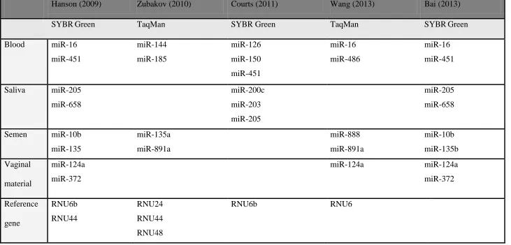
Table 1. Summary of miRNA markers and reference genes identified in forensic research. 64 Table 2. Showing an example of a calculated qPCR master mix. 81 Table 3. Showing a comparison of the standard and modified magnetic bead isolations. 88 Table 4. Showing the list of candidate miRNA markers for body fluid identification. 112 Table 5. Summarizing the final miRNA BFID panel and differentiation capabilities. 125 Table 6. Showing the reference gene selected for miRNA body fluid identification. 136 Table 7. Showing the sensitivity of KM, LMG and Phadebas® (PAT) tests. 138 Table 8. Summarizing the final miRNA BFID panel and differentiation capabilities. 180 Table 9. Showing the species specificity of the candidate miRNA markers. 223 Table 10. Showing the ∆Cq data from Chapter 4 miRNA screen in bloodstains. 239 Table 11. Showing the ∆Cq data from Chapter 4 miRNA screen in saliva swabs. 240 Table 12. Showing the ∆Cq data from Chapter 4 miRNA screen in saliva deposits. 241 Table 13. Showing the ∆Cq data from Chapter 4 miRNA screen in skin. 242 Table 14. Showing the ∆Cq data from Chapter 4 from the miRNA screen in semen. 243 Table 15. Showing the ∆Cq data from Chapter 4 miRNA screen in vaginal material. 244 Table 16. Showing the ∆Cq data from Chapter 4 miR-451 in 6 different body fluids. 245 Table 17. Showing the ∆Cq data from Chapter 4 miR-194 in 6 different body fluids. 245 Table 18. Showing the ∆Cq data from Chapter 4 miR-205 in 6 different body fluids. 245
xiii
Acknowledgements
First I would like to give an enormous thank you to my supervisor Dr. Graham Williams (Father Graham), for his unequivocal support and guidance throughout my PhD. His experience and expertise as a former Forensic Biologist with the Forensic Science Service (FSS) and as an active casework reporter and expert witness has provided me with invaluable insight into my field.
I would also like to give a hearty thank you to Professor James P. Landers at the University of Virginia, USA for first sparking my interest in forensic genetics during an industrial placement year at his research laboratory and for encouraging me to pursue doctorate.
Next, I would like to give a tremendous heartfelt thank you to my family and relatives. They have been with me from the very start and without them I would not be where I am today. Thank you for always believing in me and supporting my dreams Mom, Dad, Ken, Fuchu no oji-chan and oba-chan, Kodaira no oji-chan and oba-chan and the Matsutake family.
I would also like to give a big thank you to the University of Huddersfield Forensic Genetic Research Group for making my time as a researcher worthwhile. I would also like to give a special thank you to Dr. Chris Dunnill, Corinne Waite, Dieu van der Meer, Mitra Ameri and Rafina Bashir for keeping me sane during my write up.
xiv
List of abbreviations
BFID……….……….…..Body fluid identification cDNA………...Complementary DNA DNA……….………Deoxyribonucleic acid mRNA……….……….Messenger RNA
miRNA, miR…..……….……...MicroRNA Pri-miRNA………...…..Primary miRNA Pre-miRNA………....…..…Precursor miRNA RNA………...Ribonucleic acid
PCR…….……….……..…..Polymerase chain reaction
qPCR. ……….…..……...Quantitative polymerase chain reaction RT……….………...Reverse transcription
sl………..……...…...Stem-loop
STR.………..………....………..…Short tandem repeats
dNTPs…..……….………..…Deoxyribonucleotide triphosphates MMLV……….…..…....Moloney Murine Leukaemia Virus AQ………..….….…..Absolute quantification
Cq………..….……Quantification cycle
LOD………..………..Limit of detection LOQ………...…...Limit of quantification RQ………..Relative quantification bp……….………..Base pair
xv nt………....Nucleotide
DGCR8……….….DiGeorge syndrome critical region RISC………..………RNA-induced silencing complex
PACT...………..……….…...Protein kinase RNA activator
TRBP….………Transactivation response RNA binding protein CpG………Cytosine-phosphate-guanine
Oligo-dT….………Oligo-deoxythymine
GAPDH……....………..Glyceraldehyde 3-phosphate dehydrogenase Hb………ß–units of haemoglobin
HBB………..………..Haemoglobin ß KRT13………....………...….Keratin 13
MNDA…..…………....………...…Myeloid cell nuclear differentiation antigen
snoRNA………....………...…...Small nucleolar RNA
EDNAP………..……....………....….European DNA profiling group SOP…..………....………..….Standard operating procedure
ALS………....………..…...Alternative light source AP………....………..…….……Acid phosphatase test KM………..….……...Kastle-Meyer test
xvi SAP…...………..………...Semen acid phosphatase
VAP…..………...Vaginal acid phosphatase UV……….Ultra violet light
RSID……….………….Rapid stain identification
Fe2+…..………..………..………..Iron in an oxidation state of 2+
H2O2.………..……Hydrogen peroxide
DTT………....Dithiothreitol
EDTA……….Ethylenediamine tetraacetic acid SDS………...….Sodium dodecyl sulphate
UNG...Uracil N-glycosylase CCD………...……Charge coupled device
NIRS………...…...Near infrared Raman spectroscopy FT………..…...…..Fourier transform
SERS……….…….……Surface-enhanced Raman spectroscopy
WL……….……..……..Woods lamp
C. elegans….………...………...Caenorhabditis elegans, (roundworm) B. primigenious….……….Bos primigenious (cattle)
C. herengus……….…..…..……...……Caspius herengus (herring)
Cervidae………..…..………...Deer
G. gallus………..………...…Gallus gallus (chicken)
Phasianinae.………..………..…...Pheasant
O. aries……….……..…….………...Ovis aries (sheep)
Chapter 1
2
1.1 Body fluid identification
Body fluid identification (BFID) plays a crucial role in forensic casework. It provides valuable information regarding the body fluid origin and can provide evidentiary strength to a DNA profile. DNA profiling has become a powerful tool for forensic investigations due to the tremendous efforts that have gone behind developing this technology.
DNA profiling is one of the most common methods used for identifying an individual. Current DNA technologies utilise short tandem repeats (STRs), which are repeating DNA sequences between 100 to 400 bp in length [1]. STRs can be categorized by the repeat pattern: simple, compound and complex. Simple repeats are identical in sequence and length, compound are a combination of simple repeats while complex repeats contain a range of different repeating sequences [2]. DNA profiles utilise a combination of all three types STR markers [2]. For instance, the Life Technologies Next Generation Multiplex Select (NGM SE) kit for PCR utilises the SE33 locus, which is a complex marker [2]. The use of multiple STR markers increases discriminatory value. The NGM SE kit utilises a total of 17 loci to identify individuals. DNA profiling kits have also become very sensitive over the years. For instance the total input required for a full DNA profile using the NGM SE kits is 1.0 ng [3]. One of the major limitations of DNA profiling is that it cannot give information regarding the body fluid origin.
1.1.1
Forensic applications
3
1.1.1.1 Non-consensual intercourse
BFID would be valuable where multiple body fluids are present [4]. For example in a case of non-consensual intercourse, there may be vaginal material and semen heavily present within an item of evidence collected. Generally the identification through DNA profiling is sufficient for identifying the suspect and victim. However identification of additional body fluids could provide valuable insight into a case. For instance, the presence of blood could indicate forced entry or non-consensual vaginal intercourse [5-10]. The lack of lubrication may cause lacerations and abrasions in the vagina and labia majora and minora (skin protecting the vulva, urethra and vagina) and result in the presence of circulatory blood in the sample. Additionally, forced entry may cause tears along the frenulum (a string connecting the prepuce to the vernal mucosa) and cause blood to be released from the penis. The presence of menstrual blood also may be an indicator of forced entry or non-consensual vaginal intercourse as it is different to circulatory blood. In all three scenarios, BFID may provide important circumstantial evidential evidence to the DNA profile.
1.1.1.2 Withdrawn consent
non-4 consensual stimulation or intercourse took place where as the suspect may state that only the exchange of kisses on the mouth occurred. DNA profiling would not be able to resolve the context of this case. However BFID may give an indication of both the presence and the type of body fluids present e.g. saliva, semen or vaginal material, which could then be used to establish if fellatio or cunnilingus took place.
1.1.1.3 Digital penetration
BFID would also be very useful for cases of sexual assault through penetration [14-18]. Assault through penetration is defined in Section 2 of the Sexual Offences Act 2003 as non-consensual penetration of the vagina or anus through digital penetration or with a foreign object [19, 20]. Digital penetration can be with the fingers, tongue or toes [20]. An example of digital penetration is in a case of Regina vs. Z (2009) [20]. A suspect digitally penetrated the victim staying at her boyfriend‟s home. The victim‟s anus was penetrated three times. The suspect
was convicted in this case. However it was not determined whether penetration occurred through direct contact in the anus or through clothing. BFID of anal swabs and clothing could help establish the method of contact in addition to the DNA profiles obtained.
5 vaginal material may reflect the level of violence behind the assault. Thus providing valuable circumstantial evidence to the DNA profiles.
1.1.1.4 Bestiality
In forensic casework it is also common to encounter body fluids from different species [22]. For instance, crimes that occur in the household may contain animal body fluids. Body fluids from animals may be prevalent in homes where domesticated pets or livestock are cared for. For instance in sexual assaults involving animals, semen from a human male may be present in an animals anal or vaginal cavities. Identification of the individual through DNA profiling is powerful evidence. However the identification of the body fluids (e.g. semen, vaginal or anal swabs) would provide crucial information to the circumstances in a case. In 2009, Imbschweiler et al (2009) described such case between a human male and an ewe. Successful conviction was achieved using a combination of DNA profiling and BFID through visualisation of semen and epithelial cells [23]. In addition, body fluids from animals may be present in the home from preparing food. The presence of animal blood would need to be put into context of a case. For instance it is unlikely that the presence of Bos primigenious (cow) blood will be of significance unless the crime had taken place at a farm.
1.1.1.5 Cold cases
6 can be particularly challenging to forensic practitioners. The standard operating procedures (SOP‟s) used to recover, collect, transfer, test, record and preserve samples may have varied.
Practitioners may also have little or no sample remaining from the tests performed while the case was active. Therefore the use of tests such as DNA profiling and BFID is very important. BFID can provide valuable insight into the circumstances of a case, which can be very useful where a full, partial or no DNA profile has been obtained from the samples tested. BFID may also be useful in cases where samples have been heavily degraded by environmental factors such as ultra-violet (UV) light, temperature and humidity. DNA profiles may or may not be obtained from these types of samples. In these instances BFID can provide additional circumstantial information where DNA profiles cannot.
1.1.1.6 Trace DNA
7 Additionally there may be cases where body fluids may not be present. However this does not necessarily eliminate the possibility that a crime took place. For instance, semen will generally remain longer in the vagina than in the oral or anal cavities of individuals [31]. A victim may report non-consensual intercourse long after a crime taken place e.g. weeks, years. In these types of circumstances, samples such as clothing may not be available, making DNA profiling or BFID methods unusable. Instead, courtrooms may have to turn to other forms of evidence such as eyewitnesses [32, 33].
1.1.2 Current BFID tests
There are a number of different BFID tests currently used in forensic casework. They can be divided into two sections: presumptive and confirmatory. Presumptive BFID testing plays an important role in forensic investigations. They allow for both forensic practitioners and police to decide the most effective approach and tests to use in casework, which is often limited by both time and budgetary constraints. Presumptive tests are generally rapid, accurate, safe, easy to use, inexpensive and portable. However they also generally lack in specificity (e.g. body fluids, animal species, plants, household products) and sensitivity (e.g. limited or mixed samples) resulting in false positive and false negative results. False positives can suggest the presence of a particular body fluid where there is none. Conversely, false negatives can suggest the absence of a particular body fluid when it is present.
8 tests. They also tend to require lower sample input, which is very beneficial for in forensic casework where sample is often limited. Furthermore these qualities generally lower the chances of false positive and negative results. The most casework relevant presumptive and confirmatory BFID tests have been described in this section.
1.1.2.1 Blood
9 aggregating around the injury and releasing histamine and serotonin that will eventually form a blood clot [37]. They are produced by megakaryocytes in the bone marrow.
1.1.2.1.1 Oxidation-reduction tests
There are several different presumptive techniques used to indicate the presence of blood [38-43]. Two of the most commonly used tests in forensic casework are the Kastle-Meyer (KM) and Leucomalachite Green (LMG) tests [44, 45]. The principle of both tests is very similar. The KM and LMG reagents in the presence of haem will undergo a standard oxidation-reduction reaction. Both KM and LMG are tested indirectly onto samples using filter paper or swabs. Both reagents are in a reduced state (colourless) before performing the test. KM or LMG reagent is first added to the samples. After a few seconds hydrogen peroxide (H2O2) is
then added. In the presence of haem, the KM dye phenolphthalin will oxidise into phenolphthalein causing the sample to turn a bright pink colour. Similarly, with the LMG test, the base leuco will oxidise in the presence of haem turning the sample a vibrant green colour [46]. However it is important to mention that it is the peroxidase-like activity that causes this colour change in both of the tests. False positives can be indicated by a colour change between the addition of KM or LMG and addition of H2O2. Another common enzymatic method used
10 There have been a number of reports exploring the sensitivity and specificity of the KM, LMG and luminol tests in blood. Webb et al (2006) performed a comprehensive study comparing the sensitivity of presumptive tests for blood including the KM, LMG and luminol tests. In their findings, they found the luminol test to be the more sensitive than the KM and LMG tests. The sensitivity range of luminol was similar to other studies in this area, having sensitivity range of 1 in 5,000,000 dilutions [41]. The sensitivity range reported for the KM and LMG tests varied when compared to other research such as Cox et al (1991). For instance with the KM test, Webb et al (2006) detected blood-stained cloth down to 1 in 10,000 dilutions where as Cox et al (1991) detected it down to 1 in 100,000 dilutions. This may be down to a number of variables including reagent preparation. For instance, both Webb et al (2006) and Cox et al
(1991) prepared their reagents from scratch. The wide range of sensitivity observed may also be a reflection of the target molecule, the more specific the target the more accurate the result.
11 Both the KM and LMG tests are sensitive methods. However their sensitivity is limited by their lack of specificity towards vegetables, household products and body fluids. The luminol test is more sensitive and specific than the KM and LMG tests and can be used to indicate the presence of blood that may have been cleaned up. However it is limited by the need to visualise the stains in the dark.
1.1.2.1.2 Crystal tests
Another common test used for blood in forensic casework are the crystal tests. Crystal tests are confirmatory methods. The Takayama and Teichmanns tests are two common methods used in forensic casework. The principles of both tests are very similar, utilising a chemical reaction to identify haem. This reaction will produce specific derivatives that can later be visualised using microscopy. The Takayama test uses an alkaline solution (contains sugar and pyridine) to form pyridine ferroprotophorphyrin [46]. The Teichmanns test uses a combination of heat and glacial acetic acid to produce ferriporphyrin chloride [46]. Both techniques are limited by their low sensitivity and inability to distinguish between different species [47].
1.1.2.1.3 Immunochromatographic tests
anti-12 human Hb antibodies, which have a blue dye attached for testing. During analysis, suspected blood samples are placed onto the sample area. A buffer containing Tris is then added to the sample. In the presence of Hb, an antibody-antigen-antibody complex will form along the sample and control regions of the strip resulting in the appearance of two separate blue lines. Samples not containing Hb will only show a blue line in the control region [43]. Hochmeister
et al (1999) performed an extensive validation study on the Hexagon OBTI test® exploring factors such as sensitivity, specificity, casework and the hook effect. Their findings showed that the Hexagon OBTI test® was primate specific and sensitive down to 1 in 100,000 dilutions in water. The test was also found to be robust, showing positive reactions in all casework samples e.g. blood-stained cloth 2-15 years old [48]. The Hexagon OBTI test® also exhibited the high dose hook affect where high levels of erythrocytes were present, highlighting one of the main limitations of this test. The other main limitation of this test is that it is not human-specific and has tested positive in other primates including Pongo pygmaeus (Orangutan).
Summary
13 Their main drawback is the lack specificity towards human blood. Two confirmative tests were also described; Takayama and Teichmann. These tests are accurate however they have lower sensitivities than in other presumptive blood tests. Despite the range of different presumptive and confirmatory blood tests there are currently no confirmative tests that can differentiate between trauma and menstrual blood.
1.1.2.2 Saliva
Saliva is another commonly encountered body fluid in forensic casework (e.g. biting, licking, oral sex) [49-51]. Saliva is generally comprised of three major components: water (99.5%), organic (0.3%) and inorganic compounds (0.2%) [52-55]. The organic components of saliva consist mainly of proteins (e.g. α-amylase, albumin, histatins, mucins), acids (e.g. uric acid) and fats (e.g. cholesterol and lipids) [54]. The inorganic components of saliva comprise mainly of cations (e.g. calcium, magnesium, potassium, sodium) [54]. Three major glands in the mouth produce saliva: paratoid, sublingual and submandibular [56]. The major roles of saliva include deglutition, lubrication of deglutition (e.g. food) and protection of the enamel and from foreign viruses, bacteria and fungi of the mouth [52, 54]. Humans produce on average 1.3 L of saliva per day [46].
1.1.2.2.1 Amylase tests
14 digestion and aiding in the breakdown of complex carbohydrates (e.g. polysaccharides) into simple sugars (e.g. mono-saccharides). Humans normally have high levels of amylase in the body. However this can vary between individuals due to natural variation, disease and time of day [57, 58].
One of the main kits used to indicate the presence of saliva in forensic casework is the Phadebas® tests [57, 59-62]. The Phadebas® tests utilises the α-amylase activity to indicate the presence of saliva. In the presence of α-amylase, a water-insoluble polymer attached to a soluble blue dye will hydrolyse releasing the dye and will result in the appearance of a dark blue colour. There are liquid and paper versions of this test. The liquid version will test for α-amylase activity using aliquots of suspected samples before undergoing quantification with a spectrophotometer (620 nm). The paper Phadebas® press test is generally applied onto a piece of filter paper, moistened with water and placed onto the suspected sample regions (e.g. clothing) [58, 59, 63, 64]. The presence of α-amylase is then determined through visualisation of the stain.
15 Summary
Amylase tests form an important part of forensic investigations involving saliva. The presence of amylase can give forensic practitioners insight on the circumstances of a case and how to proceed. In addition they are also relatively inexpensive and easy to use [62]. Amylase tests are limited by a number of factors. The tests are not specific to HSA and can be mistaken for HPA found in other areas of the body (e.g. blood, semen, vaginal material) [46]. Animals (e.g. primates, some rodents), plants (e.g. potatoes) and common household products (e.g. lotions, detergent) also contain high levels of amylase and as a result can generate false positives [51, 57, 65].
1.1.2.2.2 Microscopy
Alternatively, buccal cells can be visualised using microscopy. It is relatively quick and easy to use. Epithelial cells can be stained prior to visualisation. The haematoxylin and eosin (H&E) stain can be used in forensic casework to enhance the appearance of epithelial cells. The nucleus will be dyed blue while the surrounding region of the cell will be dyed red. Microscopy can be useful for identifying epithelial cells. However its inability to differentiate the morphology of epithelial cells found in vaginal material and skin, means that it cannot definitively confirm the presence of saliva.
1.1.2.2.3 Immunochromatographic tests
16 is the rapid stain identification (RSID™) for human saliva test [50, 51]. They utilise lateral
flow strips similar to pregnancy tests, which contain both sample and control regions. Both the sample and control region contain two monoclonal antibodies. The presence of the specific antigens will result in binding to the antibodies and a red line in both sample and control regions will appear. Samples not containing these antigens will only show a red line in the control region [50, 66].
Old et al (2009) performed a study exploring different casework applications (e.g. sensitivity, specificity, stability, and mixed body fluids) using the RSID™ human saliva test. In their findings, the RSID™ tests showed greater sensitivity than with the generic amylase tests. In their specificity studies, the RSID™ saliva test showed slight cross reactivity towards milk and post-coital samples containing vaginal material but no cross reactivity in blood and semen. The RSID™ test for saliva showed α-amylase stability in samples stored at high temperatures (e.g.
37 º C) as wells as in samples stored over long periods of time (e.g. 30 days) [50, 67]. Saliva samples showed specificity in mixed samples containing blood or semen. The studies performed by Old et al (2009) reiterate the strengths of using an immunochromatographic test in forensic investigations. However their study also highlights the need for a confirmatory BFID test for identifying saliva.
1.1.2.2.4 Alternative light sources
17 instance, Polilight®, which is a common ALS source can indicate the presence of saliva when using orange goggles and a wavelength of 450 nm [26]. A study by Vandenberg and van Oorschot (2006) explored Polilight® and its applications on forensically relevant body fluids, which included saliva. In the majority of their studies (e.g. appearance on different fabrics, cleaning products, diluted and mixed samples), the presence of saliva could be indicated. However it is clear through their work that body fluid differentiation with ALS is limited by factors such as the composition of body fluids, mixed body fluids and the presence of household cleaning products [26].
Summary
18
1.1.2.3 Semen
Semen is another commonly encountered body fluid in forensic casework (e.g. sexual assaults) [65, 70-73]. It normally contains two components: 5% spermatozoa (sperm) and 95% fluid [74]. Spermatozoa have a head, which contains DNA and RNA, a tail, which is used for mobility and a complex mid-piece, which connects the two parts. The fluid consists of a number of components necessary for the survival of sperm during reproduction. The main energy sources for sperm include fructose and glycerophosphorylcholine. They are derived from storage areas such as the ampulla and epididymis, as well as the prostate glands and seminal vesicles. The environment of the sperm (e.g. pH 7-7.5) is regulated mainly by these glands and vesicles [74]. They will release a number of different enzymes, hormones, acids and elements including acid phosphatase, diamine oxidase, y-glutamyl transpeptidase, prostaglandins, semenogelins, citric and lactic acid and calcium, sodium, potassium and zinc [65, 74, 75]. In addition, the urethral glands will release mucus. The secretions produced by these glands and vesicles will also aid in the motility of sperm during reproduction. A healthy male on average releases about 3 ml of semen, which can contain up to 10 million sperm [74].
1.1.2.3.1 Acid phosphatase tests
19 easy to use. Acid phosphatase is an enzyme produced by the prostate gland. Its main function is to increase acidity of semen [82]. PAP is normally found at high levels in semen.
There are two common AP tests used in forensic casework: the α-naphthyl phosphate and the 4-methylumbelliferone phosphate test [46, 65]. Suspected casework stains are treated with moistened filter paper or swabs to preserve the original sample [81]. The filter paper is subsequently treated with these substrates and additional reagents or light sources to indicate the presence of semen.
The chemistry behind both tests is relatively similar. Acid phosphatase in the presence of α-naphthyl phosphate or 4-methylumbelliferone phosphate will cause removal of these phosphate groups through hydrolysis [46, 65]. Semen is then indicated through precipitation using Brentamine Fast Blue B, which gives a purple colour or through the examination with ultra-violet respectively.
20 1.1.2.3.2 Microscopy
Samples containing acid phosphatase then undergo confirmatory testing using microscopy [65]. Spermatozoa is visualised through the microscope directly or through cytological staining. There are three popular methods used by the forensic community; the nuclear fast red and picroindigocarmine, alkaline fuchsin and haematoxylin and eosin tests [65, 72, 85]. Spermatozoa are prepared onto slides by drying (e.g. air drying or heat drying) and treatment with chemicals such as ether or alcohol [85]. Slides are then stained with the dyes and visualised under a microscope. The current standard for conviction within forensic casework is the presence of a single sperm head in addition to a DNA profile [85, 86].
The staining process for these dyes is relatively similar. The nuclear fast red and picroindigocarmine dye will target two different regions of the spermatozoa. Nuclear fast red is basophilic and will bind to the sperm head dying it red while the picroindigocarmine is acidophilic and will bind to the sperm tail dying it green [65, 85, 87]. The haematoxylin and eosin (H&E) stain performs in a similar manner except the sperm head is dyed blue while the sperm tail is dyed red. The alkaline fuchsin stain will dye the sperm red [87].
21 body. Alternative tests have been developed for establishing the presence of semen in a sample.
1.1.2.3.3 Immunochromatographic tests
Another common confirmatory method used to indicate the presence of semen are immunochromatographic tests [71]. They are generally rapid, accurate and easy to use. A common immunochromatographic test using in forensic casework is the prostate-specific antigen (PSA, P30, Kallikrein 3). PSA is a protein produced by the prostate gland. Its main role is to add volume to semen to encourage motility of sperm during reproduction. Semen normally contains high levels of PSA (e.g. 5.0 mg/ml) [89]. Its high concentration and relative stability make it particularly useful for identifying heavily degraded semen samples (e.g. 55 years) [48, 89-91].
The seminal vesicle-specific antigen test is another common immunochromatographic test used for semen samples. Semenogelins are comprised of two main proteins (semenogelin I and semenogelin II) and are produced by the seminal vesicles. Their main role is to add viscosity to semen during ejaculation. They are found at higher levels than PSA making them particularly useful for heavily degraded samples [91].
22 antibodies and a pink line in both sample and control regions will appear. Samples not containing these antigens will show a pink line in only the control region [66].
Boward and Wilson (2013) performed a comprehensive study exploring the sensitivity, specificity and cost-effectiveness of both ABAcard® p30 and RSID™ semen test using fresh, frozen, post-coital, vasectomised and mixed samples. The overall findings from their study indicated similar levels of specificity. In terms of cost-per-analysis, ABAcard® p30 was slightly cheaper than the RSID™ semen test. Also interestingly, the ABAcard®
p30 showed varying levels of sensitivity when compared to the RSID™ semen test [66]. For instance in the mixed samples the RSID™ semen test showed greater sensitivity than with the ABAcard®
p30, which was expected as semenogelins are generally found at higher levels than PSA. However the ABAcard® p30 showed greater sensitivity in post-coital samples. This was surprising as high levels of semenogelins are released prior to ejaculation. The difference in sensitivity may be down to variation in PSA and semenogelins levels between individuals. It may also be due to the use of different commercial kits.
23 1.1.2.3.4 Alternative light sources
Other less commonly used methods to identify semen in forensic casework include ALS. Alternative light sources are presumptive methods. They are rapid, non-destructive and easy to use. ALS can be useful for visualising biological stains on individuals and crime scenes (e.g. sexual assaults) [92, 93]. As mentioned previously, ALS will absorb light from shorter wavelengths to emit light at longer wavelengths; resulting in fluorescence of a sample [26]. One common ALS used in forensic casework to identify semen is the Woods lamp (WL). The WL will emit a wavelength at 360 nm, which falls in the ultra-violet (UV) light region of light; 200-400 nm [94, 95]. Semen with the WL will generally fluoresce between 300-500 nm [26, 68, 96]. Another common ALS used in forensic casework is the Polilight®. Polilight® emits light in the range 310-610 nm, which is also in the UV light region [26].
Wawryk and Odell (2005) performed a study on ALS. They explored the fluorescence of semen and other substances on skin using the Woods lamp. They reported that the appearance of semen stains was generally more faint on skin than on items such as sheets and clothing. They also performed a study comparing the fluorescence of azoospermic, vasectomized and normal semen. However they were not able to distinguish between these sample types. ALS relies heavily on visual interpretation of a sample.
24 hand creams, soaps, detergents and ointments and must be interpreted with caution [26, 94, 96]. In addition, ALS is not a confirmatory method for identifying semen.
Summary
The identification of semen can be crucial in forensic investigations, especially in cases of sexual assault. There are a number of different presumptive and confirmatory techniques that were described for targeting different components of semen. Microscopy with staining was one of the main confirmatory methods described. Its use within forensic casework is powerful as the presence of one sperm head in addition to a DNA profile is enough to convict an individual. The main limitation of this technique is that it can only be used in the presence of sperm. The AP and immunochromatographic methods (e.g. PSA) were two other presumptive methods described. Both tests are generally used in cases where sperm is absent, with the latter method being of particular use for aged semen stains. In addition, these techniques are relatively accurate and easy to use. The main drawback of both tests is their lack of specificity towards other body fluids, plants and chemicals. Another limitation is that it can produce false negatives if the hook effect occurs. ALS is another presumptive technique that was described. Its main advantage over the other techniques is the ability to visualise the area of the stain. However it lacks in specificity towards other body fluids and common household products.
1.1.2.4 Vaginal material
25 vagina is comprised of glycogenated epithelial cells. They will secrete vaginal transudate (e.g. glycogen, mucin, potassium, sodium) that will keep the vaginal walls moist during or without stimulation as well as during different points during the menstrual cycle [102-104]. Glycogen will also serve as one of the main energy sources for microflora (e.g. lactobacilli), which are naturally present in the body. They will produce organic acids (e.g. lactic acid) that will help act as a natural barrier against antigens based on the low pH (<4.5) [98, 105]. The vaginal walls will also renew itself on a regular basis through shedding. In this way it can keep the vaginal clean from bacteria build up. The average female will produce 3 ml of vaginal transudate per day [105]. This will be combined with other lubricating fluids from the upper and lower reproductive tracts. The endometrial and tubal glands found in the cervix will produce cervical mucus [106]. This mucus is thick and rich in glycoprotein and carbohydrates, serving as a protective barrier to the uterus [101]. The sebaceous glands surrounding the labia minora will produce sebum that will help protect and lubricate the entrance to the vagina during stimulation or no stimulation [106]. All of the components found in vaginal material are responsible for reproduction and health of the vagina.
1.1.2.4.1 Microscopy
26 iodine and PAS reagent are acidophilic and will turn the cytoplasm of the glycogenated epithelial cells dark brown and red respectively [65, 87, 111].
Jones and Leon (2004) performed a study exploring the glycogen content from different body fluid types (e.g. saliva, vaginal material) using both Lugol iodine and PAS tests. In both methods, they found a low glycogen content in saliva (1-8%) when compared to vaginal material (>10%) [110]. The use of these techniques could be particularly useful for woman who are menstruating as they have been reported to have high levels of glycogenated epithelial cells in the vagina [109]. The level of glycogen in saliva was also interesting, as both tests have shown higher glycogen levels than in saliva and semen [108, 109, 112, 113]. Both the Lugol iodine and PAS tests can be useful tools for indicating the presence of vaginal material. However the use of these tests is limited by the ability to confirm the body fluid origin of glycogenated epithelial cells (e.g. saliva, vaginal material, semen), particularly where little sample is available [114]. In addition, microscopy using Lugol and PAS staining can give false negatives as non-menstruating women do not always have glycogen epithelial cells present [65]. It is also destructive to the sample. Other methods have been developed to indicate the presence of vaginal material.
1.1.2.4.2 Immunoelectrophoretic tests
27 isoelectric focusing (e.g. pH 4) [115, 116]. The gel is then stained with a dye e.g. Brentamine fast black. The bands are then visualised under a light source such as a UV light. In a gel, smaller molecules tend to migrate faster than larger molecules.
Ablett (1983) performed a study comparing the mobilities of acid phosphatase from vaginal fluid and semen using twelve isoforms of acid phosphatase. Characteristic bands were observed for vaginal acid phosphatase (VAP) and semen acid phosphatase (SAP) suggesting that Laurell immunoelectrophoretic separation could be used to differentiate the two [116].
Adams and Wraxall (1974) also performed a study on range of different acid phosphatases e.g. VAP, SAP and other body fluids and plants. Their findings showed that immunoelectrophoretic separation could be used to indicate the presence of VAP or SAP. Their work also highlighted the limitations of this test (e.g. false positives). For instance, AP was also detected in yeast and plants [79].
1.1.2.4.3 Alternative light sources
28 Summary
The identification of vaginal material is crucial to forensic investigations, especially in cases of sexual assault. There are a few presumptive tests that have been used to indicate the presence of vaginal material. These tests target different components of vaginal material. One presumptive method described was microscopy with staining. Glycogenated cells are stained using the Lugol or PAS reagent. Its use in forensic casework is limited due to its lack of specificity towards other body fluids (e.g. saliva, vaginal material and semen) and women at different developmental stages (e.g. menstruating, non-menstruating). It is also destructive to samples [97].
Another presumptive method described was immunoelectrophoretic testing, which uses isoelectric focusing to indicate the presence of VAP. The technique can distinguish between VAP and SAP but not other body fluids (e.g. saliva) and plants. It is also destructive to the sample [97]. The last presumptive method described was ALS. This method utilised light to indicate the presence of vaginal material. ALS is rapid, easy to use and non-destructive. However its main limitation was that it lacked specificity towards other body fluids and household products. The main drawback of all of these methods is that there are currently no presumptive BFID tests that can distinguish vaginal material cells from skin cells.
1.1.2.5 Skin cells
29 [117]. It is responsible for thermoregulation, sensation and protection of the skin. The epidermis is the outermost layer, consisting of keratinised skin cells (contains no nuclei). Its main role is to provide a protective barrier from the environment (e.g. bacteria, chemicals).
1.1.2.5.1 Microscopy
There are a few tests that have been used to indicate the presence of skin in forensic casework. One presumptive method that has been used is microscopy through staining (e.g. H&E). French et al (2008) performed a study developing a staining technique that could differentiate epithelial cells collected from the ear or elbow from other areas of the body e.g. oral and vaginal cavities [104]. They explored a number of different dyes (e.g. Dane‟s, Ayoub-Shklar) and fixtures (e.g. 100% methanol, 95% ethanol) to observe the morphology and colour of the cells. Dane‟s technique utilises one dye, which stains the nuclei and cytoplasm a red-orange colour. It has been used to target protein (e.g. keratin) and carbohydrates (e.g. mucin). Ayoub-Shklar method utilises two dyes, which stain nuclei blue and cytoplasm red. In their study they found that a combination of Dane‟s method and methanol could distinguish epithelial cells from all cell types. Epithelial cells from the ear and elbow gave an orange colour while the buccal cells gave red-orange colour and vaginal cells gave bright orange colour and blue hue [104].
30 explore skin collected from the mid-layer e.g. dermis, which could be of relevance where a suspect has produced a deep scratch on a victim during a sexual assault. This could affect the ability to distinguish different epithelial cells because of the skin from the dermis layer contains nuclei.
Summary
Skin cell identification can be very important in forensic investigations. It has been suggested by French et al (2008) that microscopy combined with histological staining could be powerful tool for indicating the presence epithelial cells from different cell types [104]. However their method may be limited by identification of epithelial cells collected from the epidermis. Furthermore there are currently no presumptive or confirmative methods available in forensic casework that can distinguish skin cells from cells collected from the oral and vaginal cavity.
Current BFID tests summary
31 Despite the large number of body fluid identification tests available there are still no tests that can differentiate trauma blood from menstrual blood. Similarly there are no tests that can currently differentiate epithelial cells collected from skin, saliva or vaginal material. The ability to be able to identify the origin of a body fluid can be particularly important to understanding the context of a case.
1.1.3 New BFID tests
There are a number of new techniques that are currently being developed to overcome these challenges within forensic casework. These techniques can be divided by their application: forensic or non-forensic (e.g. biology, chemistry, material science). Non-forensic methods include Raman, fluorescence, hyper spectral imaging and nanotechnology. Forensic methods include DNA methylation, messenger RNA and microRNA analysis.
1.1.3.1 Raman spectroscopy
32 There is another form of scattering known as elastic scattering. This occurs when there is no change in frequency or energy between molecules and photons. The frequency of molecules are generally higher in samples containing functional groups e.g. alkenes, alkynes. Molecules are generally excited using a laser such as an argon ion (488.0 and 514.5 nm) [118].
Raman spectroscopy has been used in a wide range of fields including biology, chemistry, material science and pharmaceuticals [118]. Its use within forensic casework has only recently been explored. Virkler and Lednev(2008) performed a handful of studies exploring the use of Raman spectroscopy for identifying blood, saliva, vaginal material and semen. In their studies they utilised near infrared (NIR) Raman spectroscopy using a confocal Raman spectrophotometer [118]. The instrument as the name suggests, uses a combination of microscopy and Raman scattering to produce a characteristic chemical spectrum that is read through a charge coupled device (CCD) camera.
33 Raman microscopy such as NIR Raman spectroscopy has a number of advantages over other presumptive and confirmatory BFID tests [65, 119]. One of its main advantages is its ability to penetrate and produce a three-dimensional image without any destruction to the sample. Casper et al (2003) performed a study exploring the composition of skin using confocal Raman spectroscopy. In their findings they were able to identify both the cellular (e.g. blood cells, epithelial cells from hypodermis, dermis and epidermis) and molecular structures of skin (e.g. amino acid levels and moisture in skin) [122]. Applications explored in Casper et al (2003) study may be very useful in resolving the issue of indicating the presence of epithelial cells from various cell types in forensic casework. For instance it could be used to indicate the presence vaginal material, skin or saliva based on the chemical composition of each body fluid type. Raman spectroscopy has also shown potential for indicating the presence of mixed, trace and animal body fluids, which could be very useful for casework [65, 118]. Other advantages include ease of use and portability [123, 124].
However despite these advantages, Raman spectroscopy is limited by the variations that can occur in the spectrum (e.g. body fluid heterogeneity and sample variation between individuals), which can cause false positive results [65, 118, 120]. Also unless Raman spectroscopy is coupled with another technique (e.g. NIR) it can be subject to fluorescence interference from other molecules [120].
1.1.3.2 Hyper spectral imaging
34 cubic (hypercubic) structure, which consists of three planes: x, y and λ (one wavelength). Samples undergo temporal scanning, which involves stacking of narrow spectral bands from x,
y and λ planes to form three-dimensional images. Samples can be scanned according to points,
lines or area. The point scanning imaging system (whiskbroom), targets two points along the
spatial planes (x, y) to produce a hypercube. The line scanning system (pushbroom), targets
one line along the spatial (x,y) axes and one line along the spectral (λ) axes to produce the
cube. The area scanning system (staredown), targets a sequence of images along the spatial planes (x, y) to produce the cube [125]. Samples are visualised along the electromagnetic
spectrum (e.g. NIR, IR, UV).
HSI has been used for a wide range of applications including satellite imaging, medicine and pharmaceuticals [125]. Its applications within forensic casework such as aged samples or trace samples are recent [126, 127]. Bo et al (2013) performed an interesting study exploring the spectral changes of dried equine blood over a 30-day period using pushbroom HSI. In their work they were able to establish the average age of bloodstains (±1.2 days) through spectroscopic changes observed when blood ages. Both α- and ß– units of haemoglobin will oxidise (HbO2) into meta-haemoglobin (met-Hb) then hemichrome (HC) [127]. Their findings
showed that the first oxidation stage, met-Hb occurred in ≤ 3 days while second oxidation stage, HC occurred in ≥ 30 days [127]
35 may add an additional challenge as samples exposed to different environmental changes (e.g. temperature, humidity) can affect the final spectra.
1.1.3.3 Nanoparticles
Another emerging technique within the forensic community is nanotechnology [128-130]. Nanotechnology can be defined as a technique that can manipulate samples at cellular or molecular levels. It uses nanoparticles to target the sample of interest (e.g. biological, chemical, mechanical, optical, physical). Nanoparticles are generally 1-100 nm in size and are also often coated with metals (e.g. Au, Ag) or polymers (e.g. chitosan, silica) to further target properties [128]. Nanoparticles have been used for a number of different applications including biology, chemistry and biomedical science [128]. Its use within forensic casework has been very limited e.g. toxicology, fingerprints.
A study by Kestell and Gabriel (2010) explored the use of gold nanoparticles to identify drugs and their metabolites in urine using surface-enhanced Raman spectroscopy (SERS). In their work they spiked benzodiazepine and metabolites (e.g. 1,4-benzodiazepam) and tested them against different agents to enhance the interaction and sensitivity of the spectra [130]. They found that magnesium chloride (MgCl2) gave the highest sensitivity amongst the spiked urine
36
1.1.3.4 DNA methylation
Another technique that is currently of forensic interest is DNA methylation [131-134]. The principle of this technique utilises the addition of a methyl group (-CH3) onto the 5‟ position of
cytosine using DNA methyltransferases [135, 136]. DNA methylation generally occurs where cytosine-phosphate-guanine (CpGs) are present. CpGs are where a cytosine is directly preceded by guanine. CpGs are often found clustered along the DNA sequence in areas known as CpG islands. CpG islands can be completely methylated or unmethylated. They can also contain a combination of the two. DNA methylation plays a fundamental role human development. It will regulate gene expression through methylated and unmethylated CpG sites. The majority of CpGs are methylated in humans (e.g. 60-90%) [136].
37 Another common method utilises methylation-sensitive restriction enzymes [136, 140]. The principle of this technique involves digestion of methylated CpG sites along a target DNA sequence. Samples then undergo PCR where primers will bind to the target regions. The forward primer will bind left of the enzyme recognition site while the reverse primer will bind right of this site. Fully digested samples will be fragmented and will show no amplification during PCR while samples that have not digested will show amplification. A commonly used methylation-sensitive restriction enzyme is the HhaI enzyme [142]. It targets the sequence 5‟…GCGC…3‟, which contains a methylated cytosine directly after the first guanine in the 5‟
direction [143]. Methylation-sensitive restriction enzymes are an inexpensive, sensitive and method for determining methylation patterns. They also can be multiplexed which can be useful in BFID. However the enzymes ability to successfully recognise and cleave the target DNA sequence and CpG sites limits this technique.
38 to perform DNA profiling and methylation separately, which results in higher sample consumption.
Wasserstrom et al (2012) performed a study expanding on Frumkin et al (2011) work. They explored the applications of DNA methylation in forensic casework using a kit they developed called the Nucleix DNA source identifier (DSI)-semen kit [131, 146]. It uses the same principles and methods as in their earlier BFID study [142]. They tested the sensitivity and specificity of this kit using casework samples containing body fluids such as semen, blood, saliva and vaginal material. In their work they were able to successfully identify and differentiate semen from these body fluids. They findings were comparable to microscopy of sperm cells. This technique is limited in its use towards mixed body fluids, as it cannot distinguish semen when it is a minor component.
DNA methylation patterns have also shown potential use in trace body fluids. Xu et al (2012) performed a study on trace bloodstains using a modified bisulfite sequencing method. They utilised a combination of Qiagen‟s micro kit and EpiTect bisulfite kit to isolate and treat
samples respectively [147]. Samples then underwent methylation-specific PCR (MSP) and direct sequencing. Their findings showed that their modified method gave a high conversion rate in the differentially methylated region of the SNRPN gene [148, 149]. Thus demonstrating its potential application for trace body fluids. However their technique is limited to trace blood samples. Further work needs to be performed to determine whether this method can be used for forensic casework.
39 methylation pattern in monozygotic twins is identical at birth. However differences in lifestyle factors, growth, diet and health can affect their methylation patterns. Li et al (2013) performed an interesting study exploring the difference in methylation profiles of peripheral blood between monozygotic twins. They collected blood samples from a total of 22 female and male monozygotic twins aged from 17 to 74 years old [152]. Blood underwent DNA isolation and bisulfite treatment. They used a Beadchip, consisting of 27,578 CpG sites to establish methylation sites in the blood samples [152]. In their study, they identified 92 CpG sites that were significantly different between each pair monozygotic twins. Their findings demonstrated the potential of using DNA methylation patterns for BFID in forensic casework. However the applications of their technique are limited by the need to explore methylation patterns within other body fluid types such as semen and vaginal material.
40
1.1.3.5 Messenger RNA analysis
Another confirmative method used for BFID is messenger RNA (mRNA) profiling [153-158]. The principle of this method utilises gene specific expressions to identify body fluids. Initially, mRNA is transcribed from DNA after which it is processed. There are several different mechanisms for processing mRNA. One of the main mechanisms is through splicing. Splicing is the removal of introns (non-coding sequences). Messenger RNA can code for single or multiple proteins depending on the method of splicing. Messenger RNA makes up approximately 4% of total RNA [154]. Mature mRNA from the 5‟ to 3‟ direction contain a 5 guanidine methyl cap, a 5‟ untranslated region, a coding region, a 3‟ UTR region and a poly(A)
tail.
41 a single stranded molecule called complementary DNA (cDNA). RT is performed using a combination of buffers, primers, deoxynucleotide triphosphates (dNTP‟s), reverse transcriptase, RNase inhibitor and PCR grade water. An example of a RT kit is Ambion‟s RETROscript® kit which utilises components such as Moloney Murine Leukaemia Virus reverse transcriptase (MMLV-RT).
42 Bauer and Patzelt (2002) performed the first study exploring mRNA analysis as a potential body fluid identification method in forensic casework. They focused primarily on developing a co-isolation technique for DNA and RNA. They used blood (venous and menstrual) and epithelial cells for their study. Samples underwent mRNA isolation, RT, PCR and gel electrophoresis. In their work they developed a suitable co-isolation method for mRNA analysis. However their study was limited to dried bloodstains and epithelial cells. Further work addressing the stability of mRNA would be useful.
43 Fleming and Harbison (2010) also developed an mRNA multiplex system for single and mixed dried blood (peripheral, menstrual), saliva and semen stains. They also explored the sensitivity and specificity of this test. Samples underwent co-isolation, RT, PCR and capillary electrophoresis. In the majority of their studies they were able to obtain both DNA and mRNA profiles from both single and mixed samples [4]. Their findings also showed the test to be both sensitive and specific. Their overall findings demonstrated the potential of using mRNA BFID in forensic casework. However their studies did not test the stability of mRNA, which is one of the main issues associated with this technique.
44 interpretation e.g. chain of custody [168]. Their findings from both studies highlight the potential of using an mRNA BFID method in forensic casework. Their work showed relatively high sensitivity and specificity. However one of the main limitations of this work, apart from developing this test is the issue of stability of mRNA. In Lindenbergh et al (2012) study, they were able to obtain both DNA and mRNA profiles from stains. However this work utilised dried stains in controlled humid-free conditions.
45 into current forensic genetic laboratories. However their studies were limited by the use of multiple technologies. The findings from these groups could not always be directly compared to one another. Furthermore their stability studies were limited to the use of dried stains and did not include factors such as humidity.
Roeder and Haas (2013) also performed a smaller study on mRNA BFID using dried blood, saliva, skin, semen and vaginal material swabs. Samples were analysed through single or co-isolation, RT, PCR and capillary electrophoresis. Their initial approach in developing a mRNA BFID test was to use one or two mRNA markers to indicate the presence of a particular body fluid [183]. However they found that this could give lead to false positives. Therefore they incorporated a minimum of five body fluid specific mRNA markers to identify each body fluid. They also included reference genes in their study. Their findings showed that the use of additional mRNA markers gave greater accuracy in BFID and lowered the number of false positives. However their findings also show that mRNA BFID could still produce false positives, which can be problematic where sample amount is limited. Furthermore their study is also limited by the stability of mRNA when exposed to environmental factors.
46 the main advantages of this technique, which is the ability to multiplex markers. However their work is still limited by the stability of mRNA.
Messenger RNA analysis has shown potential as a BFID method in forensic casework. The technique is both sensitive and specific and can be applied to mixtures. It can also be multiplexed, which can reduce overall cost and time. It also offers a quality that many of the current BFID do not have, which is compatibility with current DNA profiling techniques. There are also drawbacks to this technique including the potential to generate false positive results. The main drawback is the instability of mRNA when exposed to external factors such as UV-light, temperature, dust and humidity. Despite these challenges mRNA analysis is still being studied extensively as a BFID method in forensic casework. In fact, a number of strategies have been used to implement mRNA profiling into current DNA profiling methods. Many research groups have utilised co-analysis to obtain both DNA and mRNA profiles [157, 159, 164, 173-175, 182, 185]. In forensic casework, samples are often limited in both quality and quantity. Thus a number of research groups have also explored co-isolation of DNA and mRNA as a means of maximising sample.
47 [166].