R E V I E W
In
fl
ammatory mechanisms in the lung
B Moldoveanu1
P Otmishi1
P Jani1
J Walker1,2
X Sarmiento3
J Guardiola1
M Saad1
Jerry Yu1
1Department of Medicine, University
of Louisville, Louisville, KY, USA, 40292; 2Department of Respiratory
Therapy, Bellarmine University, Louisville, KY, USA, 40205; 3Intensive
Care Medicine Service, University Hospital Germans Trias i Pujol, Badalona, Spain 08916
Correspondence: Jerry Yu
Department of Medicine (Pulmonary), University of Louisville, ACB-3, 550 S. Jackson St. Louisville, KY 40292, USA Tel +1 502 852 5146
Fax +1 502 852 1359 Email [email protected]
Abstract: Infl ammation is the body’s response to insults, which include infection, trauma, and hypersensitivity. The inflammatory response is complex and involves a variety of mechanisms to defend against pathogens and repair tissue. In the lung, infl ammation is usually caused by pathogens or by exposure to toxins, pollutants, irritants, and allergens. During infl ammation, numerous types of infl ammatory cells are activated. Each releases cytokines and mediators to modify activities of other infl ammatory cells. Orchestration of these cells and molecules leads to progression of infl ammation. Clinically, acute infl ammation is seen in pneumonia and acute respiratory distress syndrome (ARDS), whereas chronic infl ammation is represented by asthma and chronic obstructive pulmonary disease (COPD). Because the lung is a vital organ for gas exchange, excessive infl ammation can be life threatening. Because the lung is constantly exposed to harmful pathogens, an immediate and intense defense action (mainly infl ammation) is required to eliminate the invaders as early as possible. A delicate balance between infl ammation and anti-infl ammation is essential for lung homeostasis. A full understanding of the underlying mechanisms is vital in the treatment of patients with lung infl ammation. This review focuses on cellular and molecular aspects of lung infl ammation during acute and chronic infl ammatory states.
Keywords: infl ammation, lung, infl ammatory mediators, cytokines
Introduction
Immunity involves innate and adaptive systems. Innate immunity is nonspecifi c and evokes rapid responses, including infl ammation in face of pathogen insults. Adaptive immunity is antigen-specifi c. It fi rst detects the specifi c antigen and then mobilizes infl ammatory cells to target that particular antigen. The innate and adaptive systems share components and act in concert to defend against pathogens. The lungs are exposed to constant insults from the atmosphere and also to toxic molecules circulating through the pulmonary and bronchial vasculature. Elaborate pulmonary defense mechanisms are needed for survival. These include fi rst-line fi ltration and removal systems such as the nasal vibrissae, mucociliary escalator, and cough refl ex. Secretory immunoglobulin A (IgA) in mucus and surfactant produced by alveolar cells also assist in immunity against pathogens and smaller particles, while resident immune cells within the lung parenchyma await organisms that successfully penetrate the physical barriers. Optimal lung defense requires the coordinated action of multiple cell types.
Defense mechanisms in the lung
The airway epithelium is the fi rst site of contact with inhaled agents. Its epithelial cells secrete a variety of substances such as mucins, defensins, lysozyme, lactoferrin. and nitric oxide, which nonspecifi cally shield the respiratory tract from microbial attack.1
The epithelial cells also produce a number of mediators such as reactive oxygen radicals, cytokines (TNF-α, IL-1β, granulocyte/macrophage colony-stimulating factor [GM-CSF]), and platelet-activating factor to recruit infl ammatory cells onto the site of infl ammation.2 The cytokines stimulate arachidonic acid release from membrane lipids,
Journal of Inflammation Research downloaded from https://www.dovepress.com/ by 118.70.13.36 on 24-Aug-2020
Moldoveanu et al
leading to production of eiconasoids, which further stimulate mucus secretion by goblet cells and tissue infl ammation.
Surfactant lies on the surface of alveoli and contains four surfactant proteins (SP A-D). Important for reducing lung surface tension, these proteins play a critical role in surfactant absorption into the alveolar surface. SP-A and SP-D also participate in host defense. They bind bacterial surface molecules, modulate leukocyte activity, and lead to pathogen opsonization.3
IgA secreted by plasma cells forms an additional epithelial protective barrier, which prevents microbial adherence to the epithelial surface4 and inhibits certain
viral infections (influenza and rotavirus) by interfering with their assembling processes. It also binds to pathogens, causing phagocytosis and antibody-dependent cell-mediated cytotoxicity. Selective IgA defi ciency manifests as atopy and recurrent respiratory tract infections. Moreover, patients with COPD have decreased IgA levels in saliva and bronchial secretions.5 Immunoglobulin E (IgE) induces immediate
type hypersensitivity in the respiratory tract. It produces severe reactions by binding to IgE receptors on the surfaces of mast cells, basophils, eosinophils, and B lymphocytes. Repeat exposure to the same antigen induces degranulation and the release of pro-infl ammatory mediators, including histamine, prostaglandins, leukotrienes, and tryptase.6 These
increase vascular permeability, bronchoconstriction, and infl ammatory cell infi ltration.
The foregoing pulmonary defenses maintain sterility in the lower respiratory tract, and redundancy in their mechanisms ensures overlapping protection from invading pathogens.
In
fl
ammatory cells
Dendritic cells are antigen-presenting cells (APCs), which stimulate naïve T cell proliferation. Dendritic cells and macrophages are the fi rst line of defense in recognizing various pathogens. Originating in bone marrow, dendritic cells reach tissues through blood circulation and, in the lung, reside in and below the airway epithelium, the alveolar septa, pulmonary capillaries, and airway spaces.7 Once the dendritic
cell identifi es, ingests, and processes an antigen, it migrates to the lymph nodes and presents the antigen to resident T cells, inducing the immune response.
Macrophages reside in the airways, alveoli, and lung interstitium, or migrate into the lung microvasculature. Their role is essential in modulating acute and chronic infl ammatory responses, but although macrophages can proliferate within the lung, their number is not adequate
to fi ght infection.8 Macrophage function is augmented by
dendritic cells. Together they are capable of phagocytosing bacteria, particulates, and apoptotic cells. However, macrophages are the main source of cytokines, chemokines, and other infl ammatory mediators that propagate or suppress the immune response. Following an insult, macrophages and epithelial cells secrete chemokines and cytokines, promoting neutrophil accumulation and local infl ammation.9
Neutrophils provide second-line defense. They are the fi rst cells to be recruited to sites of infection or injury, and attack fungi, protozoa, bacteria, viruses, and tumor cells. During pulmonary infection, neutrophils migrate out of the pulmonary capillaries and into the air spaces.10 After
phagocytosis, neutrophils kill ingested microbes with reactive oxygen species, antimicrobial proteins (bacteri-cidal permeability-inducing protein and lactoferrin), and degradative enzymes (elastase). Defi cits in neutrophil quantity (neutropenia) and quality (chronic granuloma-tous disease) predispose patients to opportunistic lung infections.
Lymphocytes are found throughout the airway and lung parenchyma. There are two major populations of lymphocytes: thymus-dependent T cells and bone marrow-dependent B cells. T lymphocytes provide cell-mediated immunity, while B lymphocytes produce humoral immune responses by synthesizing antibodies (immunoglobulins). T lymphocytes have two major subsets: CD4+ and CD8+. CD4+ T lymphocytes are also known as helper T cells, which are further subdivided into Th1 and Th2, with dif-ferent cytokine profi les. Th1 cells drive cellular immunity. Th1 cytokines (interferon gamma, TNF-α) produce the pro-infl ammatory responses to fi ght viruses and other intra-cellular parasites, and to eliminate cancer cells. Excessive pro-infl ammatory responses can lead to uncontrolled tissue damage. Th2 cells drive humoral immunity to up regulate antibody production to fight extracellular organisms. Th2 cytokines (IL-4, IL-5, IL-9, and IL-13) promote IgE and eosinophilic responses in atopy. Excessive Th2 responses will counteract the Th1-mediated anti-microbial actions (Figure 1). Optimally, a balanced Th1 and Th2 response is suited to the immune challenge, and a dysregulated response is linked to a variety of chronic infl ammatory conditions like asthma and chronic bronchitis.11 CD8+ T cells are mainly
cytotoxic T cells. They secrete molecules that kill infected cells and tumor cells. In addition, there is a natural killer cell (NK cell) subset of T cells with no antigen-specifi c recep-tors.12 Another subset of T cells, named NKT cells, which
have the properties of NK cells, are important in combating
Journal of Inflammation Research downloaded from https://www.dovepress.com/ by 118.70.13.36 on 24-Aug-2020
Lung infl ammation
bacteria, protozoa, and viruses.13 Furthermore, there are
regulatory T cells, which suppress the other lymphocytes. During the immune response, some antigen-activated B cells and T cells differentiate into memory cells, producing long-lasting immunity.
Mast cells reside near blood vessels and nerves in tissues throughout the body. They may be activated by a variety of stimuli through various receptors. In the airways, mast cells have receptors for IgE. Once activated, the mast cells produce histamine, leukotrienes, proteases, cytokines, chemokines, and other substances that cause immediate airway inflammation, leading to asthma symptoms. Secreted cytokine and chemokine may contribute to chronic airway infl ammation. Mast cells function in innate immunity, host defense against parasites, tissue repair, and angiogenesis.14
Eosinophils, the least common white blood cells, are often associated with parasite infections, allergic diseases (such as asthma), chronic lung infl ammatory states, and hypereosinophilic syndrome. The eosinophil is an important source of major basic proteins, lipid mediators, cytokines, and growth factors, and also secretes mast cell stem cell factor, essential for mast cell growth, activation, chemotaxis, and degranulation.15
Although the inflammatory cells take center stage, epithelial, endothelial and mesenchymal cells also participate in the infl ammatory process.2,16
Cytokines
Cytokines are secreted polypeptides produced by all kinds of cells. They have autocrine, paracrine, or endocrine func-tions to regulate immunity and infl ammation. Binding to specifi c membrane receptors, cytokines signal the cell via secondary messengers, increasing or decreasing expres-sion of membrane proteins, as well as cell proliferation and secretion. Different cell types can secrete the same cytokine and a given cytokine can act on several cell types (pleiotropy) to stimulate or suppress other cytokines. Cytokines that act as chemotactic agents or chemoattrac-tants for other cells are known as chemokines. Cytokines are redundant, that is, similar functions can be produced by different cytokines that act either synergistically or antagonistically.
Cytokines can be categorized as pro-infl ammatory or anti-infl ammatory. Major pro-anti-infl ammatory cytokines are TNF-α, IL-1β, IL-6, IL-8, and IFNγ. They activate the immune system and participate in the acute infl ammatory response. TNF-α and IL-1β are the most important pro-infl ammatory
PAMPs
PRRs
APC
APC presents antigen to naïve T cells (Th0)
in LN or BALT
Th1 cells Th2 cells
IFN-γ, TNF, IL-1, 12 Cellular immune resp.
IL-4, 5, 6, 10, 13 Humoral immune
resp. IFN-γ inhibits Th2 resp.
IL-4, 10 inhibit Th1 resp. Intracellular
organism
Extracellular organism
Figure 1 Immune response to lung infections.
Abbreviations: APC,antigen presenting cell; BALT, bronchial-associated lymphoid tissues; LN, lymph nodes; PAMPs, pathogen-associated molecular patterns; PRRs, pattern recognition receptors; resp, response; Th0, naïve T cells; Th1, type 1 helper T cells, Th2 cells, type 2 helper T cells.
Journal of Inflammation Research downloaded from https://www.dovepress.com/ by 118.70.13.36 on 24-Aug-2020
Moldoveanu et al
cytokines and stimulate antigen presentation, adhesion molecule expression on endothelial cells, infl ammatory cell activity, and expression of matrix-degrading enzymes, like collagenase. Major anti-infl ammatory cytokines include IL-10, TGF-β, and IL-1ra (a natural IL-1 receptor antagonist). Alveolar macrophages secrete the anti-infl ammatory cyto-kines to down regulate the infl ammatory response in the lungs.17,18 Receptors for TGF-β are present on virtually all
cells. TGF promotes wound healing and scar formation. IL-10 inhibits the production of pro-infl ammatory cytokines by T cells, NK cells, and monocytes.19 Increasing evidence
indicates that dysregulation of cytokines is an important step in many pulmonary diseases. For example, allergens stimu-late Th2 cells. In allergic asthma, APCs activate Th2 cells to produce IL-4 and IL-13, which in turn stimulate B cells to produce IgE and subsequent mast cell degranulation. Activation of Th2 cells also causes IL-5 production, which stimulates eosinophils.20 Mediators released by mast cells
and eosinophils produce an asthmatic attack. Many cyto-kines are associated with COPD, including TNF-α, IFN-γ, IL-1β, IL-6, and GM-CSF.21 In ARDS, pro-infl ammatory
cytokines (TNF-α and IL-1β) are increased 22 and the
incre-ment of anti-infl ammatory cytokines like IL-10 may not keep up with their production. It has been reported that IL-10 is lower in ARDS than in critically ill non-ARDS patients.23
The imbalance of the pro- and anti-infl ammatory cytokines may promote the disease.
Acute pulmonary in
fl
ammation
Macroscopically, infl ammation is characterized by redness, swelling, heat, pain, and loss of function. Microscopically, it is exhibited by vasodilation, increased vascular perme-ability, and infl ammatory cell infi ltration. Infl ammatory responses are to destroy and remove, as well as to wall off and confi ne the injurious agents. Furthermore, infl ammation stimulates the immune response to promote recovery. Acute lung infl ammation is dominated by neutrophils, whereas chronic reactions involve mainly macrophages and lympho-cytes. ARDS is an example of acute infl ammation not only persisting but also becoming amplifi ed to involve the entire organ. During acute lung insult, endothelial cells are activated to express chemotactic factors, including endothelial-derived adhesion molecules that lead to attachment and diapedesis of leukocytes in the region. A resulting chemotactic gradient fi rst directs neutrophil migration and then further chemokine production to orchestrate formation of granulation tissue comprising cellular matrix, fi broblasts, endothelial cells, and leukocytes.
Bacterial infections
When the lung is exposed to minimal bacterial loads, pathogen clearance operates through innate defenses and the event is generally subclinical. Acute infection results when higher loads of bacteria overcome the local defenses, leading to acute infl ammation involving both innate and adaptive defenses. Bacterial colonization results from abnormal innate defenses, establishing an equilibrium between bacterial replication and clearance. Chronic infection occurs when marked a infl ammatory response generated by host defense mechanisms fails to clear the bacteria, with continued tissue destruction.24
When inhaled in a signifi cant load, bacteria overcome primary host defenses by releasing ciliary toxins, pneumolysin, endotoxin, and IgA proteases, thereby disrupting mucociliary clearance.25 Ultimately, bacteria adhere to the epithelium. In
response, dendritic cells, alveolar macrophages, and epithelial cells are activated as pathogen markers are identifi ed through toll like receptors (TLRs). Recognition of the pathogen initiates infl ammation, which progresses through four phases: initiation, amplifi cation, phagocytosis, and resolution.
Initiation
TLRs are membrane-bound pattern-recognition receptors (PRRs). They recognize specifi c conserved molecular patterns broadly shared by pathogens, known as pathogen-associated molecular patterns (PAMPs). PAMPs are molecules essen-tial for microorganism survival, and innate and adaptive immunity ensue when they are engaged by PRRs.4 There
are at least 10 types of TLRs that recognize microbes on the cell surface or in endosomes. TLR-4 recognizes endotoxins and lipopolysaccharide-binding proteins from gram-negative bacteria; TLR-2 recognizes gram-positive bacteria and peptidoglycans.26 In addition, cytosolic PRRs consist mainly
of nucleotide oligomerization domain (NOD)-like receptors (NLRs) and function as regulators of innate immune response against microbial pathogens. Stimulation of NOD1 and NOD2, two prototypic NLRs, activates mitogen-activated protein kinases and NF-κΒ.27 The NLRs recognize specifi c
compo-nents of bacteria and form a cytoplasmic signaling complex with other proteins known as the infl ammasome.28 In some
cells, infl ammasome activation may lead to rapid host cell death (pyroptosis), which may protect the host by preventing bacterial replication.
Amplifi cation
After PAMPs are recognized, cell activation increases transcription factors like NF-κβ, producing growth factors,
Journal of Inflammation Research downloaded from https://www.dovepress.com/ by 118.70.13.36 on 24-Aug-2020
Lung infl ammation
chemokines, adhesion molecules and pro-inflammatory cytokines including IL-8 and TNF-α.29 IL-8 acts as a
neutrophil chemotactic agent, and TNF-α increases expres-sion of lung capillary endothelial cell adheexpres-sion molecules30
for increased neutrophil adhesion. Activated neutrophils release more IL-8,31 which in turn increases neutrophil
recruitment. Also, elastase from activated neutrophils induces epithelial IL-8 production.32 While neutrophils and alveolar
macrophages are combating the pathogen in a nonspecifi c way, dendritic cells present the T lymphocytes with the foreign antigen and cause either a Th1 or Th2 response. The activated T cells and B cells (producing antibodies) follow to defend the body against the bacterial attack. Growth factors and cytokines released from activated T cells further stimulate the macrophages.
Phagocytosis
After successfully evading mechanical barriers and ciliary removal, and surviving the actions of surfactants and antibodies, bacteria may encounter complement proteins. Complements facilitate recognition by alveolar macrophages of bacteria through PRRs and prepare bacteria for phagocy-tosis. At least two additional mechanisms can be activated to enhance killing and clearance of the microbe.33 The fi rst
involves alveolar macrophages and their ability to liberate chemotactic factors that attract nearby neutrophils and initiate infl ammatory responses. The second occurs when the bacteria trigger T cells, to release cytokines that stimulate the phago-cytic and bactericidal capacity of alveolar macrophages.
Resolution
Resolution occurs after a successful host response. Complete bacterial phagocytosis, and killing by reactive oxygen species, bacteriocidal permeability-inducing protein, lactoferrin, elastase, and neutrophil extracellular trap,34 down regulates
the host defense system. Resolving lung infl ammation depends upon apoptosis as well as timely and adequate removal of acute infl ammatory cells by macrophages. During apoptosis, neutrophils and eosinophils undergo surface changes enabling phagocytes to recognize and ingest them. The apoptotic process is modulated through extracellular signaling. For example, neutrophils cultured in the bronchial alveolar lavage fl uid from ARDS patients have a longer lifespan than those from patients with normal lungs. Pro-infl ammatory cytokines such as GM-CSF may be responsible for apoptosis delay.35 In
some circumstances, apoptosis of eosinophils and neutrophils is conversely regulated. For example, dexamethasone enhances apoptosis of eosinophils, but inhibits that of neutrophils,
prolonging neutrophil viability.5 Resolution of infl ammation
not only depends on the removal of apoptotic cells but also on suppression of infl ammatory mediator production.6 Incomplete
resolution leads to chronic infl ammation.
Viral infections
Viruses activate the innate immune system through cell surface and cytosolic PRRs, which detect viral components (especially nucleic acids). TLR-3 recognizes double-stranded RNA viruses, while TLR-7 and TLR-8 detect single-stranded RNA viruses. The activated immune cells synthesize antiviral type I interferon (IFN), pro-infl ammatory cytokines, and chemokines including TNF-α, IL-1β, IL-6, IL-8, IL-12, and monocyte chemoattractant protein 1.36
Respiratory virus infections elicit primarily a Th1 type response, which is dominated by IFNγ production. Th2 responses are ineffective or even detrimental.37 CD8+ cytotoxic
T cells as well as NK cells play a key role in viral clearance, although neutralizing antibody is also generated late in a primary infection by infl uenza and parainfl uenza.37 In a
second-ary infection, both CD4+ and CD8+ T cells respond promptly. Large populations of memory T cells persist in the airways and lung parenchyma and decrease gradually over several months, and then stabilize at a low level. This progressive loss correlates with a decline in protective cellular immunity, sug-gesting a critical role of memory T cells in the lung. Viruses are also responsible for episodes of exacerbations of COPD and asthma, where they increase the airway infl ammation (acute exacerbation or chronic infl ammation).
Parasitic infections
Many protozoa and helminthes involve the respiratory system. Our understanding of the role of TLR in parasitic diseases is just beginning. For example, TLR-2 and TLR-9 are associated with malarial infection,38 whereas TLR-4 involves
contain-ment of leishmaniasis.39 Protozoa vary greatly and stimulate
distinct immune responses. Protozoa may be phagocytized by macrophages, but many are resistant and may replicate within macrophages.40 In resolving Leishmania infections,
a protective Th1 response by host cells leads to macrophage activation to kill the organism.41 PRRs are also used to detect
molecules on protozoa, which leads to their killing by comple-ment activation and phagocytosis.42
Helminthes usually produce Th2 responses, with secretion of IgE and activation of eosinophils, basophils, and mast cells. In contrast, bacteria and viruses typically evoke IFNγ domi-nant Th1 response with activation of CD8+ cytotoxic T cells, neutrophils, and macrophages. Activated macrophages engulf
Journal of Inflammation Research downloaded from https://www.dovepress.com/ by 118.70.13.36 on 24-Aug-2020
Moldoveanu et al
and destroy microorganisms through expression of inducible nitric oxide synthesis (iNOS). However, in Th2 response to helminthes, macrophage activation is triggered by IL-4, IL-10, IL-13, and IL-21, and does not express iNOS.43 The
activated macrophages and eosinophils may contribute to the healing of damaged tissue caused by invasive helminthes. Neutrophils are rapidly recruited in response to invasive helminthes. Eosinophils migrate to the site of infection where they release mediators, help in tissue remodeling, and assist neutrophils to kill the organism.43 IgE is also very important.
It forms immune complexes with the organisms that are then eliminated by macrophages.
Chronic pulmonary in
fl
ammation
Chronic infl ammation occurs when resolution of acute infl am-mation is incomplete. Chronic infl ammatory responses clear necrotic debris and apoptotic cells from acute infl ammation; defend against and prevent the spread of persistent infec-tions; and heal and repair the lung tissue damage. The major cells involved are macrophages and lymphocytes. Cytokines are produced by a variety of immune and nonimmune cells.
The production of these cytokines can dictate the extent and type of the infl ammatory response. In chronic lung infl am-mation, profi brotic and immunoregulatory Th2 cytokines dominate. Chemokines play a pivotal role in regulating cell traffi cking, angiogenesis, and the infl ammatory response. They mediate neutrophil infi ltration into the lung parenchyma and pleural space by binding to receptors on neutrophils, lymphocytes, monocytes, and dendritic cells.44,45 In mice,
inoculation of the airways with Pseudomonas leads to per-sistent release of chemokines and time-dependent neutrophil infl ux.46 In interstitial pulmonary fi brosis (IPF), chemokine
(IL-8) is signifi cantly elevated and correlates with the pres-ence of neutrophils in bronchoalveolar lavage fl uid. As men-tioned above, apoptosis plays an important role in chronic lung infl ammation. During chronic infl ammation, abnormal function of inducer or suppressor genes decreases apoptosis of immune cells. This leads to prolongation of infl ammatory cell infi ltration of the lungs.
Chronic airway infl ammation is characteristic in both asthma and COPD, yet there are marked differences in the infl ammatory cells involved. Airway eosinophils is
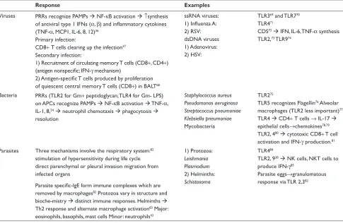
Table 1 Mechanisms in different types of infection
Response Examples
Viruses PRRs recognize PAMPs Æ NF-κB activation Æ↑synthesis of antiviral type 1 IFNs (α, β) and infl ammatory cytokines (TNF-α, MCP1, IL-6, 8, 12)36
Primary infection:
CD8+ T cells clearing up the infection37
Secondary infection:
1) Recruitment of circulating memory T cells (CD8+, CD4+) (antigen nonspecifi c; IFN-γ mechanism)
2) Antigen-specifi c T cells produced by proliferation of quiescent central memory T cells (CD8+) in BALT68
ssRNA viruses: 1) Infl uenza A: 2) RSV: dsDNA viruses 1) Adenovirus: 2) HSV:
TLR369 and TLR770
TLR471
CDS72Æ IFN, IL-6, TNF-α synthesis
TLR2,73 TLR974
Bacteria PRRs (TLR2 for Gm+ peptidoglycan; TLR4 for Gm- LPS) on APCs recognize PAMPs Æ NF-κB activation Æ TNF-α, IL-1, 8,24Æ neutrophil chemotaxis Æ phagocytosis Æ
resolution
Staphylococcus aureus Pseudomonas aeruginosa Streptococcus pneumoniae Klebsiella pneumoniae
Mycobacteria
TLR275
TLR5 recognizes Flagellin76 Alveolar
macrophages (TLR2 less important)77
TLR4 Æ CD4+ T cells → IL-17 Æ
epithelial cells→chemokines78,79
TLR2, 480Æ cytotoxic CD8+ T cell
activation and IFN-γ production.81
Parasites Three mechanisms involve the respiratory system:82
stimulation of hypersensitivity during life cycle direct parenchymal or pleural invasion migration from infected organs
Parasite specifi c-IgE form immune complexes which are removed by macrophages82 Protozoa vary in structure and
bioche-mistry Æ distinct immune responses. Helminths Æ
Th2 response and alternate macrophage activation83 Major:
eosinophils, basophils, mast cells Minor: neutrophils43
1) Protozoa:
Leishmania Plasmodium
2) Helminths:
Schistosoma
TLR484
TLR2, 938Æ NK cells, NKT cells to
produce IFN-γ85
Parasite eggs→granulomatous response via TLR 2,382
Abbreviations: APCs, antigen presenting cells; BALT, bronchial-associated lymphoid tissues; dsDNA, double-stranded DNA; HSV, herpes simplex virus; IFN, interferon; MCP1, monocyte chemoattractant protein 1; NK cells, natural killer cells; PAMPs, pathogen-associated molecular patterns; PRRs, pattern recognition receptors; RSV, respiratory syncytial virus; ssRNA, single-stranded RNA; TLR, Toll-like receptors.
Journal of Inflammation Research downloaded from https://www.dovepress.com/ by 118.70.13.36 on 24-Aug-2020
Lung infl ammation
prominent in asthma, but not in COPD except during acute exacerbations.47,48 Apoptosis is inversely correlated with
the clinical severity of asthma. Asthmatics show more submucosal “nonapoptotic” eosinophils than patients with chronic bronchitis or normal subjects. Bronchial biopsy specimens from asthmatic subjects reveal a higher infi ltration of eosinophils expressing anti apoptotic genes (bcl-2) than those expressing pro-apoptotic oncogenes (p53).49 Decreased
T cell apoptosis in asthma may lead to its increased number. CD4+ T cells are more evident in asthma, whereas CD8+ T cells are predominant in COPD.50 Moreover, CD8+ T cells
are higher in smokers than asthmatics.51,52 In smokers, the
severity of airfl ow limitation correlates with the number of neutrophils, macrophages, and NK lymphocytes in
bronchoalveloar lavage fl uid.51 Infl ammation in asthma is
only in the airways, but in COPD, infl ammation extends from the peripheral airways down to the lung parenchyma.
Chronic bronchitis is characterized by airfl ow obstruction, mucus hypersecretion, and infl ammation throughout the lungs. Neutrophils predominate in the airway lumen, although mononuclear cells, macrophages, CD8+ T cells and B cells infi ltrate the larger airway walls.53 Plausibly, the
severity of airway infl ammation can be related to the severity of the disease 51 and is a better pathologic marker of chronic
bronchitis than submucosal gland hypertrophy.
Th2 response is involved in chronic fi broproliferative disorders. Suppression of Th1 response or exaggerated Th2 response can lead to IPF.54 Interstitial lung diseases are
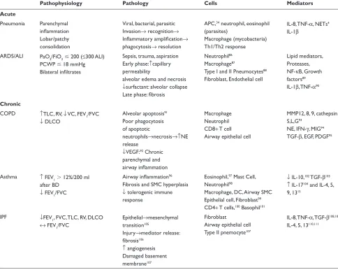
Table 2 Pathology and immune mechanisms in some common respiratory diseases
Pathophysiology Pathology Cells Mediators
Acute
Pneumonia Parenchymal infl ammation Lobar/patchy consolidation
Viral, bacterial, parasitic Invasion→ recognition→ Infl ammatory amplifi cation→ phagocytosis→ resolution
APC,34 neutrophil, eosinophil
(parasites)
Macrophage (mycobacteria) Th1/Th2 response
IL-8, TNF-α, NETs4
IL-1β
ARDS/ALI PaO2/FiO2ⱕ 200 (≤300 ALI) PCWP ⱕ18 mmHg Bilateral infi ltrates
Sepsis, trauma, aspiration Early phase:↑capillary permeability
alveolar edema and necrosis ↓surfactant: alveolar collapse Late phase: fi brosis
Neutrophil86
Macrophage87
Type I and II Pneumocytes88
Fibroblast, Endothelial cell
Lipid mediators, Proteases, NF-κB, Growth factors89
IL-1β, TNF-α90
Chronic
COPD ↑TLC, RV, ↓ VC, FEV1/FVC ↓ DLCO
Alveolar apoptosis91
Poor phagocytosis of apoptotic
neutrophils→necrosis→↑NE release
↓VEGF:92 Chronic
parenchymal and airway infl ammation
Macrophage Neutrophil CD8+ T cell Airway epithelial cell
MMP12, 8, 9, cathepsin S,L,G93
NE, IFN-γ, MIG94
TGF-β, EGF, PDGF95
Asthma ↑ FEV1⬎ 12%/200 ml after BD
↓ FEV1/FVC
Airway infl ammation96
Fibrosis and SMC hyperplasia ↓ tolerogenic immune response
Eosinophil,97 Mast Cell,
Neutrophil98
Macrophage, DC, Airway SMC Epithelial cell, Fibroblast99
CD4+ T cells,100 Basophil101
↓ IL-10,102 TGF-β103
↑ IL-17104 and IL-4, 5,
9, 1315
IPF ↓FEV1, FVC, TLC, RV, DLCO
↔ FEV1/FVC
Epithelial→mesenchymal transition105
Injury→mediator release:
fi brosis106
↑ angiogenesis Damaged basement membrane107
Fibroblast
Airway epithelial cell Type II pnemocyte107
IL-8, TNF-α, TGF-β108,109
IL-4, 5, 13110,111
Abbreviations: ALI, Acute lung injury; APC, antigen presenting cells; ARDS, Acute respiratory distress syndrome; BD, Bronchodilator; COPD, Chronic obstructive pulmonary disease; DC, Dendritic cell; DLCO, Diffusing capacity of lung for Carbon monoxide; EGF, Endothelial growth factor; FEV1, Forced expiratory volume in 1 sec; FiO2, Fraction of inspired oxygen; FVC, Forced vital capacity; IFN-γ, Interferon gamma; IL, Interleukin; MIG, Monokine induced by gamma interferon; MMP, Matrix metalloproteinases; NE, Neu-trophil elastase; NETs, NeuNeu-trophil extracellular traps; NF-κβ, Nuclear factor kappa beta; PaO2, Partial pressure of arterial oxygen; PCWP, Pulmonary capillary wedge pressure; PDGF, Platelet derived growth factor; RV, Residual volume; SMC, Smooth muscle cell; TGF-β, Transforming growth factor beta; TLC, Total lung capacity; TNF-α, Tumor necrosis factor alpha; Treg, Regulatory T cell; VC, Vital capacity; VEGF, Vascular endothelial growth factor.
Journal of Inflammation Research downloaded from https://www.dovepress.com/ by 118.70.13.36 on 24-Aug-2020
Moldoveanu et al
characterized by proliferation of fi broblasts and production of cytokines, chemokines, and glycosaminoglycans. These cytokines mediate the generation of tissue collagenase, gelatinase, and PGE2, which augment extracellular collagen and fi brinous matrix degradation. IL-1α and IL-1β stimulate production of type I and III collagen from fi broblasts and type IV collagen from epithelial cells. Moreover, production of PGE2 inhibits fi broblast proliferation.
Th1 primarily involves granulomatous infl ammation. Sarcoidosis is the best known of these, although tuberculo-sis and hypersensitivity pneumonitis are also important.55,56
Th2 drives eosinophilic granualomatous diseases, like Churg-Strauss vasculitis.57 Although the clinical presentation of
these diseases may be different, the infl ammatory processes possess a similar propagation of events in the pathogenesis of their development.
Recently, GITR (glucocorticoid-induced TNF receptor-related protein) was found to be important in acute as well as chronic infl ammation, and is closely associated with the development of pulmonary fi brosis. Neutrophil infi ltration, pro-infl ammatory cytokines, and toxic oxygen species are decreased in the pleural fl uid in GITR-/- pleurisy mice,58
and neutrophils are decreased in the bronchoalveolar lavage fl uid of GITR-/- bleomycin-induced pulmonary fi brosis mice. Moreover, lung histology also showed less fi brosis in GITR-/-mice.58
Regulation of lung in
fl
ammation
The lung provides a huge surface for gas exchange and pathogen contact. While a forceful innate immune response, such as infl ammation, is needed to fi ght off the harmful invaders, effective regulation to suppress the infl ammatory processes and limit infl ammation-induced damage is also necessary to maintain homeostasis. Such regulatory mechanisms are in place to protect lung function. For example, activation of TLRs may suppress Th1- and Th2- mediated lung infl ammation, thus providing negative feedback to prevent excessive lung infl ammation.59 In
addi-tion, cytokines produced by infl ammatory cells (IL-1060
and TGF-β61) can down regulate infl ammatory cytokine
production and infl ammatory response. The lungs provide organ-specifi c regulatory strategies to prevent excessive infl ammation during microbial invasion. For example, Type II epithelial cells in the lung communicate with alveolar mac-rophages, providing tonic inhibitory effects through TGF-β to limit potential adaptive immune-induced lung infl ammation.1
Recent evidence indicates that neural immune interaction may play an important role in controlling infl ammation in
a variety of diseases.62 Such control involves the peripheral
and central nervous systems to regulate the body’s reaction. Sensory neurons in the airways may provide a sensing mechanism to detect the infl ammatory intensity in the lung for lung-brain communication. Indeed, sensory neurons in the lung are activated during acute lung injury.63 In addition,
pro-infl ammatory cytokines and mediators, and TLR ligands can stimulate airway sensory neurons,64 which may initiate a
refl ex to suppress production of pro-infl ammatory cytokines.65
These negative feedback regulatory mechanisms are impor-tant in control of infl ammatory intensity. On the other hand, activation of sensory afferents may release neuropeptides, which can induce neurogenic infl ammation to intensify the infl ammatory response.66,67
Concluding remarks
Infl ammation is an important feature of many pulmonary diseases such as pneumonia, ARDS, asthma, and COPD. Varied and disparate strategies have been adopted to intervene in pulmonary immune responses. In addition to looking at the cytokines, cytokine receptors, and cell-surface molecules, cellular signal transduction and gene activation have been targeted for therapy. It is apparent that to understand the mechanism of upcoming future modalities of treatment, the physician should have a basic understanding of the underlying cellular mechanisms of infl ammation.
Acknowledgments
This work was partly supported by a grant from NIH HL-58727.
References
1. Oliveira ES, Hancock JT, Hermes-Lima M, et al. Implications of dealing with airborne substances and reactive oxygen species: what mammalian lungs, animals, and plants have to say? Integ Comp Biol. 2007;47:578–591.
2. Adler KB, Fischer BM, Wright DT, Cohn LA, Becker S. Interactions between respiratory epithelial cells and cytokines: relationships to lung infl ammation. Ann N Y Acad Sci. 1994;725:128–145.
3. Kuroki Y, Takahashi M, Nishitani C. Pulmonary collectins in innate immunity of the lung. Cell Microbiol. 2007;9:1871–1879.
4. Kaisho T, Akira S. Toll-like receptor function and signaling. J Allergy Clin Immunol. 2006;117:979–987.
5. Nittoh T, Fujimori H, Kozumi Y, Ishihara K, Mue S, Ohuchi K. Effects of glucocorticoids on apoptosis of infi ltrated eosinophils and neutrophils in rats. Eur J Pharmacol. 1998;354:73–81.
6. Fadok VA, Bratton DL, Konowal A, Freed PW, Westcott JY, Henson PM. Macrophages that have ingested apoptotic cells in vitro inhibit proinflammatory cytokine production through autocrine/ paracrine mechanisms involving TGF-beta, PGE2, and PAF. J Clin Invest. 1998;101:890–898.
7. Lipscomb MF, Bice DE, Lyons CR, Schuyler MR, Wilkes D. The regulation of pulmonary immunity. Adv Immunol. 1995;59:369–455.
Journal of Inflammation Research downloaded from https://www.dovepress.com/ by 118.70.13.36 on 24-Aug-2020
Lung infl ammation
8. Nakata K, Gotoh H, Watanabe J, et al. Augmented proliferation of human alveolar macrophages after allogeneic bone marrow transplanta-tion. Blood. 1999;93:667–673.
9. Bender AT, Ostenson CL, Wang EH, Beavo JA. Selective up-regulation of PDE1B2 upon monocyte-to-macrophage differentiation. Proc Natl Acad Sci U S A. 2005;102:497–502.
10. Burns AR, Smith CW, Walker DC. Unique structural features that infl u-ence neutrophil emigration into the lung. Physiol Rev. 2003;83:309–336. 11. Yazdanbakhsh M, Kremsner PG, van RR. Allergy, parasites, and the
hygiene hypothesis. Science. 2002;296:490–494.
12. Yokoyama WM. Natural killer cell immune responses. Immunol Res. 2005;32:317–325.
13. Brigl M, Bry L, Kent SC, Gumperz JE, Brenner MB. Mechanism of CD1d-restricted natural killer T cell activation during microbial infec-tion. Nat Immunol. 2003;4:1230–1237.
14. Prussin C, Metcalfe DD. 5. IgE, mast cells, basophils, and eosinophils.
J Allergy Clin Immunol. 2006;117(2 Suppl Mini-Primer):S450–S456. 15. Kariyawasam HH, Robinson DS. The role of eosinophils in airway
tissue remodelling in asthma. Curr Opin Immunol. 2007;19:681–686. 16. Suratt BT, Parsons PE. Mechanisms of acute lung injury/acute
respira-tory distress syndrome. Clin Chest Med. 2006;27:579–589.
17. Toossi Z, Hirsch CS, Hamilton BD, Knuth CK, Friedlander MA, Rich EA. Decreased production of TGF-beta 1 by human alveo-lar macrophages compared with blood monocytes. J Immunol. 1996;156:3461–3468.
18. Moore SA, Strieter RM, Rolfe MW, Standiford TJ, Burdick MD, Kunkel SL. Expression and regulation of human alveolar macrophage-derived interleukin-1 receptor antagonist. Am J Respir Cell Mol Biol. 1992;6:569–575.
19. Ding L, Linsley PS, Huang LY, Germain RN, Shevach EM. IL-10 inhibits macrophage costimulatory activity by selectively inhibiting the up-regulation of B7 expression. J Immunol. 1993;151:1224–1234. 20. Holgate ST. Pathogenesis of asthma. Clin Exp Allergy. 2008;38:872–897. 21. Sarir H, Henricks PA, van Houwelingen AH, Nijkamp FP, Folkerts G. Cells, mediators and Toll-like receptors in COPD. Eur J Pharmacol. 2008;585:346–353.
22. Suter PM, Suter S, Girardin E, Roux-Lombard P, Grau GE, Dayer JM. High bronchoalveolar levels of tumor necrosis factor and its inhibitors, interleukin-1, interferon, and elastase, in patients with adult respiratory distress syndrome after trauma, shock, or sepsis. Am Rev Respir Dis. 1992;145:1016–1022.
23. Armstrong L, Millar AB. Relative production of tumour necrosis factor alpha and interleukin 10 in adult respiratory distress syndrome. Thorax. 1997;52:442–446.
24. Stockley RA. Lung infections. 1. Role of bacteria in the pathogen-esis and progression of acute and chronic lung infection. Thorax. 1998;53:58–62.
25. Wilson R, Pitt T, Taylor G, et al. Pyocyanin and 1-hydroxyphenazine produced by Pseudomonas aeruginosa inhibit the beating of human respiratory cilia in vitro. J Clin Invest. 1987;79:221–229.
26. Menendez R, Torres A. Treatment failure in community-acquired pneumonia. Chest. 2007;132:1348–1355.
27. Shaw MH, Reimer T, Kim YG, Nunez G. NOD-like receptors (NLRs): bona fi de intracellular microbial sensors. Curr Opin Immunol. 2008;20:377–382.
28. Sutterwala FS, Ogura Y, Flavell RA. The infl ammasome in pathogen recognition and infl ammation. J Leukoc Biol. 2007;82:259–264. 29. Khair OA, Devalia JL, Abdelaziz MM, Sapsford RJ, Tarraf H, Davies RJ.
Effect of Haemophilus infl uenzae endotoxin on the synthesis of IL-6, IL-8, TNF-alpha and expression of ICAM-1 in cultured human bronchial epithelial cells. Eur Respir J. 1994;7:2109–2116.
30. Wardlaw A. Leucocyte adhesion to endothelium. Clin Exp Allergy. 1990;20:619–626.
31. Takahashi GW, Andrews DF, III, Lilly MB, Singer JW, Alderson MR. Effect of granulocyte-macrophage colony-stimulating factor and interleukin-3 on interleukin-8 production by human neutrophils and monocytes. Blood. 1993;81:357–364.
32. Nakamura H, Yoshimura K, McElvaney NG, Crystal RG. Neutrophil elastase in respiratory epithelial lining fl uid of individuals with cystic fi brosis induces interleukin-8 gene expression in a human bronchial epithelial cell line. J Clin Invest. 1992;89:1478–1484.
33. Gordon S. The macrophage: past, present and future. Eur J Immunol. 2007;37(Suppl 1):S9–17.
34. Mizgerd JP. Acute lower respiratory tract infection. N Engl J Med. 2008;358:716–727.
35. Matute-Bello G, Liles WC, Radella F, et al. Neutrophil apoptosis in the acute respiratory distress syndrome. Am J Respir Crit Care Med. 1997;156:1969–1977.
36. Wang JP, Kurt-Jones EA, Finberg RW. Innate immunity to respiratory viruses. Cell Microbiol. 2007;9:1641–1646.
37. Woodland DL. Cell-mediated immunity to respiratory virus infections.
Curr Opin Immunol. 2003;15:430–435.
38. Coban C, Ishii KJ, Uematsu S, et al. Pathological role of Toll-like receptor signaling in cerebral malaria. Int Immunol. 2007;19:67–79. 39. Kropf P, Freudenberg N, Kalis C, et al. Infection of C57BL/10ScCr
and C57BL/10ScNCr mice with Leishmania major reveals a role for Toll-like receptor 4 in the control of parasite replication. J Leukoc Biol. 2004;76:48–57.
40. rsic-Arsenijevic V, Dzamic A, Mitrovic S, Radonjic I, Kranjcic-Zec I. [Characteristics of the immune response in protozoan infections]. Med Pregl. 2003;56:557–563.
41. Liew FY, O’Donnell CA. Immunology of leishmaniasis. Adv Parasitol. 1993;32:161–259.
42. McGuinness DH, Dehal PK, Pleass RJ. Pattern recognition mol-ecules and innate immunity to parasites. Trends Parasitol. 2003;19: 312–319.
43. Anthony RM, Rutitzky LI, Urban JF Jr, Stadecker MJ, Gause WC. Pro-tective immune mechanisms in helminth infection. Nat Rev Immunol. 2007;7:975–987.
44. Zlotnik A, Yoshie O. Chemokines: a new classifi cation system and their role in immunity. Immunity. 2000;12:121–127.
45. Frevert CW, Farone A, Danaee H, Paulauskis JD, Kobzik L. Functional characterization of rat chemokine macrophage infl ammatory protein-2.
Infl ammation. 1995;19:133–142.
46. Tsai WC, Strieter RM, Mehrad B, Newstead MW, Zeng X, Standiford TJ. CXC chemokine receptor CXCR2 is essential for protective innate host response in murine Pseudomonas aeruginosa pneumonia. Infect Immun. 2000;68:4289–4296.
47. Barnes PJ, Shapiro SD, Pauwels RA. Chronic obstructive pulmo-nary disease: molecular and cellular mechanisms. Eur Respir J. 2003;22:672–688.
48. Fabbri LM, Romagnoli M, Corbetta L, et al. Differences in airway infl ammation in patients with fi xed airfl ow obstruction due to asthma or chronic obstructive pulmonary disease. Am J Respir Crit Care Med. 2003;167:418–424.
49. Vignola AM, Chanez P, Chiappara G, et al. Evaluation of apoptosis of eosinophils, macrophages, and T lymphocytes in mucosal biopsy specimens of patients with asthma and chronic bronchitis. J Allergy Clin Immunol. 1999;103:563–573.
50. Sutherland ER, Martin RJ. Airway infl ammation in chronic obstructive pulmonary disease: comparisons with asthma. J Allergy Clin Immunol. 2003;112:819–827.
51. Di SA, Capelli A, Lusuardi M, et al. Severity of airfl ow limitation is associated with severity of airway infl ammation in smokers. Am J Respir Crit Care Med. 1998;158:1277–1285.
52. O’Shaughnessy TC, Ansari TW, Barnes NC, Jeffery PK. Infl amma-tion in bronchial biopsies of subjects with chronic bronchitis: inverse relationship of CD8+ T lymphocytes with FEV1. Am J Respir Crit Care
Med. 1997;155:852–857.
53. Szilasi M, Dolinay T, Nemes Z, Strausz J. Pathology of chronic obstruc-tive pulmonary disease. Pathol Oncol Res. 2006;12:52–60. 54. Strieter RM, Keane MP. Innate immunity dictates cytokine
polariza-tion relevant to the development of pulmonary fi brosis. J Clin Invest. 2004;114:165–168.
Journal of Inflammation Research downloaded from https://www.dovepress.com/ by 118.70.13.36 on 24-Aug-2020
Moldoveanu et al
55. Gerke AK, Hunninghake G. The immunology of sarcoidosis. Clin Chest Med. 2008;29:379–390, vii.
56. Hernandez C, Cetner AS, Jordan JE, Puangsuvan SN, Robinson JK. Tuberculosis in the age of biologic therapy. J Am Acad Dermatol. 2008;59:363–380.
57. Hellmich B, Csernok E, Gross WL. Proinflammatory cytokines and autoimmunity in Churg-Strauss syndrome. Ann N Y Acad Sci. 2005;1051:121–131.
58. Nocentini G, Cuzzocrea S, Bianchini R, Mazzon E, Riccardi C. Modu-lation of acute and chronic infl ammation of the lung by GITR and its ligand. Ann N Y Acad Sci. 2007;1107:380–391.
59. Hayashi T, Beck L, Rossetto C, et al. Inhibition of experimental asthma by indoleamine 2,3-dioxygenase. J Clin Invest. 2004;114:270–279. 60. Raychaudhuri B, Fisher CJ, Farver CF, et al. Interleukin 10
(IL-10)-mediated inhibition of infl ammatory cytokine production by human alveolar macrophages. Cytokine. 2000;12:1348–1355.
61. Pittet JF, Griffi ths MJ, Geiser T, et al. TGF-beta is a critical mediator of acute lung injury. J Clin Invest. 2001;107:1537–1544.
62. Otmishi P, Gordon J, El-Oshar S, et al. Neuroimmune interac-tion in infl ammatory diseases. Clin Med Circ Respir Pulm Med. 2008;2:35–44.
63. Lin S, Walker J, Xu L, Gozal D, Yu J. Respiratory: Behaviours of pulmonary sensory receptors during development of acute lung injury in the rabbit. Exp Physiol. 2007;92:749–755.
64. Yu J, Lin S, Zhang J, Otmishi P, Guardiola JJ. Airway nociceptors activated by pro-infl ammatory cytokines. Respir Physiol Neurobiol. 2007;156:116–119.
65. Tracey KJ. The infl ammatory refl ex. Nature. 2002;420:853–859. 66. Bozic CR, Lu B, Hopken UE, Gerard C, Gerard NP.
Neuro-genic amplification of immune complex inflammation. Science. 1996;273:1722–1725.
67. Veronesi B, Carter JD, Devlin RB, Simon SA, Oortgiesen M. Neuropep-tides and capsaicin stimulate the release of infl ammatory cytokines in a human bronchial epithelial cell line. Neuropeptides. 1999;33:447–456. 68. Kohlmeier JE, Miller SC, Smith J, et al. The chemokine receptor CCR5
plays a key role in the early memory CD8+ T cell response to respiratory virus infections. Immunity. 2008;29:101–113.
69. Guillot L, Le GR, Bloch S, et al. Involvement of toll-like receptor 3 in the immune response of lung epithelial cells to double-stranded RNA and infl uenza A virus. J Biol Chem. 2005;280:5571–5580.
70. Diebold SS, Kaisho T, Hemmi H, Akira S, Reis e Sousa C. Innate antiviral responses by means of TLR7-mediated recognition of single-stranded RNA. Science. 2004;303:1529–1531.
71. Kurt-Jones EA, Popova L, Kwinn L, et al. Pattern recognition receptors TLR4 and CD14 mediate response to respiratory syncytial virus. Nat Immunol. 2000;1:398–401.
72. Nociari M, Ocheretina O, Schoggins JW, Falck-Pedersen E. Sensing infection by adenovirus: Toll-like receptor-independent viral DNA recognition signals activation of the interferon regulatory factor 3 master regulator. J Virol. 2007;81:4145–4157.
73. Kurt-Jones EA, Chan M, Zhou S, et al. Herpes simplex virus 1 interac-tion with Toll-like receptor 2 contributes to lethal encephalitis. Proc Natl Acad Sci U S A. 2004;101:1315–1320.
74. Krug A, Luker GD, Barchet W, Leib DA, Akira S, Colonna M. Herpes simplex virus type 1 activates murine natural interferon-producing cells through toll-like receptor 9. Blood. 2004;103:1433–1437.
75. Yoshimura A, Lien E, Ingalls RR, Tuomanen E, Dziarski R, Golenbock D. Cutting edge: recognition of Gram-positive bacterial cell wall components by the innate immune system occurs via Toll-like receptor 2. J Immunol. 1999;163:1–5.
76. Zhang Z, Louboutin JP, Weiner DJ, Goldberg JB, Wilson JM. Human airway epithelial cells sense Pseudomonas aeruginosa infection via recognition of fl agellin by Toll-like receptor 5. Infect Immun. 2005;73:7151–7160.
77. Xu F, Droemann D, Rupp J, et al. Modulation of the infl ammatory response to S. pneumoniae in a model of acute lung tissue infection.
Am J Respir Cell Mol Biol. 2008;39:522–529.
78. Happel KI, Zheng M, Young E, et al. Cutting edge: roles of Toll-like receptor 4 and IL-23 in IL-17 expression in response to Klebsiella pneumoniae infection. J Immunol. 2003;170:4432–4436.
79. Ye P, Rodriguez FH, Kanaly S, et al. Requirement of interleukin 17 receptor signaling for lung CXC chemokine and granulocyte colony-stimulating factor expression, neutrophil recruitment, and host defense. J Exp Med. 2001;194:519–527.
80. Means TK, Jones BW, Schromm AB, et al. Differential effects of a Toll-like receptor antagonist on Mycobacterium tuberculosis-induced macrophage responses. J Immunol. 2001;166:4074–4082.
81. Vesosky B, Flaherty DK, Turner J. Th1 cytokines facilitate CD8-T-cell-mediated early resistance to infection with Mycobacterium tuberculosis in old mice. Infect Immun. 2006;74:3314–3324. 82. Fischer GB, Sarria EE, Leite AJ, de Britto MC. Parasitic lung infection
and the paediatric lung. Paediatr Respir Rev. 2008;9:57–65. 83. Reyes JL, Terrazas LI. The divergent roles of alternatively
acti-vated macrophages in helminthic infections. Parasite Immunol. 2007;29:609–619.
84. Kropf P, Freudenberg MA, Modolell M, et al. Toll-like receptor 4 contributes to effi cient control of infection with the protozoan parasite Leishmania major. Infect Immun. 2004;72:1920–1928.
85. D’Ombrain MC, Hansen DS, Simpson KM, Schofi eld L. Gammadelta-T cells expressing NK receptors predominate over NK cells and conventional T cells in the innate IFN-gamma response to Plasmodium falciparum malaria. Eur J Immunol. 2007;37:1864–1873.
86. Ware LB, Matthay MA. The acute respiratory distress syndrome.
N Engl J Med. 2000;342:1334–1349.
87. Dong L, Wang S, Chen M, Li H, Bi W. The activation of macrophage and upregulation of CD40 costimulatory molecule in lipopolysaccharide-induced acute lung injury. J Biomed Biotechnol. 2008;2008:852571.
88. Gropper MA, Wiener-Kronish J. The epithelium in acute lung injury/acute respiratory distress syndrome. Curr Opin Crit Care. 2008;14:11–15.
89. Luh SP, Chiang CH. Acute lung injury/acute respiratory distress syndrome (ALI/ARDS): the mechanism, present strategies and future perspectives of therapies. J Zhejiang Univ Sci B. 2007;8:60–69. 90. Gropper MA, Wiener-Kronish J. The epithelium in acute lung
injury/acute respiratory distress syndrome. Curr Opin Crit Care. 2008;14:11–15.
91. Segura-Valdez L, Pardo A, Gaxiola M, Uhal BD, Becerril C, Selman M. Upregulation of gelatinases A and B, collagenases 1 and 2, and increased parenchymal cell death in COPD. Chest. 2000;117:684–694.
92. Kanazawa H, Asai K, Hirata K, Yoshikawa J. Possible effects of vascular endothelial growth factor in the pathogenesis of chronic obstructive pulmonary disease. Am J Med. 2003;114:354–358. 93. Shapiro SD. Proteolysis in the lung. Eur Respir J Suppl.
2003;44:30s–2s.
94. Grumelli S, Corry DB, Song LZ, et al. An immune basis for lung parenchymal destruction in chronic obstructive pulmonary disease and emphysema. PLoS Med. 2004;1:e8.
95. Sharafkhaneh A, Hanania NA, Kim V. Pathogenesis of emphysema: from the bench to the bedside. Proc Am Thorac Soc. 2008;5:475–477. 96. De Monchy JG, Kauffman HF, Venge P, et al. Bronchoalveolar
eosinophilia during allergen-induced late asthmatic reactions. Am Rev Respir Dis. 1985;131:373–376.
97. Bousquet J, Jeffery PK, Busse WW, Johnson M, Vignola AM. Asthma. From bronchoconstriction to airways infl ammation and remodeling.
Am J Respir Crit Care Med. 2000;161:1720–1745.
98. Koh YY, Dupuis R, Pollice M, Albertine KH, Fish JE, Peters SP. Neutrophils recruited to the lungs of humans by segmental antigen challenge display a reduced chemotactic response to leukotriene B4.
Am J Respir Cell Mol Biol. 1993;8:493–499.
99. Doucet C, Brouty-Boye D, Pottin-Clemenceau C, Canonica GW, Jasmin C, Azzarone B. Interleukin (IL) 4 and IL-13 act on human lung fi broblasts. Implication in asthma. J Clin Invest. 1998;101:2129–2139.
Journal of Inflammation Research downloaded from https://www.dovepress.com/ by 118.70.13.36 on 24-Aug-2020
Lung infl ammation
100. Robinson D, Hamid Q, Bentley A, Ying S, Kay AB, Durham SR. Activation of CD4+ T cells, increased TH2-type cytokine mRNA expression, and eosinophil recruitment in bronchoalveolar lavage after allergen inhalation challenge in patients with atopic asthma. J Allergy Clin Immunol. 1993;92:313–324.
101. Guo CB, Liu MC, Galli SJ, Bochner BS, Kagey-Sobotka A, Lichtenstein LM. Identifi cation of IgE-bearing cells in the late-phase response to antigen in the lung as basophils. Am J Respir Cell Mol Biol. 1994;10:384–390.
102. Hawrylowicz CM. Regulatory T cells and IL-10 in allergic infl ammation.
J Exp Med. 2005;202:1459–1463.
103. Scherf W, Burdach S, Hansen G. Reduced expression of transforming growth factor beta 1 exacerbates pathology in an experimental asthma model. Eur J Immunol. 2005;35:198–206.
104. Schnyder-Candrian S, Togbe D, Couillin I, et al. Interleukin-17 is a negative regulator of established allergic asthma. J Exp Med. 2006;203:2715–2725.
105. Willis BC, Liebler JM, Luby-Phelps K, et al. Induction of epithelial-mesenchymal transition in alveolar epithelial cells by transforming growth factor-beta1: potential role in idiopathic pulmonary fi brosis.
Am J Pathol. 2005;166:1321–1332.
106. Noble PW, Homer RJ. Idiopathic pulmonary fi brosis: new insights into pathogenesis. Clin Chest Med. 2004;25:749–758, vii.
107. Katzenstein AL. Pathogenesis of “fi brosis” in interstitial pneumonia: an electron microscopic study. Hum Pathol. 1985;16:1015–1024. 108. Strieter RM, Gomperts BN, Keane MP. The role of CXC chemokines
in pulmonary fi brosis. J Clin Invest. 2007;117:549–556.
109. Zhang Y, Lee TC, Guillemin B, Yu MC, Rom WN. Enhanced IL-1 beta and tumor necrosis factor-alpha release and messenger RNA expression in macrophages from idiopathic pulmonary fi brosis or after asbestos exposure. J Immunol. 1993;150:4188–4196. 110. Ando M, Miyazaki E, Fukami T, Kumamoto T, Tsuda T.
Interleukin-4-producing cells in idiopathic pulmonary fi brosis: an immunohisto-chemical study. Respirology. 1999;4:383–391.
111. Lee CG, Homer RJ, Zhu Z, et al. Interleukin-13 induces tissue fi brosis by selectively stimulating and activating transforming growth factor beta(1). J Exp Med. 2001;194:809–821.
Journal of Inflammation Research downloaded from https://www.dovepress.com/ by 118.70.13.36 on 24-Aug-2020
Journal of Inflammation Research downloaded from https://www.dovepress.com/ by 118.70.13.36 on 24-Aug-2020