Electronic Thesis and Dissertation Repository
12-12-2013 12:00 AM
Characterizing the human vaginal microbiome using
Characterizing the human vaginal microbiome using
high-throughput sequencing
throughput sequencing
Jean Megan E. Macklaim
The University of Western Ontario
Supervisor Greg Gloor
The University of Western Ontario Joint Supervisor Gregor Reid
The University of Western Ontario Graduate Program in Biochemistry
A thesis submitted in partial fulfillment of the requirements for the degree in Doctor of Philosophy
© Jean Megan E. Macklaim 2013
Follow this and additional works at: https://ir.lib.uwo.ca/etd
Part of the Bioinformatics Commons, Computational Biology Commons, Environmental Microbiology
and Microbial Ecology Commons, Female Urogenital Diseases and Pregnancy Complications Commons,
Molecular Biology Commons, and the Other Microbiology Commons
Recommended Citation Recommended Citation
Macklaim, Jean Megan E., "Characterizing the human vaginal microbiome using high-throughput sequencing" (2013). Electronic Thesis and Dissertation Repository. 1787.
https://ir.lib.uwo.ca/etd/1787
This Dissertation/Thesis is brought to you for free and open access by Scholarship@Western. It has been accepted for inclusion in Electronic Thesis and Dissertation Repository by an authorized administrator of
(Thesis format: Integrated Article)
by
Jean Megan Elizabeth Macklaim
Graduate Program in Biochemistry
A thesis submitted in partial fulfillment of the requirements for the degree of
Doctor of Philosophy
The School of Graduate and Postdoctoral Studies The University of Western Ontario
London, Ontario, Canada
© Jean M. E. Macklaim 2013
ii
Abstract
The human vaginal microbiome undoubtedly has a significant role in reproductive health and
for protection from infectious organisms. Recent efforts to characterize the bacterial species
of the vagina using molecular techniques have uncovered an unexpected diversity. Using
high-throughput sequencing I sought to describe the structure and function of the vaginal
microbiome under different physiological states including healthy, bacterial vaginosis (BV),
post-menopausal vaginal atrophy, and acute vulvovaginal candidiasis (VVC).
Partial 16S rRNA gene sequencing revealed that healthy, asymptomatic women most often
have vaginal biotas dominated by Lactobacillus iners or L. crispatus. In contrast, BV is a
heterogeneous, highly diversified condition with reduced Lactobacillus abundance. Similar
to BV, post-menopausal women experiencing vaginal dryness were depleted in lactobacilli
and had a more diverse vaginal profile. In the case of VVC, the biotas were not significantly
altered compared to healthy women despite the fungal overgrowth.
One organism, Lactobacillusiners was ubiquitously present in all conditions, and became
predominant following antibiotic and probiotic treatment of BV. To uncover the potential
role of this bacterium, I used whole genome sequencing of vaginal isolate AB-1. The genome
is predicted to be the smallest of any Lactobacillus at 1.3 Mbp, but having a higher
proportion of horizontally acquired genes. These results, along with predicted adhesins and a
cholesterol-dependent cytolysin, indicate L. iners is highly adapted for the vagina and could
have an uncharacterized role in the etiology of BV.
As BV is the most common vaginal ailment with severe implications on acquisition and
iii
sequencing. L. iners drastically modulates gene expression in response to BV, and notably
increases expression of a cholesterol-dependent cytolysin, mucin and glycerol transport and
metabolic enzymes, and genes belonging to a CRISPR system - suggestive of bacteriophage
influence in the community. Although diverse in taxonomic membership, there is evidence of
functional conservation in BV including preference for glycogen and glycerol as carbon
sources, and predicted end products of metabolism including an abundance of succinate and
short-chain fatty acids. These studies add significantly to our understanding of the role
lactobacilli can play in vaginal and reproductive health.
Keywords
iv
Co-Authorship Statement
The experiments and data analyses presented in this thesis were primarily conducted by Jean Macklaim with Greg Gloor and Gregor Reid. The published manuscripts were written by Jean Macklaim, Greg Gloor, and Gregor Reid. Exceptions are noted below.
Chapter 1: Vaginal microbiome and epithelial gene expression of post-menopausal women with moderate to severe dryness
Ruben Hummelen and Gregor Reid conceived of the study design and collected the samples. Amy McMillan extracted sample DNA and PCR amplified. Jo-Anne Hammond connected clinical samples. Rebecca Vognsa and David Koenig
contributed data analysis. Jordan Bisanz performed analysis of the human microarray data. Jean Macklaim performed analysis of the microbiota data.
Chapter 3: At the crossroads of vaginal health and disease: the genome sequence of Lactobacillus iners AB-1
Gregor Reid and Greg Gloor conceived of the study design. Kingsley Anukam prepared the samples for sequencing. Jean Macklaim and Greg Gloor performed the genome assembly, annotation, and analyses, and the protein extraction for mass spectroscopy. Jean Macklaim and Sarah Cribby performed the immunogold labeling and TEM images with Judy Sholdice.
Chapter 4: Comparative meta-RNA-seq of the vaginal microbiota and differential expression by Lactobacillus iners in health and dysbiosis
Gregor Reid, Greg Gloor, and Jean Macklaim conceived of the study design. Jean Macklaim prepared the samples. Jean Macklaim and Greg Gloor performed the sequencing analysis and annotation. Julia Di Bella provided some R code for figure generation. Andrew Fernandes developed the statistical framework (published as: Fernandes, A.F. et al. 2013 PLoS One).
Chapter 5: Effect of antimicrobial and probiotic therapy on the vaginal microbiota
The initial clinical trials were conceived and performed by Martinez, Gregor Reid, and Elaine Cristina Pereira De Martinis. The DNA extractions and PCR
v
Acknowledgments
I am very fortunate to have had two fantastic supervisors for my PhD thesis and I can't express my gratitude for their guidance on this journey. So thank you to Gregor Reid and Greg Gloor for taking the chance on me. I came in not knowing anything about microbiome, bacteria, or computational biology but your patient mentorship and guidance (and great sense of humour) made this thesis possible. You both gave me the freedom to explore and
challenge my ideas - good and bad.
Greg: You've had seemingly unlimited patience, and you've gone above and beyond as a supervisor. You are not afraid to walk on the bleeding edge of science, and your innate curiosity about the world is the core of a good scientist.
Gregor: You have a fantastic ability to draw connections and inspiration from anywhere in your research. I especially want to thank you for pushing me beyond what I thought I was capable of. Your passion for making the world a better place is something everyone should have.
A few people have had significant influence on this thesis:
Ruben: you walked into our lab with over 300 samples from Africa and changed the course of our research for the years to come. Thank for your collaborative attitude, and for the many interesting discussions.
Andrew: I remember the day I was whining (yet again) about the woeful inadequacies of the bioinformatic tools for the data we were trying to analyze, and you took it as a challenge to make something better. One day I may eventually understand ALDEx. Thank you for you mentorship over the years, and for your healthy dose of cynicism.
vi
collegial environment for research. To my committee members, John McCormick and David Edgell, I thank you for your always insightful input into my project. And to the Molecular Biology group for providing a friendly forum for scientific discussions.
To the past and present members of the Gloor lab where I spent most of my time: I thank you for your humour and friendship and the many intellectual discussions (once in a while even about science). "On second thought, let's not go to the Gloor lab - 'tis a silly place".
To the Reid lab: So many of you have impacted this thesis in small but significant ways. I consider every one of you who have passed through in my time here a friend.
On a personal note, I thank my Mom, Dad, and sister Kristy for your love and support and for encouraging me pursue the things that fascinate me.
I also thank my karate Senseis for their influence (within and outside karate) and for showing the value of commitment.
vii
Table of Contents
Abstract ... ii
Co-Authorship Statement... iv
Acknowledgments... v
Table of Contents... vii
List of Tables ... xii
List of Figures ... xiii
List of Appendices ... xv
List of Abbreviations, Symbols, and Nomenclature... xvi
Chapter 1... 1
1 General Introduction ... 1
1.1 The human microbiome ... 1
1.2 The vaginal microbiome ... 2
1.3 What is “normal”?... 3
1.4 Conditions of the vaginal microbiome... 5
1.4.1 Bacterial vaginosis (BV)... 5
1.4.2 Other microbiological disorders ... 8
1.5 Modulating the vaginal microbiome and treating aberrant conditions ... 11
1.5.1 Antimicrobials... 11
1.5.2 Probiotics ... 12
1.6 Other physiological states ... 15
1.7 Key taxa and species-specific roles ... 16
1.8 High-throughput sequencing techniques for characterizing the microbiome ... 19
1.8.1 Targeted amplicon sequencing (16S)... 20
viii
1.9 Scope and objectives of the thesis ... 29
1.10 References... 30
Chapter 2... 49
2 Vaginal microbiome and epithelial gene expression of post-menopausal women with moderate to severe dryness ... 49
2.1 Introduction... 49
2.2 Materials and Methods... 50
2.2.1 Study population ... 50
2.2.2 Bacterial DNA extraction, amplification, and sequencing ... 51
2.2.3 OTU clustering and taxonomic assignment... 51
2.2.4 Vaginal epithelial RNA extraction... 52
2.2.5 Gene expression analysis ... 53
2.2.6 Statistical analyses ... 53
2.3 Results... 56
2.4 Discussion and Concluding Remarks ... 61
2.5 References... 64
Chapter 3... 67
3 At the crossroads of vaginal health and disease: the genome sequence of Lactobacillus iners AB-1... 67
3.1 Introduction... 67
3.2 Materials and Methods... 68
3.2.1 Organism... 68
3.2.2 Genome assembly ... 68
3.2.3 Gap closure and scaffolding... 68
3.2.4 Validating genome coverage... 70
ix
CAI) ... 71
3.2.7 Comparative genomics... 71
3.2.8 Prediction of horizontally acquired genes... 72
3.2.9 Construction of phylogenetic tree of select lactobacilli... 72
3.2.10 Cell-wall protein isolation... 72
3.2.11 Mass spectrometry ... 73
3.2.12 Rabbit polyclonal antisera... 74
3.2.13 Absorption of polyclonal antisera... 74
3.2.14 Immuno-blot assay... 75
3.2.15 Transmission electron microscopy and immunogold labeling ... 75
3.2.16 Growth assays ... 75
3.3 Results and Discussion ... 76
3.3.1 Phylogeny ... 76
3.3.2 General genomic features ... 77
3.3.3 Comparative genomics... 79
3.3.4 Horizontal gene transfer (HGT)... 81
3.3.5 Cholesterol-dependent cytolysin... 82
3.3.6 Carbohydrate transport and metabolic capabilities... 84
3.3.7 Other transport and metabolism... 86
3.3.8 Amino acid biosynthesis ... 87
3.3.9 Adherence and host interaction... 87
3.3.10 Stress tolerance and environmental response ... 91
3.3.11 Defense mechanisms... 93
3.4 Concluding remarks ... 94
x
4 Comparative meta-RNA-seq of the vaginal microbiota and differential expression by
Lactobacillus iners in health and dysbiosis ... 102
4.1 Introduction... 102
4.2 Materials and Methods... 103
4.2.1 Clinical samples ... 103
4.2.2 RNA isolation, mRNA enrichment, and sequencing ... 104
4.2.3 Reference sequence library and mapping ... 104
4.2.4 Functional assignment of refseqs... 105
4.2.5 Statistical analyses for differential expression... 105
4.2.6 CRISPR spacer analysis... 105
4.2.7 cpn60 reference mapping... 106
4.3 Results and Discussion ... 106
4.3.1 Lactobacillus iners’ response to BV... 109
4.3.2 The vaginal community in BV... 114
4.4 Concluding Remarks... 116
4.5 References... 119
Chapter 5... 124
5 Effect of antimicrobial and probiotic therapy on the vaginal microbiota ... 124
5.1 Introduction... 124
5.2 Materials and Methods... 125
5.2.1 Clinical samples and study design ... 125
5.2.2 V6-targeted 16S rRNA gene sequencing... 127
5.2.3 Sequence processing ... 127
5.2.4 Data analysis ... 127
xi
5.3.2 Vulvovaginal candidiasis (VVC) study ... 130
5.3.3 Bacterial vaginosis (BV) study ... 133
5.4 Discussion ... 136
5.5 Concluding Remarks... 138
5.6 References... 138
Chapter 6... 142
6 General discussion ... 142
6.1 Defining normal ... 142
6.2 Bacterial vaginosis ... 144
6.3 The role of Lactobacillus iners... 147
6.4 What are the bacteria doing?... 149
6.5 Considerations for modulating the ecosystem ... 153
6.6 Future directions ... 154
6.7 References... 157
xii
List of Tables
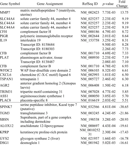
Table 2-1. The top 20 genes up- and down- regulated genes (p<0.05) in the vaginal dryness group compared to controls. ... 59
xiii
List of Figures
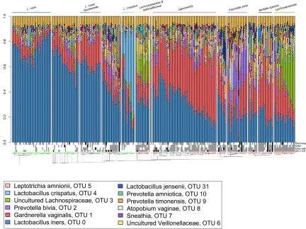
Figure 1-1. The V6-sequenced vaginal bacterial profiles of 272 samples from 132 women. .. 9
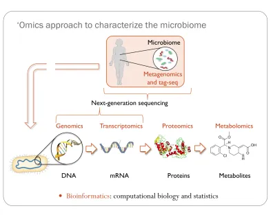
Figure 1-2. “Omics” approaches for characterizing the human microbiome. ... 20
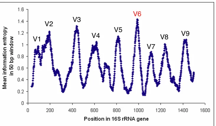
Figure 1-3. Variability within the 16S rRNA gene... 21
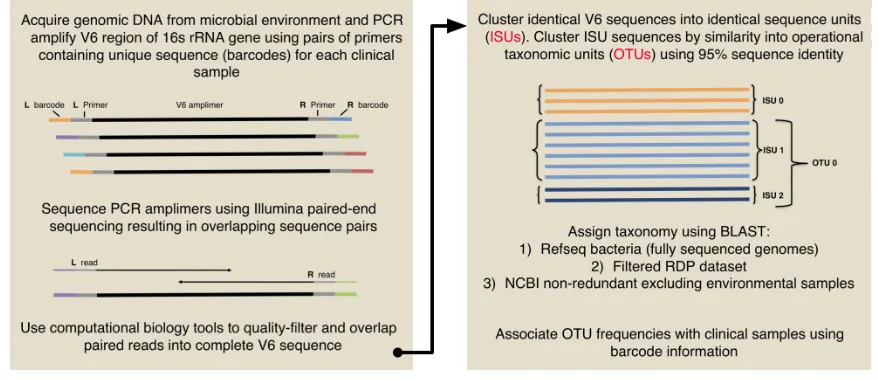
Figure 1-4. Workflow used for multiplexed Illumina V6 sequencing for studies presented in this thesis... 23
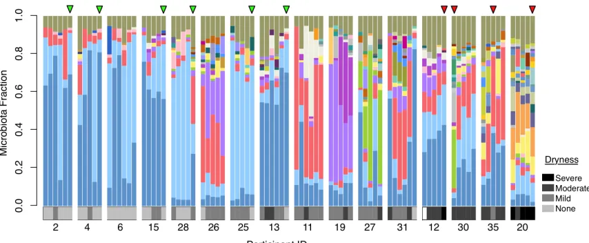
Figure 2-1. Microbiota profiles for 32 post-menopausal women clustered by biota similarity ... 54
Figure 2-2. Color taxa legend for Figure 2-1 and Figure 2-3 ... 55
Figure 2-3. 16S (V6) microbiota profiles for 16 post-menopausal women sampled every 2 weeks... 57
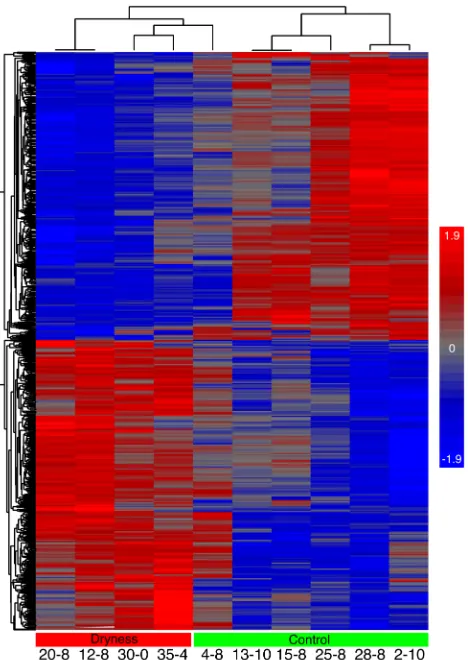
Figure 2-4. Heatmap of vaginal epithelial gene expression of 10 samples ... 58
Figure 3-1. Subset of a phylogenetic tree representing L. iners as part of the acidophilus complex... 76
Figure 3-2. Genomic atlas of Lactobacillus iners AB-1... 78
Figure 3-3. Venn diagram representing orthologous proteins between select species of the L. iners clade ... 80
Figure 3-4. Comparison of the distribution of genes by COG functional category... 81
Figure 3-5. Alignment of conserved motifs in the putative cholesterol-dependent cytolysin (LINAB1_0216) with related cytolysins ... 84
xiv
Figure 4-2. Circular representation of RNA-seq data for the Lactobacillus iners
pan-transcriptome... 110
Figure 4-3. Overview of predicted differential functions of the vaginal microbiota based on RNA-seq analysis... 118
Figure 5-1. Genus-level bar plot of vaginal bacterial abundance measured by V6 sequencing in 63 healthy, asymptomatic Brazilian women... 129
Figure 5-2. Distribution of Shannon diversity ... 130
Figure 5-3. Genus-level bar plot of vaginal bacterial abundance measured by V6 sequencing in women diagnosed with vulvovaginal candidiasis (VVC)... 131
Figure 5-4. Paired UniFrac distance between individual patients before and after treatment for BV or VVC... 132
Figure 5-5. Violin plots showing the change in bacterial abundance within patients before and after treatment ... 133
Figure 5-6. Genus-level bar plot of vaginal bacterial abundance measured by V6 sequencing in women diagnosed with bacterial vaginosis (BV) ... 134
Figure 6-1. Summary of mRNA and V6 16S rRNA gene sequence data for 28 vaginal
samples... 152
xv
List of Appendices
Appendix A: Copyright from PLoS ONE... 163
Appendix B: Copyright from PNAS... 164
Appendix C: Copyright from Microbiome/BioMed Central ... 165
Appendix D: Ethics approval for study presented in Chapter 2 ... 166
Appendix E: Ethics approval for study presented in Chapter 3... 167
Appendix F: Ethics approval for study presented in Chapter 4 ... 168
Appendix G: Ethics approval for study presented in Chapter 5 ... 169
xvi
List of Abbreviations, Symbols, and Nomenclature
ALDEx ANOVA-like differential expression (a software package implemented in R) BLAST Basic local alignment search tool
bp Base pair
BV Bacterial vaginosis
BVAB Bacterial vaginosis-associated bacterium CAI Codon adaptation index
Cas CRISPR-associated
CDC Cholesterol-dependent cytolysin CDS Coding DNA sequence
COG Clusters of orthologous groups of proteins
CRISPR Clustered regularly interspaced short palindromic repeats DNA Deoxyribonucleic acid
GIT Gastro-intestinal tract GL Glycerolipid
GLP Glycerophospholipid Gly Glycerol
Gly-3P Glycerol-3-phosphate
GO Gene Ontology
HIV Human immunodeficiency virus HTG Horizontally transferred genes
KEGG Kyoto encyclopedia of genes and genomes mRNA Messenger ribonucleic acid
NCBI National Center for Biotechnology Information nrdb Non-redundant database
nt Nucleotide
ORF Open reading frame
OTU Operational taxonomic unit PCR Polymerase chain reaction
PTS Phosphotransferase transport system
RAST Rapid annotation using subsystem technology RDP Ribosomal database project
refseq Reference sequence: referring to representative sequence in a cluster RefSeq NCBI reference sequence database
xvii SDP Sortase-dependent protein
STI Sexually transmitted infection
subsys Subsystem (referring to SEED level subsystems) VVA Vulvovaginal atrophy
Chapter 1
1
General Introduction
Sometimes referred to as our “second genome”, the human microbiome consists of all the microorganisms living on and in our human body. The number of microbial cells outnumber our own somatic cells 10 to 1 (1) and the genomic potential of these organisms is estimated to be 30X our own genome (2). These microbes exert an enormous influence on their hosts via the production of metabolic end products, acting as a line of defense against exogenous pathogens, and modulating host immune function. In the human vagina, the resident microbes reside in and on the surface of the vaginal epithelium relying primarily on host products for nutrients and in turn establishing a first line of defense against exogenous disturbances.
The broad goal of my thesis was to characterize the vaginal microbiome under healthy and altered states using high-throughput sequencing techniques. I also sought to describe the genome and transcriptome of Lactobacillus iners (Chapter 3 and Chapter 4) - the most prevalent organism of the human vaginal microbiome. Over the course of my thesis I helped develop new computational pipelines and tools for targeted 16S rRNA gene sequencing (Chapter 1 and Chapter 1), genome annotation (Chapter 3), and statistical evaluation of differential expression using mRNAseq (Chapter 4). We used these methodologies to show how the vaginal microbiota adapts upon antimicrobial and probiotic therapy of bacterial vaginosis (BV) and vulvovaginal candidiasis (VVC) (Chapter 5).
1.1 The human microbiome
immune environment of the GIT, counter pathogenesis (3), and release nutrients from otherwise indigestible components of our diet (4). Some organisms may have pathogenic potential, but it is typically thought that a healthy microbiome maintains an ecological balance whereby the pathogenic organisms are kept under control by the rest of the population. Fluctuations in abundance or structure of these populations are common but typically the system returns to homeostasis in healthy individuals (5). A large and sustained disturbance can result in, or be associated with, a dysbiotic state and lead to conditions or diseases associated with changes in the normal bacterial ecology. Examples include inflammatory bowel disease (6), dental caries (7), and obesity (5).
Our current understanding of the human microbiome has largely been due to the advancement in sequencing technologies allowing for high-throughput assessment of microbiome population structures (the sequencing techniques are reviewed in Section 1.8). These studies have shown different body sites have distinct microbiological communities, and these communities differ between individuals (8).
1.2 The vaginal microbiome
From birth and over a woman’s lifetime there are changes associated with the vaginal physiology and correspondingly with the vaginal microbiome. From birth, babies born via vaginal delivery have biota of the skin, mouth, and gut that resemble their mother’s vaginal microbiome (9). As the infant grows, the different body sites acquire unique communities that increase the overall diversity of bacteria and differentiate the sites by their composition (10). Before puberty, the vaginal microbiome is dominated by anaerobes (11), after which estrogen levels increase, the vaginal lining responds with increased glycogen production, and the vagina of most women is colonized by species of
Lactobacillus (12). The lactobacilli and other lactic acid bacteria convert the vaginal glycogen to lactic acid and acidify the vagina to a pH ≤4.5 (13). This balance is typically
maintained until menopause when a decrease in estrogen results in a loss of glycogen and a loss of the lactobacilli (14).
bacteria allows researchers to assay their biochemical abilities, most of the species that make up the human microbiome have not yet been cultured due to strict requirements that we cannot replicate in vitro (15, 16). The culture bias has carried over to the vaginal microbiota; for example, Lactobacillus iners is now known to be a major constituent of the vaginal microbiota, but it wasn’t isolated or described until relatively recently (17). Currently, there are several “bacterial vaginosis-associated bacteria” (BVAB) of which only one member has been isolated, and none have been fully described taxonomically, yet are detectable by molecular techniques (18, 19).
1.3 What is “normal”?
It’s theorized that the vagina is maintained at a slightly acidic pH to prevent the growth of harmful microorganisms (20). For most women, the production of lactic acid is driven by members of the Lactobacillus genus which dominate during healthy conditions. Vaginal lactobacilli were first described by Albert Döderlein in 1892 and their presence in discharge was quickly accepted as part of the normal status (21). Later studies reveled there were several related but distinct species of Lactobacillus in the human vagina including L. jensenii, L. crispatus, L. gasseri, L. fermentum, and L. reuteri (22, 23). Although the idea of a healthy Lactobacillus-dominated biota prevailed for some time, more recent molecular descriptions have expanded our knowledge of the species present and have indicated that the vaginal biota of up to 20% of women deemed to be healthy are not dominated by lactobacilli but have a more complex microbiota and an associated higher pH (24). Still, a Lactobacillus-dominated vaginal microbiota seems to be a uniquely human trait as sequencing studies of non-human primates show them to be rare constituents of the vaginal microbiota (25). Instead, non-human primates have low glycogen, higher pH, and a vaginal biota generally resembling human BV (25).
previously rarely detected, like Lactobacillus iners (29), Atopobium vaginae,
Megasphaera, Leptotrichia (30), Bifidobacterium, Prevotella, and Streptococcus (31, 32) as major constituents of the vaginal microbiome.
diversity with a more even mixture of species (39). This implies that low diversity in the vagina is associated with health, contrary to the GIT and mouth.
There are few longitudinal studies of the vaginal microbiome, and little is known about the short-term changes in the community structure. The most extensive, to date, by Gajer
et al. (40) sampled 32 women over a 16 week period and used partial 16S rRNA gene sequencing to track the changes in community composition. Notably, there was a lot of variation reported: some women had very stable communities, while others fluctuated rapidly over the study period. The authors theorized that most of the changes to community structure are tolerable because the functions, such as lactic acid production, are maintained. However, there were also clear perturbations to the environment represented by changes in detectable metabolic products and in response to menses. In another study using quantitative polymerase chain reaction (qPCR), Srinivasan et al. (19) described fluctuations of bacterial species during menses with an increase of L. iners and
G. vaginalis. Such environmental perturbations may therefore lead to changes in the community structure and possibly allow pathogenic, or disease-associate organisms to establish.
As a consequence of achieving a high-resolution molecular picture of the vaginal microbiome via high-throughput sequencing, the point of view shifted from organism-centric to an ecological perspective, and the next step was to understand the interactions between all the organisms, environment, and the host’s response.
1.4 Conditions of the vaginal microbiome
1.4.1
Bacterial vaginosis (BV)
lactobacilli and an overgrowth, sometimes in excess of 10 to 100-fold increase, of anaerobic species (43). However, the isolation of G. vaginalis from ~50% of those diagnosed with BV (44) from women without any signs of vaginal abnormalities (45) meant that G. vaginalis does not fulfill the criteria of Koch’s postulates (46) for causal relationship to BV. Indeed, despite many years of study, no single organism has been indicated as the causative factor in BV (47), and recent evidence has shifted the paradigm from an infectious disease to a polymicrobial dysbiosis.
The molecular descriptions of the BV microbiota using 16S rRNA gene sequencing have shown there are multiple diverse communities represented in this condition (18, 38, 48, 49). Most often, these communities show an increased abundance of anaerobic bacteria with a loss of lactobacilli. The predominant genera detected during BV include
Prevotella, Mobiluncus, Atopobium, Clostridiales, Leptotrichia, Sneathia, and
Megasphaera. Some studies have attempted to define bacterial clusters or groups of BV based on organism abundance and presence across individuals (38, 48-50) (and Figure 1-1). However, unlike healthy profiles that can generally be divided into distinct subtypes based on the dominating Lactobacillus species, BV clusters are not well defined due to the higher diversity and the difficulty in comparing data between studies. Whether different BV subtypes represent different relative risks for the host or not is still unclear.
BV affects the quality of life for women and their partners and presents an emotional and physical burden (51, 52). It is associated with more serious complications including an increased rate of HIV and other STIs (53), pelvic inflammatory disease, cervicitis (54), and higher incidence of pre-term labour (55).
The triggers that shift the microbiota to BV are not well understood, but the biggest risk factors include a past history of the condition (56), having multiple male sex partners or having female sex partners (57), and douching (58).
examination of a vaginal swab smear, 3) Vaginal fluid pH >4.5, 4) Positive “whiff” test: release of a fishy odor after adding 10% potassium hydroxide to vaginal fluid. The Nugent score is assessed from a vaginal smear viewed under 100X oil immersion microscopy. After Gram staining, the cell morphologies are counted under several fields of view. Gram-positive rods are presumptive Lactobacillus and are scored low, while small Gram-variable rods (presumptive Gardnerella), and curved Gram-variable rods (presumptive Mobiluncus) are scored higher. The final score ranges from 0-10 with 0-3 a “Normal” score, 4-6 “Intermediate”, and 7-10 as BV. Variations and alternatives to the Nugent or Amsel criteria are sometimes used to assess vaginal microbiota health clinically, such as Hays method (60), or wet mount microscopy (61). Other kits, such as the BV Blue (62) and FemExam (63) have been tried with some success but not yet widely utilized, perhaps due to cost.
Evaluating the effectiveness of BV diagnosis methods is difficult considering the lack of consensus of what BV actually is, and whether signs (such as discharge and odor) are required for the condition to exist, or if a change in microbiological profile regardless of symptomatic presentation is indicative of BV. Additionally, there is weak correlation between Amsel and Nugent criteria (38, 64, 65) - the two most widely used diagnostic methods. Srinivasan et al. (38) attempted to correlate individual taxa defined by partial 16S rRNA gene sequencing with the four components of the Amsel criteria. Only
Leptotrichia amnionii and Eggerthella sp. were correlated with all four criteria, and G. vaginalis and A. vaginae were associated with 3 of 4 criteria each. The other criteria had various taxa associations suggesting that different bacterial components could impact the symptomology of BV, which might explain some of the disassociation between Nugent and Amsel diagnoses.
Current clinical practices and diagnostic methods have not caught up with the molecular understanding of the diversity of vaginal microbiota. This has led to an association fallacy: although BV is nearly always associated with increased bacterial diversity, not all increases in bacterial diversity are associated with BV. Similarly, not all asymptomatic BV cases have Lactobacillus-dominated vaginal microbiota, but nearly all women with
1.4.2
Other microbiological disorders
BV is the most recognized and common vaginal microbiota disorder, but there is a lesser-known bacterial dysbiotic condition lesser-known as aerobic vaginitis (AV) (66). Possibly due to lacking a routine microscopy based diagnostic testing in clinical settings, this condition is not often recognized. Like BV, AV is associated with a loss of lactobacilli, but instead of anaerobes the ecosystem has an overabundance of aerobic bacilli and cocci (such as
Streptococcus species and E. coli). Unlike BV, AV is an inflammatory condition often occurring with tissue redness and inflammation, and presence of vaginal leukocytes visible under microscopic examination of vaginal smears (67). As expected, both conditions are deficient in lactate, but while BV is associated with end-products of anaerobic fermentation (mainly succinate), vaginal fluid from AV samples contain high levels of inflammatory cytokines interleukin (IL) 6, IL-8, and IL-1β (66). The medical
implications of AV are not yet clear (68), although some association with preterm labour has been reported (68).
There are several infectious conditions of the vagina caused by specific organisms invariably acquired through sexual transmission. These include human immunodeficiency virus (HIV), human papilloma virus (HPV), herpes simplex virus (HSV), Chlamydia trachomatis, Trichomonas vaginalis, Neisseria gonorrhoeae, and others that can arise spontaneously like Candida (yeast), or group B Streptococcus
infections. The vaginal mucosa and associated microbes provide the first line of defense against external or internal disturbances by other microorganisms. Thus when lactobacilli are displaced during BV, the risk for acquisition of several of these STIs increases (53).
Our recent study of the vaginal microbiota of 132 HIV-positive women in Tanzania (48) suggested that BV profiles, though diverse, could be grouped into major clusters represented by the dominating taxa (Figure 1-1). As supported by another study of HIV-positive women (34), we found that L. iners was the most commonly detected organism regardless of BV status, and that other Lactobacillus species were rare with the exception of one cluster of non-BV samples dominated by L. crispatus. The organisms associated with BV included Lachnospiraceae (later identified as BVAB1), Prevotella,
Megasphaera, and Veillonella amongst others. These profiles are similar to those of BV+ HIV-negative women and so far there has been no evidence to suggest distinct BV profiles exist in HIV-positive women (74).
Figure 1-1. The V6-sequenced vaginal bacterial profiles of 272 samples from 132 women.
the predominant organism(s). The sample numbers above the dendrogram are coloured according to Nugent categories with BV = red, intermediate = green, normal = blue. Amsel criteria are shown for each sample with present = grey, absent = white and missing data = black. Figure from (48).
After BV, vulvovaginal candidiasis (VVC) is the next most common vaginal microbial disorder (75). It is caused by Candida albicans in 70-90% of cases (75), but is also associated with C. glabrata (7-16% of cases), amongst others (76). It’s estimated that 70-75% of women will experience VVC in their lifetime (77). There is some evidence that the prevalence of VVC is higher in African-American women (78), and rare in post-menopausal women unless they are receiving estrogen therapy (77, 79). Although patients with BV and VVC present similar signs and symptoms of discharge and irritation, specific symptoms associated with VVC include inflammation, itching, pain during sex (dyspareunia) or urination (dysuria), and lack of odor and abnormally high pH associated with BV (80). Diagnosis is confirmed with positive selective Candida culture, and/or the presence of Candida by microscopy on a wet mount or Gram stain in conjunction with the associated signs and symptoms (80). However, vaginal Candida
colonization is common with one study showing 15% of unselected asymptomatic women (81), and a larger longitudinal study reporting 70% of women were positive for
Candida in at least one of four samples taken over the course of one year (82). The isolation of Candida therefore does not necessarily equate to infection.
The role of the bacterial microbiota in relation to VVC is largely unexplored. Antibiotic use is highly associated with incident of VVC with approximately 30% of treated women acquiring this condition after antibiotic use (83). The proposed mechanism of post-antibiotic VVC is thought to be due to disruption of the protective vaginal bacterial community thus allowing for Candida overgrowth. In vitro studies have suggested that
examined the association of the bacterial vaginal microbiota in relation to VVC. One by Sobel and Chaim (87) found no evidence of differential Lactobacillus abundance in women with acute VVC compared to healthy asymptomatic women. The study found no evidence of differential Lactobacillus abundance between the groups. Similarly, Vitali et al. using PCR denaturing gradient gel electrophoresis (PCR-DGGE) and real-time PCR demonstrated both healthy conditions and VVC were dominated by Lactobacillus while only BV showed a significant deviation in lactobacilli (88). A study by Zhou et al. (89) used terminal restriction fragment length polymorphisms (T-RFLP) of the 16S rRNA gene, and supplemented with cloning of 16S rRNA gene fragments for sequencing to show no evidence of a significantly altered profile in women who hadn’t had a yeast infection in the past 2 years and those with a history of VVC with most women both groups dominated by Lactobacillus species. Unfortunately these studies are limited in determining the community composition due to culture bias, low specificity, and so far lack of use of high-throughput sequencing methods.
The function of the microbiome in other vaginal infections is not well understood. Only recently have studies emerged specifically examining the bacterial composition during
Trichomonas vaginalis (90), Chlamydia trachomatis (91) and HPV (92, 93) infections. Considering the nearly 2-fold increased risk of acquiring these infections by having an abnormal BV-like profile (94, 95), and the interplay between host immunity and the vaginal microbiota (96) the composition and function of the vaginal microbiome is a key component to urogenital health and clearly needs to be investigated.
1.5 Modulating the vaginal microbiome and treating
aberrant conditions
1.5.1
Antimicrobials
associated risks of BV, especially for complications during pregnancy, many women without symptoms but who show an abnormal Gram smear may be treated with antibiotics (98). However, this treatment, especially using metronidazole, has so far failed to prevent pre-term labour (98, 99). For VVC, topical or oral anti-fungals such as fluconazole are effective in eradicating Candida overgrowth and eliminate symptoms in 90% of uncomplicated VVC cases (77), perhaps because of BV biofilms, poor specificity or rapid recovery of the pathogens (100).
For VVC, topical or oral anti-fungals such as fluconazole are effective in eradicating
Candida overgrowth and eliminating symptoms in 90% of uncomplicated VVC cases (77). VVC caused by non-albicans species are more likely to be resistant to standard treatment and are more associated with recurrent VVC (81).
Because antibiotic usage has side effects including nausea, vomiting, diarrhea, and risks like thrombophlebitis, gastrointestinal upset, development of resistant organisms, and recurrent episodes, and in the case of BV especially, the general ineffectiveness of the application, there is an urgent need for more robust treatment. This will be optimized the more we understand what is actually happening in the microbiome. With no sign of new pharmaceutical agents being developed, other options have been explored by scientists, one of which has been probiotics.
1.5.2
Probiotics
administered vaginally or orally. Strains have been selected based upon their ability to promote lactobacilli colonization, inhibit pathogen growth and adhesion and modulate host immunity(85, 103-105, 105-107). Several clinical studies have assessed the effects of Lactobacillus probiotic for treatment or prevention of BV alone or in conjunction with antibiotic therapy (reviewed by (108-110)). These have used L. acidophilus (111), L. delbrueckii subsp. lactis DM8909 (112), L. brevis CD2 + L. salivarius FV2 + L. plantarum FV9 (113), and L. rhamnosus Lcr35 (114), and some have been used to prevent recurrent BV like L. gasseri (Lba EB01-DSM 14869) + L. rhamnosus (Lbp PB01-DSM 14870) (115) and L. rhamnosus + L. acidophilus, + Streptococcus thermophilus (116). Unfortunately, many of these studies are limited in sample size, trial design, strain selection and reporting, and choice of dosage and mode of administration. However, data from well-controlled, randomized trials suggest some evidence for beneficial effects of administered lactobacilli for vaginal health – in particular, the combination of L. reuteri RC-14 and L. rhamnosus GR-1 have been effective for treatment (63, 117-119) and prophylaxis (118, 120) of BV.
For VVC, the high prevalence and number of women seeking relief from recurrent or post-antibiotic VVC have lead to over-the-counter anti-fungal medications, often incorrectly administered when the patient actually has BV or another condition (133) and seeking treatment with probiotics. Approximately 40% of women reporting post-antibiotic VVC have used probiotic or yogurt products for prevention or treatment. This is second only to over-the-counter antifungal usage for treatment (63%) or prevention (49%) of VVC (134). Of note, many women apparently use probiotics to treat VVC, when there is little evidence to suggest this is efficacious on its own. Few studies have actually assessed the effect of probiotic lactobacilli on VVC possibly due to lacking evidence that women with VVC have an altered vaginal biota as in the case of BV (77). The combination strains L. reuteri RC-14 and L. rhamnosus GR-1 with fluconazole improved the cure rate of VVC compared to fluconazole alone (135, 136). Chapter 5 presents a follow-up to the Martinez study by examining the change in the vaginal microbiota following standard and probiotic-augmented treatment. Studies of potential mechanisms show that the combination strains can directly inhibit and kill Candida albicans (137), and RC-14 can reduce IL-8 and IP-10 secretion by VK2/E6E7 cells induced by C. albicans (138). Another study showed that these two lactobacilli suppress expression of NF-κB-related inflammatory genes, and may induce IL-1α and IL-1β
expression by an alternate signal transduction pathway, such as MAPK/AP-1 (139). Still, with insufficient sample sized clinical data, it is too soon to make conclusions about the effectiveness of probiotic therapy for VVC (77).
Probiotics have had varying success for treatment of other vaginal conditions. Post-menopausal women represent a cohort deficient in vaginal lactobacilli compared to reproductive-aged women. In a double-blind, placebo-controlled study Petricevic et al.
used orally administered L. reuteri RC-14 with L. rhamnosus GR-1 to restore
(142). In general, studies using probiotics for treatment of urogenital conditions are successful when the strains are selected from the urogenital tract, and have properties allowing them to compete with the vaginal pathogens (121, 143). In the case of BV especially, probiotic lactobacilli can aid in restoring a Lactobacillus-dominated biota. Overall, probiotics are a promising therapy for vaginal and bladder health and understanding the mechanisms of actions and the interplay between host genetics, immunity, and natural microbiota will aid in designing or selecting strains with the most benefit for the individual condition.
1.6 Other physiological states
Hormone changes throughout a woman’s lifespan are associated with physiological responses that can affect the vaginal microbiota. Puberty, pregnancy and menopause are the biggest events causing hormone fluctuations. During pregnancy an influx of estrogen and progesterone from 50 to 1000 times pre-pregnancy levels (144) increases the glycogen content of the vaginal epithelial cells (12). Although the vaginal microbiota during pregnancy is largely unexplored, a recent 16S sequencing study by Aagaard et al.
Gardnerella), have been detected in amniotic fluid after complications such as intra-amniotic inflammation (147).
Conversely to pregnancy, menopause is marked by a decreased production of estradiol and progesterone by the ovaries. The resulting 7-fold drop in plasma circulating estrogen (148) has effects on the vagina including thinning of the membranes, decreased blood flow, and loss of elasticity leading to vaginal dryness and atrophy (149). Associated with these changes is a loss of lactobacilli after menopause (14, 150, 151) and elevated vaginal pH above the normal 4.5 to 6.0-7.5 (152). In general, the lactobacilli depletion does not result in colonization by BV-associated organisms (14, 151), and the Nugent scoring system is unreliable to determine BV status post-menopause (14, 153). Indeed, when these women are colonized by large numbers of lactobacilli they have lower frequencies of vaginal E. coli – an organism significantly associated with UTI (154). Hormone replacement therapy is often administered to alleviate adverse symptoms of menopause, vaginal dryness (a result of atrophy and decreased vaginal secretions), and dyspareunia (painful sexual intercourse), by re-introducing estrogen, progesterone, and/or progestin orally or vaginally. As a result lactobacilli are restored to the vaginal microbiota (150, 153, 155) and vaginal pH decreases to a normal 4.5 (156), and incidence of UTI are reduced (157).
1.7 Key taxa and species-specific roles
vaginal environment to have an antagonistic effect against other organisms (159, 160). Other than production of toxic metabolic byproducts, competitive exclusion by adherence to the mucosal surface and auto-aggregation has been proposed as an important protective strategy (161).
Revealed by comparative genomics, the genus Lactobacillus is highly diverse with evidence for extensive gene loss and gain via horizontal gene transfer (162). Species found in the vagina (including L. crispatus, L. iners, L. gasseri, L. jensenii, L. johnsonii, L. vaginalis, L. reuteri) are phylogenetically more similar than other Lactobacillus
species (163, 164).
Notably, although BV is in general a low-Lactobacillus state, several studies have indicated L. iners can remain in relatively high abundance during BV (18, 48, 165) and post-antimicrobial treatment (166). This species was overlooked due to non-standard cultivation requirements until it was cultured on blood agar and described by Falsen et al.
(17). Molecular sequencing techniques have uncovered L. iners as the most common constituent of the vaginal microbiota (24, 29, 48). Unlike other Lactobacillus species, L. iners seems to exclusively inhabit the human vagina and isn’t detectable in saliva or fecal samples (167), nor has it been reported in non-human primate vaginas (25). Although notably, the methods used in these studies may be limited in detection sensitivity.
health-associated strain. The same BV-health-associated strain had better adherence, aggregation, and biofilm formation. Biofilm formation is thought to be one of the major strategies G. vaginalis uses to persist (172) and resist antimicrobial compounds (173) and lactic acid (174). In addition, adherence of G. vaginalis biofilms to vaginal epithelial cells is responsible for the “clue cells” observed during BV and used as part of the Amsel diagnostic (175). The second comparative genomic study of four strains by Yeoman et al.
(176) found a number of conserved genes with virulent potential including exopolysaccharide (EPS) biosynthesis for biofilm formation, pili, hemolytic/cytolytic potential, and several bactericidal toxins. Of the four strains, one (409-05) associated with a healthy asymptomatic woman was lacking genes encoding enzymes for mucin degradation – a trait associated with BV (177). In comparison to other BV isolates from different genera, G. vaginalis strains were determined to have a higher virulent potential by Patterson et al. (178) by assaying adherence, cytotoxicity, and biofilm activity. One study has implicated G. vaginalis in a symbiotic relationship with another BV-associated organism Prevotella bivia whereby amino acids produced by G. vaginalis supports the growth of P. bivia that in turn produces ammonia utilized by G. vaginalis (179). A similar symbiosis has been described between P. bivia and Peptostreptococcus anaerobius (180). Prevotella and Gardnerella species, and in particular P. bivia, have been shown to have sialidase activity (181) which is associated with risk of pre-term labour (182).
different members of the community allowed access to the vaginal epithelium where attachment and lysis can occur (46). These yet to be characterized organisms reflect the complexity of the vaginal environment and the potential interactions that can occur between multiple taxa contributing to the etiology of BV and other conditions.
1.8 High-throughput sequencing techniques for
characterizing the microbiome
Figure 1-2. “Omics” approaches for characterizing the human microbiome.
The scope of this thesis uses next-generation sequencing and bioinformatics to describe the vaginal microbiome and the DNA and RNA level. Further information can be gained by examining the protein and metabolite populations in association to the microbiota.
1.8.1
Targeted amplicon sequencing (16S)
organisms to distinguish genus, species, and sometimes strain taxonomic levels using sequencing information alone (190).
Figure 1-3. Variability within the 16S rRNA gene
From pre-aligned sequenced >1200 bp downloaded from RDP, the variability, measured as Shannon information entropy, was calculated at each sequence position, using only positions without a gap in E. coli. The graph shows the Shannon entropy (y-axis) averaged over 50 bp windows, centered at each position in the gene (x-axis). Shannon entropy at position x was calculated as –Σ p(xi) log2 p(xi), where p(xi)
denotes the frequency of nucleotide i. Variable regions are marked V1 to V9, and numbering on the x-axis corresponds to the E. coli sequence. The V6 region was the target amplimer for the studies presented in this thesis. Figure is adapted from Andersson et al. (191). © 2008 Andersson et al. This is an open-access article distributed under the terms of the Creative Commons Attribution License, which permits unrestricted use, distribution, and reproduction in any medium, provided the original author and source are credited.
high-throughput sequencing allows for greater depth (more reads per sample) and thus more power for detecting rare ribotypes in the community.
The choice of variable region to sequence should depend on a number of factors: the length of the sequence read available, the complexity of the environment, and the number of closely related taxa in the environment. Though the 16S rRNA gene has become the gold standard, some groups have used other gene targets like cpn60 (193), and rpoB
Figure 1-4. Workflow used for multiplexed Illumina V6 sequencing for studies presented in this thesis
Taxonomic assignment uses sequence alignment to make presumptive classification of reads based on similarity to known sequences. This is accomplished using BLAST alignment (e.g. default Greengenes (200) classifier), global alignment (GAST) or by comparing small fragments of the read called k-mers to a database of sequences with defined k-mer frequencies (ribosomal database project (RDP) (201), Greengenes). Other than ensuring a good alignment, taxonomic assignment is only as good as the database. As a result of being able to identify more sequences by high-throughput sequencing, there is now a massive amount of sequence data in the databases that is not characterized or assigned to a taxonomic lineage. In response to this, several manually curated databases like Greengenes (200), RDP (201), and SILVA (202), have arisen in attempt to filter out unreliable uncultured and unclassified sequences. For vaginal organisms, the databases are underrepresented (203) making confident taxonomic assignments even more difficult.
dissimilar one sample is to another. UniFrac is a popular distance metric developed for 16S sequence data that can use presence/absence (unweighted) or abundance information (weighted) to create the distance matrix of sample distances (209). UniFrac builds a phylogenetic tree based on OTU sequence alignment and the sample distance is calculated based on the branch lengths shared and unshared between samples. Alternatively, sequence-independent methods, like Bray-Curtis (210), can also be used to build a distance matrix. The relative OTU abundances and distance matrix can be combined with multivariate statistics, and visualization and clustering tools in order to describe the relationship of the samples in regards to their microbiota composition (211).
There are multiple tools (e.g. QIIME (212), Mothur (213), phyloseq (214) offering multiple strategies for 16S rRNA sequence analysis since the approach will depend on the type of data and goals of the study. In addition, the complexities of multivariate analysis for these data have not been conquered. For example, both alpha and beta diversity estimates make assumptions about the data and in certain cases, such as when there are abundant rare OTUs, the calculated diversity is not representative of reality (215).
Using 16S and other tag-sequencing approaches might address “who is there”, but the data do not disclose the functional role of the detected OTUs without severe inferences, and given that strain differences are particularly hard to distinguish with partial 16S sequencing, there is considerable genomic variation not being accounted for.
1.8.2
Genome sequencing, assembly, and annotation
If 16S rRNA gene sequencing addresses “who is there?” in a microbial community, then whole genome sequencing addresses “what can they do?”. Once prohibitively expensive and time-consuming, high-throughput sequencing has allowed sequencing of bacterial genomes to become routine. As an example of the rapid pace, as of 2006 (one year after Roche released its GS20 pyosequencer) there were 10 published Lactobacillus genome sequences and 11 more in progress (216). By September 2013 there are more than 290
Shotgun sequencing is the method of choice for most whole genome sequencing projects. Briefly, pure cultures of the strain of interest are grown and the total DNA is extracted from a large number of cells. The DNA is randomly fragmented, prepared as a sequence library for the platform of choice, and then sequenced. The resulting sequenced fragments can range from 35 to 800 nt in length, and are paired or single. Paired-end sequencing overcomes some of the limitations of short read length by instead partially sequencing both ends of a longer fragment of a known length. Since the input fragment length is known, the distance (in bp) between the sequenced paired ends is determinable and can be leveraged for downstream assembly. Single unpaired reads from pyrosequencing platforms are generally longer, but with the trade-off of fewer reads produced per run and higher error rates compared to shorter paired-end sequencing on the Illumina or SOLiD platforms (217).
Once contigs or scaffolds are built, open reading frames and other genomic elements (non-coding RNA, promoters, operons, regulatory regions) can be annotated and the functions predicted. There are a large number of computational tools available for these tasks, but the challenge remains in making confident and well-supported functional predictions. Though time-consuming and labour-intensive, manually annotated genomes are considered better quality than automated strategies (220). As functional predictions are generally made by alignments to other known sequences from a database, errors are easily propagated and misannotations are common (221). Without biochemical and molecular experimental validation, the functional annotations can only be inferred. Nonetheless, bacterial genome sequencing has provided incredible insight into pathogenic function, phylogeny and diversity, genome structure, horizontal gene transfer and genome evolution, novel genes, and targets for vaccines (222).
In conjunction with high-throughput sequencing studies of the human microbiome, whole genomes are invaluable references allowing assignment of sequence information to specific organisms and functions. Whole genomes also provide contextual information of whole transcriptional units, operons, and cellular or metabolic networks for understanding how an organism functions.
1.8.3
Metagenomics and metatranscriptomics
case is reliant on the availability of sequenced and annotated genomes, while assembly is dependent on the read length and complexity of the sequences. For both cases, with the ultimate goal of assigning gene function, tools borrowed from genomic annotation can be used, or more high-throughput options such as MEGAN (224) or MG-RAST (225).
Metatranscriptomics using RNA sequencing (RNAseq) is the most recent and most challenging ‘omics sequencing method, but can potentially provide more information about the adaptations of the microbes to their environment. There are relatively few studies employing metatranscriptomics due to the difficulty in sample collection to maintain RNA integrity, and overcoming the overabundance of rRNA molecules (upwards of 95% of the total RNA content in a bacterial cell) compared to the mRNA molecules of interest (226). Many of the first studies came from marine and soil microbiome studies (227-229) and only more recently have studies emerged evaluating the metatranscriptome of microbial communities associated with human health (230, 231). With the approach only beginning to gain traction, the computational analysis tools and statistical framework are under active development and multiple methods are used to analyze transcriptional data.
Evaluating metatranscriptome data has to take in account not only the fluctuating transcript abundances, but also those of the fluctuations in the bacterial populations. Like metagenomics, metatranscriptomic reads can be assembled into larger fragments for annotation (232), but most strategies leverage sequenced genomes to map RNA reads to annotated genes, or bin sequence fragments by function. Like 16S rRNA data, once reads are binned (by OTU, gene, or function) the data are represented in a table of read counts per bin per sample.
differences between the samples or groups of samples). Tools for differential RNAseq often use parametric models to estimate variation. Poisson models have been shown to adequately model variation between technical replicates (233), but the higher biological variation is better accounted for with a dispersion estimate by the negative binomial model (234). Unfortunately for parametric tools, they only work as well as the data fits the underlying distribution. In a comparison of parametric methods PoissonSeq (Poisson model), DESeq, and edgeR (negative binomial models), Li and Tibshirani (235) found that outliers had significant impact on the performance with true false discovery rate (FDR) becoming “unacceptably high”, with the FDR being greatly underestimated. Using RNAseq data from cervical tumours and healthy control tissue, Li and Tibshirani also note many of the top significant features reported by edgeR were unlikely to follow a negative binomial distribution. Several of these features had a large number of reads in one sample of the group, while other samples in the group had few or no read counts. Although these example data come from human tissues, the same situation is arguably even more likely to occur in bacterial metaRNAseq data where, on top of biological disparity in gene expression between samples, variations in the community membership or abundances can affect the underlying genetic potential for the samples being compared. To overcome limitations of parametric approaches, a few non-parametric approaches have been proposed (NOIseq (236), SAMseq (235) which do not make assumptions about the underlying distribution of the count data. However, these methods require large sample sizes to have power (235) – a rare commodity for RNAseq experiments due to the prohibitively high cost of sample prep and sequencing. With the available tools being inadequate for the considerable variation in metaRNAseq analysis, we (myself included) developed a new approach called ALDEx (ANOVA-Like Differential Expression) (237) that explicitly identifies differential features with higher between-condition expression differences (i.e. the difference between non-BV and BV) than the within-condition expression differences (i.e. the differences between samples within the BV group).
choosing the appropriate tools and statistical framework for the type of data and the hypotheses being addressed.
1.9 Scope and objectives of the thesis
At the outset of my thesis project there were very few studies using high-throughput sequencing methodologies to describe the vaginal microbiome. I started with the general hypothesis that understanding the composition and role of the vaginal microbiome would provide insights into BV, which is a poorly understood condition. I was involved in developing a pipeline using Illumina V6 sequencing in order to describe the vaginal profiles of HIV+ African women with and without BV (48, 197). This methodology was used in Chapter 1 and Chapter 5 to characterize the vaginal microbiota under different conditions. In Chapter 1, the objective was to associate the vaginal profiles of post-menopausal women with the symptoms of vaginal atrophy – a prevalent problem for this cohort. I found that, similar to BV, abundant Lactobacillus was associated with healthy asymptomatic women while women who were experiencing symptoms of vaginal dryness had a lower abundance of lactobacilli and higher abundances of other genera (Gardnerella, Prevotella, Streptococcus, Veillonella). These dysbiotic profiles are associated with changes in gene expression of the vaginal epithelium (data contributed by Jordan Bisanz). Chapter 5 presents a follow-up to two clinical trials evaluating the effectiveness of probiotic adjunct therapy with standard antibiotic treatment for BV (132) and anti-fungal therapy for VVC (135). The goal was to evaluate the vaginal microbiota to determine if the conjoint treatment and clinical success resulted in changes to the microbiota. This is the first report using high-throughput sequencing to describe the bacterial community in relation to acute VVC. When women recovered from BV, the abundance of Lactobacillus was higher and the predominant species was L. iners.
studies targeting particular genes or evaluating the transcriptome as presented in Chapter 4.
Based on the apparent plasticity of L. iners’ genome, and its ability to persist in a variety of conditions, I hypothesized that this organism could be altering its gene expression in response to the environmental changes. Chapter 4 explores this concept by using metaRNAseq on four samples: two women with BV and two who were healthy and asymptomatic. Along with the description of the L. iners transcriptome, I sought to address the functional contribution of the whole community. In order to do so, I employed a tool developed by our group (237) that explicitly accounts for within and between condition variance and reports differences that are conserved within a condition. The goal of using this tool was to find functions that are conserved in healthy or BV states, and differentially abundant compared to the other condition. This allowed me to address the hypothesis of whether there are conserved functions in BV despite the differences in the community composition between samples.
Overall, these studies present a comprehensive picture of the vaginal microbiota composition in different conditions. The studies have resulted in several computational tools and pipelines for evaluating 16S and RNAseq data, and for functional annotation by sequence information. The importance of understanding the functional contribution of the organisms is underscored, and resultant data will provide insight into future approaches for modulating the vaginal ecosystem.
1.10 References
1. Luckey TD (1972) Introduction to intestinal microecology. Am J Clin Nutr 25(12): 1292-1294.
2. Grice EA & Segre JA (2012) The human microbiome: our second genome. Annu Rev Genomics Hum Genet 13: 151-170.
3. Round JL & Mazmanian SK (2009) The gut microbiota shapes intestinal immune responses during health and disease. Nat Rev Immunol 9(5): 313-323.
5. Ley RE, Turnbaugh PJ, Klein S & Gordon JI (2006) Microbial ecology: Human gut microbes associated with obesity. Nature 444(7122): 1022-1023.
6. Garrett WS, et al (2010) Enterobacteriaceae act in concert with the gut microbiota to induce spontaneous and maternally transmitted colitis. Cell Host Microbe 8(3): 292-300.
7. Ling Z, et al (2010) Analysis of oral microbiota in children with dental caries by PCR-DGGE and barcoded pyrosequencing. Microb Ecol 60(3): 677-690.
8. Turnbaugh PJ, et al (2007) The Human Microbiome Project. Nature 449(7164): 804-810.
9. Dominguez-Bello MG, et al (2010) Delivery mode shapes the acquisition and structure of the initial microbiota across multiple body habitats in newborns. Proc Natl Acad Sci U S A 107(26): 11971-11975.
10. Costello EK, et al (2009) Bacterial community variation in human body habitats across space and time. Science 326(5960): 1694-1697.
11. Alvarez-Olmos MI, et al (2004) Vaginal lactobacilli in adolescents: presence and relationship to local and systemic immunity, and to bacterial vaginosis. Sex Transm Dis
31(7): 393-400.
12. Farage M & Maibach H (2006) Lifetime changes in the vulva and vagina. Arch Gynecol Obstet 273(4): 195-202.
13. Boskey ER, Cone RA, Whaley KJ & Moench TR (2001) Origins of vaginal acidity: high D/L lactate ratio is consistent with bacteria being the primary source. Hum Reprod
16(9): 1809-1813.
14. Cauci S, et al (2002) Prevalence of bacterial vaginosis and vaginal flora changes in peri- and postmenopausal women. J Clin Microbiol 40(6): 2147-2152.
15. Epstein S (2013) The phenomenon of microbial uncultivability. Curr Opin Microbiol
16. Rappe MS & Giovannoni SJ (2003) The uncultured microbial majority. Annu Rev Microbiol 57: 369-394.
17. Falsen E, Pascual C, Sjoden B, Ohlen M & Collins MD (1999) Phenotypic and phylogenetic characterization of a novel Lactobacillus species from human sources: description of Lactobacillus iners sp. nov. Int J Syst Bacteriol 49(1): 217-221.
18. Fredricks DN, Fiedler TL & Marrazzo JM (2005) Molecular identification of bacteria associated with bacterial vaginosis. N Engl J Med 353(18): 1899-1911.