C A S E R E P O R T
Open Access
Total humerus replacement for osteosarcoma
with proximal part of humerus: a case report
Yukihiro Yoshida
*and Yasuaki Tokuhashi
Abstract
Incisional biopsy and intramedullary pinning were performed for pathological fracture associated with a malignant bone tumor of the proximal humerus. Osteosarcoma, for which preoperative chemotherapy had been performed, was confirmed by postoperative pathological examination. To achieve wide resection and acquire a safe resected margin, total humerus replacement was performed, and the whole humerus was reconstructed using the Howmedica Modular Reconstruction system. The patient resumed normal activities, although mild contracture of the elbow joint remains 8 years after surgery.
Keywords:Total humerus replacement, Osteosarcoma
Background
Total humerus replacement following resection of a malignant bone tumor of the upper limbs is a rarely performed procedure [1]. We performed open reduction and internal fixation simultaneously with incisional biopsy in a patient with a bone tumor of the humerus, pathological fracture of which had been caused by a metastatic bone tumor or the primary malignant bone tumor. Osteosarcoma was diagnosed, and total humerus replacement was performed after chemotherapy. Preser-vation of the affected limb was successful. In the 8 years, to date, since surgery the patient has remained recurrence-free. Herein, we present this case in details.
Case presentation
The patient was a 48-year-old male with a chief com-plaint of right upper arm pain. His past medical and familial histories were unremarkable. On February 5, 2002, severe pain occurred in the right upper arm while exercising with an iron bar, and he visited a physician. The patient was referred and admitted to our depart-ment for suspected pathological fracture associated with a metastatic bone tumor on February 20. Swelling and local heat were present in the proximal region of the right forearm, and tenderness and pain during motion were also present. No spontaneous pain was observed.
The range of motion of the right shoulder joint could not be measured because of pain. On admission, alkali-nephosphatase, at 442 and C-reactive protein at 1.05, were both markedly elevated. All tumor markers were negative, and there were no abnormal findings on other tests.
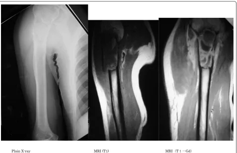
On radiographs obtained at the first examination, a pathological fracture was noted in the proximal humerus, showing hypertranslucency of bone, mainly involving osteolytic changes (Figure 1). On computed tomography (CT), the bone cortex was thinned, and showed a relatively homogenous structure. On magnetic resonance imaging (MRI), a low intensity was observed on T1-weighted imaging, while the fractured region was mainly enhanced on T1 gadolinium contrast imaging, and high intensity was observed on T2-weighted ima-ging (Figure 2). Bone scintigraphy showed signal accu-mulation in the proximal humerus. Based on these findings, pathological fracture associated with a meta-static bone tumor or primary malignant bone tumor in the right proximal humerus was diagnosed, and inci-sional biopsy and open reduction and internal fixation (ORIF) were performed on February 25, 2002. The treat-ment consisted of three Ender’s pins inserted at the pos-terior aspect of the elbow joint. Swelling of the right proximal upper arm became more severe after treat-ment. On pathological examination, tumorous immature bone tissue and osteoid were present; based on these findings, an osteoblastic-type sarcoma was diagnosed. * Correspondence: [email protected]
Department of Orthopedic Surgery, Nihon University School of Medicine, 30-1, Oyaguchikami-cho, Itabashi-ku, Tokyo 173-8610, Japan
Yoshida and TokuhashiWorld Journal of Surgical Oncology2012,10:36
http://www.wjso.com/content/10/1/36 WORLD JOURNAL OF SURGICAL ONCOLOGY
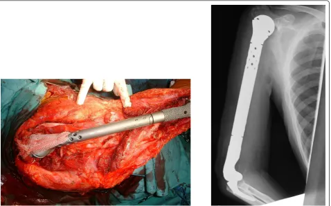
After about 4 months of preoperative chemotherapy for osteosarcoma, sclerotic changes centered within the area of bone hypertranslucency were observed on plain X-ray and CT images. In addition, the accumulation on bone scintigraphy disappeared. After preoperative che-motherapy had been judged to have achieved a complete response (CR), massive extra-capsular resection of the shoulder joint was performed, followed by total humerus placement on July 3, 2002. The humerus was entirely resected including parts of the deltoid and triceps bra-chii muscles and cuff, whereas the long head of the biceps brachii muscle, musculocutaneous nerve, and other neurovascular bundles were conserved. In recon-struction of the cuff, an artificial ligament was attached around an artificial bone head for tumor treatment, and the artificial joint was entirely covered with a latissimus dorsi muscle flap. In the postoperative specimen, the lesion was completely and widely resected. Histopatho-logically, the lesion was mostly necrotized, being close to Grade 3 employing the tissue judgment scale estab-lished by the Musculoskeletal Tumor Committee of the Japanese Orthopaedic Association (Figure 3). Postopera-tive chemotherapy was discontinued because of severe
liver dysfunction. As of 8 years after surgery, the patient’s condition has remained favorable, with no local recurrence or metastasis. Abduction, flexion and exten-sion of the shoulder joint were 10, 20 and 5°, respec-tively, external and internal rotations were -20 and 90°, respectively, and flexion at the elbow joint was 20-120°. The strengths of the biceps and triceps brachii muscles were 4 and 2, respectively. The Enneking functional score was 80%: pain, 5; function, 3; emotional accep-tance, 3; hand positioning, 3; manual dexterity, 5; and lifting ability, 3. On the International Society of Limb Salvage X-ray evaluation, all items of bone remodeling, interface, and anchorage were excellent.
Discussion
Osteosarcoma is a malignant tumor forming tumorous bone and cartilage or osteoid. In the nationwide bone tumor registry established by the Musculoskeletal Tumor Committee of the Japanese Orthopaedic Asso-ciation, 7,649 of a total of 49,768 bone tumors registered from 1972 to 2003 (32 years) were malignant (15%); 3256 of these were osteosarcomas (accounting for 42% of bone malignances). Development at 40 years of age
Plain X-ray MRI (T1) MRI㧔T㧝㧙Gd㧕
or older is reportedly rare, but 433 (13.2%) of the 3,256 osteosarcoma patients were 40 years of age or older. Regarding the developmental site, osteosarcoma most frequently arises in the femur and tibia, followed by the humerus in 262 cases (8%), being ranked the 3rd[1].
It is stated in Dahlin’s Bone Tumors that the inci-dence in those in their 40s is about 5%, and that osteo-sarcoma developed in the humerus in 154 of 1952 cases, accounting for 7.8% of all osteosarcoma cases [2].
For the differential diagnosis from metastatic bone tumor, which was a challenge in our present patient, a comprehensive judgment should be based on past medi-cal history, age, leison, and imaging findings. In patients 60 years of age and older, cancer metastasis is initially suspected. The incidence of metastatic cancer is high in the spine and pelvis, and the periosteal reaction on plain radiographs is poor in a metastatic bone tumor. The pri-mary lesion should also be simultaneously investigated. In our present patient, bone destruction was marked, and the periosteal reaction was poor. Arterial bone cyst, giant cell tumor of bone, conventional osteosarcoma,
and telangiectatic osteosarcoma are included in the dif-ferential diagnosis. Aneurysmal bone cyst was inconsis-tent with age, and the developmental site was not typical for giant cell tumor of bone [3]. Osteosarcoma, particularly, telangiectatic osteosarcoma, was strongly suspected on imaging, but a metastatic bone tumor was also considered based on the patient’s age and the higher prevalence of this type of tumor. Intramedullary pinning was selected because plate fixation was difficult due to the presence of pathological fracture and the location of the lesion in the proximal humerus.
The challenge faced by surgeons after the resection of a malignant bone tumor in the proximal humerus is reconstruction of shoulder joint function. When the cuff is included in massive resection to acquire a safe resec-tion margin, shoulder joint funcresec-tion is mostly lost. For reconstruction of the proximal end of the humerus, sev-eral methods employing an artificial bone head for tumor treatment [4], [5], allogeneic bone grafting [6], [7], processed bone [8], and vascularized bone grafting [9], [10]have been reported, but reconstruction by
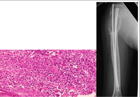
Figure 2Histopathology on biopsy and plain radiographics image at the completion of preoperative chemotherapy. Immature bone tissue and osteoid were not reactive, and were judged to be tumorous. Osteoblastic-type osteosarcoma was ultimately diagnosed.
Chemotherapy was judged to have achieved a complete response (CR).
Yoshida and TokuhashiWorld Journal of Surgical Oncology2012,10:36 http://www.wjso.com/content/10/1/36
hanging these from the acromion using an artificial liga-ment, fascia, or tendon is inevitably necessary. We con-sidered the distal humerus to be contaminated with tumorcomponents via intramedullary pinning, and adopted total humerus replacement to ensure the safety of the resection margin.
For the proximal humerus, hanging from the acro-mion using an artificial ligament was adopted. Our resection method was corresponded closely with that for type V of the Malawer classification of proximal humeral malignant bone tumors, but we opted to per-form massive resection corresponding to extra-capsu-lar resection conserving the acromion and coracoid process because chemotherapy had been sufficiently effective [11]. Very few cases of total humerus replace-ment have been reported. To our knowledge, only Grimer (1996) [12] and Fabroni (1999) [4] have reported such cases and Ayoub (1999) reported described extendable artificial joint use for the humerus [13]. Fabroni et al. reported long-term out-comes in 3 cases, all receiving custom-made artificial joints, and the mean functional score was about 65%. The characteristics of the Howmedica Modular Recon-struction System (HMRS) for the humerus used herein
include the adjustability of the stem length corre-sponding to the resected bone mass and the presence of a hole for attachment with soft tissue. In addition, a porous coating is applied, and the ulnar component of the elbow joint has an anatomical shape. However, the level of osteotomy for optimal setting of the ulnar component of the elbow joint is unclear, and improve-ment may be necessary.
Conclusion
A pathological fracture due to a metastatic bone tumor of the proximal humerus was initially diagnosed. Intra-medullary pinning was performed. However, osteosar-coma was confirmed by postoperative pathological examinations. Therefore, preoperative chemotherapy was conducted, followed by wide resection to acquire a safe margin, and total humerus replacement was then performed. The HMRS was used for reconstruction of the whole humerus. The patient resumed normal activ-ities, although mild contracture of the elbow joint remains 8 years after surgery. In Japan, HMRS is the only artificial joint for tumor treatment that covered National Health Insurance. Reconstruction of the whole humerus using HMRS is rare, but this reconstruction
method is an options, by which a favorable limb func-tion can be achieved.
Consent
Written informed consent was obtained from the patient for publication of this Case report and any accompany-ing images. A copy of the written consent is available for review by the Editor-in-Chief of this journal.
List of abbreviations
CT: Computed tomography; MRI: Magnetic resonance imaging; ORIF: Open reduction and internal fixation; CR: Complete response; HMRS: Howmedica Modular Reconstruction System
Authors’contributions
YY contributed to collection of the clinical data and writing of the manuscript. YT contributed to the writing and editing of the manuscript. Both authors read and approved the final manuscript.
Competing interests
The authors declare that they have no competing interests.
Received: 23 June 2011 Accepted: 14 February 2012 Published: 14 February 2012
References
1. JOA Musculoskeletal tumor committee. Bone Tumor Registry in Japan.
National Cancer Center2008.
2. Unni KK, Inwards CY:Dahlin’s Bone Tumors.Lippincott Wilianms&Wilikins, Sixth 2010.
3. Yarmish G, Klein MJ, Landa J, Lefkowitz RA, Hwang S:Imaging characteristics of primary osteosarcoma: nonconventional subtypes.
Radiographics2010,30(6):1653-672.
4. Fabroni RH, Castago A, Aguilera AL, Steverlynck AM, Zeballous J:Long -term results of limb salvage with the Fabroni custom made endoprosthesis.Clin Orhtop Relat Res1999,35(8):41-52. 5. Ross AC, Sneath RS, Scales JT:Endoprosthetic replacement of the
humerus and elbow joint.J Bone Joint Surg [Br]1987,69-B:652-55. 6. Mankin HJ, Doppelt SH, Sullivan TR, Tomford WW:Osteoarticular and
intercalary allograft transplantation in the management of malignant tumors of bone.Cancer1982,50:613-30.
7. Mankin HJ, Gebhardt MC, Jennings LC, Springfield DS, Tomford WW: Long-term results of allograft replacement in the management of bone tumors.Clin Orthop Relat Res 3241996, 86-97.
8. Muramatsu K, Fukano R, Iwanaga R, Taguchi T:Reconstruction of the proximal humerus by combined use of extracorporeally-irradiated osteochondral graft and free vascularized fibula following resection of Ewing sarcoma.Journal of Plastic, Reconstructive &Aesthetic Surgery2010,
63:2177-180.
9. Wada T, Usui K, Isu K, Yamawakii S, Ishii S:Reconstruction and limb salvage after resection for malignant bone tumour of the proximal humerus. A single procedure using a free vasculaized fibular graft.J Bone Join Surg1999,81B:808-13.
10. Rose PS, Shin AY, Bishop AT, Moran SL, Sim FH:Vascularized free fubula transfer for oncogenic reconstruction of the humerus.Clin Orthop Relat Res2005,438:80-4.
11. Bickels J, Wittig JC, Kollender Y, Kellar-Graney K, Meller I, Malawer MM:
Limb-sparing resections of the shoulder girdle.J Am Coll Surg2002,
194(4):422-35.
12. Grimer JG, Carter SR, sneath SR:Manegement of a huge tumour of the humerus by total replacement of the humerus:an 11-year foolow up.
Arch Orthop Trauma Surg1998,117:298-99.
13. Ayoub KS, Fiorenza F, Grimer RJ, Tilman SR, Carter RS:Extendable endoprostheses of the humerus after resection of bone tumours.J Bone Joint Surg [Br]1999,81-B:495-500.
doi:10.1186/1477-7819-10-36
Cite this article as:Yoshida and Tokuhashi:Total humerus replacement for osteosarcoma with proximal part of humerus: a case report.World Journal of Surgical Oncology201210:36.
Submit your next manuscript to BioMed Central and take full advantage of:
• Convenient online submission
• Thorough peer review
• No space constraints or color figure charges
• Immediate publication on acceptance
• Inclusion in PubMed, CAS, Scopus and Google Scholar
• Research which is freely available for redistribution
Submit your manuscript at www.biomedcentral.com/submit
Yoshida and TokuhashiWorld Journal of Surgical Oncology2012,10:36 http://www.wjso.com/content/10/1/36