1556-6811/10/$12.00 doi:10.1128/CVI.00006-10
Copyright © 2010, American Society for Microbiology. All Rights Reserved.
Highly Persistent and Effective Prime/Boost Regimens against
Tuberculosis That Use a Multivalent Modified Vaccine Virus
Ankara-Based Tuberculosis Vaccine with Interleukin-15 as
a Molecular Adjuvant
䌤
Kristopher Kolibab,
1Amy Yang,
1Steven C. Derrick,
1Thomas A. Waldmann,
2Liyanage P. Perera,
2* and Sheldon L. Morris
1*
Laboratory of Mycobacterial Diseases and Cellular Immunology, Center for Biologics Evaluation and Research,
U.S. Food and Drug Administration, Bethesda, Maryland 20892,1and Metabolism Branch, Center for
Cancer Research, National Cancer Institute, National Institutes of Health, Bethesda, Maryland 209822
Received 8 January 2010/Returned for modification 10 February 2010/Accepted 19 March 2010
Novel immunization strategies are needed to enhance the global control of tuberculosis (TB). In this study, we assessed the immunizing activity of a recombinant modified vaccinia Ankara (MVA) construct
(MVA/IL-15/5Mtb) which overexpresses five Mycobacterium tuberculosis antigens (antigen 85A, antigen
85B, ESAT6, HSP60, and Mtb39), as well as the molecular adjuvant interleukin-15 (IL-15). Homologous prime/boost studies showed that the MVA/IL-15/5Mtb vaccine induced moderate but highly persistent protective immune responses for at least 16 months after the initial vaccination and that the interval between the prime and boost did not significantly alter vaccine-induced antituberculosis protective
immunity. At 16 months, when the Mycobacterium bovis BCG and MVA/IL-15/5Mtb vaccine-induced
protection was essentially equivalent, the protective responses after a tuberculous challenge were
asso-ciated with elevated levels of gamma interferon (IFN-␥), IL-17F, Cxcl9, and Cxcl10. To amplify the
immunizing potential of the MVA/IL-15/5Mtb vaccine, a heterologous prime/boost regimen was tested using an ESAT6-antigen 85B (E6-85) fusion protein formulated in dimethyldiotacylammonium bromide/ monophosphoryl lipid A (DDA/MPL) adjuvant as the priming vaccine and the MVA/IL-15/5Mtb recom-binant virus as the boosting agent. When MVA/IL-15/5Mtb vaccine boosting was done at 2 or 6 months following the final fusion protein injections, the prime/boost regimen evoked protective responses against
an aerogenic M. tuberculosis challenge which was equivalent to that induced by BCG immunization.
Long-term memory after immunization with the E6-85-MVA/IL-15/5Mtb combination regimen was
asso-ciated with the induction of monofunctional CD4 and CD8 IFN-␥-producing T cells and multifunctional
CD4 and CD8 T cells expressing IFN-␥/tumor necrosis factor alpha (TNF-␣), TNF-␣/IL-2, and IFN-␥/
TNF-␣/IL-2. In contrast, BCG-induced protection was characterized by fewer CD4 and CD8
monofunc-tional T cells expressing IFN-␥and only IFN-␥/TNF-␣and IFN-␥/TNF-␣/IL-2 expressing multifunctional
T (MFT) cells. Taken together, these results suggest that a heterologous prime/boost protocol using an MVA-based tuberculosis vaccines to boost after priming with TB protein/adjuvant preparations should be considered when designing long-lived TB immunization strategies.
Despite being an ancient disease, tuberculosis (TB) remains one of the most devastating causes of morbidity and mortality worldwide. Each year about 9 million new cases of TB are detected and 2 million people die from this disease (12, 28). The magnitude of the disease reservoir is immense, with about 2 billion people or one-third of the world’s population thought to be infected withMycobacterium tuberculosis. In recent years, control of TB has been exacerbated by the deadly intersection of the HIV and TB epidemics and the emergence of multiple-drug-resistant tuberculosis (15, 29).
The failure of current TB immunization procedures to ade-quately protect againstM. tuberculosisinfections is largely re-sponsible for the unsuccessful global control of this disease. Although the current TB vaccine,Mycobacterium bovisBCG, has been widely used for at least 50 years, its efficacy has been shown to be highly variable in well-controlled clinical trials (3, 5). While BCG is moderately protective against disseminated TB in children, BCG is ineffective in protecting against the most prevalent form of the disease, adult pulmonary TB. The inadequacy of BCG immunization to control the TB epidemic has stimulated a worldwide effort to develop more-effective TB vaccination strategies. These immunization strategies have in-cluded live attenuated strains, viral vectored vaccine con-structs, and subunit formulations. These new approaches have potential advantages relative to BCG immunization, such as increased safety, improved stability, and decreased interfer-ence by exposure to environmental mycobacteria. However, developing a vaccination strategy based on genetic or subunit vaccines that induces strong and sustainable antituberculosis * Corresponding author. Mailing address for Sheldon L. Morris:
Laboratory of Mycobacterial, Diseases and Cellular Immunology, CBER/FDA, Building 29/Room 502, 29 Lincoln Dr., Bethesda, MD 20892. Phone: (301) 496-5978. Fax: (301) 435-5675. E-mail: sheldon [email protected]. Mailing address for Liyanage P. Perera: Metabolism Branch, Bldg. 10, Rm. 4B40, National Cancer Institute, Bethesda, MD 20892-1374. Phone: (301) 435-7518. Fax: (301) 496-9956. E-mail: [email protected].
䌤Published ahead of print on 31 March 2010.
793
on August 17, 2020 by guest
http://cvi.asm.org/
cellular immunity is a significant scientific challenge. Although immunizations with single antigen preparations have usually been inadequate for controlling intracellular infections, re-cent studies have shown that levels of pathogen-specific cellular immunity can be substantially enhanced by combin-ing different vaccines in a heterologous prime/boost regimen (2, 11, 13, 18, 23). These heterologous prime/boost vaccina-tion strategies often improve the magnitude and quality of T-cell responses and can broaden epitope coverage. Clearly the magnitude, quality, and breadth of vaccine-induced cel-lular immunity are critical for the establishment of effective antituberculosis T-cell memory.
Recombinant modified vaccinia virus Ankara (MVA) con-structs are among the strongest boosting agents used in prime/ boost protocols. MVA vaccines have consistently been shown to boost both primed CD4 and CD8 T-cell responses against intracellular pathogens. In cattle and rhesus macaques, an MVA recombinant virus expressing the M. tuberculosis antigen 85A boosted BCG-induced protection against anM. tuberculosis chal-lenge (26, 27). In humans, immunization with the MVA85A vac-cine induced high levels of antigen-specific gamma interferon (IFN-␥)-secreting T cells in BCG-naive healthy volunteers and boosted preexisting antimycobacterial immune responses elicited by previous BCG vaccination (2, 18). In earlier studies from our laboratory, we showed that a novel recombinant MVA vaccine which coexpressed five TB antigens and interleukin-15 (IL-15) (incorporated as a molecular adjuvant) induced robust antituber-culosis immune responses in mice (19, 21). IL-15 is a promising adjuvant because it plays a role in a wide range of relevant bio-logical activities, including the activation and proliferation of CD8 T cells and NK T cells, the maintenance of CD8 memory cells, and the differentiation and maturation of B cells (24). In this study, we extended our studies of the MVA/IL-15/5Mtb vaccine by showing that immunization with this viral vectored vaccine induces long-lasting antituberculosis protective immunity. Impor-tantly, we demonstrated that priming with a TB fusion protein/ adjuvant formulation followed by heterologous boosting with the MVA/IL-15/5Mtb recombinant virus evoked robust protective responses that were associated with the induction of high frequen-cies of CD4 and CD8 multifunctional T (MFT) cells.
MATERIALS AND METHODS
Animals.C57BL/6 female mice were obtained from Jackson Laboratories (Bar Harbor, ME). All mice used in this study were maintained under sterile condi-tions at the Center for Biologics Evaluation and Research, Bethesda, MD. The mice were vaccinated with selected vaccine preparations at 6 to 8 weeks old.
Vaccine preparations.MVA recombinant virus expressing ESAT6, Ag85A, Ag85B, HSP65, Mtb39A, and IL-15 was constructed as described previously (21). The E6-85 fusion protein is encoded by the ESAT6 gene fused to the region of the Ag85B gene corresponding to the N terminus without the Ag85B signal sequence. These genes were cloned by PCR using the following primers with encoded restriction enzyme sites and two alanines between the ESAT-6 and Ag85B genes: ESAT-6 forward primer, AGCATATGATGACAGAGCAGCAG TGG; ESAT-6 reverse primer, GAGAATTCTGCGAACATCCCAGTGACG TTG; Ag85B forward primer, ACGAATTCGCTGCTTTCTCCCGGCCGGGG CTGCCGGTCGAGT; Ag85B reverse primer, GCGGCCGCGCCGGCGCCT AACGAACTCTGCAG. The PCR products were ligated into the pET-23b plasmid (Novagen). The recombinant plasmid was transformed and expressed in BL21 cells. The fusion protein was purified using Ni⫹2column chromatography.
Immunization schedules.C57BL/6 female mice were vaccinated once subcu-taneously with 106
CFU of BCG Pasteur 6 weeks before challenge. For the wild-type MVA vector (as a control) or the MVA/IL-15/5Mtb vaccine, mice were administered two doses of 5⫻107
PFU subcutaneously at 1 month apart. The
E6-85 protein/adjuvant preparation was prepared by mixing 1.5 mg/ml dimeth-yldiotacylammonium bromide (DDA) (Kodak), 0.25 mg/ml monophosphoryl lipid A (MPL) (Avanti Polar Lipids, Alabaster, AL), and 25g/ml of the E6-85 protein. For this study, 0.2 ml of the E6-85 protein/DDA/MPL adjuvant vaccine and the adjuvant alone were given to mice three times at 2 weeks apart with 5g of protein per vaccination by the subcutaneous route. For the E6-85 protein/ MVA/IL-15/5Mtb heterologous prime/boost immunizations, three doses of the E6-85 protein/adjuvant formulation were given 2 weeks apart, followed at either 2 or 6 months by two doses of the MVA/IL-15/5Mtb vaccine given 1 month apart. For the 15/5Mtb/E6-85 protein prime boost, the 2 doses of MVA/IL-15/5Mtb vaccine were given 2 or 6 months prior to 3 E6-85 protein/adjuvant vaccinations. As a control, 3 doses of the DDA/MPL adjuvant were injected 2 weeks apart, followed 6 months later by 2 doses of the MVA/IL-15/5Mtb vaccine. Figure 1 gives an abbreviated timeline of the studies.
Murine aerogenicM. tuberculosisinfection model.Five mice from each group were aerogenically challenged withM. tuberculosisErdman K1 (Trudeau Myco-bacterial Culture Collection) suspended in phosphate-buffered saline (PBS) at a concentration known to deliver 200 CFU in the lungs over a 30-min exposure in a Middlebrook chamber (Glas Col, Terre Haute, IN) (7). To confirm the infec-tion dose, mice were sacrificed 4 h after exposure and organs were harvested to determine CFU numbers. The harvested lungs and spleens were homogenized separately in PBS-0.04% Tween 80 using a Seward Stomacher 80 blender (Tek-mar, Cincinnati, OH). The homogenates were serially diluted in PBS-0.04% Tween 80 and plated on Middlebrook 7H11 agar (Difco) plates containing 10% OADC enrichment (Becton Dickinson, Sparks, MD) medium, 10g/ml ampi-cillin, 50g/ml cycloheximide, and 2g/ml 2-thiophenecarboxylic acid hydride (TCH) (Sigma). The addition of TCH to the agar plates inhibits BCG growth but has no affect onM. tuberculosisgrowth. Plates were incubated at 37°C for 2 to 3 weeks, and then CFU were counted to determine the organ bacterial burden.
RNA isolation and PCR array analysis of cytokine responses from immune cells.At 2 and 16 months postvaccination, lungs were harvested from 5 mice from each experimental group 10 days after an aerogenic challenge withM. tuberculosis. Sterile PBS was added, and the lungs were sliced and shredded with a scalpel to obtain a suspension. A single-cell suspension of lymphocytes was obtained by passing the shredded lung suspension through a 70-m cell strainer, followed by a 5-min centrifugation at 1,000 rpm. The supernatant was discarded, and the remaining solution was treated with ACK lysing buffer (Lonza, Walk-ersville, MD) for 5 min, followed by 2 washes with PBS, and stored in RNA later (Qiagen, Valencia, CA). The RNeasy minikit (Qiagen) was used to isolate the total RNA from the cell suspension. Equal amounts of RNA from these samples were reverse transcribed to cDNA using a Superscript First-Strand cDNA syn-thesis kit (Invitrogen, San Diego, CA). Glyceraldehyde-3-phosphate dehydroge-nase (GAPDH) primers were used to determine the quality of the cDNA con-version of each sample by PCR. The cytokine/chemokine lung transcriptional responses were determined using the cDNA as the template for RT2
qPCR primer assays (SABiosciences, Frederick, MD) and an ABI Prism 7000 sequence detection system (Applied Biosystems, Foster City, CA). Mouse cytokine/che-mokine primers were obtained from SABiosciences (Frederick, MD). The mRNA expression levels for each cytokine were then normalized according to the manufacturer’s instructions using the formula described in the RT2qPCR
primer assay user manual. The relative cytokine expression values in immune cell cultures were determined by dividing the cytokine expression levels in vaccine candidate samples by the expression values for naive controls (16). This value represents the mean increase or decrease of RNA expression compared to that for naive controls.
Characterization of cytokine production using flow cytometry.Spleen cells from naive and vaccinated mice were isolated by disrupting the spleens in complete Dulbecco’s modified Eagle medium (cDMEM) consisting of 10 mM HEPES, 2.0 mML-glutamine, and 0.1 mM MEM nonessential amino acids with 10% fetal bovine serum (FBS) using a 3-ml syringe barrel. After passage of the spleen homogenate through a cell strainer, the resulting single-cell suspension was washed with cDMEM-FBS and treated for 1 min with 5.0 ml ACK lysis buffer (Lonza). After cells were washed with an equal volume of medium, the cells were resuspended in cDMEM-FBS, counted, and added to wells of a 24-well plate at a density of 2.5⫻106cells per well in 1.0 ml cDMEM-FBS. For
measurement of antigen-specific responses, BCG was added to the wells at a multiplicity of infection (MOI) of 0.5 bacilli per spleen cell. Wells which con-tained only spleen cells served as unstimulated controls. Infections were allowed to proceed overnight, followed by the addition of Golgiplug (BD Biosciences) (1 l per well). After 5 h of incubation, the unbound cells were removed from the wells and transferred to fluorescence-activated cell sorter (FACS) tubes, washed with PBS, and resuspended in⬃50l PBS. Live-Dead stain (Invitrogen) (10l of a 1:100 dilution) was added to each tube, followed by incubation for 30 min at
on August 17, 2020 by guest
http://cvi.asm.org/
room temperature to allow gating on viable cells. The cells were washed with PBS plus 2% FBS and resuspended in⬃50l PBS-FBS, followed by the addition of CD16/CD32 (Fc␥III/II receptor, clone 2.4G2) and an incubation at 4°C for 15 min. The cells were then stained for 30 min at 4°C by adding antibodies against CD4 (rat anti-mouse CD4 Alexa Fluor 700 [AF-700] antibody [Ab], clone RM4-5) and CD8 (rat anti-mouse CD8 peridinin chlorophyll protein complex [PerCP] Ab, clone 53-6.7) at 0.1 and 0.2g per tube, respectively. Following the incubation, the cells were washed twice with PBS and then fixed for 30 min at 4°C with 2% paraformaldehyde in PBS. After fixing, the cells were pelleted, washed twice with PBS-FBS, and stored at 4°C. Fixed cells were then washed twice with perm-wash buffer (1% FBS, 0.01 M HEPES, and 0.11% saponin in PBS) fol-lowed by intracellular staining using the following antibodies at 0.2g per tube: rat anti-mouse IFN-␥allophycocyanin [APC] Ab, clone XMG1.2; rat anti-mouse tumor necrosis factor alpha (TNF-␣) fluorescein isothiocyanate [FITC] Ab, clone MP6-XT22; and rat anti-mouse IL-2 phycoerythrin [PE] Ab, clone JES6-5H4. The cells were incubated at 4°C for 30 min and washed twice with perm-wash buffer, followed by two additional perm-washes with PBS-FBS. All antibodies were obtained from BD Biosciences.
The cells were analyzed using an LSRII flow cytometer (Becton Dickinson) and FlowJo software. For intracellular cytokine analysis, 250,000 events per sample were acquired. Then, using Flow Jo software, gating was done on live, single-cell lymphocytes. To determine the frequency of polyfunctional T cells, the gating was done on CD4⫹or CD8⫹cells stained with antibodies against TNF-␣ and IFN-␥, TNF-␣and Il-2, IFN-␥and Il-2, or all three cytokines.
RESULTS
Long-term protection againstM. tuberculosischallenge after
vaccination with a recombinant MVA virus coexpressing five
M. tuberculosis antigens and IL-15. In an earlier report, we described the creation and characterization of a multivalent MVA vectored vaccine which expresses five M. tuberculosis
antigens as well as the immunostimulatory cytokine IL-15 (21). We demonstrated that this multivalent MVA vaccine induced robust antituberculosis protective responses and conferred protection against an aerogenicM. tuberculosis challenge. In subsequent studies, we focused on evaluating the persistence
of the immune response induced by this MVA/IL-15/5Mtb vaccine candidate. For these experiments, two doses of the MVA/IL-15/5Mtb vaccine were administered because our ini-tial studies with this vaccine and work with related MVA vec-tors have shown that two MVA doses were needed to evoke optimal cellular responses (8, 9). The data summarized in Table 1 compare the protective responses detected at 2 and 12 months after two subcutaneous immunizations (1 month apart) with the MVA/IL-15/5Mtb vaccine candidate. As controls, 2 doses of wild-type MVA virus or one injection of live BCG vaccine were given. At 2 months postvaccination, modest pro-tection against anM.tuberculosisaerogenic challenge was seen in the lungs of mice immunized with the MVA/IL-15/5Mtb construct (⫺0.74 log10 CFU) relative to the stronger BCG-induced protective responses (⫺1.37 log10). In contrast,
pro-tection against dissemination to the spleen was not significantly different for the two vaccine groups at the 2-month time point. At 12 months postvaccination, highly persistent protective re-sponses were seen in the lungs of mice immunized with BCG vaccine or our MVA/IL-15/5Mtb construct. The pulmonary protective responses seen at 1 year after the immunizations were essentially unchanged for both of these vaccine groups. Importantly, the MVA virus used as a control did not induce antituberculosis protective immunity. The organ bacterial bur-dens at 2 and 12 months postvaccination were not significantly different for animals given the wild-type MVA virus relative to those for naive controls.
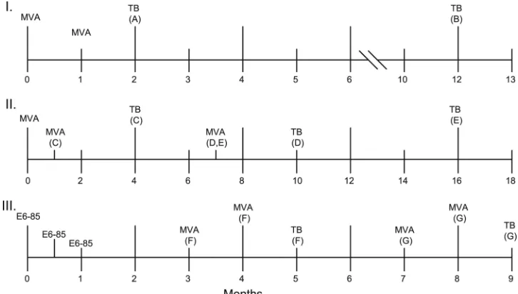
To compare the persistence following a tuberculous chal-lenge of the protective responses induced by immunization with BCG or MVA/IL-15/5Mtb vaccine, mice were vaccinated twice with MVA/IL-15/5Mtb 1 month apart and then chal-lenged by the aerosol route withM. tuberculosis1 month later. FIG. 1. Immunization schedules for three MVA/IL-15/5Mtb vaccine experiments. For study I, mice immunized with the MVA/IL-15/5Mtb construct were challenged with virulentM. tuberculosisat either 2 (A) or 12 (B) months after the priming vaccination (see Table 1). In study II, the impact of different prime/boost intervals was evaluated (Table 2). Mice in group C were boosted 1 month after the priming vaccination and then were challenged at 4 months postprime. For group D, mice were boosted at 7 months and challenged at 10 months after the prime. In group E, mice were boosted at 7 months and challenged at 16 months postprime. In study III, the heterologous boost immunizations were administered at either 3 and 4 months (group F) or 7 and 8 months following the initial E6-85 protein/adjuvant vaccination (Table 4). For study III, mice were challenged 1 month after the final booster immunization. For all of these studies, BCG vaccine was given at day 0.
on August 17, 2020 by guest
http://cvi.asm.org/
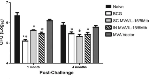
Organ bacterial burdens were evaluated at 1 and 4 months postchallenge (Fig. 2). In this study, mice were immunized with our MVA/IL-15/5Mtb vaccine by either the subcutaneous route or the intranasal route. As a control, mice were injected with BCG subcutaneously 2 months before the challenge. At 1 month postchallenge, significant (compared to that for naive animals; P ⬍ 0.05) pulmonary protection was again seen in animals vaccinated subcutaneously with MVA/IL-15/5Mtb (⫺0.74 log10CFU), while stronger protective responses were
detected in mice immunized with BCG (⫺1.20 log10). The level of protection in the lungs seen after intranasal administration of the MVA/IL-15/5Mtb vaccine (⫺0.91 log10) was also signif-icantly elevated relative to naive levels but was not different from the responses seen after the subcutaneous injections. More importantly, at 4 months postchallenge, the protection induced by our MVA/IL-15/5Mtb vaccine remained un-changed (relative to that at 1 month) while the BCG-induced protective responses decreased. At this time point, no signifi-cant differences in the protective responses between the three immunization groups were detected. Clearly, the postchallenge
protective immune responses elicited by mice vaccinated with the MVA/IL-15/5Mtb vaccine were again extremely persistent. Recent studies have suggested that increasing the time in-tervals between immunizations during a prime/boost protocol may permit increased differentiation of effector memory cells to central memory cells before boosting and thereby enhance the effectiveness of the immunizations (4, 10, 22). To investi-gate the impact of expanding the time intervals between im-munizations, mice were boosted subcutaneously with the MVA/IL-15/5Mtb vaccine at 1 or 7 months after a homologous priming vaccination and then were aerogenically challenged
withM. tuberculosis3 months later. As seen in Table 2, the
longer immunization schedule did not have a significant impact on the pulmonary protective responses generated by the prime/ boost immunization (⫺0.62 log10 CFU for 1 month versus
⫺0.74 log10for 7 months). For both MVA/IL-15/5Mtb vacci-nation regimens, the protective responses in the lung were significantly elevated compared to those for naive mice but were significantly less than the BCG-induced responses at 4 and 10 months after BCG vaccination (P⬍0.05).
In the same study, the persistence of the MVA/IL-15/5Mtb vaccine-induced protective responses was confirmed when the vaccinated mice were challenged 9 months after a 7-month MVA/IL-15/5Mtb booster immunization. Despite the long 16-month interval between the initial vaccination and challenge, a reduction of 0.81 log10CFU in lung bacterial burdens was seen
TABLE 2. Persistence of antituberculosis protective immune responses induced by MVA/IL-15/5Mtb in lungs with
different prime/boost schedules
Vaccine
Bacterial burdena
1-month boost, challenge 4 months after
primary vaccination
7-month boost, challenge 10 months after primary vaccination
7-month boost, challenge 16 months after primary vaccination
MVA/IL-15/5Mtb ⫺0.62⫾0.05* ⫺0.74⫾0.07* ⫺0.81⫾0.06* BCG ⫺1.15⫾0.13*# ⫺1.22⫾0.08*# ⫺0.70⫾0.10*
a
Mice were boosted with the MVA/IL-15/5Mtb construct at 1 or 7 months after the initial immunization. The control mice were given only a single dose of 106
CFU of BCG. At 4, 10, or 16 months after the primary immunization, the vaccinated mice were challenged by the aerosol route withM. tuberculosis Erd-man. The pulmonary bacterial burdens were evaluated at 4 weeks after the aerogenic challenge.ⴱ, significant reduction in CFU compared to results for naive animals (P⬍0.05); #, significant reduction in CFU compared to MVA/ IL-15/5Mtb-vaccinated mice.
TABLE 1. Organ bacterial burdens after an aerogenicM. tuberculosischallenge at 2 and 12 months postvaccination and 1 month postchallenge
Vaccination group
Bacterial burden (log10CFU)
a
2 months postvaccination 12 months postvaccination
Lung Spleen Lung Spleen
Naive 6.37⫾0.13 5.37⫾0.18 6.34⫾0.07 5.55⫾0.06
BCG 5.00⫾0.07* (⫺1.37)# 4.17⫾0.12* (⫺1.20) 5.01⫾0.05* (⫺1.33)# 3.94⫾0.22* (⫺1.61)#
MVA 6.10⫾0.07 5.29⫾0.10 5.96⫾0.17 5.42⫾0.39
MVA/IL-15/5Mtb 5.63⫾0.02* (⫺0.74) 4.47⫾0.06* (⫺0.90) 5.64⫾0.04* (⫺0.70) 5.15⫾0.14* (⫺0.40)
a
Mice were vaccinated twice with the MVA constructs 1 month apart or once with the BCG vaccine and then challenged by the aerosol route withM. tuberculosis
at 2 or 12 months after the primary immunization. Mean difference between naive and experimental CFU are given in parentheses..ⴱ, significant reduction in organ bacterial burdens relative to results for naive animals (P⬍0.05); #, significant reduction in organ bacterial burdens relative to those for vaccination with the MVA/IL-15/5Mtb construct.
FIG. 2. Persistence of the MVA/IL-15/5Mtb vaccine-induced pro-tective immune responses in the lung after an aerosol infection withM. tuberculosis. Mice were vaccinated subcutaneously (SC) with MVA/IL-15/5Mtb recombinant virus or wild-type MVA virus and intranasally (IN) with MVA/IL-15/5Mtb recombinant virus. As controls, other groups of mice remained nonvaccinated (naive) or were vaccinated subcutaneously with BCG. Two months after the initial vaccination, the mice were challenged with a low dose ofM. tuberculosisand then sacrificed at 1 or 4 months postchallenge. Lung homogenates were plated on Middlebrook 7H11 plates, and mycobacterial CFU were counted 2 to 3 weeks later.
on August 17, 2020 by guest
http://cvi.asm.org/
for the MVA/IL-15/5Mtb-immunized mice relative to results for naive controls. Importantly, this response was no different from the protection detected at 5 and 10 months after the initial vaccination. However, while robust protective responses (⫺1.1 log10) were seen at 4 and 10 months following the BCG vaccination, significantly lower protection was detected at 16 months post-immunization with BCG (⫺0.70 log10). As seen previously, the protective immunity induced by BCG in the mouse model clearly wanes after 16 months in the mouse model of pulmonary tuberculosis (7). At the longest time in-terval, the protection seen for both the MVA/IL-15/5Mtb and BCG vaccine groups was essentially equivalent.
To identify immune molecules associated with the MVA/IL-15/5Mtb vaccine-induced antituberculosis protection, the early pulmonary postchallenge cytokine responses induced by prime/boost vaccination with MVA/IL-15/5Mtb were com-pared to postchallenge responses evoked by immunization with BCG or wild-type MVA using real-time PCR. For these stud-ies, mice were aerogenically challenged with a low dose ofM.
tuberculosiseither at 1 month following the second
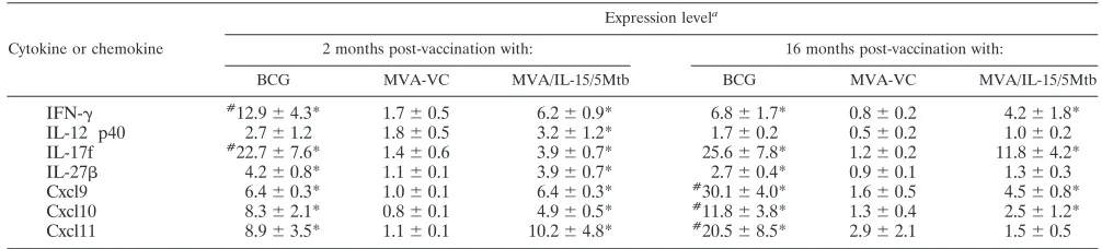
MVA/IL-15/5Mtb vaccination or wild-type MVA control vaccination (2 months after the initial vaccination) or at 9 months after 2 MVA/IL-15/5Mtb immunizations given 7 months apart (16 months postvaccination). The single BCG vaccination was ad-ministered at the time of the initial MVA injection. At 10 days postchallenge, the mice were sacrificed, the lungs were re-moved, and RNA was purified from lung cells. Subsequently, the levels of expression of 13 cytokine or chemokine genes previously shown to be up- or downregulated in vaccinated animals were assessed (16). As seen in Table 3, at 2 months postimmunization and at 10 days postchallenge, the expression of 7 of these immune mediators was increased in the lungs of mice vaccinated with either BCG or MVA/IL-15/5Mtb relative to that for naive controls (IFN-␥, IL-12p40 subunit, IL-17F, IL-27, Cxcl9, Cxcl10, and Cxcl11). In contrast, the expression of IL-1, IL-2, IL-4, Il-21, transforming growth factor (TGF-), and TNF-␣ was not elevated relative to that for naive controls. For the upregulated genes, only the pulmonary ex-pression of IFN-␥and IL-17F was significantly increased (P⬍
0.05) for BCG-vaccinated animals relative to that for those immunized with MVA/IL-15/5Mtb vaccine. Interestingly, none of the cytokine genes that were evaluated were differentially regulated, compared to results for naive mice, in mice vacci-nated with wild-type MVA virus.
At 16 months postvaccination, similar pulmonary cytokine and chemokine responses were detected in BCG-vaccinated mice at 10 days postchallenge. Again, elevated expression of IFN-␥, IL-17F, IL-27, Cxcl9, Cxcl10, and Cxcl11 transcripts were detected. Although postchallenge IL-12p40, IL-27, and Cxcl11 expression decreased in MVA/IL-15/5Mtb-immunized mice at 16 months postvaccination, the expression of IFN-␥, IL-17F, Cxcl9, and Cxcl11 were again significantly elevated compared to that for naive controls at this extended time point. Similar to the 2-month time point, the relative expression of the test genes at this later time was not significantly enhanced in mice vaccinated with wild-type MVA virus.
A heterologous prime/boost regimen confers protection
against an aerosol M. tuberculosis infection. Heterologous
prime/boost immunization strategies using poxvirus vaccines as boosting agents have been shown to induce high levels of cellular immunity (2, 11, 13, 18). To investigate whether our MVA/IL-15/5Mtb vaccine would induce substantial antituber-culosis protective immunity, mice were primed by immuniza-tion with an ESAT6/antigen 85B fusion protein (E6-85) for-mulated in DDA/MPL adjuvant and then boosted with MVA/ IL-15/5Mtb (which overexpresses ESAT6, antigen 85B, and antigen 85A along with HSP65 and Mtb39A). Since previous reports have suggested that the interval between the priming and boosting injections may impact the magnitude of the mune responses generated, the MVA/IL-15/5Mtb booster im-munizations were given at either 2 or 6 months following the final priming dose (3 or 7 months after the initial vaccination) (4, 10, 22). One month after the last booster vaccination, mice were challenged by the aerosol route with a low dose of M.
tuberculosisand then were sacrificed at 4 weeks postchallenge.
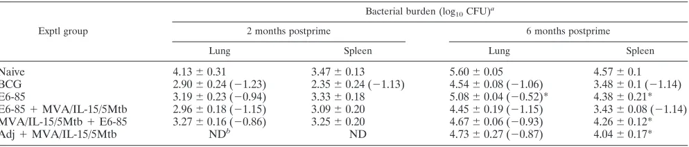
For the 2-month prime/boost interval, the animals that were vaccinated with BCG (⫺1.23 log10CFU) or the E6-85/MVA/ IL-15/5Mtb regimen (⫺1.15 log10) were highly protected in the
lung (Table 4). Less pulmonary protection was seen in groups vaccinated with only the E6-85 formulation (⫺0.84 log10) or
with the MVA/IL-15/5Mtb prime/E6-85 boost schedule (⫺0.86 log10). Interestingly, similar results were seen when the
heter-ologous vaccination was completed using a 6-month interval between the prime and the boost. Therefore, for this combi-nation, the time interval between the prime and boost did not seem to influence the magnitude of the protective responses that were evoked. Again, greater than a 90% reduction in lung CFU relative to levels for the naive controls was detected in TABLE 3. Cytokine and chemokine responses at 2 and 16 months after initial vaccination
Cytokine or chemokine
Expression levela
2 months post-vaccination with: 16 months post-vaccination with:
BCG MVA-VC MVA/IL-15/5Mtb BCG MVA-VC MVA/IL-15/5Mtb
IFN-␥ #12.9⫾4.3* 1.7⫾0.5 6.2⫾0.9* 6.8⫾1.7* 0.8⫾0.2 4.2⫾1.8*
IL-12 p40 2.7⫾1.2 1.8⫾0.5 3.2⫾1.2* 1.7⫾0.2 0.5⫾0.2 1.0⫾0.2
IL-17f #22.7⫾7.6* 1.4⫾0.6 3.9⫾0.7* 25.6⫾7.8* 1.2⫾0.2 11.8⫾4.2*
IL-27 4.2⫾0.8* 1.1⫾0.1 3.9⫾0.7* 2.7⫾0.4* 0.9⫾0.1 1.3⫾0.3
Cxcl9 6.4⫾0.3* 1.0⫾0.1 6.4⫾0.3* #30.1⫾4.0* 1.6⫾0.5 4.5⫾0.8*
Cxcl10 8.3⫾2.1* 0.8⫾0.1 4.9⫾0.5* #11.8⫾3.8* 1.3⫾0.4 2.5⫾1.2*
Cxcl11 8.9⫾3.5* 1.1⫾0.1 10.2⫾4.8* #20.5⫾8.5* 2.9⫾2.1 1.5⫾0.5
aⴱ
, significant increase in expression levels compared to results for naive animals (P⬍0.05); #, significant increase in expression levels compared to results with MVA/IL-15/5Mtb (P⬍0.05).
on August 17, 2020 by guest
http://cvi.asm.org/
the E6-85/MVA/IL-15/5Mtb (⫺1.15 log10) and BCG (⫺1.06 log10) vaccine groups. Significantly less protection in the lung
was seen for mice vaccinated with the E6-85 protein prepara-tion (⫺0.52 log10; P⬍ 0.05) than for the
MVA/IL-15/5Mtb-boosted animals, while the differences for the MVA/IL-15/5Mtb/ E6-85 (⫺0.93 log10) and adjuvant/MVA/IL-15/5Mtb (⫺0.87
log10) experimental groups approached statistical significance (P⬍0.10). Importantly, at both prime/boost intervals, the levels of antituberculosis protective immunity generated by immuniza-tion with the E6-85/MVA/IL-15/5Mtb combinaimmuniza-tion or BCG vac-cine were essentially equivalent. Also, at each time interval, the order of the prime/boost immunizations seemed to have an im-pact on the outcome. Our results suggested that the MVA/IL-15/ 5Mtb vaccine was more effective as a boosting agent because the E6-85 prime/MVA/IL-15/5Mtb boost regimen elicited higher lev-els of protective immunity in the lung than the MVA/IL-15/5Mtb prime/E6-85 boost schedule at both the 2- and 6-month prime/ boost vaccination intervals.
Recent reports have suggested that the induction of multi-functional T (MFT) cells is associated with enhanced immunity against intracellular pathogens (1, 6, 17). To determine whether immunization with the various vaccine regimens in-duced elevated MFT responses, spleen cells were removed from vaccinated mice, stimulated with BCG overnight, and then analyzed by multiparameter flow cytometry. The frequen-cies of CD4 and CD8 cells expressing IFN-␥, TNF-␣, and IL-2 were evaluated by intracellular cytokine staining (Fig. 3). Us-ing these procedures, CD4 T cells expressUs-ing only IFN-␥were the most frequent; IFN-␥-expressing CD4⫹splenocytes from mice vaccinated with the E6-85/MVA/IL-15/5Mtb combination (6.75%), BCG vaccine (3.49%), or the adjuvant/MVA/IL-15/ 5Mtb regimen (2.5%) were significantly elevated relative to naives. Among the splenocytes expressing multiple cytokines, substantially elevated levels of CD4 T cells expressing IFN-␥ and TNF-␣ were detected in all vaccine groups relative to levels for naive controls. In particular, 27- and 14-fold-in-creased frequencies (compared to those for naive controls) of CD4 T cells expressing IFN-␥ and TNF-␣ were seen in the E6-85/MVA/IL-15/5Mtb prime/boost and BCG experimental groups, respectively. Additionally, the relative frequencies of splenic CD4 T cells expressing IFN-␥, TNF-␣, and IL-2 were significantly elevated in all vaccination groups. While only 0.01% of naive CD4 T cells in the spleen expressed all three cytokines, 7- to 35-fold-increased frequencies of triple-positive
CD4 T cells were detected for the E6-85/MVA/IL-15/5Mtb (0.35%), BCG (0.16%), and E6-85/adjuvant (0.07%) groups. It is of interest that significant relative increases in splenic CD4 T cells expressing TNF-␣and IL-2 were seen only for the E6-85/ MVA/IL-15/5Mtb (0.24%, compared to 0.03% for naive con-trols) and adjuvant/MVA/IL-15/5Mtb (0.13%) immunization groups (Fig. 3A). These TNF-␣/IL-2-positive CD4 cells have been classified as possible central memory cells, and a role in the long-term memory response has been suggested for these double-positive cells (17).
Although the results were generally more variable, similar MFT results were seen with splenic CD8 T cells (Fig. 3B). Monofunctional IFN-␥-expressing cells were the most frequent CD8 T cells detected. Significantly higher frequencies of CD8 T cells expressing only IFN-␥were seen in the E6-85/MVA/ IL-15/5Mtb (9.20%), adjuvant/MVA/IL-15/5Mtb (6.80%), and BCG (5.68%) vaccination groups than for naives (1.16%). Sev-enfold-increased levels of IFN-␥/TNF-␣(⫹) CD8 T cells com-pared to results for naives were detected in the highly protec-tive E6-85/MVA/IL-15/5Mtb and BCG vaccine groups, and elevated relative concentrations of TNF-␣/IL-2 CD8 T cells were seen after the E6-85/MVA/IL-15/5Mtb heterologous prime/boost. Furthermore, the frequencies of triple-positive splenic CD8 T cells were significantly elevated in the E6-85/ MVA/IL-15/5Mtb (0.23%) and BCG (0.21%) vaccine groups relative to results for naive controls (0.04%).
In previous studies, MFT cells have been shown to make severalfold more IFN-␥than monofunctional T cells (24, 25). To evaluate the levels of IFN-␥produced by monofunctional and MFT CD4 cells, the median fluorescence intensity (MFI) was determined for IFN-␥-secreting cells within experimental groups by reviewing the flow cytometric plots. The median fluorescence for the splenocytes expressing only IFN-␥ recov-ered from animals vaccinated with the E6-85/MVA/IL-15/ 5Mtb combination or BCG was about 6,000 units. Interest-ingly, the median fluorescence intensities for the IFN-␥/ TNF-␣⫹ CD4 cells from the BCG-vaccinated (27,000) and E6-85/MVA/IL-15/5Mtb-vaccinated (24,000) mice were 4- to 5-fold higher than those for the monofunctional cells. Addi-tionally, for the triple-positive splenic CD4 T cells from BCG-and E6-85/MVA/IL-15/5Mtb-vaccinated mice, the median flu-orescence intensities were increased about 3-fold (18,000) and 4-fold (23,000), respectively, compared to results for the mono-functional IFN-␥⫹ splenic CD4 T cells induced by the same TABLE 4. Impact of time interval between priming and boosting on the bacterial burdens assessed 1 month after an
aerosolM. tuberculosischallenge
Exptl group
Bacterial burden (log10CFU)
a
2 months postprime 6 months postprime
Lung Spleen Lung Spleen
Naive 4.13⫾0.31 3.47⫾0.13 5.60⫾0.05 4.57⫾0.1
BCG 2.90⫾0.24 (⫺1.23) 2.35⫾0.24 (⫺1.13) 4.54⫾0.08 (⫺1.06) 3.48⫾0.1 (⫺1.14)
E6-85 3.19⫾0.23 (⫺0.94) 3.33⫾0.18 5.08⫾0.04 (⫺0.52)* 4.38⫾0.21*
E6-85⫹MVA/IL-15/5Mtb 2.96⫾0.18 (⫺1.15) 3.09⫾0.20 4.45⫾0.19 (⫺1.15) 3.43⫾0.08 (⫺1.14) MVA/IL-15/5Mtb⫹E6-85 3.27⫾0.16 (⫺0.86) 3.25⫾0.20 4.67⫾0.06 (⫺0.93) 4.26⫾0.12*
Adj⫹MVA/IL-15/5Mtb NDb ND 4.73⫾0.27 (⫺0.87) 4.04⫾0.17*
a
Parenthetical values are given whenP⬍0.05 compared with results for naive control.ⴱ, vaccine groups with organ bacterial burdens significantly increased (P⬍
0.05) over those for the heterologous E6-85⫹MVA/IL-15/5Mtb vaccine group.
b
ND, not done.
on August 17, 2020 by guest
http://cvi.asm.org/
vaccines. Taken together, these data strongly suggest that the MFT cells identified in this study make at least 3- to 5-fold more IFN-␥than monofunctional IFN-␥-producing cells.
DISCUSSION
To date, classical vaccination approaches have not been suf-ficient to develop highly effective vaccines against tuberculosis. Consequently, many recent experimental immunization strat-egies against this disease have focused on combining partially effective vaccines to achieve superior levels of efficacy relative to individual vaccination regimens. In particular, heterologous prime/boost regimens have been shown to induce strong and broad cellular immunity and an enhanced quality of T-cell responses (2, 11, 13, 18, 23, 26, 27). In this study, we showed that a heterologous prime/boost regimen using a TB fusion protein (E6-85) formulated in DDA/MPL adjuvant as a prime and an MVA recombinant virus expressing ESAT6, antigen 85B, antigen 85A, Hsp60, and Mtb39 with IL-15 as an adjuvant (MVA/IL-15/5Mtb) as the boosting agent elicited antitubercu-losis immunity in the mouse model which was equivalent to the
protective responses induced by BCG vaccine. Moreover, using multiparameter flow cytometry, we showed that the E6-85/ MVA/IL-15/5Mtb prime/boost regimen evoked diverse CD4 and CD8 T-cell responses. The most protective vaccines in this study, the E6-85/MVA/IL-15/5Mtb combination and live BCG vaccine, induced significantly elevated levels of multifunctional CD4 and CD8 T cells expressing IFN-␥and TNF-␣or IFN-␥, TNF-␣, and IL-2. These potent vaccines also induced a high frequency of monofunctional cells expressing only IFN-␥. In contrast, the less-protective E6-85/adjuvant formulation gen-erally elicited a more modest MFT-cell and monofunctional response than the more protective vaccination regimens. In-terestingly, the median IFN-␥response for the MFT cells de-tected in these experiments was at least 3-fold higher than the median levels of IFN-␥ in cells making only this cytokine. Earlier reports also indicated that IFN-␥concentrations were severalfold higher in MFT cells than in monofunctional T cells (6, 14, 17). The induction of MFT cells by the E6-85/MVA/IL-15/5Mtb heterologous prime/boost schedule is of interest be-cause recent studies have suggested that T cells which exert multiple effector functions, including concurrent expression of FIG. 3. Vaccine-induced multifunctional T cells. After recovery of splenocytes from mice that had been vaccinated with BCG, the E6-85 adjuvant preparation, the E6-85/MVA/IL-15/5Mtb prime-boost regimen, or the adjuvant/MVA/IL-15/5Mtb (controls), the spleen cells were incubated overnight with BCG, stained the next day for CD4 and CD8 T-cell markers and cytokine expression, and then analyzed by multipa-rameter flow cytometry. Panels A (CD4) and B (CD8) show the frequency of T cells expressing either single or multiple cytokines. These data represent the mean responses⫹SEM for 5 individual mice.
on August 17, 2020 by guest
http://cvi.asm.org/
IFN-␥, TNF-␣, and IL-2, are functionally superior to mono-functional cells (14). In mice, the induction of MFT cells by immunization correlated with the extent of protection against
Leishmania major(6). In humans, the presence of MFT cells
has been associated with the control of viremia in nonprogres-sive HIV infections (1). Several immune mechanisms have been suggested to explain the presumed superiority of MFT cells (25). First, MFT cells seem to secrete more IFN-␥than monofunctional cells. Since IFN-␥is a critical cytokine in the defense against mycobacterial infections, elevated IFN-␥ con-centrations should enhance infection control. Second, the se-cretion of IFN-␥and TNF-␣, another important antimycobac-terial immune modulator, from the same cell may increase local intracellular killing. Finally, IL-2 secretion from MFT cells should promote T-cell expansion and enhance long-term survival of memory CD4 cellsin vivo.
A clear difference in the T-cell response profile evoked by prime/boost immunization with the E6-85/MVA/IL-15/5Mtb vaccine regimen compared to BCG vaccination was the signif-icantly elevated percentages of splenic CD4 T cells making both TNF-␣and IL-2 (Fig. 3A). Lindenstrøm et al. have also reported that a TB subunit vaccine preparation induces long-lived TNF-␣/IL-2 double-positive cells, while fewer cells with the same phenotype were identified in the spleens of BCG-vaccinated animals (17). These data strongly suggest that the type of vaccine administered can impact the quality and mag-nitude of the vaccine-induced T-cell memory response. The induction of TNF-␣/IL-2-positive MFT cells and cells that make only IL-2 in response to MVA/IL-15/5Mtb vaccinations may explain the persistence of the MVA response seen in the 16-month study with the MVA/IL-15/5Mtb vaccine. The failure of BCG to induce this double–positive phenotype may contrib-ute to the waning BCG protective response detected at 16 months postvaccination in the same study. This intriguing ob-servation suggests that further studies should explore the role of the vaccine-induced TNF-␣/IL-2-positive T-cell subset in the persistence of antituberculosis protective immunity.
Recent vaccination studies have indicated that increasing the intervals between immunizations may permit enhanced maturation of effector memory cells to more-potent central memory cells prior to boosting, which ultimately should lead to improved vaccine efficacy (4, 10, 22). In our studies using the MVA/IL-15/5Mtb vaccine candidate as a boosting agent, no impact on the antituberculosis protective responses was ob-served when the time interval between priming and boosting was increased. With both homologous (1-month versus 7-month boosts) and heterologous (2-month versus 6-month boosts) regimens, the MVA/IL-15/5Mtb vaccine-induced anti-tuberculosis protection was not dependent on the immuniza-tion schedule. Even extending the postboosting vaccinaimmuniza-tion- vaccination-challenge interval to 11 months (Table 1) did not reduce the protective effectiveness of the MVA/IL-15/5Mtb vaccine. In-terestingly, in humans, the interval between an initial BCG immunization and an MVA85 booster vaccination did not in-fluence the magnitude of the antituberculosis immune re-sponse (20). Since MVA does not replicate in mammalian cells, viral persistence cannot explain the longevity of MVA/ IL-15/5Mtb-induced immunity. It is more likely that the capac-ity of the MVA/IL-15/5Mtb vaccine to induce strong and broad cellular immunity (including IFN-␥, IL-17, and IL-27
expres-sion) (Table 3) and to effectively boost memory cell popula-tions diminished any impact increasing (or decreasing) immu-nization time intervals may have on the induction of protective immunity.
In sum, we have shown that vaccination with our MVA/IL-15/5Mtb vaccine induces highly persistent antituberculosis pro-tective responses. Furthermore, immunization with a heterol-ogous prime/boost regimen using a TB fusion protein formulated with DDA/MPL adjuvant followed by boosting with MVA/IL-15/5Mtb induces substantial protective immu-nity in a mouse model of pulmonary tuberculosis. Most clinical efforts related to TB vaccines are currently focusing on the development of vaccines to boost BCG responses. While this approach sidesteps concerns about discontinuing BCG use, it also does not resolve safety issues related to BCG immuniza-tion in areas to which HIV is endemic and the continued uncertainty about the overall effectiveness of BCG-induced immunity. This heterologous E6-85 protein/MVA/IL-15/5Mtb prime/boost regimen provides an alternative strategy for com-bating the devastating TB epidemic.
REFERENCES
1.Betts, M. R., M. C. Nason, S. M. West, S. C. De Rosa, S. A. Migueles, J. Abraham, M. M. Lederman, J. M. Benito, P. A. Goepfert, M. Connors, M. Roederer, and R. A. Koup.2006. HIV nonprogressors preferentially main-tain highly functional HIV-specific CD8⫹T cells. Blood107:4781–4789. 2.Beveridge, N. E., D. A. Price, J. P. Casazza, A. A. Pathan, C. R. Sander, T. E.
Asher, D. R. Ambrozak, M. L. Precopio, P. Scheinberg, N. C. Alder, M. Roederer, R. A. Koup, D. C. Douek, A. V. Hill, and H. McShane.2007. Immunisation with BCG and recombinant MVA85A induces long-lasting, polyfunctional Mycobacterium tuberculosis-specific CD4⫹memory T lym-phocyte populations. Eur. J. Immunol.37:3089–3100.
3.Brewer, T. F.2000. Preventing tuberculosis with bacillus Calmette-Gue´rin vaccine: a meta-analysis of the literature. Clin. Infect. Dis.31:S64–S67. 4.Brice, G. T., C. Doban˜o, M. Sedegah, M. Stefaniak, N. L. Graber, J. J.
Campo, D. J. Carucci, and D. L. Doolan.2007. Extended immunization intervals enhance the immunogenicity and protective efficacy of plasmid DNA vaccines. Microbes Infect.9:1439–1446.
5.Colditz, G. A., T. F. Brewer, C. S. Berkey, M. E. Wilson, E. Burdick, H. V. Fineberg, and F. Mosteller.1994. Efficacy of BCG vaccine in the prevention of tuberculosis. Meta-analysis of the published literature. JAMA271:698– 702.
6.Darrah, P. A., D. T. Patel, P. M. De Luca, R. W. Lindsay, D. F. Davey, B. J. Flynn, S. T. Hoff, P. Andersen, S. G. Reed, S. L. Morris, M. Roederer, and R. A. Seder.2007. Multifunctional TH1 cells define a correlate of vaccine-mediated protection against Leishmania major. Nat. Med.13:843–850. 7.Derrick, S. C., A. Yang, and S. L. Morris.2004. A polyvalent DNA vaccine
expressing an ESAT6-Ag85B fusion protein protects mice against a primary infection with Mycobacterium tuberculosis and boosts BCG-induced protec-tive immunity. Vaccine23:780–788.
8.Earl, P. L., J. L. Americo, L. S. Wyatt, L. A. Eller, D. C. Montefiori, R. Byrum, M. Piatak, J. D. Lifson, R. R. Amara, H. L. Robinson, J. W. Huggins, and B. Moss.2007. Recombinant modified vaccinia virus Ankara provides durable protection against disease caused by an immunodeficiency virus as well as long-term immunity to an orthopoxvirus in a non-human primate. Virology366:84–97.
9.Earl, P. L., J. L. Americo, L. S. Wyatt, L. A. Eller, J. C. Whitbeck, G. H. Cohen, R. J. Eisenberg, C. J. Hartmann, D. L. Jackson, D. A. Kulesh, M. J. Martinez, D. M. Miller, E. M. Mucker, J. D. Shamblin, S. H. Zwiers, J. W. Huggins, P. B. Jahrling, and B. Moss.2004. Immunogenicity of a highly attenuated MVA smallpox vaccine and protection against monkeypox. Na-ture428:182–185.
10.Fuller, D. H., M. M. Corb, S. Barnett, K. Steimer, and J. R. Haynes.1997. Enhancement of immunodeficiency virus-specific immune responses in DNA-immunized rhesus macaques. Vaccine15:924–926.
11.Gilbert, S. C., V. S. Moorthy, L. Andrews, A. A. Pathan, S. J. McConkey, J. M. Vuola, S. M. Keating, T. Berthoud, D. Webster, H. McShane, and A. V. Hill.2006. Synergistic DNA-MVA prime-boost vaccination regimes for ma-laria and tuberculosis. Vaccine24:4554–4561.
12.Glaziou, P., K. Floyd, and M. Raviglione.2009. Global burden and epide-miology of tuberculosis. Clin. Chest Med.30:621–636.
13.Harari, A., P. A. Bart, W. Sto¨hr, G. Tapia, M. Garcia, E. Medjitna-Rais, S. Burnet, C. Cellerai, O. Erlwein, T. Barber, C. Moog, P. Liljestrom, R. Wagner, H. Wolf, J. P. Kraehenbuhl, M. Esteban, J. Heeney, M. J. Frachette,
on August 17, 2020 by guest
http://cvi.asm.org/
J. Tartaglia, S. McCormack, A. Babiker, J. Weber, and G. Pantaleo.2008. An HIV-1 clade C DNA prime, NYVAC boost vaccine regimen induces reliable, polyfunctional, and long-lasting T cell responses. J. Exp. Med.
205:63–77.
14.Kannanganat, S., C. Ibegbu, L. Chennareddi, H. L. Robinson, and R. R. Amara.2007. Multiple-cytokine-producing antiviral CD4 T cells are func-tionally superior to single-cytokine-producing cells. J. Virol.81:8468–8476. 15.Lawn, S. D., and G. Churchyard.2009. Epidemiology of HIV-associated
tuberculosis. Curr. Opin. HIV AIDS4:325–333.
16.Lim, J., S. C. Derrick, K. Kolibab, A. L. Yang, S. Porcelli, W. R. Jacobs, and S. L. Morris.2009. Early pulmonary cytokine and chemokine responses in mice immunized with three different vaccines against Mycobacterium tuber-culosis determined by PCR array. Clin. Vaccine Immunol.16:122–126. 17.Lindenstrøm, T., E. M. Agger, K. S. Korsholm, P. A. Darrah, C. Aagaard,
R. A. Seder, I. Rosenkrands, and P. Andersen.2009. Tuberculosis subunit vaccination provides long-term protective immunity characterized by multi-functional CD4 memory T cells. J. Immunol.182:8047–8055.
18.McShane, H., A. A. Pathan, C. R. Sander, S. M. Keating, S. C. Gilbert, K. Huygen, H. A. Fletcher, and A. V. Hill.2004. Recombinant modified vaccinia virus Ankara expressing antigen 85A boosts BCG-primed and naturally acquired antimycobacterial immunity in humans. Nat. Med.10:1240–1244. 19.Parra, M., A. L. Yang, J. Lim, K. Kolibab, S. Derrick, N. Cadieux, L. P.
Perera, W. R. Jacobs, M. Brennan, and S. L. Morris.2009. Development of a murine mycobacterial growth inhibition assay for evaluating vaccines against Mycobacterium tuberculosis. Clin. Vaccine Immunol.16:1025–1032. 20.Pathan, A. A., C. R. Sander, H. A. Fletcher, I. Poulton, N. C. Alder, N. E. R. Beveridge, K. T. Whelan, A. V. S. Hill, and H. McShane.2007. Boosting BCG with recombinant modified vaccinia Ankara expressing antigen 85A: differ-ent boosting intervals and implications for efficacy trials. PLoS One2:e1052. 21.Perera, P. Y., S. C. Derrick, K. Kolibab, F. Momoi, M. Yamamoto, S. L. Morris, T. A. Waldmann, and L. P. Perera.2009. A multi-valent vaccinia virus-based tuberculosis vaccine molecularly adjuvanted with interleukin-15 induces robust immune responses in mice. Vaccine27:2121–2127.
22.Pittman, P. R., J. A. Mangiafico, C. A. Rossi, T. L. Cannon, P. H. Gibbs, G. W. Parker, and A. M. Friedlander.2000. Anthrax vaccine: increasing intervals between the first two doses enhances antibody response in humans. Vaccine19:213–216.
23.Radosevic, K., A. Rodriguez, A. Lemckert, and J. Goudsmit.2009. Heterol-ogous prime-boost vaccinations for poverty-related diseases: advantages and future prospects. Expert Rev. Vaccines8:577–592.
24.Rodrigues, L., and C. Bonorino.2009. Role of IL-15 and IL-21 in viral immunity: applications for vaccines and therapies. Expert Rev. Vaccines
8:167–177.
25.Seder, R. A., P. A. Darrah, and M. Roederer.2008. T-cell quality in memory and protection: implications for vaccine design. Nat. Rev. Immunol.8:247– 258.
26.Verreck, F. A., R. A. Vervenne, I. Kondova, van K. W. Kralingen, E. J. Remarque, G. Braskamp, van der N. M. Werff, A. Kersbergen, T. H. Otten-hoff, P. J. Heidt, S. C. Gilbert, B. Gicquel, A. V. Hill, C. Martin, H. McShane, and A. W. Thomas.2009. MVA. 85A boosting of BCG and an attenuated, phoP deficient M. tuberculosis vaccine both show protective efficacy against tuberculosis in rhesus macaques. PLoS One4:e5264.
27.Vordermeier, H. M., S. G. Rhodes, G. Dean, N. Goonetilleke, K. Huygen, A. V. Hill, R. G. Hewinson, and S. C. Gilbert.2004. Cellular immune re-sponses induced in cattle by heterologous prime-boost vaccination using recombinant viruses and bacille Calmette-Gue´rin. Immunology112:461–470. 28.WHO.2009. Global tuberculosis control—epidemiology, strategy, financing.
WHO/HTM/TB/2009.411. WHO, Geneva, Switzerland.
29.Wright, A., M. Zignol, A. Van Deun, D. Falzon, S. R. Gerdes, K. Feldman, S. Hoffner, F. Drobniewski, L. Barrera, van D. Soolingen, F. Boulabhal, C. N. Paramasivan, K. M. Kam, S. Mitarai, P. Nunn, M. Raviglione, and the Global Project on Anti-Tuberculosis Drug Resistance Surveillance.2009. Epidemiology of antituberculosis drug resistance 2002–07: an updated anal-ysis of the Global Project on Anti-Tuberculosis Drug Resistance Surveil-lance. Lancet373:1861–1873.