Degenerative Neurological and Neuromuscular Disease
Amyotrophic lateral sclerosis: update
and new developments
Ashley J Pratt1 Elizabeth D Getzoff1 J Jefferson P Perry1,2 1Department of Molecular Biology
and The Skaggs Institute for Chemical Biology, The Scripps Research Institute, La Jolla, CA 92037, USA;
2The School of Biotechnology, Amrita
University, Kollam, Kerala 690525, India
Correspondence: J Perry The Scripps Research Institute, 10550 N. Torrey Pines Road, MB-4, La Jolla, CA 92037, USA
Tel +1 858 784 2284 Fax +1 858 784 2289 Email [email protected]
Abstract: Amyotrophic lateral sclerosis (ALS) is the most common form of motor neuron disease. It is typically characterized by adult-onset degeneration of the upper and lower motor neurons, and is usually fatal within a few years of onset. A subset of ALS patients has an inherited form of the disease, and a few of the known mutant genes identified in familial cases have also been found in sporadic forms of ALS. Precisely how the diverse ALS-linked gene products dictate the course of the disease, resulting in compromised voluntary muscular abil-ity, is not entirely known. This review addresses the major advances that are being made in our understanding of the molecular mechanisms giving rise to the disease, which may eventually translate into new treatment options.
Keywords: amyotrophic lateral sclerosis, neurodegeneration, motor neuron disease, genetics, aging
Introduction
Amyotrophic lateral sclerosis (ALS), also known as Charcot’s disease or Lou Gehrig’s disease is the most widespread type of motor neuron disease. Striking later in life, the disease causes degeneration of motor neurons and consequently progressive atrophy of associated muscle tissues and supporting cells. Unlike similar motor neuron diseases that primarily affect only a single subgroup of neurons (eg, Primary Muscular Atrophy or Primary Lateral Sclerosis), ALS patients typically have both lower motor neuron (LMN) and upper motor neuron (UMN) involvement. The symptoms of ALS com-monly are muscle weakness and wasting, especially in the limbs, cramps, twitching, and difficulties in speaking. The lifetime risk of acquiring ALS by age 70 is between 1 in 400 and 1 in 1000,1 and in general, ALS individuals succumb to the disease within
2–3 years due to respiratory failure.
A growing number of ALS-causing genes have been identified recently and are now under investigation, providing promise for increased understanding of the etiology of the disease. SOD1, encoding the highly conserved, cytosolic antioxidant enzyme Cu,Zn-superoxide dismutase (Cu,ZnSOD), was the first such gene to be identified with ALS.2,3SOD1 mutations are common in both familial ALS (FALS) and sporadic
ALS (SALS), and have been studied in the most depth. Other genes such as OPTN 4 or
TARDBP, FUS, and ANG (involved in RNA metabolism)5 were later identified as
caus-ative factors in both FALS and SALS. Suggestive of proteolytic disfunction, UBQLN2
was recently implicated in ALS,6,7 and very recently, nucleotide repeat expansions in
C9ORF72,8–10 were found to comprise the largest fraction of ALS-causing mutations
Dove
press
R E v I E w open access to scientific and medical research
Open Access Full Text Article
Degenerative Neurological and Neuromuscular Disease downloaded from https://www.dovepress.com/ by 118.70.13.36 on 21-Aug-2020
For personal use only.
Number of times this article has been viewed
known to date. The present era is an exciting time for ALS research with the major challenge of understanding how these distinct, underlying triggers lead to a common aberrant cellular dyshomeostasis phenotype, resulting in toxic protein aggregates, neuronal death, and subsequently muscle atrophy that ultimately paralyzes the ALS patient.
Only one drug, riluzole, has been approved to treat ALS, which typically provides a meager gain of a few months of survival.11 With advances in diagnostics and personalized
medicine, however, future ALS patients will hopefully find improved treatment regimes to follow for their specific ALS manifestations. In this review, we will focus on the recent breakthroughs that will likely provide new avenues to reach this outcome. These include increased understanding of the basic biology of ALS and progress toward upcoming thera-peutics in development.
Diagnosis of ALS
Epidemiology
Worldwide, the incidence rate of ALS varies from approximately 0.3–2.5 cases per year per 100,000 persons.12
Five percent or greater of all cases run in families (FALS),13
with a range from 2%–15% in different populations,14
although regional and/or ethnic variations in incidence15,16
and penetrance17 complicate the estimation,18 as do the
orga-nization of the studies themselves, being either population- or clinic-based.19 Aside from family history, the clinical
presentation of FALS and SALS can be very similar.20
The onset for FALS is typically several years before that of SALS, although an exact age is difficult to estimate. In one study, for example, the mean FALS age was 48, as compared to 66 for a population-based group,21 whereas in
another larger study the discrepancy, although still present, was not as large (52 versus 56, respectively).22 Typically, in
SALS cases, but not always in FALS,21,22 males appear to
predominate,23 but this may vary among ethnic backgrounds
and may be trending toward equality with time.24 The higher
incidence of ALS among war veterans and smokers,25–27
potentially accounts for the increased male risk, in addition to factors such as male hormones.28 Interestingly, a recent
study suggested that a lower-than-average ratio of the index to ring finger is represented in ALS patients.29 This
measure-ment (termed the 2D:4D ratio) is thought to reflect androgen exposure in the womb30,31 and therefore postulates a role
for prenatal developmental factors in the disease. Sports ( soccer and football) and sport-specific effects (soccer, but not basketball or cycling)32 have also been implicated in ALS
disease development.25,33 Finally, higher body mass index
(up to 30–35) was found to correlate to disease survival,34
possibly due to the common weight loss phenotype from muscle wasting associated with disease progression. An improved awareness of risk factors and trends for ALS might eventually establish better preventative measures or treatments, especially for those with a family history of the disease.
Symptom presentation and examination
No single test for diagnosing ALS exists; most cases are established based on symptom presentation, progression, and tests to eliminate overlapping conditions.35 ALS is typically
characterized by combined symptoms of the UMNs and LMNs. The UMNs of the central nervous systems originate in the motor cortex or brainstem and relay motor information to the LMNs. The LMNs are located in the brainstem and spinal cord and relay impulses from the UMNs to the muscles at neuromuscular synapses to innervate skeletal muscles controlling the arms and legs. UMN symptoms include weakness, speech problems, overactive reflexes, spasticity, and inappropriate emotionality; LMN symptoms also include weakness, as well as decreased reflexes, cramps, twitching and muscle wasting.36,37 Disease onset usually begins in
the limbs (termed spinal onset), although about a quarter of ALS patients have “bulbar” onset,38 the term describing
the facial, mouth/jaw, and tongue muscles controlled by the “bulb,” an early name for the lower brainstem. Associated with poorer prognosis, bulbar onset is more common in elderly patients and women.39,40 A hallmark of ALS is rapid
progression, and over time most patients will display both spinal and bulbar features (including emotionality, yawn-ing, jaw jerkyawn-ing, tongue twitchyawn-ing, wastyawn-ing, droolyawn-ing, and difficulties swallowing). The El Escorial Criteria are a set of guidelines for ALS diagnosis, frequently used to gauge clinical trial participation and clinical practice. In some cases, though, these criteria may be overly stringent when used in diagnosis.41
Diagnosis may be seen as a process of elimination, although family history can also be useful. The battery of tests performed, ie, blood tests, electromyography, magnetic resonance imaging, and nerve conduction studies, can aid in ruling out other conditions.42 For example, in some
patients, creatine kinase activity may be slightly elevated.43
Cerebrospinal fluid (CSF) examination, on the other hand, is typically normal but can aid in diagnosing conditions such as multiple sclerosis. Furthermore, muscle biopsy can rule out inclusion body myositis.44 Indeed, a central
chal-lenge in ALS diagnosis is distinguishing the many mimics.
Dovepress
Pratt et al
Degenerative Neurological and Neuromuscular Disease downloaded from https://www.dovepress.com/ by 118.70.13.36 on 21-Aug-2020
These include injuries (eg, herniated disk, spinal compres-sion, or heavy metal poisoning), cervical spondylosis, meta-bolic problems such as enzyme/vitamin deficiency (B-12 etc), copper deficiency or thyroid problems, stroke, myopathies or neuropathies, inclusion body myositis, infections such as Lyme or HIV, or diseases such as myasthenia gravis, syrin-gomyelia, cancer, Kennedy’s disease, Tay-Sachs diseases, or multiple sclerosis, among others.12,20,36,37,44–46 Misdiagnoses
are in fact very common,20,47 about 10% of patients with other
disorders are diagnosed erroneously with ALS.48,49 These
findings may result in incorrect (potentially harmful) treat-ments, and delays in obtaining the necessary therapies and support and in seizing clinical trial opportunities.
Attempts to identify ALS-specific biomarkers may prove useful. For example, a study examining blood plasma found statistically significant distinctions in a panel of several hundred metabolites among ALS patients, allowing the authors to cleanly separate control patients from diseased patients (on taking or not taking riluzole), and even to sub-classify LMN- affected patients.50 Such efforts may eventually aid the
clini-cian in more specifically diagnosing motor neuron disease.
Pathophysiology
Protein inclusions and cellular
dyshomeostasis
Typical hallmarks of ALS revealed from post-mortem exami-nations of patient brain and spinal cord sections are neuronal atrophy and the presence of cellular inclusions. Inclusions typical of affected cells include the small, cystatin-C and transferrin-immunoreactive Bunina bodies.51 Also very
common are ubiquitinated cellular inclusions, most often skein-like or of the round Lewy-body hyaline variety.52 The
presence of ubiquitin-reactive inclusions is consistent with a very recent study demonstrating that defects in the ubiq-uitin proteasome system may be a more generalized feature of ALS.6 Degenerative cellular abnormalities can afflict the
motor cortex, the brainstem, the anterior horn of the spinal cord, the lateral and/or anterior corticospinal tracts. Distinct cellular inclusions, suggested by differential protein com-position, are observed in ALS arising from different genetic backgrounds (discussed below).
Another common facet of ALS pathophysiology is irregular glutamate metabolism, targeted by riluzole, the only drug approved to treat ALS.53 Elevated synaptic
gluta-mate can lead to excessive stimulation of glutagluta-mate recep-tors (eg, AMPA and NMDA) on the postsynaptic neuron, resulting in nerve damage and death through excitotoxicity.
Interestingly, the above-described features may also occur in the supporting glia, including astrocytes in which inclu-sions and downregulation of GLT-1 (also known as EAAT2) glutamate transporter were observed.54 Other relevant cellular
abnormalities in ALS include an increase of p53-mediated apoptosis, impaired axonal transport, and cytoskeletal and mitochondrial dysfunction.55–58 Additionally, as disease
symptoms appear at mid-to-late life, cumulative damage occurring through increased levels of oxidative stress may be a significant contributor to the disease.59 A recent study
analyzing the CSF of ALS patients suggested distinct meta-bolic signatures discernible between SALS patients and those with SOD1 and non-SOD1 FALS. The metabolomes of SOD1
FALS patients were observed to be more homogeneous than those of non-SOD1 FALS patients, which were more homogeneous than those of SALS patients.60 These
observa-tions suggest that genetic contribuobserva-tions to the disease may influence ALS physiology.
FALS and SALS genes
Despite the identification of some ALS-causing genetic defects in individual families, ALS is not a single-pathway, single-gene condition. Therefore in recent years, high throughput, genome wide association studies have become a favored tactic for filling in the significant remaining space of unknown FALS-causing genes.61 Nonetheless, consistency
in reproducing candidate genes had been a problem62 until
the recent, notable exception of the C9ORF72 gene in the 9p21 locus,8,9,63 a major ALS breakthrough. The disease
sub-types associated with FALS mutations have been assigned designations of ALS1-ALS15 (Table 1). However, several known FALS mutations have now been documented in SALS cases, suggesting a broader role for these gene products in ALS pathogenesis. Although a variety of genes have been implicated in ALS (Table 1), we will focus on this subset of genes, in which genetic lesions can cause and contribute to both FALS and SALS.
SOD1
The SOD1 gene encodes the cytosolic enzyme Cu,ZnSOD, which is conserved from bacteria to humans. Cu,ZnSOD cata-lyzes the dismutation of the superoxide (O2⋅-) radical anion,
a toxic byproduct of cellular respiration, to produce molecular oxygen and hydrogen peroxide,64 with the toxicity of the
latter being removed by conversion through a peroxidase or catalase. Over 150 SOD1 mutations (Figure 1) account for a significant fraction of FALS, and are typically present in about 20% of such cases (ranging from 2.5%–23.5%), as
Dovepress ALS update
Degenerative Neurological and Neuromuscular Disease downloaded from https://www.dovepress.com/ by 118.70.13.36 on 21-Aug-2020
Table 1 Common genes involved in ALS
Gene Locus Protein Found in cellular
inclusions
ALS subtype Other
Autosomal dominant FALS genes also implicated in SALS
SOD1 21q22.1 Cu,Zn superoxide dismutase (SOD) + ALS1 Can be recessive in FALS
FUS 16p11.2 Fused in sarcoma (FUS) + ALS6 Can be recessive in FALS
ANG 14q11.1 Angiogenin (ANG) ALS9 Autosomal Dominant
or Haploinsufficient
TARDBP 1p36.22 TAR DNA Binding Protein-43
(TDP-43)
+ ALS10
OPTN 10p13 Optineurin + ALS12 Can be recessive in FALS
C9ORF72 9p21 C9ORF72 ? ‘ALS-FTD’ Newly characterized
Autosomal dominant FALS genes
ALS3 18q21 ALS3 ALS3
SETX 9q34.13 Senataxin ALS4 Can cause juvenile onset
ALS7 20p13 ALS7 ALS7
VAPB 20q13.33 vAMP-associated protein B + ALS8 Can cause juvenile onset
FIG4 6q21 Phosphoinositide 5-phosphatase ALS11
VCP 9p13.3 valosin-containing protein ALS14
Autosomal recessive FALS genes
ALS2 2q33.1 Alsin ALS2 Can cause juvenile onset
SPG11 15q15.1 Spatacsin ALS5 Can cause juvenile onset
X-linked dominant FALS gene
UBQLN2 Xp11.2 Ubiquilin-2 + ALS15 Can cause juvenile onset
Other genes
ATXN2 12q24.1 Ataxin-2 ALS13 Increases ALS susceptibility
Note: Gene products discussed in the main text, as well as additional FALS and susceptibility genes and relevant characteristics are noted.
Abbreviations: FALS, familial amyotrophic lateral sclerosis; SALS, sporadic amyotrophic lateral sclerosis.
well as in 0.44% to 7% of SALS cases.19,65 The majority of
inherited SOD1 mutations are dominant, and individuals with two copies of a mutation may have much earlier onset.66,67
The common D90A SOD1 mutation is an exception that can be inherited in either a dominant or recessive fashion, as well as appearing sporadically.68,69
SOD1 mutations do not appear to cause disease by a loss of function. For example, transgenic expression of SOD1
mutants in mice is pathogenic without altering enzyme activity.70 This is also evidenced by the fact that Cu,ZnSOD
deficient mice do not develop motor neuron disease71 and that
mutations are not restricted to the active site of the enzyme.2
Instead, mutant Cu,ZnSODs form toxic, misfolded species within neuronal and glial Lewy-body like inclusions72,73 that
usually appear before symptom presentation.73,74 Within these
aggregates, mutant Cu,ZnSOD can be associated with heat shock protein Hsc7075–77 or 14-3-3 proteins, suggesting in
the latter case that sequestration of anti-apoptotic proteins could contribute to cell death.78 In a recent report, strong
mutant Cu,ZnSOD immuno-reactivity was observed in small, granular non-ubiquitin reactive inclusions that localize to the cytosol and/or lysosomes of FALS (SOD1 and non-SOD1) and non-SOD1 SALS patients.79 Also, Cu,ZnSOD-positive
nuclear inclusions have been observed in spinal-cord derived glia from FALS and SALS patients.80 Therefore, Cu,ZnSOD
aggregates, found in tissues from distinct ALS patients, may be a component of diverse cellular inclusions in affected motor neurons and their supporting cells.
Detailed analyses of Cu,ZnSOD structures and enzy-matic mechanisms81,82 including comparisons to bacterial
Cu,ZnSOD83 and the human mitochondrial MnSOD84–86
provided an informed foundation to evaluate the diverse mutations.2,87 To explain the complex effects of Cu,ZnSOD
mutations in ALS pathogenesis, we and others have proposed a framework destabilization hypothesis.87–89 In this hypothesis
each of the diverse set of mutations can cause local unfolding events that contribute to a globally defective, self-aggregating protein, which can deleteriously co-aggregate with other cellular proteins.88 Such framework-destabilizing mutations
are associated with other neurodegenerative and cancer prone diseases as typified by mutants of the XPD helicase.90
Several studies have attempted to characterize the aggrega-tion propensity of mutant forms of Cu,ZnSOD in vitro and in cultured cells, but a direct correlation between mutant protein stability and clinical phenotype has been elusive.91–94 This lack
of correlation could be due to a multitude of contributing
Dovepress
Pratt et al
Degenerative Neurological and Neuromuscular Disease downloaded from https://www.dovepress.com/ by 118.70.13.36 on 21-Aug-2020
factors, ranging from important roles for metals in architec-tural stability,95 to aberrant oxidative modifications of the free
cysteines,96,97 to anomalous interactions of mutant Cu,ZnSOD
with other cellular components. These components likely include proteins involved in stress responses (eg, Derlin-1, Rac-1)98,99 folding/maturation (eg, Hsc70 and the Cu,ZnSOD
copper chaperone)77,100 and vesicular transport associated
proteins (eg, chromogranin, dynein heavy chain).101–103
TARDBP
The TARDBP gene encodes TAR DNA binding protein 43
(TDP-43), a modular DNA/RNA binding protein (Figure 1),
localized to the cytosol and the nucleus, which is involved in splicing and transcriptional regulation.104 In vivo, TDP-43
depletion in mice resulted in mRNA reduction and splicing errors in many mRNA transcripts and a few non-coding RNAs, particularly long intron-containing transcripts. This suggests a broad role for TDP-43 in alternative splicing and prevention of nonsense-mediated decay of transcripts expressed in neurons.105 The nearly 40 mutations identified in
the TARDBP gene encoding TDP-43 (Figure 1) may
contrib-ute to up to 6.5% of dominantly-inherited FALS cases,106,107
in addition to 0%–5% of sporadic cases.107–110 A reduced
nuclear pool of TDP-43 is associated with some mutations, Dimer
TAR DNA binding protein-43 (TDP-43; 43 kDa) Cu,Zn-superoxide dismutase 1
(Cu,ZnSOD;15.5 kDa monomer)
RRM1 RRM2 Gly rich
NLS
NLS
NLS Sec NES
NES
Monomer/Dimer?
Single mutation, insertion, deletion Hotspot site (>1)
Hotspot site (>3)
Mutations legend
QGSY rich Gly rich RG rich ZnF RG rich
Fused in sarcoma (FUS; 75 kDa)
Optineurin (OPTN; 74 kDa)
Ubiquilin-2 (Ubqln2; 66 kDa)
Monomer?
Monomer
Coiled coil domains UBD ZNF
Large oligomer?
UBL STi1 PXX UBA
Monomer/Dimer?
Approximate length (amino acids)
0 100 200 300 400 500 600 700
Angiogenin
(Ang; 14kDa)
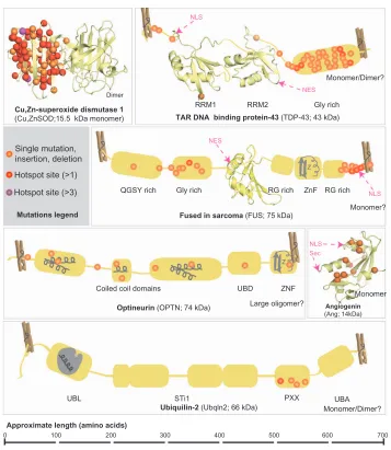
Figure 1 Known mutations in FALS and SALS-associated proteins.
Notes: Known mutations are mapped onto their corresponding proteins. Single mutations can include point mutations, premature stop codons, deletions, or insertions. For simplicity, one of the SOD dimers contains the mapped mutations. Structural and Domain Organization is indicated. Solved structures of domains or entire proteins are shown as ribbon diagrams: Cu,ZnSOD (1PU0); TDP-43 RRM1 (1CQG); TDP-43 RRM2 (1wF0); FUS RRM (1LA6); Angiogenin (1B1I). Clothespins indicate that the tertiary structure and inter-domain associations are not entirely known, so protein is stretched out to better show mutations sites. Schematic depictions of conserved domains without solved structures are shown in grey. where applicable, known or putative oligomeric state and molecular weights are indicated.
Abbreviations: FALS, familial amyotrophic lateral sclerosis; SALS, sporadic amyotrophic lateral sclerosis; NLS, nuclear localization sequence; NES, nuclear export sequence; Sec, cleaved signal sequence; RRM, RNA regnition motif; X rich, X (amino acid residue) rich motifs; UBD, ubiquitin binding domain; ZnF, zinc finger; UBL, ubiquitin like domain; STI1, heat-shock-chaperonin-binding motifs; UBA, ubiquitin associated domain.
Dovepress ALS update
Degenerative Neurological and Neuromuscular Disease downloaded from https://www.dovepress.com/ by 118.70.13.36 on 21-Aug-2020
and cytoplasmic, ubiquitin-reactive hyperphosphorylated TDP-43 inclusions are observed in tissues from frontotem-poral dementia (FTD) patients111,112 and in neuronal and glial
tissues samples from SALS and Guam ALS patients.113 The
inclusions commonly co-localize with ubiquitin and the protein p62.113 However, TDP-43 inclusions are not present
in SOD1 FALS individuals114 (with the exception of one
case113) or FUS mutant patients.115
FUS
FUS encodes fused in sarcoma (FUS, also known as Translated in Liposarcoma, TLS), a modular nucleic acid-associated protein with many similarities to TDP-43, includ-ing conservation of protein domains (Figure 1), a role in RNA processing115 and localization in both the cytosol and nucleus
in many cells. About 30 known FUS mutations account for approximately 3%–5% of FALS and ∼1% of SALS cases116,117
and all but the one known recessive variant, H517Q118 cause
a dominant phenotype. As with some TARDBP mutations, certain FUS mutations located near the nuclear localization sequence may shift the nuclear/cytoplasmic balance towards cytosolic. This imbalance occurs by impairing the transportin-mediated import of FUS into the nucleus.119 FUS-reactive
inclusions have been found in tissues from FUS mutant FALS patients but not in SOD1 mutant patients.115,117 Furthermore,
although earlier studies failed to see FUS-immunoreactivity in SALS cases 115 a more recent study did report FUS
stain-ing in inclusions from SALS patients.117 FUS inclusions are
commonly seen in FTD patients,115,118,120 in addition to ALS
patients, and these FUS-proteinopathy phenotypes might be distinguished through co-localization of other FUS family member proteins in FTD, but not in ALS.121 Furthermore,
FUS and TDP-43 inclusion phenotypes are thought to be mutually exclusive in FTD,122,123 but this may not be the case
in ALS; although TDP-43 reactivity was not observed in FUS
ALS mutant tissues,115 FUS-reactivity was later reported in
TDP-43 ALS mutant tissues.117
OPTN
A recent Italian study indicated that approximately 3.5% of SALS patients, in addition to 1.2% of FALS patients, had mutations in the OPTN gene,4 which encodes Optineurin.
About a dozen mutations in OPTN can lead to ALS, with gain of function mutations dominant and loss of function muta-tions recessive.124,125 Optineurin is a multifunctional cytosolic
and Golgi-associated coiled-coil domain- containing, ubiquitin-binding phosphoprotein (Figure 1). It is involved in vesicular trafficking and Golgi maintenance, signaling
in the tumor-necrosis factor α/NF-κB pathway,126 mGluR
signaling127,128 and autophagy.129 Optineurin has been shown
to form homo-complexes and heteromultimerize with Rab8, myosin VI, and transferrin receptor proteins. In both FALS- and SALS-affected cells, Optineurin can co-localize in inclusion bodies with FUS130 and TDP-43,124 although
the frequency of such inclusions was shown to be low in another study.131 Furthermore, Optineurin localization has
been observed in basophilic inclusions from SOD1 FALS patient tissues,124 although conflictingly this co-localization
was not observed in another study in patient-derived or mouse model tissues.132
ANG
Angiogenin (Ang, encoded by the ANG gene), a small, hypoxia- and ischemia-inducible133 ribonuclease A (Figure 1)
involved in angiogenesis, is mutated in a smaller number of FALS and SALS cases.134 Expressed in many tissues,
including motor neurons,135 where it promotes cell survival,136
Ang is required for the VEGF-mediated stimulation of angiogenesis.137 Ang is secreted and taken up by effector
cells via endocytosis, then translocated to the nucleus, to stimulate transcription of rRNA, among other roles.135 Due to
loss of ribonuclease and/or nuclear translocation activity,135 ANG mutations appear to attenuate angiogenesis although the protein stability is not compromised.138 Eighteen ANG
mutations, therefore, can cause a loss-of-function phenotype, with most ANG ALS patients presenting with bulbar onset (discussed above).134
UBQLN2
UBQLN2, a gene on the X-chromosome, was recently found
to be causative for X-linked dominant FALS.6,139 In affected
families, incomplete penetrance was noted in females, presumably due to X-inactivation. The encoded ubiquilin-2 protein (Figure 1) normally performs effector functions in the ubiquitin proteasome pathway by tethering degradation-targeted proteins (through its C-terminal ubiquitin-associated domain) to the proteasome (through association with its N-terminal ubiquitin-like domain). The intervening regions within the protein are less well characterized, and include a PXX (proline-rich) domain, where five distinct muta-tions were found. In tissues derived from UBQLN2-mutant patients, ubiquitin-positive skein-like inclusions were also reactive for ubiquilin 2. This phenotype was particularly notable in the spinal cord and hippocampus, correlating with the appearance of dementia in 20% of the X-linked ALS patients. Furthermore, these inclusions were also positive
Dovepress
Pratt et al
Degenerative Neurological and Neuromuscular Disease downloaded from https://www.dovepress.com/ by 118.70.13.36 on 21-Aug-2020
for TDP-43, FUS and OPTN, but not Cu,ZnSOD. Notably, ubiquilin-2 inclusion staining was present in all samples from a wide panel of genetically-distinct ALS patient tissues (sporadic, SOD1-mutant, TARDBP mutant, and non-FUS/
non-TARDBP/non-SOD1 FALS, and ALS with dementia) but
not in non-ALS controls.6 Expression of mutant ubiquilin-2
protein significantly slowed down proteosomal degrada-tion of a reporter substrate in Neuro-2a cells,6 suggesting
a mechanistic contribution for these mutants. Unlike the other mutations described, those in the UBQLN2 gene have not yet been implicated in SALS. However, these findings suggest ubiquilin-2 could be generally relevant to ALS pathogenesis.
C9ORF72
Very recently, two independent research groups flagged
C9ORF72 as the gene at locus 9p21 that was linked to
domi-nant cases of ALS/FTD8,9 in previous genome-wide
associa-tion studies. Strikingly, a substantial hexanucleotide repeat (GGGGCC) within an intron of this gene was identified in 24%–46% of FALS cases and 4%–21% of SALS cases, mak-ing this the most commonly mutated ALS gene. The expansion appeared to result in nuclear foci and directed preferential splicing of an alternatively spliced transcript.8 However,
pre-cisely how the aberrant RNA metabolism of C9ORF72 causes ALS is not yet known, and the protein, aside from nuclear localization,9 has no ascribed function. Interestingly,
post-mortem examination of several patients with the C9ORF72
hexanucleotide repeat, who exhibited ALS and FTD-like symptoms, also revealed neuronal TDP-43 inclusions.8
Commonalities and crosstalk
One puzzle for understanding ALS is that the known ALS-causing gene products have diverse physiological functions. However, some common themes in pathogenesis are begin-ning to emerge. For example, RNA processing defects are visible in mutants of TARDBP, FUS, and ANG (as well as a FALS gene called SETX).5 Nucleotide repeat expansions
have also now been identified in C9ORF72 (and an ALS-susceptibility protein called Ataxin-2).140 Proteinacious
cellular inclusions are also a common denominator in ALS patient-derived tissues; these can involve ubiquilin-2, as well as SOD, FUS, TDP-43, and/or optineurin. Interestingly however, different disease subtypes appear to reveal aggre-gates with distinct protein composition. Due to their roles in both ALS and FTD, TDP-43, FUS, OPTN, and ubiquilin-2 have been proposed to function in the context of a unified pathway.141 Thus, interactions among these components should
be a focus for future research. Along these lines, a recent study in zebrafish found that the expression of human FUS
could rescue the motor neuron phenotype associated with knockdown of TARDBP expression, whereas, conversely,
TARDBP could not rescue FUS knockdown, suggesting that
TARDBP is genetically upstream of FUS.142 These results are
consistent with a study showing that TDP-43 regulates the mRNA processing of FUS transcripts as well as its own.105
Genetic overlap between ALS and other
diseases
Gene products whose mutations cause ALS have been implicated in other diseases. For example, FUS, TDP-43, ubiquilin-2, and/or optineurin-positive inclusions are found in many FTD patients,131,143 and C9ORF72 is implicated also in
ALS/FTD.8,9 TDP-43-immunoreactivity is sometimes seen
in hippocampal sclerosis, Pick’s disease, and Alzheimer’s disease (AD), and ubiquitin staining can occur in the latter disease.109 Likewise, optineurin has recently been implicated
in AD due to its inclusion body staining in neurofibrillary tangles.144 Furthermore, optineurin interacts with the protein
huntingtin, suggesting some role in Huntington’s disease,145
and mutations in optineurin are associated with glaucoma146
and Paget’s disease of the bone.147 The ubiquilin-1 paralog,
with a domain structure similar to ubiquilin-2, is associ-ated with AD.6 The 14-3-3 protein isoforms co-localized in
Cu,ZnSOD inclusions have also been found in a Parkinson’s disease model, suggesting some commonalities in inclusion formation.148 Angiogenin has been implicated in a gamut
of diseases, from cancers to diabetes, asthma, and heart disease.149 Finally, nucleotide repeats (as in C9ORF72) are
known to cause a variety of neurodegerative diseases such as Huntington’s disease, Fragile X-syndrome, Kennedy’s disease and others.150 These observations underscore the
need for meaningful synergistic collaborations among researchers studying these different complex diseases that often involve protein aggregation, allowing new insights to be compounded.
Treatment of ALS
The primary goal of ALS treatment is the inhibition of disease progression, although an important secondary consideration is the treatment of damage already done. Palliative care (eg, home care and hospice) remains a significant focus of the treatment program for the ALS patient. Non-invasive ventilation, for example, can improve the quality of life and extend survival in non-bulbar patients.151 A support team,
and hospice care toward the end of life can help the ALS
Dovepress ALS update
Degenerative Neurological and Neuromuscular Disease downloaded from https://www.dovepress.com/ by 118.70.13.36 on 21-Aug-2020
patient to prepare nutritive food that is easy to swallow, provide medications for muscle spasticity, weariness, sleep and depression, and adjust ventilators, enabling the patient to adjust to lifestyle limitations.
Although domestic alterations can provide significant relief to current patients, biochemical and pharmacological advances will drive forward better therapeutics. A panel of ALS biomarkers from non-invasive analyses would be a major gain not only in diagnosis and monitoring progression, but also in identifying affected biological pathways in ALS to target therapeutically.152 Multiple studies have sought to
iden-tify protein biomarkers for ALS, including increased blood or CSF levels of TDP-43, or the cysteine protease inhibitor cystatin C, or a skewed CSF ratio of phospho-neurofilament heavy chain to complement C3.153–156 Furthermore, the
combined efforts of GC/MS (gas chromatography coupled to mass spectrometry), LC/MS (liquid chromatography coupled to mass spectrometry), and NMR (nuclear magnetic resonance) could potentially span the whole metabolome in identifying biomarker signatures.50,60 Better disease markers
could reduce the long duration, averaging 14 months, between initial symptom presentation and diagnosis,47 helping to
improve the disease trajectory.157 Such endeavors would
also provide a platform for personalized medicine for ALS patients. At present, at least one clinical trial (NCT00677768) is being organized to analyze the blood and CSF of ALS patients for biological markers.
Pharmacological interventions
The only approved medicine to treat the general symptoms of ALS is the anti-excitotoxicity drug riluzole.158 The drug
is thought to preserve motor neuron function by decreasing toxic glutamate levels at glutamatergic nerve terminals by (a) inactivating sodium channels, (b) inhibiting glutamate release, and (c) blocking postsynaptic actions of NMDA receptors.159 The safety and efficacy profiles for riluzole
are better than those for other excitotoxicity drugs, but riluzole only increases the chance of an additional year of survival by about 9%, typically prolonging survival for about 2–3 months.11 The drug serves to slightly preserve limb
and bulbar function but actual muscle strength is typically not improved.11 Recently approved for treating purely the
pseudobulbar affect symptoms less commonly observed in ALS patients is dual-acting dextromethorphan/quinine (sold as Neudexta®; Avanir Pharmaceuticals, Aliso Viejo, CA).160
Like riluzole, dextromethorphan also inhibits glutamatergic signaling, and quinine helps to increase its bioavailability, providing modest benefit to a subset of patients.160
Promising new therapeutic developments, several of which are in late-phase clinical trials, may provide strides forward in treating ALS. One such drug in phase III clinical trials (NCT00349622) is the antibiotic ceftriaxone, used to treat pneumonia and bacterial meningitis. In ALS patients, ceftri-axone appears to upregulate the GLT-1 (EAAT2) glutamate transporter, potentially correcting cellular glutamate levels.161
Another potential treatment option is high-dose methylco-balamin (vitamine B-12), currently in phase II/III studies (NCT00444613 and NCT00445172) to determine safety and efficacy for long-term use in ALS.162 This compound was
recently shown to reduce homocysteine (another excitatory amino acid)-mediated toxicity in NSC-34 cells.163 Finally, an
antioxidant targeting the mitochondria is currently in phase III trials (NCT01281189), sponsored by Biogen Idec (Westin, MA) and Knopp Biosciences LLC (Pittsburgh, PA). This drug, dex-pramipexole,164 is the R(+)-isomer of the amino-benzothiazole
drug pramipexole (currently approved to treat Parkinson’s disease and restless legs syndrome). Dexpramipexole was well tolerated in phase II clinical trials, revealing positive trends in slowing function decline and improving survivability.
SOD1
-targeting therapies
The establishment of mutant SOD1 transgenic mice in the late 1990s was a major breakthrough in the field, provid-ing the first disease models for ALS.70 Now, about a dozen
such SOD1 ALS mouse models exist.165 Other distinctive
ALS models have been developed,166,167 including the newer
TARDBP mouse models that similarly display ALS-like
symptoms such as gait abnormalities, weight loss, and spas-ticity.104 However, the use of SOD1 mouse models has
pre-dominated much of the therapeutic progress, in part because
SOD1 represents a major disease target. For example, because the SOD1 gene is predominately dispensible,71 reducing its
expression and perturbing aggregation are favored strategies for treatment of ALS. These transgenic animals are appropri-ate models in many cases, and guidelines have been suggested for standardizing studies in SOD1 mice.168
Both small molecules and siRNAs are being explored to downregulate and diminish SOD levels. The hydroxylamine drug arimoclomol (Orphazyme) is currently in stage II/III clini-cal trials (NCT00706147). This compound induces a heat shock response that resulted in a decrease in ubiquitin-positive aggre-gates in G93A SOD1 mouse models,169 and is now being tested
in SOD1 FALS patients. A free radical scavenger, edaravone (Mitsubishi Tanabe Pharma Corporation, Osaka, Japan) was recently found to ameliorate ALS symptoms and diminish SOD aggregate deposition in interior horn cells. Phase III clinical
Dovepress
Pratt et al
Degenerative Neurological and Neuromuscular Disease downloaded from https://www.dovepress.com/ by 118.70.13.36 on 21-Aug-2020
trials were recently completed (NCT00330681; NCT00424463; NCT00415519), with results pending publication, so the future success of the drug remains to be seen. Studies aimed at silenc-ing SOD1 using siRNA-based strategies in mice have met with some success,170,171 although the inability of siRNA to pass the
blood–brain barrier makes delivery a problem. Accordingly, Isis Pharmaceuticals Inc (Carlsbad, CA) has developed a CSF-infused delivery method for Isis-SOD1RX antisense oligos that recently were successful in animal models,172 and are now being
examined in phase I clinical trials (NCT01041222). Finally, an approach aimed at prevention, which is in its infancy, is immunization against mutant Cu,ZnSOD through vaccination with mutant Cu,ZnSOD or metal-free Cu,ZnSOD (exhibiting some similar pathogenic properties).173 As stable Cu,ZnSOD
polymers expected to break tolerance exist,174 and as antibodies
favor reactions with more flexible regions,175,176 such antibody
experiments may be promising.
A recent study used patient-derived progenitor cells to derive cultured astrocyte cell lines, and these were found to be toxic to motor neurons, via a mechanism involving secretion of unchar-acterized factors. Interestingly, both FALS (mutant SOD1) and SALS-derived cells, but not non-ALS derived astrocyte cells, had common pathway changes (namely NF-κB, MAPK, JNK, and AKT), and knockdown of SOD1 rescued the motor neuron killing phenotype in four of six cell lines examined.177 This
study interestingly reaffirms the use of SOD1-targeted thera-peutics in the context of SALS (although the effects on other FALS genetic backgrounds were not tested) and also suggests that such cell cultures could prove useful for therapeutic screen-ing in the absence of an all-encompassscreen-ing ALS disease model. Indeed, a few years ago, astrocyte replenishment by injection of glial precursor cells in SOD1 model rats was found to prolong life and improve motor performance.178 Similarly, a phase I
clinical trial (NCT01348451) aimed at spinal implantation of spinal cord-derived stem cells is being sponsored by Neuralstem Inc (Rockville, MD). This treatment previously extended the life of SOD1 transgenic rats by 10 days,179 and provides the first
regenerative medicine strategy for ALS.
Future directions
Where do we go from here? ALS was first described about 150 years ago180 and recent biotechnological advances have
allowed researchers to begin pinpointing the precise genet-ics and pathological mechanisms behind the disease. Yet, many questions still remain: How do the distinct pathways involved in the disease overlap and converge to cause similar phenotypes? Can diagnostics improve to the point of early screening and detection? Arguably most importantly, how
can we best treat individual patients? Fortunately, the com-plex nature of the disease also allows for many potential targets and means for therapeutic intervention.
The discovery of the role of SOD1 in ALS was a triggering event that significantly advanced our current understanding of the disease aided by the basic science of SOD structure and biochemistry.87,181 Although we now know that the mutant
proteins aggregate, we are only starting to appreciate the key architectural features of the proteins involved in triggering this aggregation and its consequences. More recently, we have realized the significant contributions of TDP-43 and FUS in ALS and other degenerative diseases.182 Indeed, RNA
metabolism appears to be a common thread. The recent iden-tification of ubiquilin-2 as a co-immunolocalized component of ALS inclusions in a wide variety of ALS cell types has also been a major breakthrough in the field.6 Thus, follow-up
work is now needed in order to determine the mechanism of this ubiquilin-mediated pathology, as well as its poten-tial contributions to other ALS-linked pathways. Finally, determining the pathogenic mechanism of action of newly identified C9ORF72 repeats may prove extremely useful in understanding a significant majority of ALS cases, both spo-radic and inherited. Newer disease models will undoubtedly play a significant role in facilitating these studies.
A critical element of progress in the ALS field will be the dissemination of genetic, epidemiologic, and therapeutic information. Fortunately, several helpful online databases and resource are now available, including the ALS online genet-ics database,183 the Genetic Association studies website,184
the ALS forum,185 and the Northeast ALS Consortium
(NEALS).186 Outreach and social networking is provided
by sites such as the Twitter-based ALS Untangled,187 which
hosts a forum for patient conversations. These assets will increase awareness and discourse among ALS patients and drive future research collaborations.
Conclusions
Currently, ALS is an unrelenting and incurable neuromus-cular disease that paralyzes its victims, eventually leaving them incapable of breathing. Gradually, thanks in part due to strides in molecular genetics, the mechanisms leading to aberrant cellular physiology and toxic inclusions are being sewn together. At present, therapeutic strategies aim to slow down the pace of the disease. Ultimately, however, future efforts will work to block the initial events leading to neu-ronal death. This will prevent damage to the patient’s motor ability before it happens, stemming from earlier diagnosis and leading to better prognosis.
Dovepress ALS update
Degenerative Neurological and Neuromuscular Disease downloaded from https://www.dovepress.com/ by 118.70.13.36 on 21-Aug-2020
Acknowledgments
This work was supported by NIH grant R03 AR059968 (to JJP) and NIH grant R01 GM39345 (to EDG). AJP is a predoctoral fellow of the National Science Foundation and the Skaggs Institute for Chemical Biology at the Scripps Research Institute.
Disclosure
The authors report no conflicts of interest in this work.
References
1. Wijesekera LC, Leigh PN. Amyotrophic lateral sclerosis. Orphanet J Rare Dis. 2009;4:3.
2. Deng HX, Hentati A, Tainer JA, et al. Amyotrophic lateral sclerosis and structural defects in Cu,Zn superoxide dismutase. Science. 1993;261(5124):1047–1051.
3. Rosen DR, Siddique T, Patterson D, et al. Mutations in Cu/Zn super-oxide dismutase gene are associated with familial amyotrophic lateral sclerosis. Nature. 1993;362(6415):59–62.
4. Del Bo R, Tiloca C, Pensato V, et al. Novel optineurin mutations in patients with familial and sporadic amyotrophic lateral sclerosis.
J Neurol Neurosurg Psychiatry. 2011;82(11):1239–1243.
5. Strong MJ. The evidence for altered RNA metabolism in amyotrophic lateral sclerosis (ALS). J Neurol Sci. 2010;288(1–2):1–12.
6. Deng HX, Chen W, Hong ST, et al. Mutations in UBQLN2 cause dominant X-linked juvenile and adult-onset ALS and ALS/dementia.
Nature. 2011;477(7363):211–215.
7. Daoud H, Rouleau GA. A role for ubiquilin 2 mutations in neurodegeneration. Nat Rev Neurol. 2011;7(11):599–600.
8. DeJesus-Hernandez M, Mackenzie IR, Boeve BF, et al. Expanded GGGGCC hexanucleotide repeat in noncoding region of C9ORF72 causes chromosome 9p-linked FTD and ALS. Neuron. 2011;72(2): 245–256.
9. Renton AE, Majounie E, Waite A, et al. A hexanucleotide repeat expan-sion in C9ORF72 is the cause of chromosome 9p21-Linked ALS-FTD.
Neuron. 2011;72(2):245–268.
10. Wood H. A hexanucleotide repeat expansion in C9ORF72 links amyo-trophic lateral sclerosis and frontotemporal dementia. Nat Rev Neurol. 2011;7(11):595.
11. Miller RG, Mitchell JD, Lyon M, Moore DH. Riluzole for amyotrophic lateral sclerosis (ALS)/motor neuron disease (MND). Cochrane Database Syst Rev. 2007;1:CD001447.
12. Sathasivam S. Motor neurondisease: clinical features, diagnosis, diagnostic pitfalls and prognostic markers. Singapore Med J. 2010;51(5):367–373.
13. Byrne SC, Hardiman O. Rate of familial amyotrophic lateral sclerosis: a systematic review and meta-analysis. Neurology. 2010;74(9):A56. 14. Conwit RA. Preventing familial ALS: a clinical trial may be feasible
but is an efficacy trial warranted? J Neurol Sci. 2006;251(1–2):1–2. 15. Haberlandt WF. Genetic aspects of amyotrophic lateral sclerosis
and progressive bulbar paralysis. Acta Genet Med Gemellol (Roma). 1959;8:369–374.
16. Murros K, Fogelholm R. Amyotrophic lateral sclerosis in Middle-Finland: an epidemiological study. Acta Neurol Scand. 1983;67(1):41–47. 17. Williams DB, Floate DA, Leicester J. Familial motor neuron
disease: differing penetrance in large pedigrees. J Neurol Sci. 1988;86(2–3):215–230.
18. Andersen PM, Al-Chalabi A. Clinical genetics of amyotrophic lateral sclerosis: what do we really know? Nat Rev Neurol. 2011;7(11): 603–615.
19. Andersen PM. Amyotrophic lateral sclerosis associated with mutations in the CuZn superoxide dismutase gene. Curr Neurol Neurosci Rep. 2006;6(1):37–46.
20. Traynor BJ, Codd MB, Corr B, Forde C, Frost E, Hardiman O. Amyotrophic lateral sclerosis mimic syndromes: a population-based study. Arch Neurol. 2000;57(1):109–113.
21. Mulder DW, Kurland LT, Offord KP, Beard CM. Familial adult motor neuron disease: amyotrophic lateral sclerosis. Neurology. 1986;36(4):511–517.
22. Li TM, Alberman E, Swash M. Comparison of sporadic and familial disease amongst 580 cases of motor neuron disease. J Neurol Neurosurg Psychiatry. 1988;51(6):778–784.
23. Johnston CA, Stanton BR, Turner MR, et al. Amyotrophic lateral sclerosis in an urban setting: a population based study of inner city London. J Neurol. 2006;253(12):1642–1643.
24. Worms PM. The epidemiology of motor neuron diseases: a review of recent studies. J Neurol Sci. 2001;191(1–2):3–9.
25. Chio A, Benzi G, Dossena M, Mutani R, Mora G. Severely increased risk of amyotrophic lateral sclerosis among Italian professional football players. Brain. 2005;128(Pt 3):472–476.
26. Horner RD, Grambow SC, Coffman CJ, et al. Amyotrophic lateral sclerosis among 1991 Gulf War veterans: evidence for a time-limited outbreak. Neuroepidemiology. 2008;31(1):28–32.
27. Wang H, O’Reilly EJ, Weisskopf MG, et al. Smoking and risk of amyotrophic lateral sclerosis: a pooled analysis of 5 prospective cohorts.
Arch Neurol. 2011;68(2):207–213.
28. Abhinav K, Stanton B, Johnston C, et al. Amyotrophic lateral sclerosis in South-East England: a population-based study. The South-East England register for amyotrophic lateral sclerosis (SEALS Registry).
Neuroepidemiology. 2007;29(1–2):44–48.
29. Vivekananda U, Manjalay ZR, Ganesalingam J, et al. Low index-to-ring finger length ratio in sporadic ALS supports prenatally defined motor neuronal vulnerability. J Neurol Neurosurg Psychiatry. 2011;82(6):635–637.
30. Manning JT, Scutt D, Wilson J, Lewis-Jones DI. The ratio of 2nd to 4th digit length: a predictor of sperm numbers and concentrations of testosterone, luteinizing hormone and oestrogen. Hum Reprod. 1998;13(11):3000–3004.
31. Berenbaum SA, Bryk KK, Nowak N, Quigley CA, Moffat S. Fingers as a marker of prenatal androgen exposure. Endocrinology. 2009;150(11):5119–5124.
32. Chio A, Calvo A, Dossena M, Ghiglione P, Mutani R, Mora G. ALS in Italian professional soccer players: the risk is still present and could be soccer-specific. Amyotroph Lateral Scler. 2009;10(4):205–209. 33. Abel EL. Football increases the risk for Lou Gehrig’s disease,
amyotrophic lateral sclerosis. Percept Mot Skills. 2007;104(3 Pt 2): 1251–1254.
34. Paganoni S, Deng J, Jaffa M, Cudkowicz ME, Willis AM. Body mass index, not dyslipidemia, is an independent predictor of survival in amyotrophic lateral sclerosis. Muscle Nerve. 2011;44(1):20–24. 35. Hardiman O, van den Berg LH, Kiernan MC. Clinical diagnosis
and management of amyotrophic lateral sclerosis. Nat Rev Neurol. 2011;7(11):639–649.
36. Lomen-Hoerth C. Amyotrophic lateral sclerosis from bench to bedside.
Semin Neurol. 2008;28(2):205–211.
37. Cristini J. Misdiagnosis and missed diagnoses in patients with ALS.
JAAPA. 2006;19(7):29–35.
38. Kiernan MC, Vucic S, Cheah BC, et al. Amyotrophic lateral sclerosis.
Lancet. 2011;377(9769):942–955.
39. Forbes RB, Colville S, Swingler RJ. The epidemiology of amyotrophic lateral sclerosis (ALS/MND) in people aged 80 or over. Age Ageing. 2004;33(2):131–134.
40. McCombe PA, Henderson RD. Effects of gender in amyotrophic lateral sclerosis. Gend Med. 2010;7(6):557–570.
41. Andersen PM, Borasio GD, Dengler R, et al. EFNS task force on management of amyotrophic lateral sclerosis: guidelines for diag-nosing and clinical care of patients and relatives. Eur J Neurol. 2005;12(12):921–938.
42. de Carvalho M, Dengler R, Eisen A, et al. Electrodiagnostic criteria for diagnosis of ALS. Clin Neurophysiol. 2008;119(3):497–503.
Dovepress
Pratt et al
Degenerative Neurological and Neuromuscular Disease downloaded from https://www.dovepress.com/ by 118.70.13.36 on 21-Aug-2020
43. Talbot K. Motor neuron disease: the bare essentials. Pract Neurol. 2009;9(5):303–309.
44. Dabby R, Lange DJ, Trojaborg W, et al. Inclusion body myositis mim-icking motor neuron disease. Arch Neurol. 2001;58(8):1253–1256. 45. Weihl CC, Lopate G. Motor neuron disease associated with copper
deficiency. Muscle Nerve. 2006;34(6):789–793.
46. Silani V, Messina S, Poletti B, et al. The diagnosis of amyotrophic lateral sclerosis in 2010. Arch Ital Biol. 2011;149(1):5–27.
47. Chio A. ISIS Survey: an international study on the diagnostic process and its implications in amyotrophic lateral sclerosis. J Neurol. 1999; 246 Suppl 3:III1–III5.
48. Davenport RJ, Swingler RJ, Chancellor AM, Warlow CP. Avoiding false positive diagnoses of motor neuron disease: lessons from the Scottish Motor Neuron Disease Register. J Neurol Neurosurg Psychiatry. 1996;60(2):147–151.
49. Ludolph AC, Knirsch U. Problems and pitfalls in the diagnosis of ALS.
J Neurol Sci. 1999;165 Suppl 1:S14–S20.
50. Rozen S, Cudkowicz ME, Bogdanov M, et al. Metabolomic analysis and signatures in motor neuron disease. Metabolomics. 2005;1(2):101–108. 51. Okamoto K, Mizuno Y, Fujita Y. Bunina bodies in amyotrophic lateral
sclerosis. Neuropathology. 2008;28(2):109–115.
52. Leigh PN, Whitwell H, Garofalo O, et al. Ubiquitin-immunoreactive intraneuronal inclusions in amyotrophic lateral sclerosis. Morphology, distribution, and specificity. Brain. 1991;114(Pt 2):775–788. 53. Plaitakis A, Caroscio JT. Abnormal glutamate metabolism in
amyo-trophic lateral sclerosis. Ann Neurol. 1987;22(5):575–579.
54. Barbeito LH, Pehar M, Cassina P, et al. A role for astrocytes in motor neuron loss in amyotrophic lateral sclerosis. Brain Res Brain Res Rev. 2004;47(1–3):263–274.
55. Barbosa LF, Cerqueira FM, Macedo AF, et al. Increased SOD1 asso-ciation with chromatin, DNA damage, p53 activation, and apoptosis in a cellular model of SOD1-linked ALS. Biochim Biophys Acta. 2010;1802(5):462–471.
56. Bilsland LG, Sahai E, Kelly G, Golding M, Greensmith L, Schiavo G. Deficits in axonal transport precede ALS symptoms in vivo. Proc Natl Acad Sci U S A. 2010;107(47):20523–20528.
57. King AE, Dickson TC, Blizzard CA, et al. Neuron-glia interactions underlie ALS-like axonal cytoskeletal pathology. Neurobiol Aging. 2011;32(3):459–469.
58. Zhu HN, Shi P, Wei YM, Zhang JY, Gal J. Mitochondrial dysfunction is a converging point of multiple pathological pathways in amyotrophic lateral sclerosis. J Alzheimers Dis. 2010;20 Suppl 2:S311–S324. 59. Miana-Mena FJ, Gonzalez-Mingot C, Larrode P, et al. Monitoring
systemic oxidative stress in an animal model of amyotrophic lateral sclerosis. J Neurol. 2011;258(5):762–769.
60. Wuolikainen A, Moritz T, Marklund SL, Antti H, Andersen PM. Disease-related changes in the cerebrospinal fluid metabolome in amyotrophic lateral sclerosis detected by GC/TOFMS. PLoS One. 2011;6(4):e17947.
61. Talbot K. Do twin studies still have anything to teach us about the genetics of amyotrophic lateral sclerosis? J Neurol Neurosurg Psychiatry. 2010;81(12):1299–1300.
62. Garber K. Genetics. The elusive ALS genes. Science. 2008;319(5859):20. 63. Daoud H, Belzil V, Dion PA, Rouleau GA. Chromosome 9p21 in
amyotrophic lateral sclerosis: the plot thickens. Lancet Neurol. 2010;9(10):945–947.
64. McCord JM, Fridovich I. Superoxide dismutase. An enzymic function for erythrocuprein (hemocuprein). J Biol Chem. 1969;244(22): 6049–6055.
65. van Es MA, Dahlberg C, Birve A, Veldink JH, van den Berg LH, Andersen PM. Large-scale SOD1 mutation screening provides evidence for genetic heterogeneity in amyotrophic lateral sclerosis. J Neurol Neurosurg Psychiatry. 2010;81(5):562–566.
66. Marucci G, Morandi L, Bartolomei E, et al. Amyotrophic lateral sclerosis with mutation of the Cu/Zn superoxide dismutase gene (SOD1) in a patient with Down syndrome. Neuromuscul Disord. 2007;17(9–10):673–676.
67. Hayward C, Brock DJ, Minns RA, Swingler RJ. Homozygosity for Asn86Ser mutation in the CuZn superoxide dismutase gene produces a severe clinical phenotype in a juvenile onset case of familial amyo-trophic lateral sclerosis. J Med Genet. 1998;35(2):174.
68. Jonsson PA, Backstrand A, Andersen PM, et al. CuZn-superoxide dismutase in D90A heterozygotes from recessive and dominant ALS pedigrees. Neurobiol Dis. 2002;10(3):327–333.
69. Mancuso M, Filosto M, Naini A, et al. A screening for superoxide dismutase-1 D90A mutation in Italian patients with sporadic amyo-trophic lateral sclerosis. Amyotroph Lateral Scler Other Motor Neuron Disord. 2002;3(4):215–218.
70. Gurney ME, Pu HF, Chiu AY, et al. Motor-neuron degeneration in mice that express a human Cu,Zn superoxide-dismutase mutation. Science. 1994;264(5166):1772–1775.
71. Reaume AG, Elliott JL, Hoffman EK, et al. Motor neurons in Cu/Zn superoxide dismutase-deficient mice develop normally but exhibit enhanced cell death after axonal injury. Nat Genet. 1996;13(1):43–47.
72. Shibata N, Hirano A, Kobayashi M, et al. Intense superoxide dismutase-1 immunoreactivity in intracytoplasmic hyaline inclusions of familial amyotrophic lateral sclerosis with posterior column involvement.
J Neuropathol Exp Neurol. 1996;55(4):481–490.
73. Bruijn LI, Becher MW, Lee MK, et al. ALS-linked SOD1 mutant G85R mediates damage to astrocytes and promotes rapidly progressive disease with SOD1-containing inclusions. Neuron. 1997;18(2): 327–338.
74. Kato S, Shimoda M, Watanabe Y, Nakashima K, Takahashi K, Ohama E. Familial amyotrophic lateral sclerosis with a two base pair deletion in superoxide dismutase 1: gene multisystem degeneration with intracytoplasmic hyaline inclusions in astrocytes. J Neuropathol Exp Neurol. 1996;55(10):1089–1101.
75. Zetterstrom P, Graffmo KS, Andersen PM, Brannstrom T, Marklund SL. Proteins that bind to misfolded mutant superoxide dismutase-1 in spinal cords from transgenic amyotrophic lateral sclerosis (ALS) model mice. J Biol Chem. 2011;286(23):20130–20136. 76. Watanabe M, Dykes-Hoberg M, Culotta VC, Price DL, Wong PC,
Rothstein JD. Histological evidence of protein aggregation in mutant SOD1 transgenic mice and in amyotrophic lateral sclerosis neural tissues. Neurobiol Dis. 2001;8(6):933–941.
77. Wang J, Farr GW, Zeiss CJ, et al. Progressive aggregation despite chaperone associations of a mutant SOD1-YFP in transgenic mice that develop ALS. Proc Natl Acad Sci U S A. 2009;106(5):1392–1397. 78. Okamoto Y, Shirakashi Y, Ihara M, et al. Colocalization of 14-13-3
proteins with SOD1 in Lewy body-like hyaline inclusions in familial amyotrophic lateral sclerosis cases and the animal model. PLoS One. 2011;6(5):e20427.
79. Forsberg K, Jonsson PA, Andersen PM, et al. Novel antibodies reveal inclusions containing non-native SOD1 in sporadic ALS patients. PLoS One. 2010;5(7):e11552.
80. Forsberg K, Andersen PM, Marklund SL, Brannstrom T. Glial nuclear aggregates of superoxide dismutase-1 are regularly present in patients with amyotrophic lateral sclerosis. Acta Neuropathol. 2011;121(5):623–634.
81. Tainer JA, Getzoff ED, Richardson JS, Richardson DC. Structure and mechanism of copper, zinc superoxide dismutase. Nature. 1983;306(5940):284–287.
82. Parge HE, Hallewell RA, Tainer JA. Atomic structures of wild-type and thermostable mutant recombinant human Cu,Zn superoxide-dismutase.
P Natl Acad Sci U S A. 1992;89(13):6109–6113.
83. Bourne Y, Redford SM, Steinman HM, Lepock JR, Tainer JA, Getzoff ED. Novel dimeric interface and electrostatic recognition in bacterial Cu,Zn superoxide dismutase. P Natl Acad Sci U S A. 1996; 93(23):12774–12779.
84. Borgstahl GE, Parge HE, Hickey MJ, Beyer WF Jr, Hallewell RA, Tainer JA. The structure of human mitochondrial manganese superoxide dismutase reveals a novel tetrameric interface of two 4-helix bundles.
Cell. 1992;71(1):107–118.
Dovepress ALS update
Degenerative Neurological and Neuromuscular Disease downloaded from https://www.dovepress.com/ by 118.70.13.36 on 21-Aug-2020
85. Guan Y, Hickey MJ, Borgstahl GE, et al. Crystal structure of Y34F mutant human mitochondrial manganese superoxide dismutase and the functional role of tyrosine 34. Biochemistry. 1998;37(14):4722–4730.
86. Perry JJ, Hearn AS, Cabelli DE, Nick HS, Tainer JA, Silverman DN. Contribution of human manganese superoxide dismutase tyrosine 34 to structure and catalysis. Biochemistry. 2009;48(15):3417–3424. 87. Perry JJ, Shin DS, Getzoff ED, Tainer JA. The structural
bio-chemistry of the superoxide dismutases. Biochim Biophys Acta. 2010;1804(2):245–262.
88. DiDonato M, Craig L, Huff ME, et al. ALS mutants of human superox-ide dismutase form fibrous aggregates via framework destabilization.
J Mol Biol. 2003;332(3):601–615.
89. Shin DS, DiDonato M, Barondeau DP, et al. Superoxide dismutase from the eukaryotic thermophile alvinella pompejana: structures, stability, mechanism, and insights into amyotrophic lateral sclerosis.
J Mol Biol. 2009;385(5):1534–1555.
90. Fan L, Fuss JO, Cheng QJ, et al. XPD helicase structures and activities: insights into the cancer and aging phenotypes from XPD mutations.
Cell. 2008;133(5):789–800.
91. Wang Q, Johnson JL, Agar NY, Agar JN. Protein aggregation and protein instability govern familial amyotrophic lateral sclerosis patient survival. PLoS Biol. 2008;6(7):e170.
92. Vassall KA, Stubbs HR, Primmer HA, et al. Decreased stability and increased formation of soluble aggregates by immature superoxide dismutase do not account for disease severity in ALS. Proc Natl Acad Sci U S A. 2011;108(6):2210–2215.
93. Bystrom R, Andersen PM, Grobner G, Oliveberg M. SOD1 muta-tions targeting surface hydrogen bonds promote amyotrophic lateral sclerosis without reducing apo-state stability. J Biol Chem. 2010;285(25):19544–19552.
94. Prudencio M, Hart PJ, Borchelt DR, Andersen PM. Variation in aggrega-tion propensities among ALS-associated variants of SOD1: correlaaggrega-tion to human disease. Hum Mol Genet. 2009;18(17):3217–3226. 95. Lindberg MJ, Tibell L, Oliveberg M. Common denominator of Cu/
Zn superoxide dismutase mutants associated with amyotrophic lateral sclerosis: decreased stability of the apo state. Proc Natl Acad Sci U S A. 2002;99(26):16607–16612.
96. Bosco DA, Morfini G, Karabacak NM, et al. Wild-type and mutant SOD1 share an aberrant conformation and a common pathogenic pathway in ALS. Nat Neurosci. 2010;13(11):1396–1403.
97. Proctor EA, Ding F, Dokholyan NV. Structural and thermodynamic effects of post-translational modifications in mutant and wild type Cu, Zn superoxide dismutase. J Mol Biol. 2011;408(3):555–567. 98. Nishitoh H, Kadowaki H, Nagai A, et al. ALS-linked mutant
SOD1 induces ER stress- and ASK1-dependent motor neuron death by targeting Derlin-1. Genes Dev. 2008;22(11):1451–1464. 99. Harraz MM, Marden JJ, Zhou W, et al. SOD1 mutations disrupt
redox-sensitive Rac regulation of NADPH oxidase in a familial ALS model. J Clin Invest. 2008;118(2):659–670.
100. Kato S, Sumi-Akamaru H, Fujimura H, et al. Copper chaperone for superoxide dismutase co-aggregates with superoxide dismutase 1 (SOD1) in neuronal Lewy body-like hyaline inclusions: an immu-nohistochemical study on familial amyotrophic lateral sclerosis with SOD1 gene mutation. Acta Neuropathol. 2001;102(3): 233–238.
101. Urushitani M, Sik A, Sakurai T, Nukina N, Takahashi R, Julien JP. Chromogranin-mediated secretion of mutant superoxide dismutase proteins linked to amyotrophic lateral sclerosis. Nat Neurosci. 2006;9(1):108–118.
102. Kieran D, Hafezparast M, Bohnert S, et al. A mutation in dynein res-cues axonal transport defects and extends the life span of ALS mice.
J Cell Biol. 2005;169(4):561–567.
103. Zhang F, Strom AL, Fukada K, Lee S, Hayward LJ, Zhu H. Interaction between familial amyotrophic lateral sclerosis (ALS)-linked SOD1 mutants and the dynein complex. J Biol Chem. 2007;282(22):16691–16699.
104. Da Cruz S, Cleveland DW. Understanding the role of TDP-43 and FUS/TLS in ALS and beyond. Curr Opin Neurobiol. 2011. 2011;21(6):904–919.
105. Polymenidou M, Lagier-Tourenne C, Hutt KR, et al. Long pre-mRNA depletion and RNA missplicing contribute to neuronal vulnerability from loss of TDP-43. Nat Neurosci. 2011;14(4):459–468.
106. Van Deerlin VM, Leverenz JB, Bekris LM, et al. TARDBP mutations in amyotrophic lateral sclerosis with TDP-43 neuropathology: a genetic and histopathological analysis. Lancet Neurol. 2008;7(5):409–416. 107. Kuhnlein P, Sperfeld AD, Vanmassenhove B, et al. Two German
kindreds with familial amyotrophic lateral sclerosis due to TARDBP mutations. Arch Neurol. 2008;65(9):1185–1189.
108. Sreedharan J, Blair IP, Tripathi VB, et al. TDP-43 mutations in familial and sporadic amyotrophic lateral sclerosis. Science. 2008;319(5870):1668–1672.
109. Rutherford NJ, Zhang YJ, Baker M, et al. Novel mutations in TARDBP (TDP-43) in patients with familial amyotrophic lateral sclerosis. PLoS Genet. 2008;4(9):e1000193.
110. Kabashi E, Valdmanis PN, Dion P, et al. TARDBP mutations in individuals with sporadic and familial amyotrophic lateral sclerosis.
Nat Genet. 2008;40(5):572–574.
111. Neumann M, Sampathu DM, Kwong LK, et al. Ubiquitinated TDP-43 in frontotemporal lobar degeneration and amyotrophic lateral sclerosis. Science. 2006;314(5796):130–133.
112. Arai T, Hasegawa M, Akiyama H, et al. TDP-43 is a component of ubiquitin-positive tau-negative inclusions in frontotemporal lobar degeneration and amyotrophic lateral sclerosis. Biochem Biophys Res Commun. 2006;351(3):602–611.
113. Maekawa S, Leigh PN, King A, et al. TDP-43 is consistently co-localized with ubiquitinated inclusions in sporadic and Guam amyotrophic lateral sclerosis but not in familial amyotrophic lat-eral sclerosis with and without SOD1 mutations. Neuropathology. 2009;29(6):672–683.
114. Mackenzie IR, Bigio EH, Ince PG, et al. Pathological TDP-43 distin-guishes sporadic amyotrophic lateral sclerosis from amyotrophic lateral sclerosis with SOD1 mutations. Ann Neurol. 2007;61(5):427–434. 115. Vance C, Rogelj B, Hortobagyi T, et al. Mutations in FUS, an RNA
processing protein, cause familial amyotrophic lateral sclerosis type 6.
Science. 2009;323(5918):1208–1211.
116. Neumann M, Mackenzie IR, Rademakers R. TDP-43 and FUS in amyotrophic lateral sclerosis and frontotemporal dementia. Lancet Neurol. 2010;9(10):995–1007.
117. Deng HX, Zhai H, Bigio EH, et al. FUS-immunoreactive inclusions are a common feature in sporadic and non-SOD1 familial amyotrophic lateral sclerosis. Ann Neurol. 2010;67(6):739–748.
118. Kwiatkowski TJ Jr, Bosco DA, Leclerc AL, et al. Mutations in the FUS/TLS gene on chromosome 16 cause familial amyotrophic lateral sclerosis. Science. 2009;323(5918):1205–1208.
119. Dormann D, Rodde R, Edbauer D, et al. ALS-associated fused in sar-coma (FUS) mutations disrupt Transportin-mediated nuclear import.
EMBO J. 2010;29(16):2841–2857.
120. Snowden JS, Hu Q, Rollinson S, et al. The most common type of FTLD-FUS (aFTLD-U) is associated with a distinct clinical form of frontotemporal dementia but is not related to mutations in the FUS gene. Acta Neuropathol. 2011;122(1):99–110.
121. Neumann M, Bentmann E, Dormann D, et al. FET proteins TAF15 and EWS are selective markers that distinguish FTLD with FUS pathology from amyotrophic lateral sclerosis with FUS mutations.
Brain. 2011;134(Pt 9):2595–2609.
122. Neumann M, Rademakers R, Roeber S, Baker M, Kretzschmar HA, Mackenzie IR. A new subtype of frontotemporal lobar degeneration with FUS pathology. Brain. 2009;132(Pt 11):2922–2931.
123. Woulfe J, Gray DA, Mackenzie IR. FUS-immunoreactive intra-nuclear inclusions in neurodegenerative disease. Brain Pathol. 2010;20(3):589–597.
124. Maruyama H, Morino H, Ito H, et al. Mutations of optineurin in amyotrophic lateral sclerosis. Nature. 2010;465(7295):223–226.
Dovepress
Pratt et al
Degenerative Neurological and Neuromuscular Disease downloaded from https://www.dovepress.com/ by 118.70.13.36 on 21-Aug-2020