1071-412X/02/$04.00
⫹
0
DOI: 10.1128/CDLI.9.2.388–393.2002
Copyright © 2002, American Society for Microbiology. All Rights Reserved.
Neutralizing Antibody Responses to Human Herpesviruses 6 and 7 Do
Not Cross-React with Each Other, and Maternal Neutralizing
Antibodies Contribute to Sequential Infection with
These Viruses in Childhood
Mariko Yoshida,
1* Sadayoshi Torigoe,
2Kumiko Ikeue,
1and Masao Yamada
1Department of Virology, Okayama University Graduate School of Medicine and Dentistry, Okayama,
1and Yokkaichi Social Insurance Hospital, Mie,
2Japan
Received 8 August 2001/Returned for modification 2 November 2001/Accepted 28 November 2001
Seroprevalence of human herpesvirus 6 (HHV-6) and HHV-7 infections is very high throughout the world,
and almost all people are exposed first to HHV-6 and second to HHV-7 in their childhood. However, it is not
clear whether the neutralizing (NT) antibody response between each virus is cross-reactive or not. To elucidate
the NT antibody response between each virus, 55 serum samples from an adult group (subjects 22 to 88 years
old) and 60 serum samples from a young group (subjects 2 to 18 years old) were examined by a dot blot method
for detecting viral late antigen. Thirty-nine serum samples obtained from cord bloods and a few serum samples
obtained from pediatric patients with exanthem subitum were also examined to assess the maternal transferred
NT antibodies against each virus. The NT antibody titers against HHV-7 in the adult group remained high
throughout all the individuals, and none were negative. Those against HHV-6 were high values in the young
group but low values, including negative values (three samples), in the adult group. These results suggested
that the NT antibody response to either HHV-6 or HHV-7 in each individual was specific to each virus and did
not cross-react with each other. In the adult group, the NT antibody response to HHV-6 decreased, while that
to HHV-7 remained high throughout all the individuals. Maternal transferred NT antibody titers against
HHV-7 were higher and remained longer after birth than those of HHV-6, and these findings were in accord
with the clinical observation that HHV-6 infection usually occurs earlier than HHV-7 infection.
Human herpesvirus 6 (HHV-6) (19) and HHV-7 (9) have
recently been discovered as etiologic agents of exanthem
subi-tum (roseola). HHV-6 and HHV-7 are T-lymphotropic viruses
and have been classified as betaherpesviruses. HHV-6 was first
isolated from the peripheral blood lymphocytes of patients
with AIDS (19) and has been divided into two variants,
HHV-6A and HHV-6B (1, 2). HHV-7 was first isolated from
the peripheral blood lymphocytes (9) and the saliva of healthy
adults (5, 10, 12, 23, 27). Clinically, HHV-6B and HHV-7 are
the common etiologic agents of exanthem subitum (roseola)
(24, 29), but diseases caused by HHV-6A are less apparent.
While diseases caused by primary infection of either HHV-6 or
HHV-7 in childhood are usually not fatal, HHV-6 and HHV-7,
as well as the other members of the herpesviruses, are thought
to establish latent, life-long infection. It has been reported that
HHV-6 may contribute to life-threatening diseases in
immu-nosuppressed conditions such as organ transplant and AIDS
(3, 4, 6, 7, 16) and to drug-induced hypersensitivity syndrome
(8, 21, 22, 25). Several investigators have reported that HHV-7
is easily isolated from the saliva of individuals who have
anti-bodies to HHV-7 (10, 23). However, it is unknown which
diseases can be caused by reactivated HHV-7.
Serologic studies showed that seroprevalence of HHV-6
and HHV-7 infections are very high throughout the world
and that almost all people are exposed first to HHV-6 and
second to HHV-7 in their childhood (17). Several serologic
studies for detection of antibodies to either HHV-6 or HHV-7
were performed by indirect immunofluorescent (IF) antibody
assay (IFA), enzyme-linked immunosorbent assay (ELISA),
neutralization, radioimmunoprecipitation, and Western
blot-ting (11, 17, 28). The neutralizing (NT) antibody response is
thought to be important in preventing infection from these
viruses. However, there have been few comparative studies
among these reports on the humoral antibody response
be-tween HHV-6 and HHV-7, and none has reported on the
cross-reactive response based on the NT antibodies between
HHV-6 and HHV-7 in individuals. These facts prompted us to
investigate the cross-reactive response of NT antibodies
be-tween each virus and to assess the maternal transferred NT
antibodies. In this report, we thought that it was important to
determine the degree of immunological cross-reactivity
be-tween HHV-6 and HHV-7 based on the NT antibodies, which
have taken an important role in the prevention of infection. In
order to assess the antibody response to each virus, we
estab-lished a dot blot method for detecting the NT antibody (26, 28)
and an ELISA method for detecting the immunoglobulin G
(IgG) and IgM antibodies (32). Here, we describe the
follow-ing. (i) NT antibody responses between HHV-6 and HHV-7
are specific and do not cross-react to each other. (ii) NT
an-tibody response to HHV-6 decreases with aging, while that to
HHV-7 is maintained highly throughout all individuals of all
ages. (iii) Maternal transferred NT antibodies against HHV-6
and HHV-7 contribute to the sequential infection between
each virus.
* Corresponding author. Mailing address: Department of Virology,
Okayama University Graduate School of Medicine and Dentistry,
2-5-1 Shikatacho, Okayama 700-8558, Japan. Phone: 81-86-235-7167.
Fax: 81-86-235-7169. E-mail: [email protected].
388
on August 17, 2020 by guest
http://cvi.asm.org/
MATERIALS AND METHODS
Serum samples.Sixty serum samples were selected from healthy individuals in different age groups from 2 to 18 years old (as the young group) who had had a medical examination within the 3 months from February to May in 1998 at Shingu Municipal Hospital, Shingu, Japan. Fifty-five serum samples were also selected from healthy individuals in different age groups from 22 to 89 years old (as the adult group) who had had a medical examination within the 3 months from February to May in 1996 at Tsukazaki Hospital, Himeji, Japan. Thirty-nine serum samples obtained from cord blood specimens were selected to evaluate the maternal NT antibody, which was collected in January 2001 at Sun Clinic, Okayama, Japan under informed consent from the mothers. Sequential serum samples were obtained from five pediatric patients with exanthem subitum who were 1 month to 1.5 years old and who had had a medical examination within the 4 years from 1992 to 1996 at Shingu Municipal Hospital under informed consent from their mothers. Thirty serum samples from pediatric patients from 5 months to 1.7 years old who were negative for either anti-HHV-6 or -7 antibody described below were selected as negative reference samples for determining the cutoff values for the ELISA for IgG [ELISA(IgG)] and ELISA(IgM). These samples were also collected at Shingu Municipal Hospital under informed consent from
the mothers. All of the serum samples were stored at⫺20°C before use.
Host cells.Fresh cord blood mononuclear cells (CBMCs) were prepared by centrifugation through a Ficoll-Conray gradient from heparinized samples and cultured for 3 days in RPMI 1640 medium containing 10% heat inactivated fetal bovine serum, recombinant human interleukin-2 (0.1 U/ml; GIBCO BRL Life
Technology Inc., Grand Island, N.Y.), and phytohemagglutinin (5g/ml; Sigma
Chemical Co., St. Louis, Mo.) at 37°C in a 5% CO2incubator. After 3 days, the
CBMCs were infected with the virus and cultured for different experiments in RPMI 1640 medium containing the above reagents.
Preparation of virus stocks.The Z29 strain of HHV-6B and the SB strain of HHV-7 were used throughout this study and prepared as described elsewhere (26, 31). The cells infected with each virus were cocultivated with uninfected cells at a ratio of 1:5 for 7 days. Virus stocks were prepared by centrifugation of the
culture fluids at 2,000⫻gfor 10 min and stored at⫺80°C. Titration of the virus
stocks was performed by an end point dilution method using a dot blot assay (30,
31). In brief, 25l of the CBMCs (adjusted to 5⫻106
cells/ml) was divided into each well of a 96-well microtiter U-bottom plate. The CBMCs were then infected
with 25l of the virus preparation (in 10-fold dilution series) and incubated for
7 days at 37°C in a 5% CO2incubator. After incubation, the supernatant medium
was removed and the cells were washed with phosphate-buffered saline (PBS) (pH 7.4). A dot blot assay to detect the viral antigens is described below. After treatment with Lumi-Phos 530, the membrane was exposed to Fuji RX-U film. The 50% cell culture infectious dose was calculated according to the method of Reed and Muench (18).
IFA.An IFA described elsewhere (30) was performed to determine the titer of
either anti-HHV-6 or -7 antibody in human serum samples. In brief, HHV-6- or
-7-infected CBMCs were mounted on a 14-well slide, dried, and kept at⫺20°C
in a freezer before use. Each serum was diluted 1:10 in PBS to prevent nonspe-cific reactions, and then twofold serial dilutions of the 1:10-diluted samples were made. The diluted samples were applied to each well of the slide fixed with acetone. The slides were incubated at 37°C for 30 min and washed in PBS for 15 min three times. The fluorescein isothiocyanate-conjugated goat anti-human IgG
F(ab⬘)2antibody (Cappel, West Chester, Pa.) was diluted 1:100 in PBS and
added to each well of the slide. After 30 min of incubation at 37°C, the slides were washed and examined with a fluorescence microscope. The end point of the positive fluorescence was determined visually at a low magnification, and the IFA antibody titers were calculated as the reciprocal of the serum dilution.
Dot blot method for determining the NT antibody titers to HHV-6 and HHV-7.
A dot blot method described elsewhere (26, 32) was performed to determine the
titers of NT antibody to HHV-6 and -7. In brief, 25l of serial twofold serum
dilutions or medium without sera (control) were prepared on each well of a 96-well microtiter U-bottom plate and mixed with equal volumes of virus strains
of HHV-6 or HHV-7 containing 2⫻10450% tissue culture infectious doses/ml.
After the microtiter plate was incubated for 1 h at 37°C in a CO2incubator, 50
l of CBMCs (2.5⫻106
cells/ml) was added to each well and centrifuged at 800
⫻gfor 1 h. For removing the added sera, 100l of medium was added to each
well of the plate and centrifuged at 800⫻gfor 5 min, and about 180l of the
supernatant was aspirated. Two hundred microliters of medium was added to each well of the plate, and the microtiter plate was incubated for 7days at 37°C
in a CO2incubator.
To monitor the virus growth in each well, a dot blot antigen detection was performed as described previously (26, 30). In brief, after 7 days of incubation
the microtiter plate was centrifuged at 500⫻gfor 10 min. The supernatant
medium was removed and the cells were washed with PBS including Mg2⫹and
Ca2⫹[PBS(⫹)] (pH 7.4). One hundred and eighty microliters of lysis buffer (20
mM Tris, 0.5 M NaCl, and 0.5% Nonidet P-40 [pH 7.5]) was added to each well. The cell lysates were spotted onto a nylon membrane (Boehringer Mannheim Biochemica, Indianapolis, Ind.), washed with blocking solution, and incubated with one of the monoclonal antibodies (MAb) described below overnight at 4°C. The membrane was washed, incubated with anti-mouse IgG-alkaline phospha-tase conjugate (Sigma Chemical Co.), treated with Lumi-Phos 530 (Boehringer Mannheim Corporation), and exposed to Fuji RX-U film.
MAb.MAb to HHV-6 and HHV-7 were established in our laboratory (26) and
characterized by radioimmunoprecipitation. For monitoring HHV-6 replication, MAb TK-2, which recognizes a 135-kDa late polypeptide, was used. MAb TK-2 reacts to both variants of HHV-6. For HHV-7, MAb IK-3, which recognizes a 125-kDa corresponding polypeptide, was used. MAb IK-3 is specific to HHV-7.
Antigen preparation for ELISA.The CBMCs infected with each virus of HHV-6 (Z29 strain) or HHV-7 (SB strain) were cultured for 5 to 7 days and harvested in 15-ml conical tubes at the period when the numbers of IF positive cells had reached almost 80%. The infected cells were pelletted down by
cen-trifugation at 800⫻gfor 10 min, washed with PBS(⫹) (pH 7.4), and then
centrifuged. The lysis buffer (20 mM Tris, 0.5 M NaCl, and 0.5% Nonidet P-40 [pH 7.5]) was added to each pellet at four times the pellet volume. The unin-fected CBMCs were cultured for 6 days and prepared for control antigen the same way as described above.
ELISA.Wells of a microplate (Nunc Immunoplate II; Nunc, Aarhus,
Den-mark) were coated with 50l of appropriate dilutions in PBS(⫹) of each virus
or control antigen overnight at 4°C. After coating, the wells were washed and
blocked for 30 min at room temperature by adding 200l of the blocking
solution [PBS(⫹) containing 5% skim milk]. After discarding the blocking
solu-tion, 50l of serum diluted in the blocking solution (1:100 for IgG and 1:25 for
IgM) was added to wells of the plate and incubated at 37°C for 2 h. The plate was
washed five times with 200l of the blocking solution. Then, 50l of alkaline
phosphatase-conjugated goat anti-human IgG (␥-chain specific) F(ab⬘)2
frag-ment (Sigma Chemical Company) or alkaline phosphatase-conjugated goat
anti-human IgM (-chain specific) F(ab⬘)2fragment (Sigma Chemical Company)
diluted in the blocking solution (1:1,000) was added to each well of the plate.
After 2 h of incubation at 37°C, the plate was washed five times with 200l of
the blocking solution and twice with PBS(⫹). Seventy-five microliters of
sub-strate,p-nitrophenylphosphate in diethanolamine buffer (alkaline phosphatase
substrate kit [Bio-Rad]) was added to each well of the plate, and the plate was
incubated at 37°C for 1 h. Then, the reaction was stopped by adding 75l of 0.4
M NaOH, and the absorbance of each well at 405 nm was read with the micro-plate reader (model 550; Bio-Rad). The results (net absorbance) were expressed as the absorbency reading of each of the viral-antigen-coated wells minus the absorbency reading for the wells coated with control antigen.
For ELISA(IgG) antibody, the absorbance readings (mean⫾standard
devi-ation [SD]) for HHV-6 and HHV-7 obtained from about 30 negative reference
serum samples of pediatric patients were 0.052⫾0.067 and 0.027⫾0.034,
respectively. All of the samples studied were negative for IF and NT antibody titers against HHV-6. These samples were also negative for IF antibody against HHV-7, and for eight of these samples the titer of NT antibody against HHV-7
was under 1:8. Studied samples with net absorbance readings ofⱖ3 SD above the
mean absorbance of the negative reference sera were considered reactive for anti-HHV-6 or HHV-7 antibody. The cutoff values for anti-HHV-6 and -7 were determined with the absorbance readings of 0.26 and 0.13, respectively. For ELISA(IgM) antibody, about 20 negative reference serum samples which were negative for IF, NT, and ELISA(IgG) antibodies against HHV-6 and HHV-7 were selected from
reference sera for ELISA(IgG) antibody. The absorbance readings (mean⫾SD)
for HHV-6 and HHV-7 obtained from these sera were 0.049⫾0.030 and 0.033
⫾0.027, respectively, and the cutoff values for HHV-6 and -7 were determined
with the absorbance readings of 0.14 and 0.12, respectively.
RESULTS
NT antibody responses against HHV-6 and HHV-7 do not
cross-react with each other.
First, we compared the NT, IF,
and ELISA(IgG) antibody titers against HHV-6 and HHV-7 in
115 serum samples, including 55 serum samples from the adult
group and 60 serum samples from the young group. A
histo-gram based on each antibody titer against these viruses is
shown in Fig. 1. The NT antibody responses against HHV-6
and HHV-7 showed different distributions between each virus.
V
OL. 9, 2002
NT RESPONSES BETWEEN HHV-6 AND -7 DO NOT CROSS-REACT
389
on August 17, 2020 by guest
http://cvi.asm.org/
The NT antibody titers against HHV-7 were high throughout
individuals of all ages, ranging from 1:8 to 1:
⬎
512 (geometric
mean titer
⫽
2
5.64). A few samples were negative for those
antibodies in the young group, but none were negative in the
adult group. In contrast, the NT antibody titers against HHV-6
in the young group (geometric mean titer
⫽
2
4.52) were high
and comparable to those of HHV-7 in the young group
(geo-metric mean titers
⫽
2
5.63), while significantly low titers,
in-cluding negative titers (ranging from 1:
⬍
1 to 1:64), were
ob-served in the adult group (geometric mean titer
⫽
2
2.64). No
significant difference in distribution patterns of either IF
anti-body or ELISA(IgG) antianti-body titers was observed between
HHV-6 and HHV-7, although these distribution patterns were
generally higher in the young group than in the adult group.
Second, to further elucidate the humoral antibody responses
between HHV-6 and HHV-7 in each individual, we analyzed
the scattergrams based on the NT, IF, and ELISA(IgG)
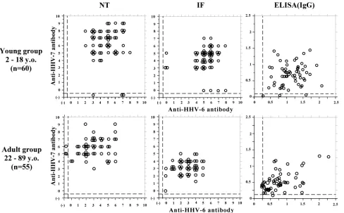
anti-bodies between each virus (Fig. 2). The NT antibody responses
between each virus were distinct in each individual. The sera
with high titers of anti-HHV-7 antibodies do not always have
high titers of anti-HHV-6 antibodies, while some sera negative
for anti-HHV-6 NT antibodies have high titers for anti-HHV-7
antibodies. Namely, the NT antibodies between HHV-6 and
HHV-7 did not correlate in each individual (Wilcoxon signed
rank test,
P
⬍
0.01; Spearman rank correlation,
P
⬎
0.01).
Figure 2 shows that the IF and ELISA(IgG) antibody titers in
each individual did not apparently correlate either.
These results revealed that the NT antibody response to
either HHV-6 or HHV-7 in each individual was specific to each
virus and that they did not cross-react with each other.
Maternal transferred NT antibodies against HHV-6 and
HHV-7 contribute to the sequential infection between each
virus.
First, 39 serum samples obtained from cord blood
spec-imens were used to evaluate the NT antibody titers against
HHV-6 and HHV-7 transferred from mothers to children
(Fig. 3). The transferred NT antibody titers against HHV-7
(geometric mean titer
⫽
2
7.03) were higher than those against
HHV-6 (geometric mean titer
⫽
2
5.22) among almost all the
samples. The ELISA(IgG) antibody titers of antibodies to
HHV-6 and HHV-7 did not represent a significant difference
(data not shown). These agree well with the results described
above.
Second, we monitored the NT antibody titers against
these viruses in five children with exanthem subitum. The
ELISA(IgM) antibodies, as well as the IF antibody and
ELISA(IgG) antibodies, were also examined to determine
the primary infection of these viruses, and the results are
shown in Fig. 4. The titers of NT antibody against HHV-7 in
early time point samples obtained sequentially were
main-tained higher than those of HHV-6 among three samples
(sam-ples 1, 15, and 22, shown in Fig. 4). Among them, two sam(sam-ples
(samples 1 and 22) represented seroconversion against HHV-6
after the NT antibody titers decreased to almost negative
val-ues. The ELISA(IgM) antibody response against HHV-6 was
detected temporally at the same time of elevation of the NT
and IF antibodies and just before elevation of the ELISA(IgG)
antibodies. One sample (sample 15, shown in Fig. 4) revealed
reversion of antibody titers between the NT and IF antibodies.
The titers of NT antibody against HHV-6 were lower than
those of HHV-7 and almost negative, while the titers of IF
antibody against HHV-6 were higher than those against
HHV-7. These data suggest that the IF antibody response is
not useful to elucidate the transferred antibody that prevent
infection with these viruses. In the case of two samples
(sam-ples 10 and 27, shown in Fig. 4), the NT antibodies against
HHV-6 were maintained higher than those of HHV-7. It was
difficult to determine which was the transferred antibody from
the mother or the primary infection of HHV-6. However, these
two samples revealed an apparent seroconversion against
HHV-6, because the ELISA(IgM) antibodies were found in
FIG. 1. Comparative study of NT antibody titers, IF antibody titers, and ELISA(IgG) antibody titers against HHV-6 and HHV-7. A histogram
based on the titers of each antibody against these viruses were illustrated for 60 serum samples from the young group (subjects 2 to 18 years old
[y.o.]) and 55 serum samples from the adult group (subjects 22 to 88 y.o.), respectively. A histogram based on ELISA(IgG) antibody titers was
plotted by intervals of 0.2 optical density units. Secondary antibody used for IF antibody detection was directed against whole IgG (heavy plus light
chain) of human immunoglobulins. Secondary antibody used for ELISA(IgG) antibody detection was directed against
␥
-chain specific IgG of
human immunoglobulins.
on August 17, 2020 by guest
http://cvi.asm.org/
the first point of the sequentially obtained samples. In the case
of sample 10, the patient was diagnosed with exanthem
subi-tum twice. These data revealed that this patient experienced
exanthem subitum caused first by HHV-6 infection and second
by HHV-7 infection.
It is not easy to collect serum samples from subjects within
6 months after birth. In this work, only 15 serum samples were
obtained from eight patients in this periods. We have
calcu-lated the geometric mean titers of these samples. Among them,
7 serum samples were apparently negative for HHV-6 and 11
FIG. 2. Comparative study of titers of NT antibody, IF antibody, and ELISA(IgG) antibody against HHV-6 and HHV-7 in each individual.
Scattergrams based on each antibody titer between each virus were illustrated for 60 serum samples from the young group (subjects 2 to 18 years
old [y.o.]) and 55 serum samples from the adult group (subjects 22 to 88 y.o.), respectively. Negative values are shown under the dashed line.
Secondary antibody used for IF antibody detection was directed against whole IgG (heavy plus light chain) of human immunoglobulins. Secondary
antibody used for ELISA(IgG) antibody detection was directed against
␥
-chain-specific IgG of human immunoglobulins.
FIG. 3. Maternal transferred NT antibody titers against HHV-6 and HHV-7 were compared in serum obtained from cord blood specimens.
V
OL. 9, 2002
NT RESPONSES BETWEEN HHV-6 AND -7 DO NOT CROSS-REACT
391
on August 17, 2020 by guest
http://cvi.asm.org/
serum samples were also negative for HHV-7. The geometric
mean titers of antibodies against HHV-6 and HHV-7 were
2
1.10and 2
2.14, respectively. We also selected serum samples
within 3 months after birth which were apparently negative for
both viruses and calculated the geometric mean titers. The
geometric mean titers were 2
1.26and 2
4.04for HHV-6 (three
serum samples) and HHV-7 (five serum samples), respectively.
Within 6 months after birth, two patients experienced
exan-them subitum caused by HHV-6. In case of one patient (data
not shown), it occurred within 3 months, and HHV-6 was
isolated from peripheral blood mononuclear cells. Although
sample numbers were not enough to conclude that the
protec-tive effect of HHV-7 maternal transferred NT antibody plays a
role, these results suggested that the average titer of NT
anti-bodies against HHV-7 in cord blood was approximately 2 logs
higher than those against HHV-6 and that HHV-7 maternal
transferred NT antibodies were maintained higher than those
of HHV-6 within 6 months after birth.
These results suggested that the maternal transferred NT
antibody response to HHV-7 was maintained at a high level
from just after birth and was delayed in decreasing, while those
to HHV-6 were maintained at considerably lower levels from
just after birth and decreased faster than those to HHV-7.
These findings were in accord with the clinical observation that
HHV-6 infection usually occurs earlier than HHV-7 infection.
DISCUSSION
Seroepidemiology of HHV-6 and HHV-7 has been based on
the antibody titers determined by the IFA or ELISA methods.
There have been some reports on the NT antibody assay for
HHV-6, conventionally determining the end points by
cyto-pathic changes or IF. A few of these were comparative studies
of the titers of NT antibodies against HHV-6 and HHV-7 in
each individual (26, 32). In the present study, we performed a
dot blot method for viral late antigen detection to assess the
NT antibodies described elsewhere (26, 32), because this
method takes advantage of a reliable, reproducible and visible
end point. This report precisely demonstrated the humoral
immune responses based on the NT antibodies between
HHV-6 and HHV-7 in each individual. The results suggest that
the immunological cross-reactivity between HHV-6 and
HHV-7 is not exhibited in the NT antibodies and that the
neutralizing epitopes of these two viruses are apparently
dis-tinct (14, 20). Serologic studies also showed that almost all
individuals are exposed first to HHV-6 and second to HHV-7
in their childhood, acquire the NT antibodies against these
viruses, and keep them at high levels for 2 or 3 decades after
primary infection (11, 13, 17, 28, 32). However, an interesting
result in the present report was that the NT antibody response
to HHV-6 was kept at significantly lower level than that to
FIG. 4. Changes of NT antibody titers, IF antibody titers, and ELISA(IgG) and ELISA(IgM) antibody titers against HHV-6 and HHV-7 in five
infants diagnosed with exanthem subitum. Shaded symbols represent negative values for ELISA(IgG) and ELISA(IgM) antibody titers for each
virus. Dotted lines with short intervals represent the cutoff value for HHV-6, while dotted lines with long intervals represent the cutoff value for
HHV-7.
on August 17, 2020 by guest
http://cvi.asm.org/
HHV-7. The continuation at high levels of anti-HHV-7 NT
antibody titers may contribute to the finding that it was easy to
isolate HHV-7 from adult saliva (10). In contrast, it was
diffi-cult to isolate HHV-6 from any source in healthy adults, while
HHV-6 DNA was detected in adult saliva by PCR as well as
HHV-7. That is, these results suggested that the continuous
reactivation of HHV-7 might have an important role for
keep-ing the NT antibody response at a high level. Serological
in-vestigation based on the NT antibody against HHV-6 will
be-come a useful tool to determine the reactivation of HHV-6,
because of the lower levels of anti-HHV-6 NT antibody
re-sponse. Recently, drug hypersensitivity syndrome (DHS), which
is a severe idiosyncratic reaction associated with drug therapy,
has focused on the relationship with the reactivation of HHV-6
(8, 21, 22, 25). We plan a retrospective study to investigate the
reactivation of HHV-6 associated with DHS using sera
ob-tained from DHS patients.
In this report, we demonstrated that maternal transferred
NT antibodies have an important role in preventing infection
with each virus and that the levels of these transferred-NT
antibody titers contributed to the sequential infection with
each virus; namely, HHV-6 infects first and HHV-7 infects
second. We also used the sera obtained from cord blood
spec-imens to examine the maternal transferred NT antibody
against HHV-6 and HHV-7 and demonstrated that maternal
transferred NT antibodies against HHV-7 are maintained at
much higher levels than those against HHV-6. These data are
in accord with the results in paired sera obtained from mothers
and cord blood specimens based on the IFA (15). These facts
support the notion that infants after birth are protected from
infection with these viruses by maternal transferred NT
anti-bodies and acquire the primary infection after a decrease in
these transferred NT antibodies.
ACKNOWLEDGMENTS
We thank T. Yamagata and colleagues for helpful support and
discussion.
Part of this work was supported by Grants-in-Aid for Scientific
Research from the Ministry of Education, Culture, Science, and
Tech-nology of Japan.
REFERENCES
1.Ablashi, D. V., H. Agut, Z. Berneman, G. Campadelli-Fiume, D. Carrigan, L. Ceccerini-Nelli, B. Chandran, S. Chou, H. Collandre, R. Cone, T. Dam-baugh, S. Dewhurst, D. Diluca, L. Foa-Tomasi, B. Fleckenstein, N. Frenkel, R. Gallo, U. Gompels, C. Hall, M. Jomes, G. Lawrence, M. Martin, L. Montagnier, F. Neipel, J. Nicholas, P. Pellet, A. Razzaque, G. Torrelli, B. Thomson, S. Salahuddin, L. Wyatt, and K. Yamanishi.1993. Human
her-pesvirus-6 strain groups: a nomenclature. Arch. Virol.129:363–366.
2.Ablashi, D. V., N. Balachandran, S. F. Josephs, C. L. Hung, G. R. Krueger, B. Kramarsky, S. Z. Salahuddin, and R. C. Gallo.1991. Genomic polymor-phism, growth properties, and immunologic variations in human
herpesvi-rus-6 isolates. Virology184:545–552.
3.Akashi, K., Y. Eizuru, Y. Sumiyoshi, T. Minematsu, S. Hara, M. Harada, M. Kikuchi, Y. Niho, and Y. Minamishima.1993. Brief report: severe infectious mononucleosis-like syndrome and primary human herpesvirus 6 infection in
an adult. N. Engl. J. Med.329:168–171.
4.Asano, Y., T. Yoshikawa, S. Suga, T. Yazaki, K. Kondo, and K. Yamanishi.
1990. Fatal fulminant hepatitis in an infant with human herpesvirus-6
infec-tion. Lancet335:862–863.
5.Black, J. B., N. Inoue, K. Kite-Powell, S. Zaki, and P. E. Pellett.1993.
Frequent isolation of human herpesvirus 7 from saliva. Virus Res.29:91–98.
6.Buchwald, D., P. R. Cheney, D. L. Peterson, B. Henry, S. B. Wormsley, A. Geiger, D. V. Ablashi, S. Z. Salahuddin, C. Saxinger, R. Biddle, et al.1992. A chronic illness characterized by fatigue, neurologic and immunologic dis-orders, and active human herpesvirus type 6 infection. Ann. Intern. Med.
116:103–113.
7.Challoner, P. B., K. T. Smith, J. D. Parker, D. L. MacLeod, S. N. Coulter, T. M. Rose, E. R. Schultz, J. L. Bennett, R. L. Garber, M. Chang, P. A. Schad, P. M. Stewart, R. C. Nowinski, J. P. Brown, and G. C. Burmer.1995. Plaque-associated expression of human herpesvirus 6 in multiple sclerosis.
Proc. Natl. Acad. Sci. USA92:7440–7444.
8.Descamps, V., F. Bouscarat, S. Laglenne, E. Aslangul, B. Veber, D. Des-camps, J. L. Saraux, M. J. Grange, M. Grossin, E. Navratil, B. Crickx, and S. Belaich.1997. Human herpesvirus 6 infection associated with anticonvul-sant hypersensitivity syndrome and reactive haemophagocytic syndrome.
Br. J. Dermatol.137:605–608.
9.Frenkel, N., E. C. Schirmer, L. S. Wyatt, G. Katsafanas, E. Roffman, R. M. Danovich, and C. H. June.1990. Isolation of a new herpesvirus from human
CD4⫹T cells. Proc. Natl. Acad. Sci. USA87:748–752.
10.Fujiwara, N., H. Namba, R. Ohuchi, H. Isomura, F. Uno, M. Yoshida, S. Nii, and M. Yamada.2000. Monitoring of human herpesvirus-6 and -7 genomes in saliva samples of healthy adults by competitive quantitative PCR. J. Med.
Virol.61:208–213.
11.Grose, C.1996. Childhood infections with human herpesviruses types 6, 7,
and 8. Adv. Pediatr. Infect. Dis.12:181–208.
12.Hidaka, Y., Y. Liu, M. Yamamoto, R. Mori, C. Miyazaki, K. Kusuhara, K. Okada, and K. Ueda.1993. Frequent isolation of human herpesvirus 7 from
saliva samples. J. Med. Virol.40:343–346.
13.Levy, J. A.1997. Three new human herpesviruses (HHV6, 7, and 8). Lancet
349:558–563.
14.Lusso, P., P. Secchiero, R. W. Crowley, A. Garzino-Demo, Z. N. Berneman, and R. C. Gallo.1994. CD4 is a critical component of the receptor for human herpesvirus 7: interference with human immunodeficiency virus. Proc. Natl.
Acad. Sci. USA91:3872–3876.
15.Ohashi, M., M. Ihira, K. Suzuki, S. Suga, Y. Asano, T. Yoshikawa, Y. Saito, and H. Sakui.2001. Transfer of human herpesvirus 6 and 7 antibodies from
mothers to their offspring. Pediatr. Infect. Dis. J.20:449–450.
16.Okuno, T., K. Higashi, K. Shiraki, K. Yamanishi, M. Takahashi, Y. Kokado, M. Ishibashi, S. Takahara, T. Sonoda, K. Tanaka, and et al.1990. Human
herpesvirus 6 infection in renal transplantation. Transplantation49:519–522.
17.Pellet, P. E., and J. B. Black.1996. Human herpesvirus 6, p. 2587–2608.In
B. N. Field, D. M. Knipe, and P. M. Howley (ed.), Fields virology, 3rd ed., vol. 2. Lippincott-Raven Publishers, Philadelphia, Pa.
18.Reed, L. J., and H. Muench.1938. A simple method of estimating fifty
percent endpoints. Am. J. Hyg27:493–497.
19.Salahuddin, S. Z., D. V. Ablashi, P. D. Markham, S. F. Josephs, S. Stur-zenegger, M. Kaplan, G. Halligan, P. Biberfeld, F. Wong-Staal, B. Kramar-sky, et al.1986. Isolation of a new virus, HBLV, in patients with
lympho-proliferative disorders. Science234:596–601.
20.Santoro, F., P. E. Kennedy, G. Locatelli, M. S. Malnati, E. A. Berger, and P. Lusso. 1999. CD46 is a cellular receptor for human herpesvirus 6. Cell
99:817–827.
21.Sullivan, J. R., and N. H. Shear.2001. The drug hypersensitivity syndrome:
what is the pathogenesis? Arch. Dermatol.137:357–364.
22.Suzuki, Y., R. Inagi, T. Aono, K. Yamanishi, and T. Shiohara.1998. Human herpesvirus 6 infection as a risk factor for the development of severe
drug-induced hypersensitivity syndrome. Arch. Dermatol.134:1108–1112.
23.Takahashi, Y., M. Yamada, J. Nakamura, T. Tsukazaki, J. Padilla, T. Kita-mura, M. Yoshida, and S. Nii.1997. Transmission of human herpesvirus 7 through multigenerational families in the same household. Pediatr. Infect.
Dis. J.16:975–978.
24.Tanaka, K., T. Kondo, S. Torigoe, S. Okada, T. Mukai, and K. Yamanishi.
1994. Human herpesvirus 7: another causal agent for roseola (exanthem
subitum). J. Pediatr.125:1–5.
25.Tohyama, M., Y. Yahata, M. Yasukawa, R. Inagi, Y. Urano, K. Yamanishi, and K. Hashimoto.1998. Severe hypersensitivity syndrome due to sulfasala-zine associated with reactivation of human herpesvirus 6. Arch. Dermatol.
134:1113–1117.
26.Tsukazaki, T., M. Yoshida, H. Namba, M. Yamada, N. Shimizu, and S. Nii.
1998. Development of a dot blot neutralizing assay for HHV-6 and HHV-7
using specific monoclonal antibodies. J. Virol. Methods73:141–149.
27.Wyatt, L. S., and N. Frenkel.1992. Human herpesvirus 7 is a constitutive
inhabitant of adult human saliva. J. Virol.66:3206–3209.
28.Yamada, M.2001. Human herpesviruses 6 and 7: effects on hematopoiesis
and mode of transmission. Jpn. J. Infect. Dis.54:47–54.
29.Yamanishi, K., T. Okuno, K. Shiraki, M. Takahashi, T. Kondo, Y. Asano, and T. Kurata.1988. Identification of human herpesvirus-6 as a causal agent
for exanthem subitum. Lanceti:1065–1067.
30.Yoshida, M., M. Yamada, S. Chatterjee, F. Lakeman, S. Nii, and R. J. Whitley.1996. A method for detection of HHV-6 antigens and its use for
evaluating antiviral drugs. J. Virol. Methods58:137–143.
31.Yoshida, M., M. Yamada, T. Tsukazaki, S. Chatterjee, F. D. Lakeman, S. Nii, and R. J. Whitley.1998. Comparison of antiviral compounds against human
herpesvirus 6 and 7. Antivir. Res.40:73–84.
32.Yoshida, M., S. Torigoe, and M. Yamada.2002. Elucidation of the cross-reactive immunoglobulin M response to human herpesviruses 6 and 7 on the
basis of neutralizing antibodies. Clin. Diagn. Lab. Immunol.9:394–402.