University of South Carolina
Scholar Commons
Theses and Dissertations
2017
Synergism of Quercetin and Sodium Butyrate for
Controlling Growth of Glioblastoma
Matthew Alan Taylor University of South Carolina
Follow this and additional works at:https://scholarcommons.sc.edu/etd Part of theBiomedical and Dental Materials Commons
This Open Access Thesis is brought to you by Scholar Commons. It has been accepted for inclusion in Theses and Dissertations by an authorized administrator of Scholar Commons. For more information, please [email protected].
Recommended Citation
SYNERGISM
OF
QUERCETIN
AND
SODIUM
BUTYRATE
FOR
CONTROLLING
GROWTH
OF
GLIOBLASTOMA
by
Matthew Alan Taylor
Bachelor of Science
University of Central Florida, 2015
Submitted in Partial Fulfillment of the Requirements
For the Degree of Master of Science in
Biomedical Science
School of Medicine
University of South Carolina
2017
Accepted by:
Swapan Ray, Director of Thesis
Anindya Chanda, Reader
Susan Lessner, Reader
ACKNOWLEDGEMENTS
Faculty:
Swapan K. Ray, PhD – Research Mentor
John Fuseler, PhD – Fluorescent Microscopy
Udai Singh, PhD – Flow Cytometry
Anindya Chanda, PhD – Thesis Committee Member
Susan Lessner, PhD – Thesis Committee Member
Students:
Firas Khathayer – PhD Student; Ray Lab
Alex Sougiannis – PhD Student; Murphy Lab
Narayan Raghava – Undergraduate
ABSTRACT
Glioblastoma multiforme (GBM), or simply glioblastoma, is the most common
and aggressive primary brain tumor, with a prevalence of approximately 20,000 new
cases per year in the United States and a 3-year survival rate of just 2%. Quercetin
(QCT) is a dietary flavonoid that can be found in common foods such as red kidney
beans, cilantro, and onions. Despite little evidence showing any benefits through dietary
intake of QCT, various studies show its promising anti-cancer results in vitro. In
glioblastoma, QCT is able to cause significant amounts of apoptosis through a variety of
mechanisms. These include activation of caspase-9 and caspase-3, deactivation of matrix
metalloprotease-2 (MMP-2), inhibition of heat-shock protein-27 (HSP-27) and HSP-72,
and increased p53 activity. Autophagy is a natural occurring survival mechanism that is
induced when cells are subjected to environmental stressors like nutrient deprivation,
high heat, or hypoxia. As a result, many synergistic studies are being performed with
QCT in order to find out adequate autophagy down regulation that could complement
QCT for enhancing its apoptosis inducing capabilities. Synergism is the concept of two
substances providing a greater affect than the sum of their individual affects. Drugs
acting in synergism with each other is a promising investigative avenue for many
alternative cancer treatments, including GBM.
The drugs QCT and Sodium Butyrate (NaB) were investigated under autophagic
growth of glioblastoma cells in-vitro due to the reduction of autophagy. The three
glioblastoma cell lines tested are C6 (rat), T98G (human), and LN18 (human). The
results of this study showed a marked increase in apoptosis in all three GBM cell lines,
with the most occurring in T98G. Apoptosis levels were determined via Wright Staining
and Annexin V/PI staining. QCT + NaB combination treatments were also found to
reduce serum-starved induced autophagy in all cell lines with the most prominent
occurring in T98G. QCT alone was also found to be an autophagy inhibitor at a 25 µM
concentration. These results were confirmed via acridine orange staining and western
blotting. QCT + NaB was also found to act in synergism to reduce poly (ADP-ribose)
polymerase-1 (PARP-1), a DNA repair enzyme, and survivin, an anti-apoptotic protein,
expression in C6 cells, which further confirmed the potential efficacy of QCT + NaB to
TABLE OF CONTENTS
ACKNOWLEDGEMENTS ... iii
ABSTRACT ... iv
LIST OF TABLES ... viii
LIST OF FIGURES ... ix
CHAPTER 1: INTRODUCTION ...1
CHAPTER 2:PROSPECTSOFENHANCINGANTI-CANCERACTIVITIESOF QUERCETININTHETREATMENTOFGLIOBLASTOMA ...6
2.1INTRODUCTION ...8
2.2OVERVIEWOFCURRENTTREATMENTSFORGLIOBLASTOMA ...12
2.3QCTAGLYCONESYNTHESISANDBIOAVAILABILITY ...14
2.4MECHANISMSOFACTIONOFQCTFORINDUCINGAPOPTOSIS ...16
2.5AUTOPHAGYENIGMAINQCTTHERAPYFORGLIOBLASTOMA...20
2.6QCTSYNERGISMFORCONTROLLINGGLIOBLASTOMA ...22
2.7QCTDELIVERYTOTHEBRAIN ...25
2.8CONCLUSION ...27
CHAPTER 3:SYNERGISTICEFFECTOFQUERCETINANDSODIUMBUTYRATEIN INHIBITIONOFAUTOPHAGYANDINCREASEINAPOPTOSISINRATAND HUMANGLIOBLASTOMACELLS ...28
3.1INTRODUCTION ...30
3.3RESULTS&DISCUSSION...55
3.4FUTUREDIRECTIONS ...77
3.5CONCLUSION ...80
LIST OF TABLES
Table 3.1 Western Blot Buffers ...54
Table 3.2 C6 Trypan Blue Data ...57
Table 3.3 Wright Stain Data ...60
Table 3.4 Annexin V Data ...64
LIST OF FIGURES
Figure 1.1 Glioblastoma MRI Image ...3
Figure 2.1 Quercetin Chemical Structure ...11
Figure 2.2 Quercetin Metabolism ...14
Figure 2.3 Quercetin Apoptosis Pathway ...18
Figure 3.1 Experimental Design ...33
Figure 3.2 Trypan Blue Example ...36
Figure 3.3 Morphological Signs of Apoptosis-1...37
Figure 3.4 Morphological Signs of Apoptosis-2...39
Figure 3.5 Annexin V Example ...41
Figure 3.6 Annexin V Compensation ...43
Figure 3.7 Annexin V FITC Example...44
Figure 3.8 Annexin V PI Example ...45
Figure 3.9 Autophagy In-Vitro ...46
Figure 3.10 Acridine Orange Histograms ...49
Figure 3.11 96-Well Plate Design ...51
Figure 3.12 Protein Normalization Formula ...52
Figure 3.13 C6 Trypan Blue ...56
Figure 3.14 Wright Staining ...58
Figure 3.15 Wright Stain Data Graph ...59
Figure 3.17 Annexin V Data Graph ...63
Figure 3.18 Acridine Orange Stain ...65
Figure 3.19 Acridine Orange Data Graph ...66
Figure 3.20 Bax and Bcl-2 ...70
Figure 3.21 Caspase-3 ...71
Figure 3.22 PARP-1 ...72
Figure 3.23 Survivin ...74
Figure 3.24 LC3 ...76
CHAPTER
1
Glioblastoma multiforme (GBM), also known as simply glioblastoma, is the
deadliest and most aggressive malignant primary brain tumor. Typically existing as an
astrocytoma, glioblastoma tumors most commonly reside in the cerebral hemispheres;
however, they can emerge anywhere throughout the central nervous system. Although it
accounts for only 14.9% of all primary brain cancers, it has the highest prevalence of
malignancy out of any brain tumor. These tumors become more frequent with age and
more commonly affect men than women. Patients diagnosed with GBM have a median
survival of 14.6 months with the current standard treatments. According to the American
Brain Tumor Association, children with high grade tumors have a better prognosis than
adults; with a 5 year survival rate of 25%, whereas adults have a 5 year survival rate of
10%.
The most prevalent symptoms include seizures, nausea, headaches, and vomiting.
Upon diagnosis, first line therapies include surgical resection in conjunction with
simultaneous radiotherapy and chemotherapy. In most cases, complete surgical resection
is difficult due to the undefined borders of GBM tumors. They often spread throughout
the cerebral hemispheres with tentacle-like extensions, which makes total resection
without the loss of essential brain functioning nearly unattainable. Standard
chemotherapy utilizes Temozolomide (TMZ), which is a DNA methylating/alkylating
agent that targets O6 guanine residues, resulting in a cessation of growth in the G-2 phase
(Sang et al., 2014). Radiation therapy has been shown to increase overall median
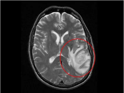
Figure 1.1: MRI image showing tentacle-like extensions of a GBM tumor located in the right hemisphere (Courtesy of Medscape).
After approximately 12 months of TMZ treatment, most GBM tumors become
resistant to the tumor DNA alkylation. This occurs due to the increased activity of O6
-methylguanine DNA methyltransferase (MGMT) repair enzyme in GBM tumors, which
leads to chemoresistant phenotypes. This mechanism, along with rapid growth and
undefined borders, are the core cause of the low survival rates of GBM patients. Because
of this, alternative therapies are being widely researched. The only FDA approved
second-line GBM treatment is Bevacizumab, which is an antibody targeting vascular
epithelial growth factor (VEGF). VEGF is highly upregulated in GBM and is responsible
therapies include Cediranib, Vatalanib, Aflibercept, and Cilengitide; however, they have
shown little-to-no efficacy in clinical trials.
The aim of the current study is to explore the therapeutic potential of a
second-line therapy using QCT and NaB synergistically with each other. QCT is a natural
bioflavonoid that is most widely known for its anti-inflammatory properties. But in
recent years, QCT has proven to contain anti-cancer properties in a variety of tumors,
including glioblastoma. QCT has been shown to induce apoptosis in GBM through
multiple mechanisms that include activation of caspase-9 & caspase-3 and
downregulation of heat shock proteins (HSP) 72 & 27 (Taylor and Ray, 2017). But
despite its anti-cancer potential, QCT is not without its downsides. QCT has also been
shown to alter autophagy in various GBM cell types. It was demonstrated in U373MG
cells that QCT upregulated autophagy (Kim et al., 2013), whereas QCT was able to block
autophagy in U87 and U251 when paired with the drug t-AUCB (Li et al., 2016).
Autophagy is considered a cell survival mechanism during cancer progression. By
pairing QCT with another drug, it is hypothesized that autophagy will be downregulated
and apoptosis will be synergistically potentiated.
Sodium butyrate (NaB) is a well-known histone deacetylase inhibitor (HDACi)
that is widely used in cancer treatment studies. Histone acetylation is important for gene
expression due to acetylated lysine residues allowing a DNA strand to reside in the
relaxed position. This allows for cell transcription machinery to access genes and
transcribe the corresponding mRNA. The role of a histone deacetylase (HDAC) is to
remove the acetyl groups from lysine residues; thus, returning the DNA into its tightly
HDAC overexpression. This results in repression of essential tumor suppressor and DNA
repair genes (West and Johnstone, 2014). Over the past decade, many clinical trials have
been underway to test the efficacy of HDACi in combination with standard chemo- and
radiotherapies. Valproic Acid in combination with radiation therapy has been shown to
be a radiosensitizer, and Vorinostat (SAHA) in combination with several chemotherapies
has shown synergistic cell death (Lee et al. 2017). But despite NaB being a
well-established HDACi, few studies have been performed investigating its impact on GBM.
Because of its relatively unknown mechanism and the success of other HDACi drugs in
combination therapy, it was determined that NaB would be the most ideal candidate for
this study.
Autophagy is a cellular response triggered by environmental stressors such as
high heat or nutrient starvation. In recent years, autophagy has become an area of interest
in cancer research due to it serving two opposite roles in the cancer cell cycle. During
tumor development autophagy is a tumor suppressive mechanism; however, during tumor
progression autophagy is a pro-survival mechanism (Yang and Klionsky, 2010). This
enigma opens a door to a new avenue of cancer therapy by downregulating autophagy
during cancer treatment. The effect QCT has on autophagy in glioblastoma remains
controversial. It has been shown to both upregulate and downregulate autophagy in
different GBM cell types. The impact of NaB on autophagy has been largely
undetermined. In order to develop viable cancer therapies, autophagy and its role in
various types of cancer must be better understood. By investigating the synergism of
QCT + NaB on GBM cells under autophagic conditions, the roles of each of these drugs
CHAPTER 2
PROSPECTS OF ENHANCING ANTI-CANCER ACTIVITIES OF
QUERCETIN IN THE TREATMENT OF GLIOBLASTOMA
_____________________
Taylor, M. A., & Ray, S. K. (2017). Prospects of Enhancing Anti-Cancer Activities of Quercetin in the Treatment of Glioblastoma. Horizons in Cancer Research,66, 173-192.
ABSTRACT
Quercetin (QCT) is a dietary flavonoid that can be found in common foods such
as red kidney beans, cilantro, and onions. It is most commonly known as a
neuroprotective agent; but at doses between 12.5 - 100 µM, QCT has been proven to
possess potent anti-cancer activities. Despite little evidence showing any benefits
through dietary intake of QCT, various studies show its promising anti-cancer results
in vitro. The most common and deadly brain cancer is glioblastoma that remains
incurable with any standard chemotherapy. In glioblastoma, QCT is able to cause
significant amounts of apoptosis through a variety of mechanisms. These include
activation of caspase-9 and caspase-3, deactivation of matrix metalloprotease-2
(MMP-2), inhibition of heat-shock protein-27 (HSP-27) and HSP-72, and increased p53
activity. In addition to induction of apoptosis, QCT upregulates autophagy in most
glioblastoma cell lines. Autophagy is a naturally occurring survival mechanism that is
induced when cells are subjected to environmental stressors like nutrient deprivation,
high heat, or hypoxia. As a result, many synergistic studies are being performed with
QCT in order to find out adequate autophagy down regulation that could complement
QCT for enhancing its apoptosis inducing capabilities. The most significant hurdle to
overcome with QCT treatment in glioblastoma is developing an efficient delivery
method into the brain to allow its prolonged accumulation in the affected brain tissue.
QCT is rapidly degraded when injected in vivo, which explains why dietary intake is
unable to produce significant results. However, recent studies have shown that QCT is
able to be delivered to the brain, liver, kidneys, and lungs in both mice and pig models
through the use of nanoparticles. Even though much still remains undiscovered, recent
opened a window of opportunity into future use of QCT as a viable alternative or
addition to standard treatment for glioblastoma.
2.1 - INTRODUCTION
Glioblastoma multiforme, which is also simply called glioblastoma, is one of the
most common and aggressive primary brain tumors. According to the American Brain
Tumor Association, glioblastoma accounts for about 15% of all primary brain tumors
with a median survival of 14.6 months from the time of diagnosis in spite of current use
of surgery, radiotherapy, and chemotherapy. Standard treatment for glioblastoma includes
first surgical resection followed by adjuvant chemotherapy with temozolomide (TMZ)
and radiotherapy. The molecular mechanism of action of TMZ shows that it acts by
methylating the O6 position of guanine nucleotides in genomic DNA, resulting in cell
cycle arrest and cell death (Sang et al., 2014). Recent Phase III clinical trials have shown
that TMZ treatment in conjunction with radiotherapy increased median survival time
from 12.1 to 14.6 months in glioblastoma patients (Hottinger et al., 2014). In most cases,
the brain tumors become resistant to TMZ treatment within a year due to development of
intracellular protective mechanisms such as altered DNA repair enzyme activity, p53
mutations, and overexpression of epidermal growth factor receptor (EGFR) (Haar et al.,
2012; Messaoudi et al., 2015). The negligible success with the current adjuvant therapies
has impelled the necessity for development of alternative treatments in order to improve
the outcomes in glioblastoma patients.
Studies in our laboratory have shown that various flavonoids possess remarkable
Chakrabarti and Ray, 2015a; Chakrabarti and Ray, 2015b) and also in vivo (Chakrabarti
and Ray, 2016). Quercetin (QCT) is an abundant dietary flavonoid commonly found in
foods such as red kidney beans, chili pepper, cranberry, onion, and tomatoes (Konar,
2013; Dajas et al., 2015). QCT has a basic flavonoid structure consisting of two separate
phenolic rings A and B joined by a 3-carbon heterocyclic ring C and with all the
substitutions in its rings, it is chemically called 3,3,’4,’5,7-pentahydroxyflavone (Figure
2.1). In nature, many flavonoids including QCT are bound to sugars and thus occur in
O-glycoside forms due to glycosylation at any hydroxyl group of the flavonoids. The
glycosylated structures of a flavonoid are the most common in nature, not the aglycone or
parent molecule. The most common QCT glycoside has a sugar group at the 3-hydroxyl
position, such as QCT-3-O-glucoside. QCT is highly regarded as a potent anti-oxidant
that acts through its ortho-dihydroxy substitutions in its ring B and also the
2,3-unsaturation and 4-carbonyl in its ring C (Dajas et al., 2015). However, recent studies
have clearly shown that QCT possesses significant anti-cancer properties. QCT acts
through multiple molecular mechanisms to induce apoptosis in many types of cancers. In
glioblastoma, QCT triggers the intrinsic apoptotic pathway that includes activation of
caspase-9 and caspase-3 (Badziul et al., 2014), inactivation of heat-shock proteins (HSPs)
(Sang et al., 2014), suppression of PI3K/Akt survival signaling (Pan et al., 2014), and
inhibition of mitogen-activated protein kinase (MAPK)/extracellular signal-regulated
kinase (ERK) kinase (MEK) 1 and Raf1 kinase activities (Lee et al., 2008). This
multi-directional approach to induction of apoptosis gives a great potential to QCT to act as an
Despite the widely proven pro-apoptotic capabilities of QCT in cell culture
studies, many obstacles still have to be overcome before it can be used as a viable
alternative therapy for glioblastoma. The greatest challenge facing QCT therapy is its
instability due to metabolic degradation in vivo. High molecular mass and low water
solubility have hindered the ability of this naturally occurring dietary QCT to accumulate
in the target tissues at therapeutically significant concentrations (Blasina et al., 2015). As
a result, current research efforts have been focused on developing nanosomes that can be
used to prevent metabolic degradation of QCT, allow penetration through the
blood-brain-barrier (BBB), and maintain QCT tissue concentrations at a therapeutically
effective level (Priprem et al., 2008).
Another obstacle with QCT therapy is the current understanding of autophagy and
modulation of induction of autophagy by QCT. Autophagy is a cellular response to
nutrient starvation and other metabolic stressors that prompt recycling of existing
cytoplasmic components in order to be used as an alternative energy source (Cui et al.,
2015). Present studies describe autophagy as either a pro-survival or pro-death
mechanism; therefore, exact role of autophagy in cancer has been difficult to determine
(Moon et al., 2015). QCT is a known up-regulator of autophagy in glioblastoma (Kim et
al., 2013, Cui et al., 2015, Moon et al., 2015). The current understanding of autophagy in
glioblastoma is that it acts as a pro-survival mechanism during glioblastoma progression
(Kim et al., 2013). This emphasizes the need to further investigate capability of QCT to
act synergistically or at least additively when combined with one of the drugs known for
The anti-cancer capabilities of QCT both in vitro and in vivo have been clearly
demonstrated over the past decade. The high prevalence, poor prognosis, and ineffective
therapies for glioblastoma have made the development of alternative therapies a
significant priority. Despite the current obstacles associated with QCT, its profound
therapeutic potential makes it an alternative agent that cannot be ignored. This chapter
will provide a comprehensive overview of the past findings, current research, and future
development paths that can be taken into consideration to use QCT as a viable alternative
therapy for glioblastoma.
2.2 - OVERVIEW OF CURRENT TREATMENTS FOR
GLIOBLASTOMA
Glioblastoma is a malignant, highly aggressive primary brain tumor that almost
always carries a poor prognosis. The incidence of glioblastoma is 3 to 4 in 100,000
people per year (Hottinger et al., 2014). However, the incidence increases with age and is
more common in men. According to the American Brain Tumor Association, about 3%
of the childhood brain tumors diagnosed are considered as glioblastoma. Without
treatment, average survival time is usually less than 1 year. With treatment median
survival ranges around 14 months. Despite a poor prognosis, many patients choose to
undergo surgery and standard treatment with the hopes of extending their survival as
much as possible.
Upon diagnosis of glioblastoma, the first priority is the surgical resection of the
tumor. Glioblastoma typically reside in the cerebral hemispheres, but tumors also have
some potential to appear anywhere throughout the central nervous system. Due to the
highly aggressive nature of glioblastoma, complete surgical resection is often
unattainable. Without complete surgical resection, adjuvant chemotherapy or
radiotherapy is a necessary next step (Hottinger et al., 2014). After surgical resection of
the tumor, radiotherapy combined with simultaneous oral TMZ chemotherapy is the
currently used treatment strategy. TMZ is a DNA alkylating agent that is currently the
standard chemotherapy for treatment of glioblastoma. TMZ induces apoptosis by
methylating O6 guanine residues in genomic DNA in cancer cells, which ultimately leads
to cell cycle arrest at the G2 phase (Jakubowicz-Gil et al., 2013). Standard treatments for
glioblastoma patient involve a daily oral TMZ dose of 75 mg/m2 of body surface area
2015). This is followed by a TMZ dose of 150 - 200 mg/m2 BSA for five consecutive
days every 28 days. Usually within one year, glioblastoma becomes resistant to TMZ and
ultimately cause recurrence of the tumor. TMZ resistance is mainly attributed to many
glioblastoma cells containing O6-methylguanine DNA methyltransferase, which
counteracts the primary cell death mechanism of TMZ. Other resistance mechanisms
include an overexpression of EGFR and p53 gene mutations (Messaoudi et al., 2015).
Due to highly aggressive nature of glioblastoma and poor outcomes of standard
treatment regimen, many alternative targeted therapies are being developed. Currently the
most prevalent second-line glioblastoma treatment is Bevacizumab (Bev), which is a US
Food and Drug Administration (FDA) approved monoclonal antibody that targets
vascular epithelial growth factor (VEGF) (Wang et al., 2016). VEGF and its variants are
highly expressed in glioblastoma cells and other brain tumors (Kloepper et al., 2016).
However, after multiple Phase II and Phase III clinical trials, Bev has been unable to
show a consistent favorable outcome in glioblastoma patients when compared with the
standard regimen of TMZ and radiotherapy (Hottinger et al., 2014). Other alternative
drugs that target factors such as EGFR, BCR-Abl, PI3K/Akt, and mTOR are currently in
clinical trials, but so far these drugs have shown little to no efficacy in treating
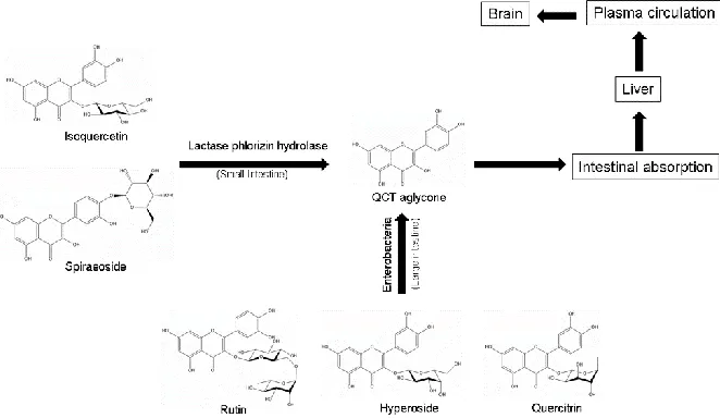
Figure 2.2: Processes and pathways for synthesis of QCT aglycone from various QCT glycosides and its biodistribution in animals. Various QCT glycosides are catabolized to QCT aglycone in small and large intestines. Absorption of QCT aglycone may ultimately transport it to some extent to the brain.
2.3 - QCT AGLYCONE SYNTHESIS AND BIOAVAILABILITY
QCT is one of the most common dietary flavonoids and it is found in many fruits
and vegetables. In Western countries, it is estimated that an average person has an
average dietary intake of 16 - 25 mg of QCT per day (Dajas et al., 2015). In vivo, QCT
aglycone synthesis and its bioavailability are shown (Figure 2.2). Most of the QCT found
in the diet is in its glycosylated forms, which include isoquercetin (QCT-3-O-glucoside),
spiraeoside (QCT-4’-O-glucoside), rutin rutinoside), hyperoside
(QCT-3-O-galactoside), and quercitrin (QCT-3-O-rhamnoside). Once ingested, QCT glycosides are
hydrolyzed by lactase phlorizin hydrolase in the small intestine and then absorbed via the
sodium-dependent glucose transporter-1 (SGLT-1). After SGLT-1 mediated import,
intestinal processing results in QCT glycosides for their conversion to QCT aglycone,
aglycone is quickly processed through secondary metabolism by the liver and then
distributed throughout the bloodstream (Kashino et al., 2015). QCT metabolites then
attach to serum albumin in order to be circulated throughout the blood plasma. These
metabolites have a half-life of 11 to 28 hours, which allow for aggressive
supplementation to increase QCT plasma concentrations to high nanomolar to low
micromolar range (Dajas et al., 2015). Normal dietary plasma QCT levels are typically in
the low nanomolar range.
In order for QCT to be a viable treatment for glioblastoma, it must be able to pass
through the BBB. A recent study performed in rats investigated the ability of dietary
QCT to accumulate in the brain after heavy supplementation by either direct QCT
injection or dietary intake (Ishisaka et al., 2011). The aim of this investigation was to
analyze the neuroprotective effects of QCT and found that QCT was detectable in the rat
brain tissue at the pmol/g level (Ishisaka et al., 2011). An earlier study determined that
rats subjected to an 11-week 1% QCT diet accumulated 330 and 680 pmol/g of brain
tissue (de Boer et al., 2005).
Despite the ability of QCT to penetrate the BBB and accumulate in brain tissue, no
studies have shown dietary or intravenous administration of QCT aglycone or its
metabolites to be a viable method for saturating brain tissue to cancer therapeutic levels.
Some recent in vitro studies, however, have determined that 25 - 100 µM QCT can be an
effective pro-apoptotic concentration in various glioblastoma cell lines (Kim et al., 2013;
Pan et al., 2014). Therefore, it can be implied that QCT dietary supplementation is not an
effective method for achieving sufficient level of QCT in vivo for treating or preventing
2.4 - MECHANISMS OF ACTION OF QCT FOR INDUCING
APOPTOSIS
Over the last decade, QCT has been shown to be a potent anti-cancer compound.
In addition to its free radical scavenging properties, QCT can also induce apoptosis in
glioblastoma cells while showing negligible side effects on normal tissues (Pozsgai et al.,
2013). This unique property has demonstrated that QCT has the potential to be a viable
second-line therapy for inducing apoptosis in human glioblastoma cells (Pan et al., 2014).
Although the complete mechanism of QCT for induction of apoptosis in glioblastoma has
yet to be fully discovered, QCT has been proven to act through an intrinsic
multi-mechanistic pathway leading to final phase of apoptosis. The mechanisms for ultimate
induction of apoptotic cell death due to QCT include down regulation of PI3K/Akt
survival signaling (Pan et al., 2014), inactivation of HSP-27 and HSP-72 (Sang et al.,
2014), inhibition of MAPK pathways (Lee et al., 2008), and also suppression of the
JAK/STAT3 signaling pathway (Michaud-Levesque et al., 2012), and activation of
caspase-9 and caspase-3 (Badziul et al., 2014) in glioblastoma cells (Figure 2.3). This
type of multi-targeted mechanisms of QCT in glioblastoma may hold the key to
combating the chemoresistance issue associated with TMZ treatment and also key to
providing hope of improving patient outcomes.
The most prevalent mechanism of QCT for induction of apoptosis in glioblastoma
is the activation of caspase-9 and caspase-3, as mentioned above. In a recent study, it has
been shown that QCT in a dose dependent manner can increase activation of caspase-9
QCT, while maximum activation of caspase-3 happened at 75 µM QCT in human
glioblastoma U373MG cells (Kim et al., 2013). Caspases play a crucial role in induction
of apoptosis by their contributions to apoptotic pathways that are manifested in
morphological and biochemical features. Caspase-9 is considered an initiator caspase in
the intrinsic mitochondrial pathway of apoptosis (McIlwain et al., 2013). After QCT acts
through the intrinsic mitochondrial pathway, cytochrome c is released from mitochondria
into the cytosol and then cytosolic cytochrome c is combined with Apaf-1 to form an
apoptosome, which recruits pro-caspase-9 and ultimately leads to activation of caspase-9
(Badziul et al., 2014). QCT activates caspase-9 that then proceeds to activate caspase-3
by causing a conformational change to expose active sites on caspase-3 (McIlwain et al.,
2013). Caspase-3 then proceeds to activate various pro-apoptotic proteins including a
DNase that ultimately degrades genomic DNA causing cell death. Activation of
caspase-9 and caspase-3 by QCT has been widely demonstrated both in single drug (Kim et al.,
2013) and multi-drug combination (Jakubowicz-Gil et al., 2013; Badziul et al., 2014)
studies.
Many other studies have also shown that QCT acts in glioblastoma cells through
the inhibition of the PI3K/Akt survival pathway (Pozsgai et al., 2013). Since its
discovery, the PI3K/Akt pathway has been of great interest due to its crucial role in cell
survival in cancers. Many cancer types have the upregulated PI3K/Akt signaling, which
may be a major contributing factor to chemoresistance (Vara et al., 2004). A recent study
investigating QCT treatments alone or in combination with TMZ in two glioblastoma cell
lines U-251 and DBTRG-05 demonstrated the ability of QCT to down regulate the
phospho-Akt levels in glioblastoma cells both in single and TMZ combination treatments.
In a more recent study, it has been demonstrated that PI3K/Akt pathway contributes to
mitochondrial health by promoting anti-apoptotic Bcl-2 stabilization (Pan et al., 2014).
Bcl-2 is an important oncoprotein and well-known for its role in preventing
mitochondrial pathway of apoptosis in cancer cells. Down regulation of the pro-survival
PI3K/Akt signaling pathway enables QCT to induce mitochondrial pathway of apoptosis
in glioblastoma cells. Interestingly, QCT has also been shown to drastically increase
phospho-ERK levels (Pozsgai et al., 2013), which may result in increased Bcl-2
translation. In contrast, a more recent study demonstrated that QCT in a dose dependent
manner decreased phospho-ERK1 levels (Pan et al., 2015). The reason for this
discrepancy has yet to be determined. Obviously, a decrease in phospho-ERK1 is highly
desirable for decreasing the Bcl-2 translation.
Figure 2.3: Molecular mechanisms of action of QCT for inhibition of growth and induction of apoptosis in glioblastoma cells. Various studies have confirmed the capability of QCT in down regulating cell migration, PI3K/Akt survival signaling, cell cycle progression, anti-apoptotic Bcl-2 protein, and HSPs. QCT promotes pro-apoptotic Bax homodimerization and inhibition of HSPs (HSP-27 and HSP-72) to trigger
mitochondrial release of cytochrome c and facilitate formation of apoptosome,
Another mechanism involved in the multi-targeted approach of QCT for induction
of apoptosis in glioblastoma cells is the inactivation of the HSP-27 and HSP-72. These
HSPs play very important roles in cell survival when exposed to stressful environments.
In cancers, HSPs can hinder treatment by inhibiting the intrinsic mitochondrial pathway
of apoptosis. HSP-27 inhibits apoptosis by binding to cytosolic cytochrome c; thus,
stopping it from binding to Apaf-1 to form the apoptosome (Takayama et al., 2003).
HSP-72 functions by acting on Apaf-1 and preventing Apaf-1 from binding to cytosolic
cytochrome c (Badziul et al., 2014) and HSP-72 can also migrate to the nucleus to
prevent nuclear DNA degradation due to caspase-3 mediated activation of a DNase
(Jakubowicz-Gil et al., 2013). Indeed, QCT is considered to be one of the best HSP
inhibitors and it is known to down regulate both HSP-27 and HSP-72 in glioblastoma
cells (Badziul et al., 2014). The ability of QCT to inhibit HSP activity enhances its ability
to induce apoptosis in glioblastoma cells through the intrinsic pathway by further down
regulating anti-apoptotic factors. Due to the oncogenic nature of HSP expression in
cancer cells, inhibition of their activity by QCT demonstrates a novel approach to future
cancer therapies.
QCT also has the ability to inhibit the STAT3 signaling pathway in glioblastoma.
The STAT3 pathway is one of the most studied signal transduction pathways due to its
prevalent and potent roles in inflammation and cancer. It has been demonstrated that
there is a significant correlation between STAT3 activation and tumor malignancy
(Birner et al., 2010). QCT reduced glioblastoma cell growth due to inhibition of the
STAT3 signaling pathway by blocking the phosphorylation of GP130 and simultaneously
STAT3 pathway is responsible for Bcl-2 upregulation, promoting cell migration through
MMP-2 activation and cell cycle progression through cyclin D1 expression. Results
showed that QCT in a dose dependent manner inhibited MMP-2 secretion in presence of
IL-6. This suggests that QCT is a suitable therapy for preventing glioblastoma invasion
and metastasis. It was also observed that QCT stopped cell cycle progression by
inhibiting cyclin D1 expression. However, inhibition of cyclin D1 expression occurred
only in presence of IL-6 (Michaud-Levesque et al., 2012). It has been demonstrated that
glioblastoma cells heavily secrete IL-6 (Hong et al., 2007) and this provides an
opportunity to QCT to enable its inhibitory effect on cyclin D1 expression for blocking
glioblastoma cell growth.
2.5 - AUTOPHAGY ENIGMA IN QCT THERAPY FOR
GLIOBLASTOMA
Autophagy is a cellular response to environmental stressors such as nutrient
starvation, high heat, or metabolic stress. This process involves intracellular degradation
of cytoplasmic components via acidic lysosomes in order to provide additional amino
acids and other building blocks for essential cellular processes (Cui et al., 2015). In
cancers, autophagy is an important topic because of its ability to either act as a protective
mechanism or promoter of apoptosis. During cancer development, autophagy is
considered to be tumor suppressive mechanism; while during cancer progression,
autophagy promotes cancer cell survival (Yang and Klionsky, 2010). Most cancers are
need to be developed to make QCT get around autophagy and put forward apoptosis for
slowing the progression of glioblastoma cell growth (Kim et al., 2013).
Despite the impression that autophagy pathway progresses in a simplistic manner,
its physiological effects remain largely misunderstood. In vitro studies show that
autophagy is most easily triggered through nutrient starvation of the cells in culture. An
autophagic cell then encompasses its organelles into a specialized vesicle, termed the
autophagosome. The autophagosome fuses with acidic lysosome, thereby the engulfed
components are digested and the resulting amino acids and other building blocks are
reused for essential cellular processes (Yang and Klionsky, 2010). Detection of
autophagy is performed by measuring the amount of increase in LC3B II (which is
present in the autophagosome) or the amount of decrease in sequestosome 1 (SQSTM1),
also known as the ubiquitin binding protein p62 (which is a substrate for lysosomal
degradation in the autophagosome), in the cell (Moon et al., 2015).
Autophagy is enigmatic in the use of many alternative and also standard
chemotherapeutic agents for glioblastoma. Several studies have so far demonstrated that
QCT is an up-regulator of autophagy in glioblastoma as well as in other forms of cancer
cells (Kim et al., 2013; Cui et al., 2015; Moon et al., 2015). This property is one of the
major pitfalls of QCT in the treatment of cancer, revealing autophagy being a cellular
protection mechanism during cancer progression. A well-designed study demonstrated
how QCT could induce protective autophagy in human glioblastoma U373MG cell line
(Kim et al., 2013). The same study also showed that QCT was unable to induce
autophagy in human glioblastoma T98G cell line. This is an example of the unique
the need to develop therapeutic strategies that are effective in many glioblastoma cell
types. Presence of protective autophagy was shown in U373MG cells by measuring the
amount of decease in apoptosis in the cells following treatments with QCT alone and
QCT combined with chloroquine (an anti-malarial drug known to block autophagy). The
results showed that when combined with chloroquine, QCT was capable of inducing
autophagy by utilizing new mechanisms. In addition to activation of caspase-9 and
caspase-3, caspase-7 was also activated in apoptotic cells (Kim et al., 2013). It can be
stipulated that QCT has unforeseen mechanistic approaches to induction of apoptosis that
may be inhibited by autophagy. In order for QCT to be a viable therapy for glioblastoma,
it must be paired with another complimentary drug for acting synergistically or additively
for promoting induction of both extrinsic and intrinsic pathways of apoptosis in various
glioblastoma cell types.
2.6 - QCT SYNERGISM FOR CONTROLLING GLIOBLASTOMA
Synergism, by definition, is the combination of two or more therapeutic agents
that accentuate a greater therapeutic effect, greater than the sum of their individual
effects. This is an important topic in development of successful cancer therapy because
not all individual drugs are perfect for promoting apoptosis and achieving desirable
therapeutic outcomes. QCT, as previously mentioned, is a known autophagy inducer in
some glioblastoma cells (Kim et al., 2013), which may promote overall cell survival
during glioblastoma progression. Studies suggest that pairing QCT with another
compound to inhibit autophagy will heighten the therapeutic effects of QCT in
While investigating the anti-cancer capabilities of QCT in glioblastoma, several
studies were conducted to gauge which drugs might pair best with QCT. So far the most
common pairing is QCT with TMZ, which is the most common chemotherapeutic drug
used for treatment of glioblastoma, as demonstrated in two recent studies
(Jakubowicz-Gil et al., 2013; Sang et al., 2014). Both studies emphasized the ability of QCT to inhibit
activities of HSP-27 and HSP-72. One of these groups showed that HSP-27 could
contribute to glioblastoma malignancy and its chemoresistance to TMZ (Sang et al.,
2014). It was suggested that increased HSP-27 phosphorylation might be an indicator for
TMZ chemoresistance. Their investigation also demonstrated that glioblastoma U251 and
U87 cell lines were insensitive to TMZ. However, QCT in combination with TMZ was
able to sensitize the glioblastoma cells to TMZ by inhibition of HSP-27 phosphorylation
and an increase in caspase-3 activity. The earlier study provided a more comprehensive
look at the molecular mechanisms of the treatment of combination of QCT and TMZ in
T98G cells (Jakubowicz-Gil et al., 2013). Even though some studies suggest autophagy
being a cell survival mechanism (Kumar et al., 2016), other studies imply that high
enough levels of autophagy may eventually lead majority of the cancer cells decide to die
by apoptosis (Jakubowicz-Gil et al., 2013). Both QCT and TMZ were able to induce
apoptosis in T98G cells, but neither drug increased levels of autophagy (Jakubowicz-Gil
et al., 2013).
Another synergistic study combined QCT and imperatorin for treatment of T98G
cells to further investigate the roles of HSPs in glioblastoma tumorigenesis (Badziul et
al., 2014). Imperatorin is a furanocoumarin and a phytochemical that can be used as an
reported that imperatorin has potent anti-cancer properties. This study found that
combination of 50 µM QCT and 50 µM imperatorin caused more apoptosis than when
the drugs were used separately. The investigators also concluded that inhibition of
HSP-27 and HSP-72 was highly crucial in sensitizing glioblastoma cells to various forms of
treatment (Badziul et al., 2014).
Further study showed the consequences of the combination of QCT and sorafenib
in the treatment of astrocytoma MOGGCCM cell line and glioblastoma T98G cell line
(Jakubowicz-Gil et al., 2014). Sorafenib was originally developed to inhibit Raf kinase in
order to treat malignant brain tumors. However, clinical trials did not yield significant
therapeutic results. It was hypothesized that sorafenib in combination with QCT would
produce greater therapeutic effects than with sorafenib alone. This study observed that
sorafenib alone was a potent autophagy inducer in T98G cells, but not in MOGGCCM
cells. When QCT was used in combination with sorafenib, synergistic amounts of
apoptosis were seen in MOGGCCM cells. In the T98G cells, QCT showed little increase
in amounts of apoptosis when compared with sorafenib treatment alone. When levels of
expression of HSP-27 and HSP-72 were blocked by the short interfering RNA (siRNA),
the most commonly used RNA interference (RNAi) technology, both tumor cell lines
became increasingly more sensitive to QCT treatment (Jakubowicz-Gil et al., 2014).
Thus, all the above mentioned studies demonstrate how inactivation of HSPs through
QCT synergism can be a promising strategy in the development of alternative therapies
2.7 - QCT DELIVERY TO THE BRAIN
The most prevalent obstacle hindering the development of QCT-based
glioblastoma therapy is the inability of QCT to be efficiently delivered to the brain. This
issue contains two components: (1) poor bioavailability of QCT in the plasma and (2)
difficulty of many compounds including QCT to penetrate the BBB. Both of these
disadvantageous components are addressed by furthering the development of
QCT-loaded nanocarriers. A nanocarrier is a nano-sized compound that is used to carry various
compounds throughout the body. They are typically created by inserting a biologically
unstable compound (e.g., QCT) into a biologically stable structure (e.g., micelles,
liposomes, various polymers) (Liu et al., 2014). QCT makes an excellent candidate for
utilization of nanocarriers due to its low water solubility and rapid metabolic degradation
(Blasina et al., 2015).
Currently, many studies are being performed with the hopes of determining the
most efficient QCT-nanocarrier that utilizes efficient delivery and provides therapeutic
potency. First of all, in order for QCT to be a viable anti-cancer treatment, it must be
adequately distributed to the brain tissues. A study investigating the relationship between
QCT and cognition demonstrated that the intranasal administration of QCT-loaded
liposomes provided more efficient brain delivery than orally administered liposomes
(Priprem et al., 2008). This is due to the olfactory pathway bypassing the BBB and
providing QCT direct access to the brain. A very recent study expanded on the use of
lipid-based nanocarriers and their ability to deliver QCT to the brain (Kumar et al., 2016).
These investigators compared the use of nano lipidic carriers (NLCs) with solid lipid
tocopherol acetate, and glyceryl behenate (a monoester of glycerin and behenic acid).
SLNs, on the other hand, utilize a solid lipid structure that is stabilized by various
emulsifiers. This study found that the use of lipid-based nanocarriers increased the
biodistribution of QCT to the brain 3.2 times (SLN) and 5.6 times (NLC) when compared
with the unaltered QCT (Kumar et al., 2016).
With oral administration being the most convenient form of drug delivery for
many types of medication, this route cannot be overlooked. Indeed, a recent study
investigated the ability of QCT-loaded cationic liposomes to assess distribution of QCT
throughout the body (Liu et al., 2014). Cationic liposomes are known to be advantageous
for oral administration of a drug due to their ability to interact with negatively charged
intestinal mucosa. This study found that these QCT-loaded cationic nanostructured lipid
carriers (QR-CNLC) were able to prolong exposure time in the gastrointestinal tract,
which subsequently improved intestinal absorption of the medication. QCT accumulated
in the lung, kidney, and liver. But insignificant amounts of QCT were found in the brain
tissue (Liu et al., 2014).
The next step after optimization of delivery is to ensure the therapeutic potency of
the nanoparticle-delivered QCT. A group of investigators examined the ability of QCT to
induce apoptosis in rat glioblastoma C6 cells in vivo following delivery of the
PEG2000-DPS-coated QCT nanoparticles to the rat brains (Wang et al., 2013). They were able to
conclude from their results that sustained QCT concentrations in rat brain tissues led to
significant amounts of apoptosis through the induction of intrinsic pathway. They
measured increase in cytosolic cytochrome c levels, which eventually led to increases in
we outlined here, many more studies need to investigate efficient methods for achieving
abundant biodistribution of QCT to the target tissues. Overall, significantly more research
must be conducted that can bridge the gap among QCT delivery, biodistribution, and
anti-cancer potential in glioblastoma in vivo.
2.8 - CONCLUSION
QCT is one of the most abundant dietary flavonoids that it is found in many fruits
and vegetables. QCT and many other flavonoids are known as potent anti-oxidants. But
over the past decade the anti-cancer potential of QCT has also been widely recognized. In
the treatment of glioblastoma, QCT has great potential in being an alternative second-line
therapy. It initiates high amounts of apoptosis in glioblastoma cells by activation of
caspase-9 and caspase-3, inhibition of HSPs, and down regulation of MAPK, PI3K/Akt,
and STAT3 signaling pathways. However, several hurdles must be overcome before QCT
can be used as a viable therapy for glioblastoma. Our current understanding of autophagy
and how it affects various cancer subtypes must be expanded. Some recent studies
suggest that QCT in combination with an appropriate chemotherapeutic agent can
circumvent autophagy and act synergistically to promote induction of apoptosis in
different glioblastoma cells. Also an efficient in vivo QCT delivery method must be
developed to penetrate BBB and sustain sufficient QCT concentrations in the brain in
order to combat progression of glioblastoma growth. Despite the long road of discovery
and delivery that await, we think that QCT is a promising flavonoid that in a synergistic
combination has the high potentials to improve the treatment outcomes in the near future
CHAPTER 3
SYNERGISTIC EFFECT OF QUERCETIN AND SODIUM BUTYRATE
IN INHIBITION OF AUTOPHAGY AND INCREASE IN APOPTOSIS IN
ABSTRACT
Glioblastoma is the most common and aggressive primary brain tumor, with a
prevalence of approximately 20,000 new cases per year in the United States and a 3-year
survival rate of just 2%. Surgery alone does not cure glioblastoma because the diffuse
residual tumor cells, which are never eliminated by surgery, eventually cause recurrence
of the tumor. Due to the poor prognosis and limited current treatment options for this
malignant disease, new therapeutic strategies must be investigated. Nutrient deficiency
and hypoxia in the tumor foster autophagy, which acts as a process of recycling of
building blocks of cells to promote survival and proliferation of glioblastoma cells.
Different molecular attributes contribute to prevention of apoptosis in glioblastoma cells.
We explored the synergistic efficacy of quercetin (QCT) and sodium butyrate (NaB) in
rat C6 as well as in human T98G and LN18 glioblastoma cell lines using Trypan Blue
dye staining. The results indicated that 25 µM QCT and 1 mM NaB exhibited the
greatest synergistic effect on apoptosis. The synergistic effect of QCT + NaB was
measured in inhibition of autophagy in the 48-h serum-starved glioblastoma cells using
acridine orange staining. We found down regulation of autophagy due to treatment with
combination of drugs, when compared with a single drug. Next, the morphological
feature of apoptosis was measured by Wright Staining, which showed occurrence of
approximately 50% apoptosis in each cell line; with the greatest impact being on T98G.
Exposure of cell membrane phospholipids, an early biochemical feature of apoptosis, was
quantified via Annexin V staining and flow cytometry. We found that glioblastoma cells
treated with QCT + NaB increased induction of apoptosis (between 50 and 60%),
Our study is designed for understanding the morphological and biochemical features of
inhibition of autophagy and enhancement of apoptosis in the serum-starved glioblastoma
cells due to synergistic effect of QCT and NaB.
3.1 - INTRODUCTION
Glioblastoma Multiforme is the most common and aggressive primary brain
tumor with a median survival time with treatment of 15 months. Currently there is no
known cure. Modern treatments include simultaneous chemotherapy and radiotherapy
with second line treatments, such as Bevacizumab, having little impact on prognosis.
Since GBM has such a bleak prognosis, continued alternative therapy development is
crucial. Today, two widely pursued treatment development avenues exist for GBM,
immunotherapy and synergism. This study focuses on analyzing the synergism between
the bioflavonoid quercetin and histone deacetylase inhibitor sodium butyrate and their
effects on apoptosis and autophagy.
Drug synergism is a common therapeutic avenue to treat complicated pathologies
such as hypertension, diabetes, and cancer. Many conventional chemotherapeutic agents
prove to be unsuccessful in many cancer treatments due to the molecular and genetic
variations in cancer subtypes, leading to eventual drug resistance (Jang et al., 2015). To
combat these poor outcomes, methods such as targeted nanoparticle delivery, monoclonal
antibodies, and drug synergism are actively being developed. Although finding a suitable
synergistic drug combination for a certain disease has been difficult to ascertain, when
that combination is found it can give a detailed mechanistic view into disease progression
highly specific treatment avenues with lower overall side effects. Many chemotherapies
have proven to be highly cytotoxic with unspecific cellular targets. Temozolomide
(TMZ), the most common first-line chemotherapy treatment for GBM, is an orally
delivered DNA alkylating agent that eventually induces GBM chemo-resistance (Winkler
et al., 2014). Synergism aims to lower the overall chemotherapy drug dose by combining
the therapy with a less cytotoxic drug while simultaneously increasing the anticancer
effects on a patient’s specific cancer subtype (Yin et al., 2014).
A 2014 clinical study conducted on GBM patients showed that TMZ
administration in conjunction with chloroquine synergistically increased the
chemosensitivity of GBM to TMZ by chloroquine-induced autophagy downregulation
(Golden et al., 2014). Autophagy in cancer is generally considered to be a protective
mechanism, but can also inhibit growth during tumor development (Yang and Klionsky,
2010). Similarly to the Golden et al. study, this study aims to study the synergism
between QCT and NaB on glioblastoma by measuring its impact on apoptosis and
autophagy. QCT, a known autophagy upregulator, is also a potent anti-cancer agent.
NaB is a HDACi that has been shown to induce autophagy in various forms of cancer,
but little investigation has been performed in GBM. More importantly, NaB has the
potential to make epigenetic changes in GBM to allow QCT to further potentiate its
3.2 - MATERIALS AND METHODS
Reagents. HycloneTM RPMI-1640 Medium, HycloneTM trypsin-EDTA, phosphate
buffered saline (Fisher Scientific), fetal bovine serum (BioABChem),
penicillin/streptomycin (cellgro®), 0.4% trypan blue solution (Sigma Aldrich),
Kwik-DiffTM Solution #1, Kwik-DiffTM Solution #2, Kwik-DiffTM Solution #3, acridine orange
(InvitrogenTM), Annexin V-FITC (BD Biosciences), propridium iodide (BD Biosciences),
Annexin V buffer solution (BD Biosciences), caspase-3 polyclonal antibody (Santa Cruz
Biotechnology), LC3B monoclonal antibody (Cell Signaling Technologies), cleaved
PARP-1 monocloncal antibody (Cell Signaling Technologies), survivin monocloncal
antibody (Santa Cruz Biotechnology), Bcl-2 polycloncal antibody (Santa Cruz
Biotechnology), Bax polycloncal antibody (Santa Cruz Biotechnology), β-Actin
polycloncal antibody (Santa Cruz Biotechnology), 4X Laemmli Sample Buffer
(Bio-Rad), ClarityTM Western ECL Substrate (Bio-Rad), quercetin (Sigma Aldrich), sodium
butyrate (Sigma Aldrich).
Cell Culture. C6 rat glioblastoma cells were provided by ATCC # CCL-107.
T98G human glioblastoma cell line was provided by ATCC # CRL-1690. LN18 human
glioblastoma cell line was provided by ATCC # CRL-2610. All cell lines were grown in
HycloneTM RPMI-1640 medium with 10% fetal bovine serum and 1%
penicillin/streptomycin. Serum starved samples were grown in HycloneTM RPMI-1640
medium with 0% fetal bovine serum (FBS) and 1% penicillin/streptomycin (P/S). Cells
were grown in a humidified incubator at 37°C and 5% CO2.
Experimental Procedure. Appropriate amounts of GBM cells were plated on
experimental protocol. Samples were grown for 24 hours in RPMI-1640 medium
containing 10% FBS and 1% P/S for 24 hours to allow cell adhesion and recovery to
occur. Samples were then serum starved for 24 hours with RPMI-1640 medium
containing 0% FBS and 1% P/S for 24 hours in order to induce autophagy. Samples were
then treated for 24 hours with either 25 µM QCT, 1 mM NaB, or a combination of 25 µM
QCT + 1 mM NaB. Control samples were grown in replenished serum-starve medium
(RPMI-1640 w/ 0% FBS + 1% P/S). Samples were then tested with the appropriate
experimental protocols.
Figure 3.1: A visualization of the cell culture protocol for all experiments conducted throughout the study.
Statistical Analysis. All experiments were performed with a minimum of n ≥ 3.
Standard error was calculated with the equation: Standard Error =
StDev(n1,n2,..nx)/Sqrt(n). Only upward error bars were used because the lower error bars
are assumed. Statistical significance was calculated via One-way ANOVA and p-values
were calculated by a Fisher post-hoc test with the Minitab Express software.
Trypan Blue Exclusion Assay
The first step in this study is to determine the cell viability for each GBM cell line
at varying doses of both QCT and NaB. In order to do this, a trypan blue exclusion assay
(TBEA) was performed. Trypan blue is a diazo dye characterized by its impermeability
surgeries such as corneal transplants (Jhanji et al., 2011). Laboratory testing for cell
viability is just as profound. TBEA for cell viability uses the premise that dead or dying
cells lose their membrane integrity, which allows the dye to infiltrate the cells. The
samples can then be read under a microscope and counted in order to determine cell
viability by using the formula:
Viable cells (%) = 100 - (# of apoptotic cells ÷ total # of cells × 100)
Despite its simplicity and short experimetal timeframe, TBEA has two caveats (Strober,
1997). This test assumes that every cell with a permeable membrane is apoptotic, which
is sometimes not the case (Cooper & McNeil, 2015). Another issue is that data collection
relies on the precision and accuracy of manual experimenter quantification . But despite
these downfalls, TBEA is a simple, reliable, and efficient method for determining cell
viability.
The TBEA was used in order to calculate synergism. Synergism is when two
separate compounds produce an effect that is greater than the sum of their individual
effects. This concept identified the most ideal doses of QCT and NaB to be determined
in order to clearly demonstrate the impact of the interaction of these two drugs on GBM
cells. Cell viability data was inputted into the program CompuSyn (ComboSyn,
Paramus, NJ) to calculate the combination index (CI) value of QCT and NaB at varying
dosages. A CI value is a concept developed by Paul Talalay and Ting-Chao Chou in
1984 that aims to quantify synergism. They developed an equation called the
median-effect equation that utilizes concepts from the Henderson-Hasselbalch equation, Hill
values to quantify synergism (Chou, 2010). The mathmatical formulas for calculating
synergism is as follows:
f
a/f
u= (D/D
m)
mMedian-Effect Equation
D
x= D
m[f
a/(1-f
a)]
1/mCI = ∑
(D)𝑗 (Dx)j 𝑛
𝑗=1
D = Dose, fa = fraction affected, fu = fraction unaffected, Dm = median-effect dose, m =
slope or kinetic order
A CI value less than 1 indicates synergism, with doses being more synergistic as their CI
value approaches zero. CI values greater than 1 indicate antagonism, which means the
combined drugs are working against each other. A CI value equal to 1 indicates the
drugs are additive (Chou, 2010). The doses used were QCT 12.5 µM, QCT 25 µM, QCT
50 µM, QCT 100 µM, NaB 1 mM, NaB 3 mM, NaB 5 mM, NaB 8 mM, and
combinations of each single dose.
GBM cell lines C6, T98G, and LN18 were grown and treated using the previously
described experimental protocol. The samples were harvested via trypsinization for two
minutes. Trypsin was neutralized by adding 1 ml RPMI-1640 10% FBS 1% P/S medium
to 0.5 ml trypsin. Samples were spun at 3000 RPM for 4 minutes and washed twice with
1X phosphate buffered saline (PBS). One part cell suspension (10µl) was mixed with
one part 0.4% trypan blue (10µl) for 3-5 minutes at room temperature. Then 10µl of cell
+ trypan blue mixture was added to a hemocytometer and read at a 10X magnification.
Samples were counted and calculated according to the previously described equation and
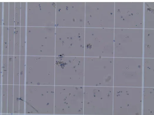
Figure 3.2: Trypan blue exclusion assay with T98G cells drugged with QCT 25 µM + NaB 1 mM visualized at 10X on a hemocytometer using an Olympus BX53 microscope.
Wright Stain
Wright staining was used to visualize the morphological signs of apoptosis in
GBM cells. The technique is more commonly used to stain peripheral blood smears and
bone marrow, but adequately stains most types of cells. Eosin dyes are used in histology
to stain cytoplasmic components such as amino acids and proteins. Methylene blue is a
basic thiazine dye that is able to stain astrocytic nuclei purple. Wright Staining uses a
combination of eosin (red) and methylene blue dyes to give a multi-dimensional picture
of cell morphology (Yue, 2014).
This technique was utilized to visualize the morphological changes that occur
when GBM cells are underoging apoptosis. The most prevalent change that occurs to
apoptotic cells is condensation and fragmentation of the nucleus. One of the greatest
hallmarks in the apoptosis molecular pathway is the caspase activation cascade. After the
apoptosome is formed through the intrinsic mitochondrial apoptosis pathway, caspase-9
activates caspase-3 (Taylor and Ray, 2017). Caspase-3 activation then causes cleavage of
structure. Early apoptosis shows the nucleus forming a crescent shape, followed by
complete nuclear condensation in the later stages of apoptosis. It is important to note that
cell membrane integrity is still in tact. This is a process called karyorrhexis (Ziegler &
Groscurth, 2004).
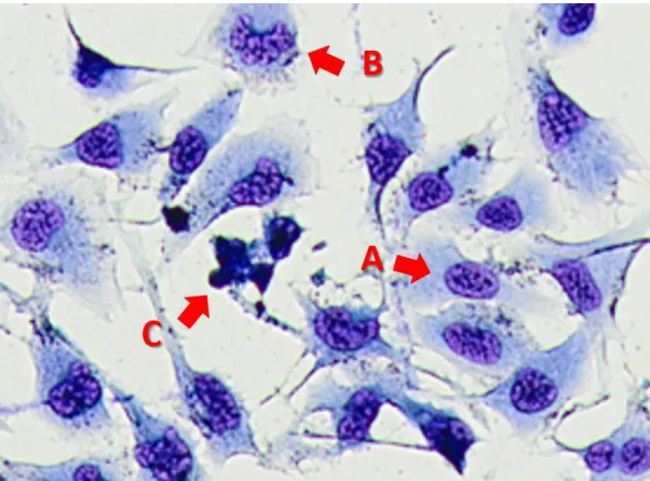
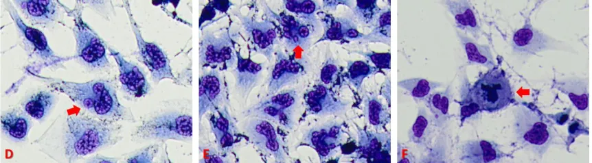
Figure 3.3: Wright staining of T98G GBM cells dosed with QCT 25 µM + NaB 1 mM. (A) A normal, non-apoptotic cell (B) Cell undergoing early apoptosis showing a crescent shaped nucleus (C) Late apoptotic cells showing complete nuclear condensation and cell shrinkage.
Another morphological sign of apoptosis is cellular membrane degredation and
blebbing. Blebbing is a protrusion of the cell membrane caused by degredation of
membrane structural proteins such as actin and β-catenin; along with gelsolin, Gas2, and
PAK2, which help maintain organization and attachment of the cytoskeleton. Due to
membrane degredation, the cell will begin to lose volume during the middle to late
apoptotic stages. Blebbing will occur due to the increased pressure of the cytoplasm
caused by the membrane shrinkage. Decreased membrane integrity will allow
As blebs begin to separate, nuclear fragmentation will start to initiate. These
fragments have lost membrane integrity and will expose intracellular contents to the
external environment. Under most circumstances, these intracellular components will be
phagocytosed; thus, not causing an inflammatory response (Ziegler and Groscurth, 2004).
Phagocytosis is initiated by the externalization of phosphatidylserine, which is a
glycerophospholipid that resides on the internal side of the cellular membrane. Upon
initiation of apoptosis, the enzyme flippase in inactivated which allows for the
externalization of phosphatidylserine (Verhoven et al., 1995). Phosphatidylserine signals
macrophages to phagocytose the apoptotic cells. If apoptotic cells are not phagocytosed
by the host immune system, their degradation patterns will begin to resemble necrosis.
Necrosis is a form of cell death that does not undergo the normal apoptosis
mechanism. It is often initiated by factors such as high heat or a toxic environment.
Morphologically the greatest difference between apoptosis and necrosis is necrotic cells
lose membrane integrity in the very early stages of cell death. Necrotic cells can be
distinguished by massive swelling of the cell, caused by polar molecules passing through
the porous cell membrane. Necrotic cells are often not quantified when measuring
amounts of apoptosis in a sample due to it being a form a cell death not caused by the
desired drug treatments (Ziegler and Groscurth, 2004).
Figure 3.4: Image D shows cell membrane blebbing in T98G GBM cells treated with a 24 hour dose of QCT 25 µM. Image E shows nuclear fragmentation in T98G GBM cells treated with a 24 hour dose of NaB 1 mM. Image F shows necrosis in T98G GBM cells in a 48 hour serum-starved control sample.
Approximately 100,000-300,000 C6, T98G, and LN18 GBM cells were grown in
preparation for wright staining using the previously mentioned experimental protocol in
6-well plates. Upon sample readiness, treated RPMI-1640, 0% fetal bovine serum (FBS),
1% penicillin/streptomycin (P/S) medium is removed by a Pasteur pipette with careful
consideration taken to not aspirate free-floating cells. Immediately after medium
removal, 1 ml of Kwik-DiffTM Solution #1 is added to each well for 25 seconds in order
to fixate the cells to the plates. Solution #1 is removed by a Pasteur pipette and 1 ml
Kwik-DiffTM Solution #2 is added to each well for approximately 20 seconds. Solution
#2 is removed via Pasteur pipette and approximately 0.7 ml Kwik-DiffTM Solution #3 is
added to each well for approximately 20 seconds and then removed with a Pasteur
pipette. Samples are then washed with 1 ml dH2O to remove any excess dye. The
samples are immediately read on an Olympus BX53 microscope at 10X magnification.
Images were taken using an Olympus SC30 camera mounted on the BX53 microscope.
Apoptosis was quantified by manually counting approximately 300 cells, differentiating
between apoptotic and non-apoptotic. Apoptotic cells were characterized by the previous
Percent Apoptosis (%) = [# of Apoptotic Cells ÷ Total # of cells] x 100
These results were then plotted in Microsoft Excel 2016. Each experiment was
performed in triplicate.
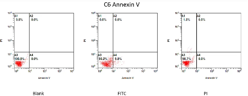
Annexin V
A common and efficient way to quantify apoptosis in a cell population is the
Annexin V test. It utilizes a dual Annexin V-FITC + Propidium Iodide (PI) stain to
determine the physiological state of samples undergoing a form of cell death. Annexin V
is a Ca2+ dependent phospholipid binding protein that has a high affinity to
phosphatidylserine, a cellular membrane phospholipid that becomes externalized in the
early stages of apoptosis. PI is a fluorescent dye that is unable to permeate viable cell
membranes. It is commonly used in flow cytometry of fluorescent microscopy to
evaluate whether a cell is live, apoptotic, or necrotic. A combination of both Annexin
V-FITC and PI paints a clear picture of the stages of cell viability in a sample population
(Hingorani et al., 2011).
A flow cytometer is a powerful tool that is used to measure many different
properties of a cell population. It is based off the premise of shooting a laser at an
individual cell, then analyzing the resulting light scatter patterns. Flow cytometers have
the ability to measure any property of a cell, as long as the fluorescent probes are
conjugated to bind to the desired property. This versatility allows flow cytometers to
measure anything from DNA, RNA, cytoplasmic acidity, membrane permeability,
various proteins, receptors, and many more. In addition, multiple fluorescent probes can
be analyzed simultaneously on the same cell. This allows for precise categorization of
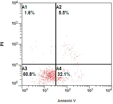
Figure 3.5: This is a flow cytometry graph of C6 cells treated for 24 hours under 48 hour serum starvation with QCT 25 µM + NaB 1 mM. The graph is divided into 4
subpopulations: A1 = AnnexinV-/PI+, A2 = AnnexinV+/PI+, A3 = AnnexinV-/PI-, A4 = Annexin V+/PI-. Each graph represents 10,000 cells.
These four subpopulations display cells in different physiological states.
Quadrant A3 represents AnnexinV-/PI-, which means neither Annexin V or PI dyes
bound to or penetrated the cell. This indicates that phosphatidylserine has not been
externalized and the cell membrane is fully intact; thus, determining that the cells are
fully viable. Quadrant A4 represents AnnexinV+/PI- cells, which means Annexin V is