Research Article
1
Conformation and Domain Movement Analysis of
2
Human Matrix Metalloproteinase-2: Role of Associated
3
Zn
2+and Ca
2+Ions.
4
Leah Voit-Ostricki1, Sándor Lovas2, and Charles R. Watts1,3,*
5
1 Department of Neurosurgery, Mayo Clinic Health System-Franciscan Healthcare in La Crosse, Wisconsin,
6
54601 USA; [email protected]
7
2 Department of Biomedical Sciences, Creighton University, Omaha, Nebraska, USA, 68178;
8
9
3 Department of Neurologic Surgery, Mayo Clinic, Rochester, Minnesota, USA, 55905;
10
11
12
* Correspondence: [email protected]; Tel.: +1-952-993-3200, Fax: +1-952-993-7407
13
Abstract: Matrix Metaloproteinase-2 (MMP-2) is an extracellular Zn2+ protease specific to type I and IV
14
collagens. Its expression is associated with several inflammatory, degenerative, and malignant diseases.
15
Conformational properties, domain movements, and interactions between MMP-2 and its associated
16
metal ions were characterized using a 1.0 s molecular dynamics simulation. Dihedral principle
17
component analysis revealed 10 families of conformations with the greatest degree of variability
18
occurring in the link region connecting the catalytic and hemopexin domains. Dynamics cross
19
correlation analysis indicated domain movements corresponding to opening and closing of the
20
hemopexin domain in relation to the fibronectin and catalytic domains facilitated by the link region.
21
Interaction energies were calculated using the MMPBSA-interaction entropy analysis method and
22
revealed strong binding energies for the catalytic Zn2+ ion 1, Ca2+ ion 1, and Ca2+ ion 3 with significant
23
conformational stability at the binding sites of Zn2+ ion 1 and Ca2+ ion 1. Ca2+ ion 2 diffuses freely away
24
from its crystallographically defined binding site. Zn2+ ion 2 plays a minor role in conformational
25
stability of the catalytic domain while Ca2+ ion 3 is strongly attracted to the highly electronegative
26
sidechains of the Asp residues around the central -sheet core of the hemopexin domain.
27
Keywords: molecular dynamics; matrix metalloproteinase; domain movement; zinc binding protein;
28
calcium binding protein.
29
30
1. Introduction
31
Matrix Metaloproteinase-2 (MMP-2) is a 550 amino acid residue extracellular Zn2+ protease that
32
degrades type I and IV collagens [1,2]. It is related to a family of 24 known endopeptidases with an active
33
site Zn2+ ion. On the basis of evolutionary relationships and the structure of their domains, the family is
34
divided into 4 subfamilies [3-5]. MMP-2 expression is associated with normal physiology as well as
35
several inflammatory, degenerative, and malignant diseases [6-12]. As shown in Figure 1; MMP-2 has
36
three domains, catalytic (Cat), fibronectin (Fib), and hemopexin (Hpx) and five crystallographically
37
assigned divalent cations (two Zn2+ and three Ca2+) [13]. The Cat domain (Tyr110 through Tyr445) is a
38
conserved matrixin fold consisting of five -sheets and three -helices. The Fib domain (Glu217 through
39
Gln393) is inserted within the catalytic domain between the 5-sheet and 2-helix and contains three type
40
II fibronectin fingers consisting of two antiparallel -sheets connected by a short -helix forming a three
41
Preprints (www.preprints.org) | NOT PEER-REVIEWED | Posted: 4 July 2019 doi:10.20944/preprints201907.0085.v1
© 2019 by the author(s). Distributed under a Creative Commons CC BY license.
prong treble hook-like arrangement. This domain and its arrangement may play a crucial role in substrate
42
binding and presentation to the catalytic site. The Hpx domain (Leu461-Cys660) is a four bladed propeller
43
fold that is partially oriented toward the catalytic domain. This domain binds an endogenous inhibitor
44
TIMP-2, however, its role in enzymatic function is unknown. The Hpx and Cat domains are connected
45
by a 16 amino acid proline rich Lnk region (Gly446 through Thr460) which is unresolved in the 1CK7 X-ray
46
crystal structure and has been shown to be highly flexible in other molecular dynamics investigations
47
[14,15].
48
49
Figure 1. The ribbon diagram of the X-ray crystal structure of 1CK7. The pro-peptide (Pro31-Gln109) region
50
is removed and the unresolved link (Asp450-Thr460) connecting the Cat and Hpx domains built with
51
YASARA [16]. Domains, subdomains, and secondary structural features of the catalytic domain are labeled
52
accordingly (blue, -helix; red, -sheet; green, -turn/bend; and aqua, coil) (A). The associated ions are
53
shown as van der Waals radii with Zn2+ pink and Ca2+ yellow. The catalytic Zn2+ ion 1 is bound to His403,
54
Glu404, His407, His413, and catalytic water (B). Zn2+ ion 2 is bound to His178, Asp180, His193, and His206 (C). Ca2+
55
ion 1 is bound to Asp185, Gly186, Asp188, Leu198, Asp208, and Glu211 (D). Ca2+ ion 2 is bound to Glu166, Ala167,
56
Asp168, Gly200, Gly202, and Asp204 (E). Ca 2+ ion 3 is bound to Asp 476, Asp521, Asp569, and Asp618 (F).
57
Table 1. Distances between ions and the interacting MMP-2 residue atoms and associated binding geometry
58
identified from the 1CK7 X-ray crystal structure. Binding geometries were determined for those peptide
59
3 of 19
Protein Atom Geometry
Distance / nm
Zn2+ ion 1 Zn2+ ion 2 Ca2+ ion 1 Ca2+ ion 2 Ca2+ ion 3 His403:N2 tetrahedral 0.23
His407:N2 tetrahedral 0.22
His413:N2 tetrahedral 0.25
Water:O tetrahedral 0.25
Glu404:O1 0.83
Glu404:O2 0.76
His178:N2 trigonal planar 0.21
Asp180:O1 0.35
Asp180:O2 trigonal planar 0.23
His206:N1 trigonal planar 0.21
His193:N2 trigonal planar 0.21
Asp185:C=O octahedral 0.29
Asp185:O1 0.38
Asp185:O2 0.61
Gly186:C=O octahedral 0.24
Asp188:C=O octahedral 0.26
Leu198:C=O octahedral 0.25
Asp208:O1 0.44
Asp208:O2 octahedral 0.25
Glu211:O1 0.49
Glu211:O2 octahedral 0.27
Glu166:O1 0.42
Glu166:O2 pentagonal pyramidal 0.32
Ala167:C=0 pentagonal pyramidal 0.29
Asp168:C=O pentagonal pyramidal 0.29
Gly200:C=O pentagonal pyramidal 0.27
Gly202:C=O pentagonal pyramidal 0.34
Asp204:O1 pentagonal pyramidal 0.35
Asp204:O2 0.40
Asp476:C=O seesaw 0.25
Asp521:C=O seesaw 0.28
Asp569:C=O seesaw 0.27
Asp618:C=O seesaw 0.27
a Zn2+ ion 1 is the catalytic ion.
61
b Glu404 is critical to catalytic activity.
62
c Location and orientation of the catalytic water was derived from the sidechain of Cys102 and the X-ray crystal
63
structure of MMP-13 (PDB ID: 1XUD).
64
Expanded views of the bound divalent cations to MMP-2 residues are shown in Figures 1B through
65
1F with the X-ray crystal structure 1CK7 coordination geometry and interatomic metal cation to MMP-2
66
residue distances given in Table 1 [13,17]. The catalytic Zn2+ ion 1 is bound by the conserved MMP
67
extended zinc binding motif [5]:
68
HExxHxxGxxH⁄D (1)
consisting of the sidechains of His403 and His407 from the 2-helix, and His413 from the -loop (Figure 1B).
69
The structural Zn2+ ion 2 is bound in a tetrahedral arrangement involving the sidechains of His193 and
70
His206 from the 5- and 4-sheets respectively and Asp180 and His178 of the long S-loop of the Cat domain
71
which is a conserved motif in the MMP family (Figure 1C). Two of the Ca2+ ions are bound near the Cat
72
domain with Ca2+ ion 1 bound by the sidechains of Asp208 and Glu211 of the interim loop connecting the
73
Cat and Fib domains and the carbonyl oxygens of Asp185, Gly186, Asp188, and Leu190 of the loop connecting
74
Preprints (www.preprints.org) | NOT PEER-REVIEWED | Posted: 4 July 2019 doi:10.20944/preprints201907.0085.v1
the 3- and 5-sheets (Figure 1D). Ca2+ ion 2 is bound by the sidechains of Glu166 and carbonyl oxygens
75
of Ala167 and Asp168 arising from the loop connecting the 1- and 3-sheets, the carbonyl oxygen of Gly200
76
of the loop connecting 5- and 4-sheets, and the sidechain of Asp204 arising from the 4-sheet, Figure
77
1E. The third structural Ca2+ ion 3, is bound by the carbonyl oxygens of Asp476, Asp521, Asp569, and Asp618
78
at the edge of the central cavity of the Hpx domain (Figure 1F).
79
In the present study we examine the domain movements within MMP-2 and evaluate the structural
80
stability of the bound (2 Zn2+ and 3 Ca2+) divalent ions using 1.0 s NPT MD simulations. Protein-metal
81
cation distances and MMPBSA-interaction entropy binding energies (G) were calculated, metal cation
82
hydration evaluated and the sampled conformational space analyzed with dihedral Principle
83
Component Analysis (dPCA) and Dynamic Cross-Correlation Matrix (DCCM) analysis.
84
2. Results and Discussion
85
2.1 System Equilibration and Conformational Stability.
86
In order to evaluate convergence of the trajectory, the configurational entropy of MMP-2 together
87
with the associated divalent ions was calculated as a function of time (Figure S1) [18-20]. After a sharp
88
rise in the configurational entropy over the first 100 ns, the value plateaus prior to 200 ns. Based on these
89
results, we used the 200 ns to 1000 ns portion of the trajectory for our analysis with a sampling frequency
90
of 0.1 ns. The radius of gyration (Rg) and inter-domain center-of mass (COM) distances: Cat-Hpx and
Fib-91
Hpx, were analyzed with k-means clustering and the associated means and standard deviations of each
92
population calculated (Figures S2 and S3) [21,22]. Five different distributions of protein conformation
93
were identified with Rg: 2.65±0.02 nm, 2.70±0.06 nm, 2.80.±0.05 nm, 2.83±0.04 nm, and 3.09±0.13 nm. The
94
COM distances mirror the Rg results, identifying five distributions of Cat-Hpx COM distances: 3.55±0.10
95
nm, 3.70±0.17 nm, 3.79±0.06 nm, 3.86±0.13 nm, and 4.20±0.23 nm. Five distributions of Fib-Hpx COM
96
distances: 3.70±0.06 nm, 3.84±0.18 nm, 4.32±0.22 nm, 4.48±0.18 nm, and 5.52±0.48 nm, were also identified.
97
The Rg and COM distance data are consistent with inter-domain motions between Cat/Fib and Hpx and
98
the presence of inter-domain motions and the sampling of more extended conformations of MMP-2 in
99
solution compared to the more compact X-ray crystal structure (PDB ID: 1CK7) which has Rg: 2.77 nm,
100
Cat-Hpx COM distance: 3.81 nm, and Fib-Hpx COM distance: 2.00 nm [13-15].
101
Table 2. Average RMSD of the C-trace of MMP-2 as a whole and divided into its individual domains:
102
Cat, Fib, and Hpx with the Lnk region
103
All Cat w/ Fib Fib Cat w/o Fib Hpx Lnk
RMSD / nm 3.20±0.14 3.34±0.02 3.69±0.03 0.50±0.03 0.34±0.04 0.54±0.08
The average C-trace RMSD data for MMP-2 as a whole and divided into its individual domains are
104
given in Table 2. The C-trace RMSF with the >50% sampled secondary structure assigned by the DSSP
105
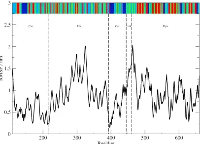
method are shown in Figure 2 [23]. Those regions with defined rigid secondary structure (-sheets and
106
-helices) have lower RMSF values compared to more flexible -turn/bend and coil regions. The greatest
107
degree of conformational variability occurs within the Fib domain while the most stable regions of the
108
protein are within the Cat and Hpx domains. The stability of the Cat and Hpx domains is most likely
109
secondary to the presence of the three long -helices of the Cat domain and the prominent -sheets and
110
ordered arrangement of the Hpx domain. Although ordered secondary structure is present within the
111
Fib domain, it consists of three separate subdomains with significant portion of the fold consisting of
112
flexible -turn/bend and coils. Flexibility within the treble hook arrangement of the Fib domain may play
113
an important role in collagen binding and unraveling [24-27].
5 of 19
115
Figure 2. The C-trace RMSF. The DSSP assigned secondary structure (sampled >50%) is shown at the top
116
of the graph (blue, -helix; red, -sheet; green, -turn/bend; and aqua, coil). The Cat, Fib and Hpx domains
117
and Lnk regions are demarcated with dotted lines.
118
2.2 Conformational Analysis.
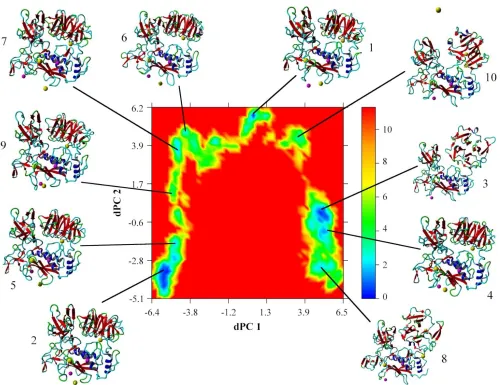
119
The free energy landscape created by the first two dihedral principle components (dPC) is shown in
120
Figure 3 with the corresponding lowest energy centroid conformations as determined by k-means
121
clustering. The most conformationally stable region is the Cat domain in which the active site Zn2+ ion 1
122
is bound by His residues of the 1-helix and the -loop (Figures 3 and 4). The structure around Ca2+ ion
123
1 also is stable with the cation bound within a pocket created by the distal portion of the S-loop and the
124
interim loop connecting the Cat and Fib domains. This stability is also confirmed by the low RMSD and
125
RMSF values for the Cat domain and those of Zn2+ ion 1 and Ca2+ ion1 with both ions staying closely
126
associated with the Cat domain of the protein and approximate to their crystallographically defined
127
positions during simulation (Tables 2 and 3 and Figure 3 and 4). The other associated metal ions of the
128
Cat domain do not share this degree of stability (Table 3). Zn2+ ion 2 remains associated with the S-loop
129
but loses contact with the crystallographically demonstrated interactions with the His residues on the 4-
130
and 5-sheets. This may not be unexpected since conformational flexibility within the S-loop region, as
131
was reported previously [28] and may allow for changes in the binding pocket conformation necessary
132
for substrate recognition. Ca2+ ion 2 does not remain in close contact with the 1-3-loop and diffuses out
133
of the binding pocket (Figures 3 and 4). The Hpx associated Ca2+ ion 3 remains close to its
134
crystallographically observed binding site (Figures 3 and 4) with a relatively low RMSF (Table 3) but to
135
a lower degree that either Zn2+ ion 1 or Ca2+ ion 1.
136
Preprints (www.preprints.org) | NOT PEER-REVIEWED | Posted: 4 July 2019 doi:10.20944/preprints201907.0085.v1
137
Figure 3. Free energy landscape (kJ mol-1) as a function of dPC1 and dPC2; the lowest energy conformations of each family as identified by k-means clustering are shown.
138
7 of 19
140
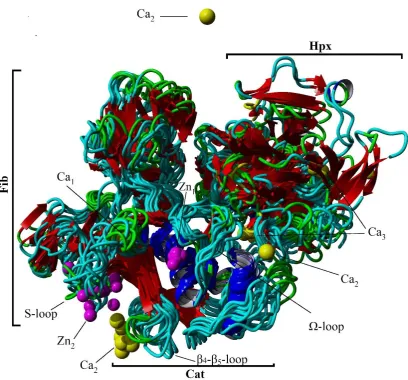
Figure 4. Overlay of 10 cluster centroid structures from the cluster analysis of dPC1 and dPC2.
141
Secondary structural motifs and ions are shown (blue, -helix; red, -sheet; green, -turn/bend; aqua,
142
coil; pink, Zn2+; and yellow, Ca2+).
143
Table 3. Average RMSF of the associated divalent cations to the C-trace of MMP-2
144
Zn2+ ion 1* Zn2+ ion 2 Ca2+ ion 1 Ca2+ ion 2 Ca2+ ion 3
RMSF / nm 0.593 0.882 0.571 2.906 1.062
The majority of the conformational fluctuations within MMP-2 are within the Fib domain
145
(Figures 3 and 4 and Table 2). The three type II Fib subdomains are highly flexible due in part to the
146
large amount of -turn/bend and coil structure within the subdomains. This degree of flexibility may
147
be important for interactions between MMP-2 and its collagen substrates [24-27].There is also clearly
148
an inter-domain interaction that occurs between Hpx and the Cat and Fib domains mediated by the
149
Lnk region. The Lnk region acts as a complex hinge allowing the COM distance between the Cat/Fib
150
and Hpx domains to open and close while changing the orientation of the Hpx domain from an edge
151
view to an end on view (Figure 3).
152
The catalytic Zn2+ ion 1 is hydrated with a single water molecule within 0.5 nm (Table 4). This is
153
consistent with its catalytic function and prior computational studies [29,30]. Ca2+ ion 1 is the most
154
solvent sequestered of the bound divalent cations. The remaining divalent cations (Zn2+ ion 2, Ca2+
155
ion 2 and Ca2+ ion 3) have increased solvent accessibility (Table 4).
156
157
Preprints (www.preprints.org) | NOT PEER-REVIEWED | Posted: 4 July 2019 doi:10.20944/preprints201907.0085.v1
Table 4. Probability () of a water molecule within 0.5 nm of the associated divalent cations.
158
Zn2+ ion 1 Zn2+ ion 2 Ca2+ ion 1 Ca2+ ion 2 Ca2+ ion 3
ρ(g(r)) 0.056 0.071 0.035 0.063 0.067
2.3 Domain Movement Analysis.
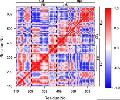
159
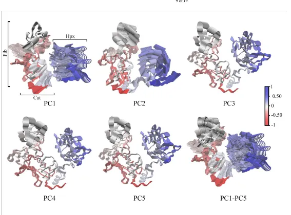
DCCM (Figure 5) demonstrated that motions within Cat, Fib and Hpx domains and Lnk region
160
are for the most part highly correlated with a few exceptions. The first five principle components (PC)
161
account for total of 93.7% of the total domain motions (Figure 6). The plots of the C-trace RMSF of
162
the projection of the trajectory onto the first five eigenvectors are shown (Figure S4). For PC1, the
163
majority of the contributions arises from the first two subdomains of Fib, the Hpx domains and Lnk
164
region. PC2, has significant motions in the third subdomain of Fib and Hpx. There are also
anti-165
correlated motions with the Cat domain involving the 1-helix and 1- through 5-sheets that contain
166
the active site. Motions along PC3 through PC5 represent minor fluctuations within the domains and
167
global conformation. The distal portion of the Cat domain which contains the active site has
168
correlated motions with the first and third subdomains of the Fib domain and the first and second
169
blades of the Hpx domain. There are also anti-correlated motions between the active site on the Cat
170
domain and the second subdomain of the Fib domain and the third and fourth blades of the Hpx
171
domain.
172
173
Figure 5. Dynamic Cross Correlation Matrix. Values range from -1 (complete anti-correlation) to +1
174
(complete correlation).The Cat, Fib, and Hpx domains and Lnk regions are demarcated with dashed
175
9 of 19
177
Figure 6. Cα-trace of PC1 through PC5 and an overlay of all principle components PC1-PC5. Correlated domain movements are indicated in blue and anti-correlated are in
178
red.
179
Preprints (www.preprints.org) | NOT PEER-REVIEWED | Posted: 4 July 2019 doi:10.20944/preprints201907.0085.v1
2.4 Protein-Metal Ion Interaction Energies.
180
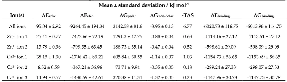
MMP-2 has a high affinity for the bound divalent cations (Table 5) with major contribution from
181
the electrostatic interactions. The solvation term Gpolar is unfavorable particularly for the catalytic
182
Zn2+ ion 1, the structural Ca2+ ion 1, and Ca2+ ion 3 and the Gnon-polar is only weakly favorable. For the
183
weakly bound Zn2+ ion 2 and the freely diffusing Ca2+ ion 2, the Gpolar is significantly smaller. The
184
entropic contributions to MMP-2 divalent cation interaction energies are very small but positive
185
indicating decreased system entropy with binding of the metal ions to the peptide. The entropic term
186
is lowest for those ions that for those metal ions (Ca2+ ion 2 and Ca2+ ion 3) that either diffuse freely
187
away from their crystallographically determined binding sites or do not form close associations and
188
stable binding geometries with the electronegative backbone and sidechain atoms of MMP-2 [31].
189
Table 5. Binding energies between MMP-2 and the associated divalent cations as determined by the
190
MMPBSA and interaction entropy methods with its associated components.
191
Mean ± standard deviation / kJ mol-1
Ion(s) Evdw Eelec Gpolar Gnon-polar -TS Ebinding Gbinding
All ions 95.04 ± 2.92 -9264.45 ± 194.34 3142.58 ± 81.6 -3.95 ± 0.13 6.77 -6020.73 ± 116.75 -6013.96 ± 116.75 Zn2+ ion 1 25.41 ± 0.77 -2427.66 ± 72.19 1291.3 ± 42.75 -0.88 ± 0.04 0.63 -1114.16 ± 27.12 -1113.51 ± 27.12
Zn2+ ion 2 13.79 ± 0.96 -799.35 ± 63.45 188.73 ± 35.14 -0.47 ± 0.04 0.52 -598.61 ± 29.09 -598.09 ± 29.09
Ca2+ ion 1 38.15 ± 1.90 -1796.42 ± 89.21 605.84 ± 30.55 -1.14 ± 0.07 1.03 -1154.73 ± 56.65 -1153.69 ± 56.65
Ca2+ ion 2 6.52 ± 0.58 -367.21 ± 36.96 73.71 ± 9.94 -0.35 ± 0.05 0.18 -289.24 ± 27.33 -298.07 ± 27.33
Ca2+ ion 3 14.94 ± 0.57 -1480.59 ± 42.61 320.38 ± 11.31 -1.32 ± 0.05 0.23 -1147.96 ± 30.78 -1147.73 ± 30.78
Residue contribution to the binding energy with their associated interatomic distances and
192
geometries are given in Table 6. The catalytic Zn2+ ion1 maintains interactions with His403, His407, and
193
His413 similar to what is observed in the x-ray crystal structure. The Glu404 sidechain O atoms are in
194
closer proximity than what is observed in the crystal structure. The binding geometry for the bound
195
His residues is trigonal pyramidal however, if a coordinated hydration water is considered, this
196
geometry would be tetrahedral. Other important interactions are also noted between Zn2+ ion 1 and
197
Asp and Glu residues within the Cat domain. These residues contribute significantly to MMP-2 to
198
Zn2+ ion 1 interaction energy despite being outside what is considered to be the normal coordination
199
sphere of the ion. Zn2+ ion 2 loses contact with His178, His193, and His206 shifting to a more linear
200
coordination geometry that is depended on a strong interaction with the O atoms of Asp180.
201
Ca2+ ion 1 has strong interactions with the adjacent O atoms of Asp and O atoms of Glu of
202
adjacent residues. There is a shift from the divalent cation to backbone carbonyl oxygen interactions
203
that are observed in the crystal structure to interactions dominated by the acidic sidechain groups.
204
The coordination geometry changes from pentagonal pyramidal to a seesaw geometry. The strong
205
interaction and coordination with these sidechains is expected and has been previously observed for
206
other systems [32]. In general the favorability of interaction is Glu>Asp with the difference attributed
207
to increased flexibility of the Glu residue secondary to presence of the extra methyl group. The
208
binding of this ion is also similar to that of the catalytic Zn2+ ion 1 in that adjacent but non-coordinated
209
electronegative Asp and Glu residues make significant contributions to its binding energy. Ca2+ ion 2
210
freely diffuses out of its binding site and although it has strong interactions with several electrostatic
211
sidechains, these interactions are mostly transient. Ca2+ ion 3 is more stable in its RMSF compared to
212
Ca2+ ion 2 (Table 3) however, it is still much more variable that the other associated ions. The
213
interaction energies and geometry are again dominated by the electronegative sidechains of Asp
214
residues surrounding the central core of the Hpx domain.
11 of 19
Table 6. Distances of ions to the interacting protein residue atoms identified from the 1CK7 X-ray
217
crystal. Statistically significant protein residue atoms as identified by outlier analysis with the
218
associated per residue interaction energies and binding geometry. 1CK7 identified interactions are
219
marked with a dagger (†), statistically significant interactions are marked with an asterisk (*), and
220
binding geometries were determined for those atoms within 0.35 nm. Distance are given in nm and
221
energies in kJ mol-1.a,b
222
Protein Atom Ebinding Geometry Zn2+ Ion 1 Zn2+ ion 2 Ca2+ ion 1 Ca2+ ion 2 Ca2+ ion 3
†*Glu404:O1 -154.8223 0.46 ± 0.05
†*Glu404:O2 0.48 ± 0.06
*Glu412:O1 -88.9662 0.96 ± 0.13
*Glu412:O2 0.96 ± 0.13
*Asp188:O1 -73.8901 1.46 ± 0.18
*Asp188:O2 1.47 ± 0.18
†*His403:N2 -72.1511 trigonal
pyramidal 0.21 ± 0.01 *Asp437:O1 -67.5468 1.29 ± 0.11
*Asp437:O2 1.29 ± 0.11
*Asp185:O1 -66.7783 1.65 ± 0.20
*Asp185:O2 1.68 ± 0.16
†*His407:N2 -66.3450 trigonal
pyramidal 0.21 ± 0.01 *Asp208:O1 -65.6438 1.60 ± 0.12
*Asp208:O2 1.49 ±0.09
*Glu211:O1 -64.0824 1.57 ± 0.13
*Glu211:O2 1.58 ± 0.11
†*His413:N2 -61.5998 trigonal
pyramidal 0.21 ± 0.01 *Asp416:O1 -61.3612 1.52 ± 0.05
*Asp416:O2 1.52 ± 0.05
†*Asp180:O1 -92.3424 linear 0.19 ± 0.01
†*Asp180:O2 linear 0.19 ± 0.01
*Glu177:O1 -42.684 0.71 ± 0.27
*Glu177:O2 0.71 ± 0.27
†His178:N2 -1.1803 1.13 ± 0.17
†His206:N1 -1.0497 1.00 ± 0.42
†His193:N2 -0.2562 1.34 ± 0.51
†*Asp185:C=O -167.499 0.70 ± 0.09
†*Asp185:O1 seesaw 0.29 ± 0.16
†*Asp185:O2 seesaw 0.28 ± 0.16
†*Glu211:O1 -147.6924 seesaw 0.33 ± 0.11
†*Glu211:O2 seesaw 0.35 ± 0.10
†*Asp208:O1 -142.7074 seesaw 0.34 ± 0.09
†*Asp208:O2 seesaw 0.24 ± 0.02
*Asp210:O1 -112.8532 seesaw 0.33 ± 0.14
*Asp210:O2 seesaw 0.35 ± 0.14
*Asp209:O1 -61.7053 0.77 ± 0.16
*Asp209:O2 0.76 ± 0.14
†*Asp188:C=O -57.9066 0.98 ± 0.14
*Asp188:O1 1.27 ± 0.19
*Asp188:O2 1.27 ± 0.19
*Glu404:O1 -50.3849 1.37 ± 0.14
*Glu404:O2 1.38 ± 0.15
*Asp134:O1 -49.6054 1.19 ± 0.13
*Asp134:O2 1.19 ± 0.14
*Asp392:O1 -49.3403 1.37 ± 0.17
*Asp392:O2 1.36 ± 0.17
*Asp180:O1 -46.489 1.50 ± 0.25
*Asp180:O2 1.51 ± 0.25
†Gly186:C=O -7.1579 0.75 ± 0.17
†Leu198:C=O -0.4421 2.68 ± 0.17
a Zn2+ ion 1 is the catalytic ion.
223
b Glu404 is critical to catalytic activity.
224
Preprints (www.preprints.org) | NOT PEER-REVIEWED | Posted: 4 July 2019 doi:10.20944/preprints201907.0085.v1
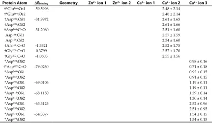
Table 6 Continued. Distances of ions to the interacting protein residue atoms identified from the
225
1CK7 X-ray crystal. Statistically significant protein residue atoms as identified by outlier analysis
226
with the associated per residue interaction energies and binding geometry. 1CK7 identified
227
interactions are marked with a dagger (†), statistically significant interactions are marked with an
228
asterisk (*), and binding geometries were determined for those atoms within 0.35 nm. Distance are
229
given in nm and energies in kJ mol-1.a,b
230
Protein Atom Ebinding Geometry Zn2+ ion 1 Zn2+ ion 2 Ca2+ ion 1 Ca2+ ion 2 Ca2+ ion 3
†*Glu166:O1 -59.5996 2.48 ± 2.14
†*Glu166:O2 2.48 ± 2.14
†Asp204:O1 -31.9972 2.61 ± 1.65
†Asp204:O2 2.61 ± 1.66
†Asp168:C=O -31.2060 2.51 ± 1.60
Asp168:O1 2.57 ± 1.59
Asp168:O2 2.54 ± 1.60
†Ala167:C=O -1.3321 2.52 ± 1.75
†Gly200:C=O 0.3799 2.57 ± 1.70
†Gly202:C=O -1.0605 2.55 ± 1.56
*Asp521:O2 0.98 ± 0.16
†*Asp569:C=O -79.0260 0.71 ± 0.18
*Asp569:O1 0.92 ± 0.15
*Asp569:O2 0.91 ± 0.15
*Asp490:O1 -69.0106 1.19 ± 0.11
*Asp490:O2 1.19 ± 0.11
*Asp615:O1 -68.1150 1.29 ± 0.14
*Asp615:O2 1.30 ± 0.14
*Asp153:O1 -63.3125 2.52 ± 0.96
*Asp153:O2 2.51 ± 0.95
*Asp472:O1 -54.3377 1.54 ± 0.15
*Asp472:O2 1.54 ± 0.15
a Zn2+ ion 1 is the catalytic ion.
231
b Glu404 is critical to catalytic activity.
232
3. Materials and Methods
233
3.1 Matrix Metalloprotease-2 Starting Conformation.
234
Initial coordinates were obtained from the X-ray l structure of the Glu404 to Ala404 mutant of the
235
human MMP-2 (PDB ID: 1CK7) [13]. Crystallographically resolved waters and sulfate ions were
236
removed while the protein bound Zn2+, Ca2+, Na+, and Cl- ions where retained. Residues 31-109 were
237
removed as in the biologically active form of the enzyme. The crystallography non-resolved loop
238
from residues Asp450-Thr460 was build using the homology modelling script of YASARA [16]. The
239
coordinates for the sidechain of Glu404 and the Zn2+ coordinated water molecule that replaces Cys102
240
at the enzyme active site were derived from the MMP-13 crystal structure (PDB ID: 1XUD) by a least
241
squares fitting of the backbone and Zn2+ atoms of both x-ray structures [33]. Sidechain protonation
242
states of the Zn2+ associated His residues were assigned based on the 1CK7 crystal structure as
243
follows: HND1 for His178, His403, His193, His407, His413, and HNE2 for His206. The remaining Histidine
244
residues (His163, His276, and His628) were assigned automatically with HNE2 atoms using the pdb2gmx
245
module of GROMACS version 5.1.2 [34,35]. The protonation state and charges of all other residues
246
within the protein were set to correspond to a pH of 7.0.
247
4.2 Molecular Dynamics.
248
Simulations were performed using the CHARMM36m force field with modified TIP3Pm water
249
model and the CM model of divalent metal cation parameters of Li et al. as implemented in
250
GROMACS version 5.1.2 [Zn2+ ( = 0.226466454151 nm, = 0.01381916624 kJ•mol-1) and Ca2+ ( =
251
13 of 19
hydration free energy by optimizing the ion-oxygen distance in the first solvation shell. The metal
253
ions are represented with a standard Lennard-Jones and coulomb potential energy function [42]:
254
𝑬𝒊𝒐𝒏= ∑ 𝟒𝜺𝒊𝒋 𝑵
𝒋
[(𝝈𝒊𝒋 𝒓𝒊𝒋 )
𝟏𝟐 − (𝝈𝒊𝒋
𝒓𝒊𝒋 )
𝟔
] + ∑ 𝒒𝒊𝒒𝒋
𝟒𝝅𝜺𝟎𝒓𝒊𝒋 𝑵
𝒋 (2)
where,
255
𝝈𝒊𝒋=
(𝝈𝒊+ 𝝈𝒋) 𝟐
(3)
and
256
𝜺𝒊𝒋= √(𝜺𝒊𝟐 • 𝜺𝒋) (4)
and i and j are atom indices. The system was solvated in a truncated dodecahedron with 45105
257
TIP3Pm water molecules. The minimal distance of the protein to the edge of the dodecahedron was
258
1.4 nm. The system was neutralized with 141 and 132, Na+ and Cl- ions, respectively, so that the final
259
concentration of the NaCl was set to 150 mM; the initially retained protein bound Zn2+, Ca2+, Na+, and
260
Cl- ions are not included. . The system was subjected to 5000 steps of steepest descent energy
261
minimization allowing all bond distances and angles to relax. This was followed with 10 ns of NVT
262
simulation at 310 K so that the position of the protein heavy atoms and retained Zn2+, Ca2+, Na+, and
263
Cl- ions and catalytic Zn2+ associated water were constrained to their energy -minimized coordinates
264
with force constant of 1000 kJ•mol-1. The solvent and non-restrained Na+ and Cl- ions were then
265
subjected to 10 ns of NPT simulation at 310 K and 101.325 kPa using Berendsen temperature and
266
pressure scaling with a relaxation constant of 0.1 ps and 4.5 × 10⁻⁵ bar-1 isothermal compressibility
267
[43]. The heavy atom restraints were then removed and the entire system was subjected to 10 ns of
268
NPT at 310 K and 101.325 kPa using the stochastic velocity-rescaling method of Bussi and Berendsen
269
pressure coupling with a relaxation constant of 1.0 ps and 4.5 × 10⁻⁵ bar-1 isothermal compressibility
270
[44]. The production run consisted of 1.0 s NPT simulation at 310 K and 101.325 kPa. The integration
271
step was 2 fs, the LINCS algorithm was used to constrain all bonds to their correct length, with a
272
warning angle of 30° [45,46]. The long-range electrostatic interactions were calculated using the PME
273
method with 1.2 nm cutoff distance and 0.15 nm Fourier spacing [47]. For the calculations of Van der
274
Waals interactions the short-range and long-range cutoff, respectively, was 1.0 and 1.2 nm. The
275
protein and solvent with ions were separately coupled to a 1 bar Parrinello-Rahman barostat and the
276
temperature was maintained by the velocity-rescaling method [44,48].
277
4.3 Biophysical Properties
278
The C-trace root mean square deviation (RMSD) and per-residue root mean square fluctuation
279
(RMSF) from the average sampled peptide conformation were calculated with the g_rmsd and g_rmsf
280
utilities of GROMACS, respectively [35]. The >50% sampled per-residue DSSP assigned [23]
281
secondary structure (-helix, -sheet, -bend/turn, and coil) were determined using the do_dssp utility
282
of GROMACS and an in house perl script to calculate sampling statistics. The hydration of the Zn2+
283
and Ca2+ was determined by calculating the radial distribution function for the oxygen atom of the
284
surrounding water molecules using the g_rdf utility in GROMACS [35]. The integral of the radial
285
distribution function is equal to the probability of finding water molecules within the defined radius
286
(0.5 nm for the first hydration shell) [49,50].
287
Preprints (www.preprints.org) | NOT PEER-REVIEWED | Posted: 4 July 2019 doi:10.20944/preprints201907.0085.v1
4.4 Conformational Analysis.
288
The time-dependent dihedral angles from residues 111 to 659 of the protein were extracted
289
from the trajectories using the g_rama utility of GROMACS. An in-house python script was used to
290
transform the data for the input to the dPCA program provided by Dr. Yuguang Mu [51,52]. Lowest
291
energy conformations were identified by projecting the trajectories of the first two principal
292
components (dPC1 and dPC2) onto a two-dimensional free energy (G) landscape:
293
𝜟𝑮 = −𝑹 ⋅ 𝑻 ⋅ 𝒍𝒏 𝝆𝒙,𝒚
𝝆𝒎𝒂𝒙
(5)
where R is the universal gas constant, T is the temperature, x and y are the first two dihedral
294
principal components from the trajectory. The free energy (G) landscape was calculated by
295
dividing the dPC1-dPC2 subspace into grids creating a 2D histogram of the sampled phase space
296
and calculating the probability x,y where max corresponds to the grid with the maximum
297
probability of occurrence. Results were visualized using the scatterplot3D, akima, and latticeExtra
298
packages in the R software environment with conformations and secondary structural elements
299
rendered using YASARA [16,21,53-55]. Families of low energy conformations were identified using
300
k-means clustering as implemented in the cluster package in R and the identified lowest energy
301
conformations extracted for further analysis [21,22]. The optimal number of clusters was
302
determined by visual inspection, sum of squared error (SSE), average silhouette width (SAVG),
303
silhouette coefficient (SC) and distribution plots [56-59].
304
4.5 Dynamic Cross-Correlation Matrix.
305
Correlated motions within and between domains were evaluated by calculating dynamic cross
306
correlation matrices (DCCM) from the principal components of the C-trace covariance matrix
307
using the GeoStaS method as implemented in the Bio3D package written in R [60-62]. The results are
308
displayed as a color coded matrix of Pearson correlation coefficients with a value of -1 indicated
309
completely anti-correlated motions and a value of +1 indicating completely correlated motions
310
[63,64].
311
4.6 Interaction Energy.
312
The free energy of binding between the metal cations and protein was calculated using the
313
g_mmpbsa program [65]. The polar component of the solvation energy was calculated using the
314
Poisson-Boltzmann equation and non-polar component calculated from the solvent-accessible
315
surface area approximation [66,67]. Dielectric constants for the solute and water were 4 and 80,
316
respectively. The entropic contribution to the binding energy was determined using the interaction
317
entropy method of Zhang and coworkers [31,68]. The trajectory was sampled every 0.1 ns for the
318
equilibrium phase (200 ns to 1000 ns). A bootstrap analysis (n = 5000) was performed to obtain
319
standard errors and the residue contributions to the binding energy were also calculated.
320
The residue contributions to binding were deconvoluted. To determine the most significant
321
residue interactions between MMP-2 and the divalent cations, an outlier analysis was performed to
322
identify statistically significant interactions. The distribution of interaction energies were not
323
Gaussian (normal). In the setting of non-normal distributions, the method of Tukey’s fences can be
324
used to identify those observations that are outside of the expected fluctuations within the data [69].
325
Tukey’s fences defines minimum and maximum values of the interaction energy:
326
[𝑀𝑖𝑛𝑖𝑚𝑢𝑚𝑉𝑎𝑙𝑢𝑒, 𝑀𝑎𝑥𝑖𝑚𝑢𝑚𝑉𝑎𝑙𝑢𝑒] (6)
such that measurements less than or equal to the minimum value or greater than or equal to the
327
maximum value are considered statistical outliers. The respective minimum and maximum values of
328
the fences, are defines as:
15 of 19
[𝑄1− 𝑘(𝑄3− 𝑄1), 𝑄3+ 𝑘(𝑄3− 𝑄1)] (7)
where Q1, Q3 are the interquartile values, and k is the constant that defines the outlier range (k = 1.5
330
is an outlier, k = 3.0 is an extreme outlier) [69].
331
5. Conclusions
332
Here, we report a microsecond scale molecular dynamics analysis of the full biologically active
333
protein (Cat, Fib, and Hpx domains with Lnk region) with its crystallographically associated
334
(structural) divalent metal ions (Zn2+ and Ca2+). So far, most of the simulations were done on the
335
truncated structure of MMP-2 protein [28-30]. Diaz et al. had previously investigated the role of the
336
structural metal ions using a truncated version of the protein with the AMBER force field, a
non-337
bonded model of Ca2+ and a bonded tetrahedral model of Zn2+ using 10 ns simulations [29]. Their
338
results are in agreement with ours: the Zn2+ in 1 (catalytic ion) and Ca2+ ion 1 demonstrate the lowest
339
RMSFs and are tightly bound within their crystallographically defined pockets. Zn2+ ion 2 was more
340
flexible and associated with the S-loop which has demonstrated increased flexibility in both prior
341
simulations and our own however, the use of short simulation time (10 ns) and a bonded potential
342
representing the interaction between the Zn2+ ion and the associated His and Asp/Glu residues may
343
have artificially stabilized the protein-Zn2+ ion 2 interaction. This is an important point since prior
344
investigators have identified variations in the stoichiometry of the MMP-2-Zn2+ interaction that are
345
strongly dependent on the purification procedure used [70]. The current non-bonded model of Li et
346
al. with the CHARMM36m force field appears to be a reasonable model of protein metal cation
347
interactions and is in agreement with prior studies [71,72].
348
Dihedral principle component analysis demonstrates that only minor structural fluctuations
349
occur within the Cat domain. There is increased fluctuations within the Fib domain however, this is
350
expected given the large number of -turn/loop structures connecting the individual subdomains.
351
The three lowest energy populations of conformations from differ predominantly in the orientation
352
of the Hpx domain in relation to the Cat and Fib domains. This is confirmed by the DCCM analysis
353
where the difference in orientation of the Hpx to the Cat and Fib domains comprises the first two
354
principle components. These inter domain movements are facilitated by the flexible linker region
355
Gly446 through Asp476 and may play and important role in collagen substrate binding, unravelling and
356
subsequent catalysis [24-27].
357
Supplementary Materials: Supplementary materials can be found at www.mdpi.com/xxx/s1.
358
Author Contributions: conceptualization, C.R.W and S.L.; methodology, C.R.W and S.L.; software, C.R.W.,
L.V.-359
O., and S.L.; validation, C.R.W. and L.V.-O.; formal analysis, C.R.W. and L.V.-O.; investigation, L.V.-O.;
360
resources, C.R.W.; data curation, L.V.-O.; writing—original draft preparation, C.R.W.; writing—review and
361
editing, C.R.W, L.V.-O., and S.L.; visualization, L.V.-O.; supervision, C.R.W. and S.L. ; project administration,
362
C.R.W.; funding acquisition, C.R.W.
363
Funding: Funding for this investigation was provided in part through a grant from Mayo Clinic Health
System-364
Franciscan Healthcare Foundation Inc. and Mayo Foundation for Medical Education and Research.
365
Acknowledgments: The molecular dynamics simulations and subsequent analysis were completed using the
366
High Performance Cluster, Resource Computing Services, Mayo Clinic, Rochester, Minnesota.
367
Conflicts of Interest: Dr. Charles R. Watts is a consultant for Medtronic Spine and Biologics. The remaining
368
authors have disclosed that they do not have any conflicts of interest. The funders had no role in the design of
369
the study; in the collection, analyses, or interpretation of data; in the writing of the manuscript, or in the decision
370
to publish the results”.
371
Abbreviations
372
dPCA Dihedral Principle Component Analysis DCCM Domain Cross-Correlation Matrix MD Molecular Dynamics
Preprints (www.preprints.org) | NOT PEER-REVIEWED | Posted: 4 July 2019 doi:10.20944/preprints201907.0085.v1
MMP-2 Matrix Metalloproteinase-2 PDB Protein Databank
RMSD Root Mean Square Deviation RMSF Root Mean Square Fluctuation SASA Solvent Exposed surface Area
References
373
1. Basbaum, C.B.; Werb, Z. Focalized proteolysis: spatial and temporal regulation of extracellular matrix
374
degradation at the cell surface. Curr Opin Cell Biol. 1996, 8(5), 731-738, DOI: 10.1016/S0955-0674(96)80116-5.
375
2. Birkedal-Hansen, H.; Moore, W.G.; Bodden, M.K.; Windsor, L.J.; Birkedal-Hansen, B.; DeCarlo, A.; Engler,
376
J.A. Matrix metalloproteinases: a review. Crit Rev Oral Biol Med. 1993, 4(2), 197-250, DOI:
377
10.1177/10454411930040020401.
378
3. Maskos, K. Crystal structure of MMPs in complex with physiological and pharmacological inhibitors.
379
Biochimie. 2005, 87(3-4), 249-263, DOI: 10.1016/j.biochi.2004.11.019.
380
4. Ra, H.-J.; Parks, W.C. Control of Matrix Metalloproteinase Catalytic Activity. Matrix Biol. 2007, 26(8),
587-381
596, DOI: 10.1016/j.matbio.2007.07.001.
382
5. Verma, R.P.; Hansch C. Matrix metalloproteinases (MMPs): chemical-biological functions and (Q)SARs.
383
Bioorg. Med. Chem. 2007, 15 (6), 2223–68. doi:10.1016/j.bmc.2007.01.011. PMID 17275314.
384
6. van Meurs, J.; van Lent, P.; Holthuysen, A.; Lambrou, D.; Bayne, E.; Singer, I.; van den Berg, W. Active
385
matrix metalloproteinases are present in cartilage during immune complex mediated arthritis: a pivotal
386
role for stromelysin-1 in cartilage destruction. J Immunol. 1999; 163(10), 5633-5639,
387
http://www.jimmunol.org/content/163/10/5633.
388
7. Nagase, H.; Woessner, J.F. Jr. Matrix metalloproteinases. J Biol Chem.1999; 274(31), 21491-21494, DOI:
389
10.1074/jbc.274.31.21491.
390
8. Steffensen, B.; Häkkinen, L.; Larjava, H. Proteolytic events of wound-healing-coordinated interactions
391
among matrix metalloproteinases (MMPs), integrins, and extracellular matrix molecules. Crit Rev Oral Biol
392
Med. 2001, 12(5), 373-398, DOI: 10.1177/10454411010120050201.
393
9. Egeblad. M; Werb, Z. New functions for the matrix metalloproteinases in cancer progression. Nat Rev
394
Cancer. 2002; 2(3), 163-176, DOI: 10.1038/nrc745.
395
10. Fingleton, B. Matrix metalloproteinases: roles in cancer and metastasis. Front Biosci. 2006, 11, 479-491, DOI:
396
10.2741/1811.
397
11. Butler, G.S.; Overall, C.M. Updated biological roles for matrix metalloproteinases and new "intracellular"
398
substrates revealed by degradomics. Biochemistry. 2009, 48(46), 10830-10845, DOI: 10.1021/bi901656f.
399
12. Rodríguez, D.; Morrison, C.J.; Overall, C.M. Matrix metalloproteinases: what do they not do? New
400
substrates and biological roles identified by murine models and proteomics. Biochim Biophys Acta.2010,
401
1803(1), 39-54, DOI: 10.1016/j.bbamcr.2009.09.015.
402
13. Morgunov, E.; Tuuttila, A.; Bergmann, U.; Isupov, M.; Lindqvist, Y.; Schneider, G.; Tryggvason, K.;
403
Structure of Human Pro-Matrix Metalloproteinase-2: Activation Mechanism Revealed. Science. 1999,
404
284(5420), 1667-1670, DOI: 10.1126/science.284.5420.1667.
405
14. Díaz, N.; Suárez, D.; Valdés, H. From the X-ray Compact Structure to the Elongated form of the Full-Length
406
MMP-2 Enzyme in Solution: A Molecular Dynamics Study. J Am Chem Soc. 2008, 130(43), 14070-14071, DOI:
407
10.1021/ja806090v.
408
15. Díaz. N.; Suárez, D. Alternative Interdomain Configurations of the Full-Length MMP-2 Enzyme Explored
409
by Molecular Dynamics Simulations. J Phys Chem B. 2012, 116(9), 2677-2686, DOI: 10.1021/jp211088d.
410
16. Krieger, E.; Vriend, G. YASARA View - molecular graphics for all devices - from smartphones to
411
workstations. Bioinformatics. 2011, 30(20), 2981-2982., DOI: 10.1093/bioinformatics/btu426.
412
17. Petrucci, R.H.; Herring, F.G.; Madura, J.D.; Bissonnette, C. General Chemistry: Principles and Modern
413
Applications, 11th ed.; Pearson: Don Mills, Ontario, Canada, 2017; p 411-465, ISBN-13: 978-0132931281.
414
18. Amadei, A.; Ceruso, M.A.; Di Nola, A. On the convergence of the conformational coordinates basis set
415
obtained by the essential dynamics analysis of proteins’ molecular dynamics simulations. Proteins.1999,
416
36(4), 419-424, DOI: 10.1002/(SICI)1097-0134(19990901)36:4<419::AID-PROT5>3.0.CO;2-U.
417
19. Hayward, S.; de Groot, B.L. Normal modes and essential dynamics. In: Methods in Molecular Biology:
418
Molecular modeling of proteins, Kukol, A., Ed.; Humana Press, New York City, New York, USA, 2008; Volume
419
17 of 19
20. Andricioaei, I.; Karplus, M. On the calculation of entropy from covariance matrices of the atomic
421
fluctuations. J Chem Phys. 2001, 115(14): 6289–6292, DOI: 10.1063/1.1401821.
422
21. R Core Team. R: A language and environment for statistical computing. Available online:
http://www.R-423
project.org/ (accessed on 2 Jan 2019).
424
22. Maechler, M.; Rousseeuw, P.; Struyf, A.; Hubert, M.; Hornik, K.; Studer, M.; Roudier, P.; Gonzalez, J.;
425
Kozlowski, K. cluster: methods for cluster analysis. Available online:
https://cran.r-426
project.org/web/packages/cluster/ (accessed on 2 Jan 2019).
427
23. Kabsch, W.; Sander, C. Dictionary of protein secondary structure: pattern recognition of hydrogen-bonded
428
and geometrical features. Biopolymers. 1983, 22(12), 2577-2637, DOI: 10.1002/bip.360221211.
429
24. Steffensen, B.; Wallon, U.M.; Overall, C.M. Extracellular matrix binding properties of recombinant
430
fibronectin type II-like modules of human 72-kDa gelatinase/type IV collagenase. High affinity binding to
431
native type I collagen but not native type IV collagen. J Biol Chem. 1995, 270(19), 11555−11566, DOI:
432
10.1074/jbc.270.19.11555.
433
25. Gehrmann, M.L.; Douglas, J.T.; Bányai, L.; Hedvig, T.; Patthy, L.; Llinás, M.; Modular autonomy, ligand
434
specificity, and functional cooperativity of the three in-tandem fibronectin type II repeats from human
435
matrix metalloproteinase 2. J Biol Chem. 2004, 279(45), 46921−46929, DOI: 10.1074/jbc.M408859200.
436
26. Xu, X.; Wang, Y.; Lauer-Fields, J.L.; Fields, G.B.; Steffensen, B. Contributions of the MMP-2 collagen binding
437
domain to gelatin cleavage. Substrate binding via the collagen binding domain is required for hydrolysis
438
of gelatin but not short peptides. Matrix Biol. 2004; 23(3), 171−181, DOI: 10.1016/j.matbio.2004.05.002.
439
27. Xu, X.; Mikhailova, M.; Llangovan, U.; Chen, Z.; Yu, A.; Pal, S.; Hinck, A.P.; Steffensen, B. Nuclear magnetic
440
resonance mapping and functional confirmation of the collagen binding sites of matrix
metalloproteinase-441
2. Biochem. 2009, 48(25), 5822−5831, DOI: 10.1021/bi900513h.
442
28. Díaz N, Suárez D, Molecular dynamics simulations of the active matrix metalloproteinase-2: Positioning of
443
the N-terminal fragment and binding of a small peptide substrate. Proteins. 2008, 72(1), 50-61, DOI:
444
10.1002/prot.21894.
445
29. Díaz, N.; Suárez, D. Molecular Dynamics Simulations of Matrix Metalloproteinase 2: Role of the Structural
446
Metal Ions. Biochem. 2007, 46(31), 8943-8952, DOI: 10.1021/bi700541p.
447
30. Díaz, N.; Suárez, D. Peptide Hydrolysis Catalyzed by Matrix Metalloproteinase 2: A computational Study.
448
J Phys Chem B. 2008, 112(28), 8412-8424, DOI: 10.1021/jp803509h.
449
31. Duan, L.; Liu, X.; Zhang, J.Z.H. Interaction Entropy: A New Paradigm for Highly Efficient and Reliable
450
Computation of Protein-Ligand Binding Free Energy, J Am Chem Soc. 2016, 138(17), 5722-5728, DOI:
451
10.1021/jacs.6b02682.
452
32. Cates, M.S.; Teodoro, M.L.; Phillips, G.N. Jr., Molecular Mechanisms of Calcium and Magnesium Binding
453
to Parvalbumin. Biophys J. 2002, 82(3), 1133-1146, DOI: 10.1016/S0006-3495(02)75472-6.
454
33. Engel, C.K.; Pirard, B.; Schimanski, S.; Kirsch, R.; Habermann, J.; Klingler, O.; Schlotte, V.; Weithmann,
455
K.U.; Wendt, K.U. Structural basis for the highly selective inhibition of MMP-13. Chem.Biol. 2005, 12(2),
181-456
189, DOI: 10.1016/j.chembiol.2004.11.014
457
34. Abraham, M.J.; Murtola, T.; Schulz, R.; Páll, S.; Smith, J.C.; Hess, B.; Lindahl, E. GROMACS: High
458
performance molecular simulations through multi-level parallelism from laptops to supercomputers.
459
SoftwareX. 2015, 1-2, 19-25, DOI: 10.1016/j.softx.2015.06.001
460
35. Abraham, M.J.; van der Spoel, D.; Lindahl, E.; Hess, B. and the GROMACS development team, GROMACS
461
User Manual ver. 5.1.2 2016, http://www.gromacs.org, (accessed Jan 2, 2019).
462
36. Huang, J.; Rauscher, S.; Nawrocki, G.; Ran, T.; Feig, M.; de Groot, B.L.; Grubmüller, H.; MacKerell, A.D. Jr.
463
CHARMM36m: an improved force field for folded and intrinsically disordered proteins, Nat Methods. 2017,
464
14(1), 71-73, DOI: 10.1038/nmeth.4067.
465
37. Best, R.B.; Zhu, X.; Shim, J.; Lopes, P.E.; Mittal, J.; Feig, M.; Mackerell, A.D. Jr. Optimization of the Additive
466
CHARMM All-Atom Protein Force Field Targeting Improved Sampling of the Backbone , and
Side-467
Chain 1 and 2 Dihedral Angles, J Chem Theory Comput. 2012, 8(9), 3257-3273. DOI: 10.1021/ct300400x
468
38. MacKerell, A.D. Jr.; Feig, M.; Brooks, C.L. III. Extending the treatment of backbone energetics in protein
469
force fields: limitations of gas-phase quantum mechanics in reproducing protein conformational
470
distributions in molecular dynamics simulations, J Comp Chem. 2004, 25(11), 1400-1415, DOI:
471
10.1002/jcc.20065
472
39. MacKerell, A.D. Jr.; Bashford, D.; Bellott, M.; Dunbrack, R.L.; Evanseck, J.D.; Field, M.J.; Fischer, S.; Gao, J.;
473
Guo, H.; Ha, S.; Joseph-McCarthy, D.; Kuchnir, L.; Kuczera, K.; Lau, F.T.; Mattos, C.; Michnick, S.; Ngo, T.;
474
Preprints (www.preprints.org) | NOT PEER-REVIEWED | Posted: 4 July 2019 doi:10.20944/preprints201907.0085.v1
Nguyen, D.T.; Prodhom, B.; Reiher, W.E.; Roux, B.; Schlenkrich, M.; Smith, J.C.; Stote, R.; Straub, J.;
475
Watanabe, M.; Wiórkiewicz-Kuczera, J.; Yin, D.; Karplus, M. All-atom empirical potential for molecular
476
modeling and dynamics Studies of proteins, J. Phys. Chem. B 1998, 102(18), 3586-3616, DOI:
477
10.1021/jp973084f.
478
40. Bjelkmar, P.; Larsson, P.; Cuendet, M.A.; Bess, B.; Lindahl, E. Implementation of the CHARMM force field
479
in GROMACS: Analysis of protein stability effects from correction maps, virtual interaction sites, and water
480
models, J Chem Theory Comput. 2010, 6(2), 459-466, DOI: 10.1021/ct900549r.
481
41. Li, P. Roberts, B.P.; Chakravorty, D.K.; Merz, K.M. Jr. Rational Design of Particle Mesh Ewald Compatible
482
Lennard-Jones Parameters for +2 Metal Cations in Explicit Solvent. J. Chem Theory Comput. 2013, 9(6),
2733-483
2748, DOI: 10.1021/ct400146w.
484
42. Stote, R.H.; Karplus, M. Zinc Binding in Proteins and Solution: A Simple but Accurate Nonbonded
485
Representation. Proteins. 1995, 23(1), 12-31, DOI: 10.1002/prot.340230104.
486
43. Berendsen, H.J.C.; Postma, J.P.M.; van Gunsteren, W.F.; DiNola, A.; Haak, J.R. Molecular dynamics with
487
coupling to an external bath. J Chem Phys 1984, 81(8), 3684–3690, DOI: 10.1063/1.448118.
488
44. Bussi, G.; Donadio, D.; Parrinello, M. Canonical Sampling through Velocity Rescaling. J. Chem. Phys. 2007,
489
126(1), 014101, DOI: 10.1063/1.2408420.
490
45. Hess, B.; Bekker, H.; Berendsen, H.J.C.; Fraaije, J.G.E.M. LINCS: A Linear Constraint Solver for molecular
491
simulations. J Comp Chem. 1997, 18(12), 1463-1472, DOI:
10.1002/(SICI)1096-987X(199709)18:12<1463::AID-492
JCC4>3.0.CO;2-H.
493
46. Hess, B. P-LINCS: A Parallel Linear Constraint Solver for Molecular Simulation. J Chem Theory Comput.
494
2008, 4(1), 116-22, DOI: 10.1021/ct700200b.
495
47. Essmann, U.; Perera, L.; Berkowitz, M.L.; Darden, T.; Lee, H.; Pedersen, L.G. A smooth particle mesh Ewald
496
method. J Chem Phys. 1995, 103(19), 8577-8592, DOI: 10.1063/1.470117.
497
48. Parrinello, M.; Rahman, A. Polymorphic Transitions in Single Crystals: A New Molecular Dynamics
498
Method. J Appl Ph. 1981, 52(12), 7182-7190, DOI: 10.1063/1.328693.
499
49. White, A.D.; Keefe, A.J.; Ella-Menye, J.R.; Nowinski, A.K.; Shao, Q.; Pfaendtner, J.; Jiang, S. Free energy of
500
solvated salt bridges: A simulation and experimental study. J. Phys. Chem. B 2013, 117, 7254–7259 DOI:
501
10.1021/jp4024469
502
50. Nguyen, B.L.; Pettitt, B.M. Effects of Acids, Bases, and Heteroatoms on Proximal Radial Distribution
503
Functions for Proteins. J. Chem. Theory Comput. 2015, 11, 1399–1409, DOI: 10.1021/ct501116v
504
51. Mu, Y.; Nguyen, P.H.; Stock, G. Energy landscape of a small peptide revealed by dihedral angle principal
505
component analysis. Proteins. 2005, 58(1), 45-52, DOI: 10.1002/prot.20310.
506
52. Altis, A.; Nguyen, P.H.; Hegger, R.; Stock, G. Dihedral Angle Principal Component Analysis of Molecular
507
Dynamics Simulations. J Chem Phys. 2007, 126(24), 216-225, DOI: 10.1063/1.274633.
508
53. Ligges, U.; Mӓchler, M. Scatterplot3d - an R package for visualizing multivariate data. J Stat Software 2003,
509
8(11), 1-20, Available online, https://cran.rproject.org/web/packages/scatterplot3d/ (accessed on 2 Jan 2019).
510
54. Akima. H.; Gebhardt, A.; Petzold, T.; Maechler, M. akima: interpolation of irregularly and regularly spaced
511
data. Available online, https://CRAN.R-project.org/package=akima (accessed on 2 Jan 2019).
512
55. Sarkar. D.; Andrews, F. latticeExtra: extra graphical utilities based on lattice. Available online,
513
https://CRAN.R-project.org/package=latticeExtra (accessed on 2 Jan 2019).
514
56. Tan, P.-N.; Steinbach, M.; Kumar, V. Chapter 8, Cluster Analysis: Basic Concepts and Algorithms in
515
Introduction to Data Mining. 2nd ed.; Pearson Press, New York, New, York, USA, 2005. pp. 487-568,
ISBN-516
13: 978-0321321367
517
57. Tan, P.-N.; Steinbach, M.; Kumar, V. Chapter 9, Cluster Analysis: Additional Issues and Algorithms in
518
Introduction to Data Mining. 2nd ed; Pearson Press, New York, New, York, USA, 2005. pp. 569-650,
ISBN-519
13: 978-0321321367
520
58. Rousseeuw, P.J. Silhouettes: a graphical aid to the interpretation and validation of cluster analysis. J Com.
521
Appl Math. 1987, 20(1), 53-56, DOI: 10.1016/0377-0427(87)90125-7.
522
59. Thinsungnoena, T.; Kaoungkub, N.; Durongdumronchaib, P.; Kerdprasopb, K.; Kerdprasopb, N. The
523
Clustering Validity with Silhouette and Sum of Squared Errors. Proc. 3rd Int Conf Ind Appl Eng. 2015; 3(1),
524
44-51, DOI: 10.12792/iciae2015.012.
525
60. Romanowska, J.; Nowínski, K.S.; Trylska, J. Determining Geometrically Stable Domains in Molecular
![Figure 1. The ribbon diagram of the X-ray crystal structure of 1CK7. The pro-peptide (Pro31-Gln109) region is removed and the unresolved link (Asp450-Thr460) connecting the Cat and Hpx domains built with YASARA [16]](https://thumb-us.123doks.com/thumbv2/123dok_us/7907063.1312903/2.612.105.523.182.560/figure-diagram-crystal-structure-unresolved-connecting-domains-yasara.webp)