University of Windsor University of Windsor
Scholarship at UWindsor
Scholarship at UWindsor
Electronic Theses and Dissertations Theses, Dissertations, and Major Papers
11-6-2015
Cognitive-Affective Processing, Sleep Quality, and Mood in
Cognitive-Affective Processing, Sleep Quality, and Mood in
Obstructive Sleep Apnea
Obstructive Sleep Apnea
Ciaran Michael Considine
University of Windsor
Follow this and additional works at: https://scholar.uwindsor.ca/etd
Recommended Citation Recommended Citation
Considine, Ciaran Michael, "Cognitive-Affective Processing, Sleep Quality, and Mood in Obstructive Sleep Apnea" (2015). Electronic Theses and Dissertations. 5522.
https://scholar.uwindsor.ca/etd/5522
Cognitive-Affective Processing, Sleep Quality, and Mood in Obstructive Sleep Apnea
by
Ciaran Michael Considine, M.A.
A Dissertation
Submitted to the Faculty of Graduate Studies through Psychology
in Partial Fulfillment of the Requirements for the Degree of Doctor of Philosophy at the
University of Windsor
Windsor, Ontario, Canada 2015
Cognitive-Affective Processing, Sleep Quality,
and Mood in Obstructive Sleep Apnea
by
Ciaran Michael Considine
APPROVED BY:
__________________________________________________ Dr. B. Waldron-Perrine, External Examiner
Wayne State University School of Medicine
__________________________________________________ Dr. E. Donnelly
School of Social Work
__________________________________________________ Dr. A. Pascual-Leone
Department of Psychology
__________________________________________________ Dr. A. Baird
Department of Psychology
__________________________________________________ Dr. C. Abeare, Advisor
Department of Psychology
DECLARATION OF ORIGINALITY
I hereby certify that I am the sole author of this dissertation and that no part of this dissertation has been published or submitted for publication.
I certify that, to the best of my knowledge, my dissertation does not infringe upon anyone’s copyright nor violate any proprietary rights and that any ideas, techniques, quotations, or any other material from the work of other people included in my dissertation, published or otherwise, are fully acknowledged in accordance with the standard referencing practices. Furthermore, to the extent that I have included
copyrighted material that surpasses the bounds of fair dealing within the meaning of the Canada Copyright Act, I certify that I have obtained a written permission from the copyright owner(s) to include such material(s) in my dissertation and have included copies of such copyright clearances to my appendix.
ABSTRACT
OBJECTIVES : Extant experimental research implicates sleep disturbance as causal to
dysregulation of emotional processes and neurocognitive functioning. Clinical research
with psychiatric samples suggests that sleep disturbance may be an etiological or
sustaining factor in certain conditions, rather than solely a symptom. Recently proposed
models have hypothesized cognitive-affective processing (CAP) as a potential mediator
for the relationship between sleep disturbance and depressed mood. This study
investigated relevant neuropsychological and sleep-physiological variables to explore the
applicability of this type of model within a sleep apnea referral sample.
METHODS: 61 participants referred for polysomnogram also completed self-report
measures of mood and sleep, as well as a neuropsychological battery consisting of
standard neurocognitive measures and novel cognitive-affective processing counterpart
measures.
RESULTS: Correlational and ANCOVA analyses suggested cognitive-affective
processing measures were potentialy more sensitive toward dysfunction secondary to
sleep-disordered breathing than standard neurocognitive measures. Regression analyses
were mixed, while most of the a priori model was confirmed, unexpected null findings
between sleep physiology and depression suggested poor fit for this sample. Exploratory
analyses suggest there may be a more complex model relating the three constructs of
interest, incorporating related sleep physiology and affective state constructs.
CONCLUSIONS: Within our sample, findings suggest dysfunctional sleep-breathing
physiology impacts the affective valence of previously identified subcortical-frontal
retrieval dysfunction. The relative absence of findings within standard measures suggests
that cognitive-affective processing measures may be more sensitive to finer gradients of
sleep disturbance severity. Additionally, this finding is independent of the incidental
findings that the cognitive-affective processing is sensitive to negative mood and
psychological distress about lack of sleep, suggesting the neurocognitive measure is
sensitive to both physiological and psychological sequelae. This study provides initial
support for a neuropsychological measure of how humans process emotionally-laden
information, which has significant potential for use in research and clinical fields. Future
research will generate normative data for the novel cognitive-affective processing
measures, as well as explore the original and expanded model of negative mood within
other psychiatric and neurological samples.
Keywords: Sleep, Cognitive Processing, Emotional Processing, Depression, Obstructive Sleep Apnea
ACKNOWLEDGEMENTS
First and foremost, I thank my advisor, Dr. Chris Abeare. His intellectual curiosity enthused mine. I will remember fondly the excitement that would build when we discussed possible research projects or debated clinical questions. Our collaborations embodied the adage, “a man’s reach should exceed his grasp.” While our reach often led me to feel on the verge of being overwhelmed, he never let me sink. This built pride and confidence within me. He provided a role model for my personal, professional, and research development – from him I learned to make my actions thoughtful and my perspective balanced. Chris’ influence on my clinical, research, and personal growth will continue as I leave with my degree. He represents a convergence of
supervisor, collaborator, and friend. I hope to pass on some semblance of what he provided me, to those that I care for and train in the future.
I also thank my departmental committee members, Drs. Anne Baird and Antonio Pascual-Leone, for their academic mentorship during my training and their research guidance during my
dissertation project. Dr. Baird provided kind, supportive, and encouraging guidance. Dr. Pascual-Leone encouraged inquisitiveness, challenging me when necessary, and grounded my language and thought in order to foster understanding. I thank Dr. Elizabeth Donnelly for her balanced and nuanced input into a project that, without her voice, might have drifted too far away from the importance of applicability. I also appreciate and thank Drs. Anil Dhar and Winston Rajkumar for facilitating the project's data collection at the Windsor Regional Hospital. Finally, I thank Dr. Brigid Waldron-Perrine for providing her expert perspective upon final review of this project.
Additional gratitude extends to labmates and peers who provided intellectual and emotional support during the dissertation process. Thank you Eva Keatley, Sabrina Freund, Jenny Carstens, Ashley Danguecan, Sam Iskandar, Andrea Coppens, and Elmar Gardizi for listening to me complain, spitballing ideas, and reminding me of deadlines.
I am also indebted to the team of dedicated and sharp research assistants that worked on this project. Katelyn Roberts, Alexa Garant, and Ashley Seguin all dedicated dozens of hours to recruitment and assessment. They were joined by Tara Mcauley, Holly Echlin, and Sadeer Peters in generating quality professional research presentations based on the data collected.
Thank you to Drs. Sara Weisenbach and Scott Langenecker for providing personal, research, and professional guidance from the very beginning of my neuropsychological career. Thank you to my clinical supervisors, Drs. Axelrod, Hanks, LaBuda, Gola, Millis, Merker, Torres, Funk, and Meyers for guiding and supporting my integration of research into practice.
TABLE OF CONTENTS
DECLARATION OF ORIGINALITY ...iii
ABSTRACT ...iv
ACKNOWLEDGEMENTS ...vi
LIST OF TABLES ...ix
LIST OF FIGURES ...x
CHAPTER 1: Introduction ...1
Relevance & Importance ...4
Sleep ...14
Disturbed Sleep & Neurobehavioral Functioning ...12
Sleep & Affect ... 19
Obstructive Sleep Apnea ... 30
CHAPTER 2: The Present Study: Rationale, Objectives, & Hypotheses ...37
CHAPTER 3: Design & Methodology ...41
Participants ...41
Measures ...43
Procedure ...61
CHAPTER 4: Analysis of Results ...65
Statistical Analyses ...65
Assumptions ...66
Demographic and Descriptive Data ...69
Correlational Analyses ...70
Mean Comparisons for Sleep Apnea Severity Subgroups ...82
Mean Comparisons for Subjective Sleep Report Clinical Groups ...89
Regression Pathway Analysis ...90
Exploratory ...91
CHAPTER 5: Discussion, Conclusions, and Recommendations ...96
The Relationship of Sleep with Cognition and Mood ...98
Cognitive-Affective Processing Findings ...101
Relating Findings to Theory and Clinical Use ...107
Relating Findings to Research Utility ...112
Limitations and Future Direction of Research ...116
Conclusions ...119
REFERENCES/BIBLIOGRAPHY ...121
APPENDICES ...149
Appendix A ...149
Appendix B ...154
Appendix C ...161
Appendix D ...164
Appendix E ...165
Appendix F ...166
Appendix G ...167
Appendix I ...169
Appendix J ...170
Appendix K ...174
Appendix L ...175
Appendix M ...176
Appendix N ...178
Appendix O ...179
LIST OF TABLES
TABLE 1: Summary of EEG, Neurochemical, and Neurofunctional Characteristics of Sleep Stages ...5
TABLE 2: Sample Demographic Descriptives Divided by OSA Severity Group ….….70
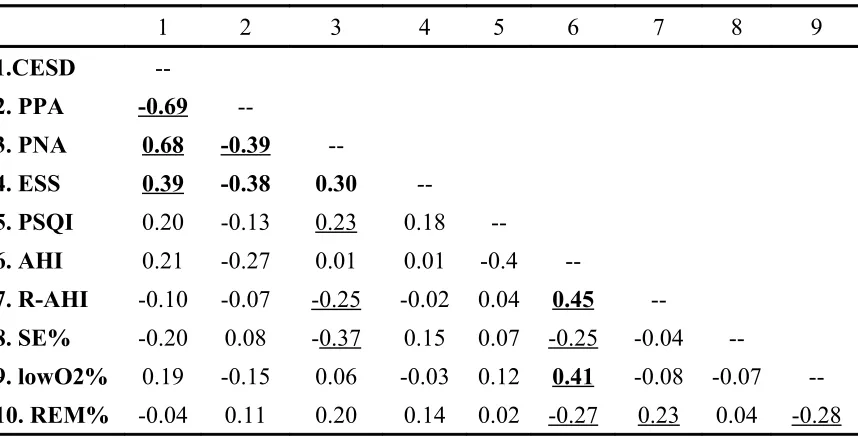
TABLE 3: Partial Correlation Matrix (controlling for age) of Mood, Subjective Sleep Self-Report Measures, and Sleep Study Indicators ...73
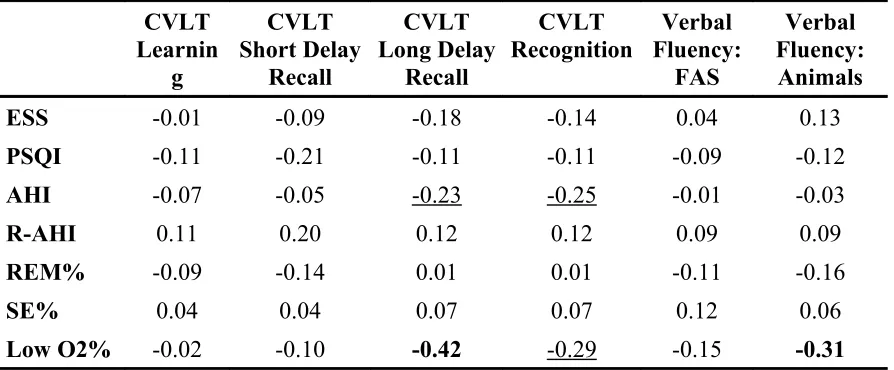
TABLE 4: Partial Correlation Matrix (controlling for age) of Subjective Sleep Self-Report Measures, Sleep Study Indicators, and Standard Cognitive Measure Performance …...74
TABLE 5: Partial Correlation Matrix (controlling for age) of Subjective Sleep Self-Report Measures, Sleep Study Indicators, and Cognitive-Affective Processing
Performance Measures …...77
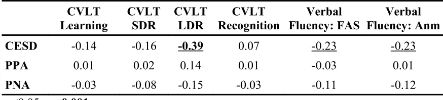
TABLE 6: Mood and Cognitive Performance Partial Correlation Matrix (controlling for age) ...78
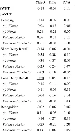
TABLE 7: Partial Correlation Matrix (controlling for age) for Self-Reported Mood Scores and Cognitive-Affective Processing Performance ...80
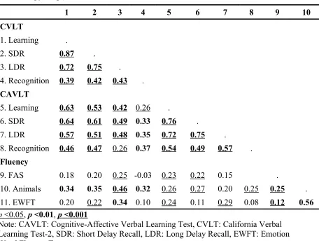
TABLE 8: Cognitive and Cognitive-Affective Processing Performance Partial
Correlation Matrix (controlling for age) ...82
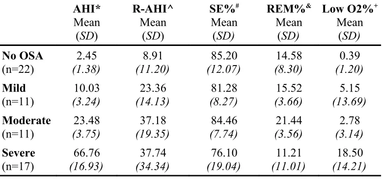
TABLE 9: ANCOVA (controlling for age) Comparison of Polysomnogram Sleep Quality Indicators by AHI Diagnostic Category ...84
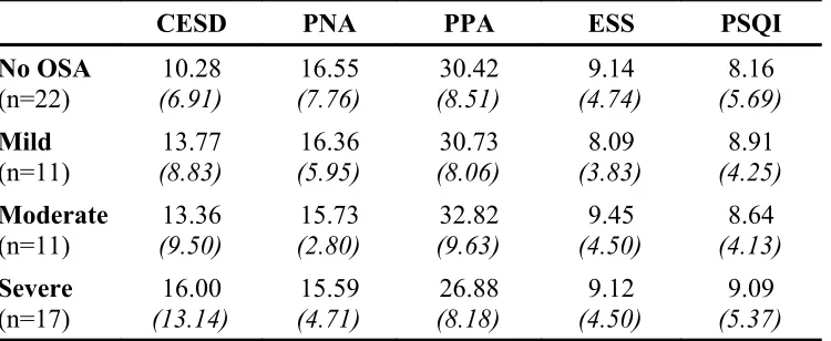
TABLE 10: ANCOVA (controlling for age) Comparison of Self-Reported Mood and Sleep Quality Scores by AHI Diagnostic Category ...85
TABLE 11: ANCOVA (controlling for age) Comparison of Cognitive Raw Score by OSA Diagnostic Severity Category ...86
TABLE 12: ANCOVA (controlling for age) Comparison of Cognitive-Affective Raw Scores by OSA Diagnostic Category ...88
TABLE 13: Regression Statistics: Significant Sleep Study Indicators and Self-reported Mood Scores that Predict CAVLT Short Delay Recall Valence Factor ...94
TABLE 14: Regression Statistics: Significant Sleep Study Indicators and Self-reported Mood Scores that Predict CAVLT Long Delay Recall Valence Factor ...94
LIST OF FIGURES
FIGURE 1: Two Conceptualizations of the Interaction between Depression, Sleep, and Cognitive Impairments ...28
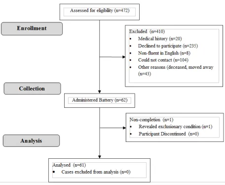
FIGURE 2: Flow-chart Detailing the Recruitment Process ...63
FIGURE 3: Proposed Mediation of Relationship between Sleep Study Indicators and Mood Measures via Cognitive-Affective Processing Valence Bias ...91
FIGURE 4: Graphical Representation of Significant Correlation Findings between Five Construct Domains ...103
CHAPTER 1
INTRODUCTION
Relevance & Importance
Deficits in sleep quality and associated sleep loss are experienced by nearly all people at
some point during life, whether from lifestyle or disorder, and whether chronic or acute.
These periods of disturbed sleep are usually limited in their length and incidence, but for
many individuals they can last for extended lengths of time, and often re-emerge at
various points of a lifetime. Even relatively mild and limited periods of disturbed sleep
are associated with a myriad of daytime behavioral impairments. Research has estimated
that the overall prevalence rate of adults obtaining insufficient sleep is 20% (Hublin,
Kaprio, Partinen, & Koskenvuo, 2001). Additionally, a study of over 1,000 young adults
over 5.5 years found that the degree of this partial sleep deprivation was proportional to
the amount of daytime sleepiness experienced (Breslau, Roth, Rosenthal, & Andreski,
1997). Neurocognitive functioning is thought to mediate the relationship between
sleepiness and behavioral performance decrements, which in turn are directly related to
the occurrence of functional accidents in everyday life.
Overall, accidents related to some degree of sleep deprivation have been
estimated to have an economic impact ranging between $43 and $56 billion (U.S.; Leger,
1994). Motor vehicle collisions are the most commonly associated cost of sleep
deprivation, yet are thought to be highly underestimated (Horne & Reyner, 1999;
McCartt, Ribner, Pack, & Hammer, 1996; Mitler, Carskadon, Czeisler, Dement, Dinges,
& Graebner, 1988). In addition to motor vehicle collisions, sleep deprivation research has
Coblentz, 2003; Price & Holley, 1990), truck drivers (Lyznicki, Doege, Davis, &
Williams, 1998; McCartt, Rohrbaugh, Hammer, & Fuller, 2000), medical residents
(Landrigan, Rothschild, Cronin, Kaushal, Burdick, Katz, et al., 2004; Lockley, Barger,
Ayas, Rothschild, & Czeisler, 2007), shift workers (Richardson, Miner, & Czeisler,
1989-1990), and other professions are at high risk for making high-damage accidents due to
sleepiness and its related sequelae. Blood alcohol content (BAC) is a useful comparison
metric for sleep deprived populations. Driving performance for those deprived of one
night of sleep was found to be equivalent to those non-sleep deprived individuals driving
with a BAC of 0.07% (Fairclough & Graham, 1999); for comparison, driving in the state
of Michigan or Ontario with a BAC of 0.08% or higher is illegal. Multiple other studies
have found that as uninterrupted waking periods exceed 16 hours, psychomotor
performance impairments progressively increase to levels comparable to BACs ranging
between 0.05% and 0.1% (Dawson & Reid, 1997; Williamson & Feyer, 2000).
In addition to functional accidents, disturbed sleep is demonstrated to cause
significant quality of life decrements, which strongly drive patients to seek assistance.
Reimer and Flemons (2003) conducted a literature review investigating how sleep quality
and quantity correlated with a wide range of domains that contribute to quality of life.
They found that across all measures and etiologies of disturbed sleep, quality and
quantity of sleep were related to some or all measures. For instance, the large Sleep Heart
Health Study (n = 5816), in which 90% of participants received an in-home
polysomnograph, found that those suffering from excessive daytime sleepiness had
significantly poorer quality of health in all subscales measured (Baldwin, Griffith, Nieto,
populations (of which obstructive sleep apnea is the predominant diagnosis),
symptomatic fragmented sleep (meaning many sub-conscious arousals from deep sleep
throughout a night) has been associated with increased mortality, abnormal waking
electroencephalograms (EEG), metabolic and endocrine dysregulation, decreased
immune and inflammatory responsivity, and cardiovascular sequelae (Dinges, Rogers, &
Baynard, 2005). In sum, sleepiness secondary to poor sleep is associated with
neurocognitive dysfunction, behavioral accidents, and quality of life decrements (both
mood and functional).
Relatively recent improvements in the methodology available for sleep research
(e.g. polysomnogram, functional imaging) have allowed for more detailed investigation
of the relationship between quality and quantity of sleep and daytime behavioral
outcomes. The following sections will briefly review the current understanding of sleep
physiology, explain how it relates to neurocognitive processing, cognitive-affective
processing, and mood, and finally introduce a published cognitive model of
sleep-dependent emotional processing in order to guide proposed investigation of
neuropsychological functioning in obstructive sleep apnea.
Sleep
The following section is a significantly condensed overview of sleep physiology,
emphasizing the introduction and definition of terminology that is relevant to the present
study. An expanded version of this section is available in Appendix A.
Sleep in mammalian species has been generally categorized into two separate types -
rapid eye movement (REM) sleep, and non-rapid eye movement (NREM) sleep, which
has predominantly been further subdivided in primates into four, progressively deeper,
stages (Rechtschaffen & Kales, 1968). Research in human sleep patterns has identified a
90 minute alternating, ultraradian cycle between NREM and REM sleep. The American
Academy of Sleep Medicine (AASM; Iber, 2007) recently updated the terminology used
to break sleep down based on electroencephalograph (EEG) readings into REM sleep,
and NREM stages labeled N1, N2, N3.
Table 1 summarizes the EEG (AASM, 2007; Steriade & Amzica, 1998),
neurochemical (Rosenthal, 1998; Saper, Chou, & Scammell, 2001), and functional
Table 1
Summary of EEG, Neurochemical, and Neurofunctional Characteristics of Sleep Stages
State EEG markersEEG Sleep Characteristics Neurochemical Functional
Awake Desynchronized beta waves (12-30 Hz)
RAS efferents to HT, THAL, BFB
Use CA, HTM, ACh, Asp, Glu
N1
Transition from alpha waves (8-12 Hz) to theta waves (4-7 Hz)
Drowsiness
If woken, will report not having been asleep
GABAergic neurons in cortex, THAL, and HT highly active Decreased subcortical cholinergic systems of forebrain and brainstem Reduced activity of the PFC, TL, BG, THAL, brainstem Reduction level intensifies with progression through N1-N3 N2 11-16 Hz, but predominately
12-14 Hz Sleep spindles K-complexes Tranquil state maintained
50% of total sleep time
N3 0-4 Hz, at least 20% delta waves (0.5-2 Hz) Slow-wave sleep (SWS) Mass cortical synchronization (organization processing related to daytime cognition) REM Theta wave reemergence (4-7 Hz) High frequency gamma waves (30-80 Hz) PGO waves (originating from pons, LGNT, & OC)
Rapid eye movement bursts in rhythm with PGO waveforms
20-25% of total sleep over 4-5 periods
Descending muscle atonia & increased variability of
heart/breathing rate & temperature
ACh neurons of PT = "REM-on cells," highly active
5HT & NE neurons of Raphe & LC = "REM-off cells," are
deactivated
Increased activity of the mbPFC, OC, Thalamic nuclei, PT, ACC, AMYG, HPC
Decreased activity of the PCC, PL, dlPFC
Sleep-Wake Cycle
Three separate yet networked neuroanatomical systems regulate the sleep-wake cycle in
humans (Borbely & Achermann, 1999; Pace-Schott & Hobson, 2002).
A homeostatic regulation system is responsible for intensity, length, and quantity
of sleep. Adenosine has been identified as a molecular-level somnogen integral to this
system at the cellular level. During wakeful periods, it is hypothesized to naturally
accumulate to levels that impact sleep/wake related areas of the brain. This nucleoside
has an activating effect on ventrolateral preoptic area neurons bordering the basal
forebrain and an inhibitory effect on wake-promoting areas of the basal forebrain
(Porkka-Heiskanen, Strecker, Thakkar, Bjorkum, Greene, & McCarley, 1997). Thus,
during wakeful accumulation of adenosine, a drive towards sleep accrues. Preoptic neural
circuitry has been associated with the homeostatic functions.
A circadian system manages the timing of sleep and wake periods within the
overall day-night cycle, promoting both wakefulness and sleep - at opposite phases. The
anterio-hypothalamic elements are associated with circadian functions. The circadian
timing system (CTS) is comprised of three components, the central of which is the
suprachiasmatic nucleus (SCN) of the anterior hypothalamus, which acts as a circadian
pacemaker - coordinating circadian oscillator subcomponents via control over melatonin
secretion by the pineal gland (Rossenwasser & Turek, 2005). The SCN is responsible for
establishing the sleep-wake circadian rhythm. The SCN is entrained (i.e., synchronized)
via physiological and environmental signals. The subcomponent circadian-oscillators in
peripheral tissues are in turn entrained by physiological signals from the pacemaker
cellular-level pacemaker "clock cells," independent of the circadian system as a whole
(Herzog, 2007). The SCN is thus thought to entrain the various peripheral cellular
oscillators rather than sustain their rhythmic activity (Okamura, 2004). The third
component of the CTS are the efferent projections that serve to regulate otherwise
non-rhythmic physiological and behavioral systems (e.g., body temperature,
autonomic/endocrine systems, feeding, sleep/wake state, locomotor activity).
The cyclical vacillation between REM and NREM sleep within each sleep period
is controlled by an ultradian system. Mesencephalic and pontine rostral brainstem areas
are associated with REM/NREM regulation; Table 1 offers more details.
Saper, Chou, and Scammell (2001) reviewed recent literature on sleep regulation
and identified a substantial amount of evidence that a reciprocal inhibition model of sleep
and arousal systems exists; they termed it a sleep-wake switch. GABAergic and
galaninergic neurons of the ventrolateral preoptic nucleus (VLPO) are active and
necessary for normal sleep. In contrast, hypocretin/orexin (exchangeable names) neurons
within the posterior lateral hypothalamus (PLHT) are necessary for maintaining normal
wakefulness. These two systems are thought to exist in a sustained state of balanced
reciprocal inhibition (a bi-stable feedback loop) when not influenced from external
pressures. Once either of the systems is excited, it inhibits the other, thereby resulting in
further excitation due to decreased inhibitory afferents from its partner. In sum, human
sleep physiology can be conceptualized as three gears nested within each other, with the
transition between sleep and wakefulness occurring in rapid fashion when the
homeostatic and circadian gears align.
Basal sleep need is defined as habitual sleep duration in the absence of any sleep debt.
Sleep debt is conceptualized as "the fundamental duration of sleep below which waking
deficits begin to accumulate" (Dinges, Rogers, & Baynard, 2005, p. 68). Both
experimental and epidemiologic research has found high interindividual variance in
amount of sleep habitually obtained. A large study found that after long-duration sleep
designed to eliminate sleep debt, average sleep length stabilized at 8.17 hours (Wehr,
Moul, Barbato, Giesen, Seidel, Barker, & Bender, 1993); another large, dose-dependent
sleep deprivation study statistically estimated a daily requirement of 8.16 hours of sleep
to avoid negative impacts on functioning during the subsequent wake period (Van
Dongen, Maislin, Mullington, & Dinges, 2003). The following section will discuss a
number of past and present theoretical frameworks for understanding sleep deprivation.
Models
In the 1980's, a two component hypothesis of sleep gained popularity - core and optional
sleep (Horne, 1988). The analogy of appetite was cited by its proponents, in which
hunger cues consumption of food until satiation, but additional food can be consumed
beyond the body's immediate need. Core sleep is composed of primarily slow wave sleep,
and it is the quality and length of this period of sleep that determines the degree of
daytime cognitive functioning and alertness. Optional sleep is conceptualized as
superfluous, or a luxury without confirmed function; one proposed possibility is that
optional sleep serves as an evolutionary behavioral carry-over meant to keep the
individual withdrawn and safe during the remaining hours of darkness. Proponents
redefined the average amount of core sleep required each night from 4-5 hours to 6 hours.
result in detrimental functional impacts in humans, which is not the case (Van Dongen,
Maislin, Mullington, & Dinges, 2003). That study will be addressed in more detail below.
Another hypothesis proposes that at the onset of a chronic restriction of sleep
time, acute neurobehavioral functioning decrements occur, but that over time individuals
are able to adapt to the new, reduced sleep period. Research has shown that self-reports
of sleepiness drop after an initial spike when sleep time is chronically restricted to 4-6
hours per night, up to 8 months (Belenky, Wesensten, Thorne, Thomas, Sing, Redmond,
et al., 2003; Lubin, 1967). Abruptness of sleep restriction is an important moderating
factor within this hypothesis, with research demonstrating that gradual (versus
precipitous) accumulation of a set amount of sleep debt resulted in neurobehavioral
performance decrements smaller in magnitude (Van Dongen, Maislin, Mullington, &
Dinges, 2003). Other research suggests that adaptation to sleep deprivation may differ
depending on which neurobehavioral outcomes are measured. It appears that waking
EEG and non-REM slow-wave sleep (SWS) show the most and quickest adaptation,
subjective sleepiness shows slower and less adaptation, and that neurocognitive
functioning shows the slowest and least adaptation to sleep deprivation (Van Dongen,
Rogers, Dinges, 2003; Van Dongen, et al., 2003).
A bio-mathematical two-process model taps the well-researched regulation
components of sleep: (1) the homeostatic process that exponentially builds during waking
periods and exponentially declines during SWS periods, and (2) a near-24 hour circadian
regulation process. The hypothesis proposes that wakeful cognition is primarily based on
alertness (A), and that this construct could be mathematically modeled as the quantitative
a blind study, this model was found to accurately predict neurobehavioral responses
based on total sleep deprivation, but failed to accurately predict cognitive performance
and sleepiness across a chronic sleep deprivation paradigm (Borbely & Achermann,
1999).
All of the prior hypotheses and models share a common feature in their
conceptualization of chronic sleep deprivation, which is an emphasis on cumulative sleep
time lost. The "wake extension" hypothesis approaches alterations of the sleep-wake
cycle from a different angle, instead emphasizing cumulative wake-time cost. Proponents
attempted to reconcile the neurocognitive results found in complete sleep deprivation and
those from chronic sleep deprivation by positing that during periods of wakefulness,
neurobiological costs/consequences accumulate (Van Dongen, Maislin, Mullington, &
Dinges, 2003).
Researchers used a sleep dose-response experiment (8, 6, or 4 hours in bed across
two weeks, and a 0 hours in bed condition across 3 days) to investigate their hypothesized
model. Results showed a near-linear accumulation of cognitive performance deficits
across all conditions. The rate (slope) of deterioration was inversely related to amount of
sleep time; the 0 hour condition demonstrated the highest rate of deterioration. At the two
week period, the 4 hours time in bed (TIB) condition was performing with similar
cognitive decrements to the 0 hours TIB at the three day mark. Calculating the
cumulative sleep loss for each condition reveals that the 4 hour TIB group had lost
approximately 55 hours of sleep, whereas the 0 hours TIB group had lost approximately
25 hours. To reconcile this inconsistent observation, the wake-extension time (defined as
This approach resulted in the same values across the restricted sleep conditions as the
previous approach. However, for the full sleep deprivation condition, each day after the
end of the first "habitual wake duration" added a full 24 hours to the wake extension
time, thereby bringing the observed deficits into alignment with quantification of
sleep/wake disruption (for visual representation of this phenomenon, see Figure 4 within
the Van Dongen et al., 2003 paper). This study clearly demonstrates that cumulative
wake extension and cumulative sleep loss are different constructs with different
quantitative values dependent on how sleep loss occurs. Further, it suggests that the field
should conceptualize sleep debt as an accrual of wakefulness beyond roughly 16 hours
with an associated neurobiologic cost.
Disturbed Sleep & Neurobehavioral Functioning
Sleep deprivation results in increased sleep propensity, as measured by reduced sleep
onset latency and reduced latency between the transition from lighter NREM sleep to
SWS on polysomnograms (PSG; Carskadon & Dement, 1987). After one night of
complete sleep deprivation, average sleep onset latency drops to less than 1-2 minutes,
and the time to progress from sleep onset to deep SWS is halved (Dinges, 1986).
Progressive reductions in daytime sleep onset latency has also been demonstrated in a
week long, 5 hour sleep restriction paradigm (Carskadon & Dement, 1981). Increased
sleep propensity, even when being resisted, results in the occurrence of microsleeps
intruding into wakefulness (Akerstedt, 1987). The state instability hypothesis posits that
neurocognitive performance becomes progressively more variable as homeostatic
begins to become more dependent on compensatory mechanisms (Doran, Van Dongen, &
Dinges, 2001). Behavioral examples of these compensatory mechanisms include
sleep-deprived individuals falling asleep while walking and "semidreaming" while performing
verbal cognitive tasks (Kleitman, 1963; Dinges, 1990). Errors of commission are
explained as ineffective compensatory efforts initiated during the resistance of sleep
(Durmer & Dinges, 2005). Thus, at any given moment, sleep deprived individuals
produce widely varied neurocognitive and neurobehavioral performance.
Effects of Disturbed Sleep on Cognitive & Behavioral Functioning
There are several broad findings in the research addressing cognitive performance in
sleep deprived and partial sleep deprived healthy individuals that must be considered
before reviewing domain-specific findings. Research suggests that sleep deprivation often
has unexpectedly measure-specific performance impacts; for example, a study found that
after a 5 night, 40% reduction of habitual sleep time, performance decrements on a
measure of vigilance and simple reaction time were observed but no deficits were noted
on a measure of choice reaction time (Herscovitch & Broughton, 1981). This may be due
to psychometric properties of different measures, or perhaps that the impact of sleep
deprivation is nuanced and focal rather than broad, even within traditionally internally
consistent cognitive domains (Dinges, Rogers, & Baynard, 2005). Cognitive decrements
have generally been found to be dose-dependent to the amount and length of sleep
restriction (Belenky, Wesensten, Thorne, Thomas, Sing, Redmond, et al., 2003). As
previously discussed, extended periods of restricted sleep have an accumulating
impairing effect that can eventually become equivalent to acute sleep deprivation (Van
deficits associated with sleep deprivation vary significantly between individuals, above
and beyond differences in sleep histories. In fact, it appears that vulnerability or
resiliency to sleep deprivation is a trait, though no neurobiological correlates have been
identified thus far (Van Dongen, Baynard, Maislin, & Dinges, 2004). Though average
deficits scores are stable within-subjects, a hallmark of sleep deprivation is a significant
increase in variability of test performance, between and within measures; this is thought
to reflect the transitory nature of attentional lapses (Waters & Buck, 2011).
To offer a launching point for the following discussion, consider the following. A
large meta-analysis has concluded that upon collapsing measures of three areas of
functional level (cognition, mood and fatigue, and motor functioning), "any
sleep-deprived individual is estimated to be comparable to the 9th percentile of
non-sleep-deprived subjects" (p. 120, Durmer & Dinges, 2005). Effect sizes for the impact of sleep
loss have generally been classified in the moderate range (Lim & Dinges, 2010).
Meta-analysis also found that cognition and mood were affected worse by partial sleep
deprivation than total, though the reverse is true for behavior (Pilcher & Huffcutt, 1996).
In general, performance on cognitive tasks becomes progressively worse as task
engagement time is extended, in an exacerbated "fatigue" effect phenomenon (Kribbs &
Dinges, 1994). Conversely, brief measures with an emphasis on speed or time also are
sensitive (Dinges, 1992). Finally, early theories on cognitive decrements following
disrupted sleep hypothesized that decreased motivation mediated the performance
deficits. However, sleep deprived populations have been demonstrated to pass
neuropsychological measures of adequate effort, as well as perform poorly on high
not significant etiological factors in explaining cognitive performance declines (Harrison
& Horne, 2000; Wilkinson, 1961).
Durmer and Dinges (2005) reviewed the literature on the neuropsychological
performance impact of sleep deprivation and identified a number of reliably affected
cognitive processes. This section summarizes the majority of their findings, and includes
more recent research findings to expand upon their review. First, there will be a
discussion of the cognitive findings associated with large amounts of sleep deprivation
(i.e. 4 or less hours per night), then these will be related to partial sleep deprivation and
fragmented sleep.
Individuals who have been deprived of sleep demonstrate slowing on
subject-paced tasks, and make increased errors when a time pressure component is present. Not
unexpectedly, this processing speed deficit is also reliably demonstrated in reaction time
measures. Speed of information processing has been demonstrated to be reliably
impacted by any disruption of normal sleep, with a 27 study meta-analysis finding that
speed was the most impacted cognitive construct, followed by accuracy (Koslowsky &
Babkoff, 1992; Waters & Bucks, 2011). Eye-hand coordination and psychomotor
performance consistently show decrements of roughly 30% in speed and accuracy after
sleep deprivation (Williamson & Feyer, 2000).
Tasks that require continued attention and vigilance are negatively impacted by
sleep deprivation, with increases in omission and commission errors. The functional
impact of increased attentional fatigue negatively affects performance on sustained
attention measures, as well as on other neuropsychological measures that require
extended simple attention is more significantly impacted than performance on tasks
requiring divided attention (Lim & Dinges, 2010). For example, on cognitive tasks with a
learning component, sleep deprived individuals are less efficient at reaching equivalent
levels of acquisition, though often can reach normative expected performance levels
when given additional time and exposure to the stimuli. Short-term recall for successfully
encoded information has also been shown to suffer post-sleep deprivation. After 1 night
of sleep deprivation, individuals scored worse on measures of visual and verbal
short-term memory, and performance was related to the magnitude of abnormally decreased
intraparietal sulcus and hippocampal activity (Chee & Chuah, 2007; Chen, Hardy, Zhang,
LaHoste, & Bazan, 2006; Van der Werf, Altena, Schoonheim, Sanz-Arigita, Vis, De
Rijke, et al., 2009). Working memory tasks that require maintenance and manipulation of
information from multiple modalities are compromised as well, resulting in difficulty
with temporal organization of information, decreased ability to maintain flexible
thinking, and decreased ability to filter distractions and maintain focus on relevant
information and cues. One study estimated working memory performance drops
averaging 37% after large amounts of sleep deprivation (Turner, Drummond, Salamat, &
Brown, 2007).
Neuropsychological measures thought to rely on more complex cognitive
processes have often been considered insensitive to partial sleep deprivation. It is thought
that problem-solving and critical thinking based tasks allow for convergent skills being
tapped in parallel, permitting intact performance via compensatory support from less
affected systems (Waters & Bucks, 2011). However, consistent with the weaknesses
impaired performance on certain complex cognitive tasks that require executive functions
such as mental flexibility and multitasking, perhaps reflecting the increased variability in
general cognitive performance following sleep deprivation. More specifically, divergent
thinking tasks that require lateral thinking, assimilation and utilization of feedback, and
risk assessment all show decrements after sleep deprivation. Executive processing errors
such as decreased insight into performance decrements, suppression of inappropriate
responses, and increased ineffective response perseveration are also well-documented.
Associated with the decrements in executive functioning, sleep deprived individuals rely
more heavily on compensatory effort to maintain adequate performance; this comes at the
cost of situational awareness, as neglect for activities and stimuli judged to be
nonessential increases (Durmer & Dinges, 2005).
The deficits discussed have led researchers to conclude that deprivation of sleep
negatively impacts performance on tasks believed to originate from or mediated through
the prefrontal cortex (PFC) - for those tasks that are attention-rich, more specifically, the
dorsolateral PFC (Kane & Engle, 2002). When sleep restriction is increased to complete
deprivation for 36 hours, research found that young participants produced
neuropsychological deficit profiles comparable to an elderly habitual sleep group
(Harrison & Horne, 2000). This is consistent with current attribution of documented
neurocognitive deficits in aging to declines in PFC functioning (Corey-Bloom,
Wiederholt, Edelstein, Salmon, Cahn, & Barrett-Connor, 1996). Functional neuroimaging
research suggests that two elements of the functional network connected to the PFC are
disrupted after sleep deprivation (Harth, 1995; Posner, 1994). The first is an anterior
in selective attention and the mental maintenance of a memory for immediate
manipulation (i.e. working memory). The second is a posterior network consisting of the
superior parietal lobes, pulvinar, and superior colliculus, which is thought to control
attentional switching and divided attention. The anterior cingulate has afferent
projections to the superior parietal lobes, and is innervated by the PFC, connecting the
three network components.
Significant-to-complete sleep deprivation is a useful paradigm to research the
neurocognitive correlates of decreased sleep; however, the ecological application is
limited. Sleep decrements in vivo generally take the form of partial sleep deprivation or
fragmented sleep. Recent research that has improved methodological controls for sleep
history and external influencing factors has found that 4 or more days of 7 or less hours
of sleep restriction per night results in measureable decrements in neurobehavioral
functioning and performance (Belenky, Wesensten, Thorne, Thomas, Sing, Redmond, et
al., 2003; Dinges, Pack, Williams, Gillen, Powell, Ott, et al., 1997). Restriction between 6
and 3 hours per night results in increased sleep propensity, working memory deficits, and
impaired sustained attention and vigilance (Carskadon & Dement, 1981; Drake, Roehrs,
Burduvali, Bonahoom, Rosekind, & Roth, 2001). The most extensive study on sleep
deprivation and restriction to date, conducted by Van Dongen and colleagues (2003), was
previously discussed, confirming that sleep restriction induced neurocognitive
performance decrements accumulate to levels equivalent to acute complete sleep
deprivation. While occupational research on partial sleep deprivation is relatively
common (e.g., for air traffic control, heavy machinery operators, etc.), a relatively
potential risk, such as driving. An epidemiological study found an elevated occurrence of
sleep-related vehicle crashes in those individuals who reported an average of less than 7
hours of sleep per night (Strutts, Wilkins, Osberg, Vaughn, 2003). Driving simulator
research has found that 1 night of restricted sleep (5 hours) results in decrements of
performance in simulated normal driving conditions and situations requiring emergency
maneuvers (Horne & Baulk, 2003). Another two studies found that chronic restriction of
sleep (time in bed between 4 and 6 hours) is associated with a significant increase in
number of accidents, and rates increase further after 2 nights of this degree of sleep
restriction (Dorrian, Dinges, Rider, Price, & Rogers, 2003; Rupp, Arnedt, & Carskadon,
2003).
Fragmented sleep refers to repeated arousals (3+ seconds of disrupted EEG
frequency in NREM or increased electromyographic frequency during REM) occurring
throughout a sleep period. Arousals do not result in awakenings. However, multiple
studies have demonstrated that persistent fragmentation of sleep results in the same
effects on daytime somnolence, mood alteration, and cognitive performance decrements
as partial sleep deprivation (Bonnet, 1985; Bonnet, 1986; Bonnet, 1989; Martin,
Engleman, Deary, & Douglas, 1996). In fact, arousals occurring at an average rate of
once per minute throughout a sleep period of normal duration result in neurocognitive
performance decrements equivalent in pattern and magnitude to that of 1 night of
complete sleep deprivation (Bonnet, 1986; Downey & Bonnet, 1987). This is not an
unusual fragmentation pattern for those suffering from intrinsic sleep disorders such as
obstructive sleep apnea (Durmer & Dinges, 2005). These findings hold true in
fragmentation of sleep, as well as experimentally fragmented sleep using aural
stimulation (Martin, Wraith, Deary, & Douglas, 1997). A detailed discussion of
cognitive, behavioral, and mood decrements in the obstructive sleep apnea (OSA)
population is provided later in this review. Finally, of considerable importance is the
finding that across all domains, cognitive deficits are reversible following a period of
normal sleep, which means that cognitive dysfunction attributed to disrupted sleep should
be framed in remediable terms and highlights the clinical importance of sleep
intervention (Waters & Buck, 2011).
Sleep & Affect
Affective Processing
The ability to effectively and efficiently process affective-based stimuli is crucial for
human functioning from a socio-evolutionary perspective, as suggested first by Charles
Darwin in 1872 (Norris & Cacioppo, 2007). The generation and regulation of emotions
and the guidance provided by emotional content and cues is fundamental to individual
mental health, interpersonal functioning, and societal structure. In the past decade,
cognitive neuroscience and clinical neuropsychology have rapidly embraced and
investigated the domain of emotional or affective processing as a critical element of
normal and abnormal cognition, and recognized the relationship between affective
processing and clinical mental health (Labar & Cabeza, 2006). The following will
provide a brief outline of a systems-level framework of affective informational
To discuss affective processing, a few terms and constructs should be clarified.
For the purposes of this review, the term emotion is meant to represent a complex
physiological, behavioral, and cognitive experience associated with the onset and
maintenance of mood/s. Affective is a descriptor meant to refer to characteristics that
conveys emotionally-relevant information, and affect is meant to refer to the process or
state of consciously labeling and experiencing internal emotional states (generally
resulting in phenotypic expression of the emotion, as often commentated on clinically).
Thus, affective processing is a form of information processing whereby
emotionally-relevant information is gleaned from a stimulus, analyzed, and then utilized in order to
facilitate correct/adaptive/appropriate reaction. Suchy (2011) posits that there are three
theoretical properties that are necessary for an affective processing system (APS). First,
the brain's APS needs the ability to detect emotionally-germane stimuli and judge the
affective qualities and characteristics (e.g., valence and intensity) quickly (likely
incorporating aspects of preconscious detection) in order to facilitate an immediate,
adaptive reaction. Second, an APS must also be able to initiate and maintain
physiological, behavioral, and cognitive events that comprise the response. Third, an
association and memory component is required to learn emotionally relevant
characteristics of a stimulus that was initially emotionally-neutral. Two related neural
circuits are involved in triggering emotional responses to stimuli - the amygdala
processes and responds to external stimuli, and the hypothalamus processes and responds
to disruption to internal homeostasis. The following will focus on the amygdala, as this
Two information processing routes exist, with both sharing an input pathway and
the amygdala acting as a mediator for behavioral and physiological responses (Suchy,
2011). The "fast route" (≈10-12 msec.) of affective information processing begins with
the sensory organ input projecting to the thalamus and primary visual cortex, where
crude, basic information (i.e., valence and intensity) regarding the stimuli is perceived.
The fast route next consists of this affective information being received by the basolateral
amygdala which projects to the orbitofrontal cortex and striatum involved with emotional
learning circuitry, and also to the central nucleus of the amygdala which in turn projects
to the hypothalamus and brainstem nuclei to generate physiological and behavioral
responses. The "slow route" (≈30-40 msec.) involves primary association cortices that
supply information about the perceived stimuli and supramodal association cortices that
supply more abstract and contextual meanings about the stimuli. These include the
secondary sensory cortex, tertiary sensory cortex, and hippocampus.
The integration of affective processing and cognitive research has resulted in
some domain-specific findings. Attentional blink paradigms have shown that responding
to a target during rapid stimuli presentation causes momentary depletion of attentional
resources, resulting in missed targets that immediately follow a first target. Strong
affective valence of the second target ameliorates this depletion. However, individuals
with amygdala damage cannot benefit from this phenomenon (Anderson & Phelps,
2001). Significantly faster psychomotor response speed has also been found for
negative/threatening valenced stimuli, above and beyond that which can be explained by
increased provision of attentional resources to the stimuli (Flykt & Caldara, 2006; LoBue
affectively salient stimuli (Frank & Tomaz, 2000). This occurs due to improved encoding
via amygdala modulation of the perceptual cortical areas and improved consolidation of
affective stimuli in proportion to the relative survival importance of the associated
outcome (Phelps, 2004). Individuals with damaged or dysfunctional amygdala do not
benefit from the memory facilitation by affective characteristics of emotional stimuli as
found in healthy controls (Dolan & Fullam, 2010).
The Role of Sleep in Affective-cognitive Processing
The domain of learning and memory is a useful cognitive domain to research as it relates
to the affective processing system (APS), as it is understood to tap elements of attention,
working memory, and executive functions such as organization. Impact of sleep quality
on memory recall has focused on two stages, the initial formation of new memories
(encoding), and then the subsequent solidification of the memories (consolidation). Both
will be addressed with an emphasis on encoding as it is more germane to this dissertation
(Marshall & Born, 2009; Walker, 2009).
Affective Memory Encoding and Sleep
The elicitation of emotional states can strongly modulate the initial stages of learning
(i.e., encoding). Stimuli with emotionally arousing affective traits are recalled better than
those considered neutral (Buchanan & Lovallo, 2001; Heuer & Reisberg, 1990; Phelps,
2004). The modulation effect of emotionally arousing stimuli on encoding occurs in
different ways depending on the affective stimuli's valence (positive, neutral, negative)
and arousal level (calm to excitement). High arousal affective characteristics enhance
memory encoding through the adrenergic system. Introduction of propanolol (a
emotionally arousing stimuli (narratives, individual words) results in a disappearance of
the enhancing effects of affect on learning found in control groups (Cahill, Prins, Weber,
& McGaugh, 1994; Strange, Hurlemann, & Dolan, 2003). Lesion and functional
neuroimaging has identified the amygdala, anterior hippocampus, ipsilateral
parahippocampus, and ventrolateral prefrontal cortex as being involved with the
formation of memory for affectively-valenced information (Cahill, Haier, Fallon, Alkire,
Tang, & Keator, 1996; Dolcos, Labar, Cabeza, 2004; Kilpatrick & Cahill, 2003). In the
absence of high arousal, valence of stimuli (versus neutrality) still positively modulates
encoding, though this is primarily governed by frontally-mediated strategic and semantic
processes outside of the amygdala and paralimbic system (Labar & Cabez, 2006). High
arousing stimuli with a negative valence have been found to have the strongest enhancing
effect on encoding compared to positive and neutral stimuli (Kensinger & Corkin, 2003).
The above findings relate to individuals under normal sleeping conditions.
Beginning at a cellular level, in rodent studies REM sleep deprivation (24-72 hours) has
been found to reduce hippocampal neuron excitability and significantly impair long-term
potentiation (LTP), a process demonstrated to be a critical mechanism for memory
formation (Davis, Harding, & Wright, 2003). The LTP observed after REM sleep
deprivation decays within 1.5 hours, suggesting significant impairment in hippocampal
plasticity. Behaviorally, avoidance learning, passive avoidance learning, and taste
aversion processes are all significantly impaired in rodents following both general sleep
deprivation and specific REM deprivation (McGrath & Cohen, 1978; Smith, 1985).
continued practice trials do not result in performance improvements (Gruart-Masso,
Nadal-Alemany, Coll-Andreu, Portell-Cortes, & Marti-Nicolovius, 1995).
In one thorough human study, participants were either deprived of sleep for 36
hours or allowed a habitual sleep period, then exposed to emotionally arousing and
neutral stimuli (Walker, unpublished results, most recently cited in Saletin & Walker,
2012). All participants then were allowed two habitual sleep periods before recall was
tested, thereby controlling for impaired recall due to sleep deprivation confounds;
retention of material was thus deemed to represent encoding processes. Control group
individuals demonstrated significantly superior retention for positive and neutral stimuli
relative to the experimental group. Sleep deprived individuals had significantly impaired
retention for neutral stimuli, but even worse retention of positively valenced stimuli (59%
reduction relative to control group for positive stimuli) compared to controls. Retention
of negative stimuli was not significantly different from that of the control group. In
effect, sleep deprivation led to skewed encoding sessions, with the experimental group
ending the session with a prevailing dominance of memory for negative material, and far
fewer neutral and positive memories. Explanations for this sleep deprivation and affective
valence interaction range from suggestions that the arousal levels differ significantly at
the neural level of the stimuli (rather than the reported level) which interact with the
post-sleep deprivation hypo-activation of the prefrontal cortex and hyper-activation of the
amygdala to bias toward successful encoding of negative stimuli versus neutral/positive
stimuli (Chee & Chuah, 2008; Kensinger & Corkin, 2004; Yoo, Gujar, Hu, Jolesz, &
sleep-deprived individuals could lead to a mood-congruent encoding bias (Lewis, Critchley,
Smith, & Dolan, 2005).
Affective Memory Consolidation & Sleep
Currently, research suggests that emotion and affective characteristics of stimuli have a
modulating impact on subsequent memory consolidation. Behavioral human studies have
found decreased forgetting of affectively valenced material versus neutral material. This
retrieval benefit emerges more strongly as the delay between encoding and retrieval
attempt increases, as demonstrated in a variety of different delay contrast methodologies:
immediate versus 1 hour or 24 hours (LaBar & Phelps, 1998; Sharot & Phelps, 2004,
respectively), 20 minutes versus 1 week (Kleinsmith & Kaplan, 1963), and 15 minutes
versus 2 weeks (Anderson, Yamaguchi, Grabski, & Lacka, 2006). Research has
demonstrated that using pain manipulation and stress hormone introduction (adrenaline
and corticosterone) post-learning trials increases amygdala activity and selectively
improves long-term memory for affectively valenced stimuli (Cahill & Alkire, 2003;
Cahill, Gorski, & Le, 2003). Neurotransmitter research in this area has identified
adrenergic transmitters and acetylcholine as co-regulators of the consolidation of memory
for affective stimuli. Acetylcholine augments amygdala-reliant memory consolidation;
antagonist and agonist introduction into the amygdala of rodents impairs and enhances
(respectively) memory for previously learned, valenced material (e.g., fear-conditioning
and alteration of reward magnitude paradigms; Power & McGaugh, 2002; Schroeder &
Packard, 2002).
In humans, REM sleep characteristics have been noted to be altered after
mechanisms impact sleep-stage characteristics (Sanford, Silvestri, Ross, & Morrison,
2001; Sanford, Tang, Ross, & Morrison, 2003). Next-day memory retention has been
demonstrated to be impaired after sleep deprivation between learning and testing, which
is thought to reflect a disruption of memory consolidation (Walker & Stickgold, 2004).
Sensitivity to fear-conditioning consolidation deficits appears to be limited to paradigms
that disrupt/deprive sleep 0-6 hours after the learning task, suggesting that negatively
valenced memory consolidation occurs shortly following learning (Graves, Heller, Pack,
& Abel, 2003; Ji, Wang, & Li, 2003). Emerging research suggests a
REM-sleep-dependent hypothesis of affective human memory consolidation, elements of which were
proposed by both Cahill (2000) and McGaugh (2004). Consolidation of affectively
arousing stimuli across a 12 hour day, or a 12 hour period including a habitual sleep
period, results in a benefit in consolidation of the affective information only when sleep
occurs between learning and testing (Hu, Stylos-Allan, & Walker, 2006). Total sleep
deprivation for only the first night after exposure to neutral and valenced visual stimuli
resulted in significant decrements in retention for all stimuli at testing 1 week later,
however, the greatest decrement was for neutral stimuli (Atienza & Cantero, 2008). The
authors hypothesized that the consolidation process for emotionally-relevant memories
may be intrinsically more resilient to sleep disruption. Speed of recognition for
affectively valenced facial stimuli is increased after a period of sleep, in proportion to the
amount of REM sleep experienced. In a separate study the power of right-dominant
prefrontal theta activity during REM sleep in a nap following a learning task for
neutral/negatively valenced stimuli was also proportionally related to emotional memory
Born, 2007). This supports the conclusion of neurophysiological reviews that posit REM
sleep representing a brain environment especially amenable to consolidation of memory
for affective material, based on its pro-cholinergic (forebrain ACh levels up to four times
higher when compared to NREM) characteristics (Marrosu, Portas, Mascia, Casu, Fa,
Giagheddu, et al., 1995; Pare, Collins, & Pelletier, 2002; Walker, 2009). One limitation
of this area of research is the absence of human investigation using positively valenced
stimuli (van der Helm & Walker, 2009).
Emotional Experience, Regulation, & Sleep
Research on the impact of sleep deprivation, or loss, on emotion is limited, despite the
fact that nearly all psychiatric and neurological condition involving disturbed mood also
have documented, co-occurring sleep disruption. Affective volatility, lability, and
irritability are subjectively increased following sleep deprivation (Horne, 1985).
Emotional disturbance due to chronic restricted sleep (5 hours per night across 1 week)
has been demonstrated to have an accruing impact (Dinges, Pack, Williams, Gillen,
Powell, Ott, et al., 1997). Furthermore, restricted sleep has been found to blunt intrinsic
positive reactions to rewarding/goal-oriented activities and increase negative emotional
reactions toward experiences that disrupt the achievement of goals or rewards (Zohar,
Tzischinsky, Epstein, & Lavie, 2005).
Only recently have researchers investigated the interaction between sleep and
psychophysiological/emotional reactivity. One functional MRI study exposed controls
and sleep deprived (1 night) individuals to images spanning a gradient between negative,
neutral, and positive valences (Yoo, Gujar, Hu, Jolesz, & Walker, 2007). Both groups
images, but the sleep deprived group showed a 60% increase in extent of activation and
three times the volume of amygdala recruitment compared to the control group.
Researchers noted decreased functional connectivity between the medial prefrontal cortex
and the amygdala, a pathway normally thought to represent frontal inhibition of
amygdala reactivity. This finding suggests that sleep is crucial to appropriate top-down
inhibitory functioning within the mPFC-amygdala circuit, and that this circuit
significantly governs appropriate emotional responses to affectively loaded stimuli.
Functional imaging research on populations with mood disorders that commonly have
co-occurring disturbed sleep patterns have also identified abnormalities in this circuit
(Davidson, Pizzagalli, Nitschke, & Putna, 2002). Little conclusive research findings exist
for positively valenced material (van der Helm & Walker, 2009).
Current conceptualization of mood disorders almost universally includes sleep
disturbance as a common feature or formal symptom (American Psychiatric Association,
2013), with reviews of the research into the relationship generally concluding mood and
sleep disturbances are bi-directional (Bliwise, 2004; Harvey, 2001). However, a massive
(N = 18,631) longitudinal twin-study offered strong evidence that poor sleep predates
onset of depression, though the methodology could not confirm a mechanistic causal
relationship (Paunio et al., 2009). Depression is a clinical condition that is highly relevant
to the intersect between abnormal affective processing and disturbed sleep. Depression
has comorbidity rates of disturbed sleep as high as 90%, with polysomnogram (PSG)
profiles indicating increased sleep latency and arousals, along with decreased REM sleep
latency and increased REM time and density (American Psychiatric Association, 2013;
Gottesmann, 2007). Depressed individuals exhibit heightened activity in the anterior
paralimbic cortex and midbrain reticular formation while awake, which researchers have
suggested may reflect a predisposition to encode negatively valenced experiences and
consolidate them more easily (Nofzinger, Price, Meltzer, Buysse, Villemagne, Miewald,
et al., 2000).
Walker and Van der Helm (2009) proposed a clinical model of sleep-dependent
emotional processing, based upon their literature review, concluding that under
conditions of sleep loss, the brain has a tendency toward encoding negatively valenced
stimuli and emotional memories, hyper-active limbic reactivity toward negatively
valenced events, and that negative memory consolidation is increased during REM sleep
(which tends to be higher density post-sleep deprivation). Their model proposes a "sleep
to forget and sleep to remember (SFSR)" hypothesis to explain the consistent finding that
emotional memory initially is comprised of both affective (generally
amygdala-associated activity) and informational (hippocampal-amygdala-associated activity) components, but
that over time (many months) the affective component is stripped from the informational
component of the memory (Dolcos, LaBar, Cabeza, 2004, 2005). The SFSR hypothesis
posits that this decoupling predominately takes place during sleep, with the end result
being sleeping to forget the affective/emotional component but sleeping to remember the
informational component. Failure to strip the affective component due to disturbed sleep
results in potential mood impacts during wakeful hours. Further, their model argues that
REM sleep offers a distinctly advantageous neurobiological environment for the
information-association facilitation of core memories and the depotentiation and
that the findings that affective valence impacts cognitive processing and memory across
waking periods absent of sleep suggest that sleep may be a preferential period for
consolidation but that a mechanism similar to REM sleep or completely independent
occurs during waking periods. How this segment of affective-processing relates to sleep
in both healthy controls and mood disorder populations requires further elucidation.
However, in effect, these authors suggest that there may be an alternate or
complementary conceptualization of how depression relates to sleep and neurocognitive
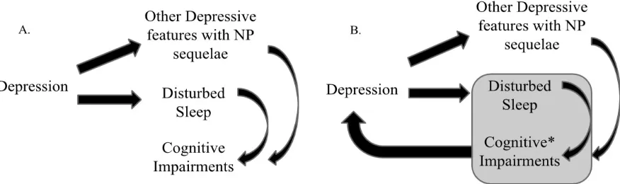
processes (see Figure 1).
Figure 1. Two Conceptualizations of the Interaction between Depression, Sleep, and Cognitive Impairments.
Note, NP: Neuropsychological. A) Common neuropsychological conceptualization of cognitive difficulties for a chronic depressive patient. B) Modified neuropsychological conceptualization, *cognitive processing dysfunction includes a negative bias in processing emotionally-valenced information (i.e., cognitive-affective processing).
Obstructive Sleep Apnea
Obstructive sleep apnea (OSA) is a common sleep disorder, estimated to affect roughly 2
Ancoli-1995). Base rates vary slightly based on demographics; 4% of middle-aged men versus
2% of middle-aged women are thought to suffer, with a large spike after the age of 65
resulting in estimates of up to 42% of this age group suffering (Ancoli-Israel, Kripke,
Klauber, Mason, Fell, & Kaplan, 1991; Young, Dempsey, Skatrud, Weber, & Badr,
1993). The pathophysiology of OSA is characterized by repeated episodes of momentary
(10+ seconds) incomplete (hyponea event, 50+%) or complete (apnea event, 100%
reduction) cessation of airflow. In OSA, the decreases in airflow are attributable to
obstruction or restriction of the breathing airway by the tongue and/or soft palate. The
near complete attenuation of skeletal muscle tone that occurs in N2 (intermittently) and
REM sleep is true for neck musculature as well, making these stages a common period
for the breathing events to occur. During apnea/hyponea events, blood oxygen saturation
(SaO2) can fall to dangerous levels and physical exertion to breathe by the diaphragm and
chest muscles increases. These trigger neurological mechanisms that cause a neurological
arousal to resume muscle tone and breathing almost never completely awakens an
individual, but does result in fragmented sleep and disturbed sleep architecture (Bassiri &
Guilleminault, 2000). Diagnosis of OSA is usually done with a polysomnography (PSG),
which also allows for an apnea-hyponea index (AHI; based on average number of apnea
or hyponea events occurring each hour) to be assigned as an indicator of severity; a score
of <5 is considered normal, a score of 30+ is severe.
OSA is associated with a cluster of cardiovascular health complications such as
hypertension, heart disease and stroke (Guilleminault & Robinson, 1997). Individuals
diagnosed with OSA are estimated to have annual health care costs twice as high as
1999). Finally, this population also has a higher accident morbidity rate, thought to in
part be related to attentional performance comparable to mildly to moderately intoxicated
controls (George & Smiley, 1999; Powell, Riley, Schechtman, Blumen, Dinges, &
Guilleminault, 1999). Unfortunately, the compliance rates for the primary treatment
option for OSA (Continuous Positive Airway Pressure; CPAP) is low; non-compliance
(less than 4 hours per night) rates range from 46-83% (Weaver & Grunstein, 2008). The
same study found that patient perception of symptoms is a significant factor in
non-compliance. The sub-awakening arousals and fragmented sleep found in OSA patients are
inherently difficult for the patients to perceive. Alternative routes of educating identified
OSA patients about the health and functioning impacts of their condition is a prime area
for the clinical neuropsychologist to contribute to treatment.
Neurocognitive Deficit Correlates
One meta-analysis of peer reviewed OSA articles with neuropsychological findings
between the years of 1985 and 2002 identified 37 peer-reviewed articles related to
neuropsychological performance in OSA populations (excluding non-clinically
diagnosed, cognition-related comborbid, non-adult, and central apnea populations or
studies using validated measures), which were subsequently divided into
non-mutually exclusive pre-treatment, treatment efficacy, and correlational study groups
(Aloia, Arnedt, Davis, Riggs, & Byrd, 2004). Pre-treatment results suggest spared global
cognition and language functioning, consistent with sleep deprivation findings that
crystallized knowledge remains intact and compensatory recruitment allows for
well-preserved general cognitive functioning. Attention (especially vigilance), memory, and