R E S E A R C H
Open Access
RNA interference targeting human integrin
α
6
suppresses the metastasis potential of
hepatocellular carcinoma cells
Guannan Lv
1†, Tianjing Lv
2†, Shifeng Qiao
1, Wei Li
1, Weiran Gao
1, Xiaohui Zhao
1and Jikun Wang
1*Abstract
Background:Increased metastasis has been proved to be associated with a poor prognosis for hepatocellular carcinoma (HCC). There are higher-level expressions of integrinα6 in the tissues of HCC patients with a higher fatality rate. The aim of this study is to investigate the effect of short hairpin RNA (shRNA) silencing integrinα6 expression on the proliferation and metastasis in HCC cell lines.
Methods:Two human HCC cell lines, HepG2 and Bel-7402 were transfected with shRNA targeting human integrin α6. Protein and mRNA expression level were determined by western blot and real-time quantitative reverse transcription-polymerase chain reaction (qRT-PCR) to detect the transfected efficacy. The metastasis potential of HCC cells was evaluated by their proliferation, adhesion and invasion abilities. Cell proliferation was measured by MTT assay. Adhesion ability was measured by adhesion and spreading assays. The expression of matrix metallopro-teinases (MMPs) was measured by qRT-PCR. The potential of invasion was measured by qRT-PCR and Transwell chamber assay. PI3K inhibitor LY294002 was used to explore the signal pathways of integrinα6 in HCC cells. Results:Western blot and qRT-PCR detection showed that over 75% of integrinα6 expression in HCC cells was through knockdown by shRNA. Proliferation, adhesion, spreading and invasion of HepG2 and Bel-7402 cells were dramatically decreased in cells transfected with shRNA compared to the control cells. P-ERK and p-AKT were reduced by shRNA targeting integrinα6 and PI3K inhibitor LY294002.
Conclusion:Knockdown integrinα6 can inhibit the proliferation and metastasis of HCC cells through PI3K/ARK and MAPK/ERK signal pathways by shRNAin vitro. Integrinα6 can mediate the metastasis potential, and can be used as a candidate target for therapy in HCC resulting in improved patients’survival.
Keywords:Hepatocellular carcinoma, integrinα6, Short hairpin RNA, Metastasis
Background
Hepatocellular carcinoma (HCC) is a highly lethal can-cer with a poor prognosis. The occurrence of HCC has recently shown a worldwide increase [1], mainly because of its high metastasis potential [2].
Integrins are heterodimeric transmembrane receptors composed of non-covalently associatedαandβsubunits. At least 18 α and 8 β subunits have been identified so far, generating more than 24 members of the integrin family. Increasing evidence suggests that integrins are
the most important receptors for cell metastasis [3]. Re-cently, it has been reported in many researches that in-tegrinα6β1 andα6β4 were associated with metastasis of HCC [4,5] and patients with high levels of expression of integrin α6β1 have a poorer prognosis [4,6]. Higher levels of expression of integrinα6β4 in patients is associ-ated with increased invasive potential of HCC, as well as a higher fatality rate [5,7]. Integrinα6β1, as an important kind of cell surface receptor, can mediate the adhesion between HCC cells and extracellular matrix (ECM) [8,9]. Owenset al. [10] demonstrates that integrinα6β4 could regulate the migration and invasion of laminin (LN) to stimulate the metastasis potential of HCC.
* Correspondence:[email protected] †Equal contributors
1
The First Affiliated Hospital of Liaoning Medical College, Jinzhou 121001, China
Full list of author information is available at the end of the article
However, few research studies have focused on the sin-gle action of integrin α6 alone in the progression of HCC metastasis. Furthermore, the metastatic mecha-nisms under high levels of expression of integrinα6 are still unclear. A better understanding of the molecular mechanisms underlying integrin α6 affecting HCC me-tastasis may facilitate the development of targeted therapy.
In the current study, in order to explore the effect of integrin α6 in the process of HCC metastasis without the influence of β subunits and the molecular mecha-nisms involved, two human HCC cell line, HepG2 and Bel-7402 were transfected with short hairpin RNA (shRNA) targeting human integrin α6. The metastasis potential of HCC cells was evaluated by proliferation, adhesion and invasion abilities. PI3K inhibitor LY294002 was also used to explore the signal pathway of integrin
α6 in HCC cells.
Methods
Cell culture and plasmids preparation
Two hepatocellular cell lines, HepG2 and Bel-7402 were purchased from the Chinese Academy of Medical Sci-ence (Beijing, China). All cells were cultured in RPMI 1640 (Life Technologies Corporation, 5791 Van Allen Way, Carlsbad, CA 92008, US) with 10% FBS, 200μg/ml penicillin and streptomycin at 37°C in 5% CO2. Integrin
α6 shRNA plasmids (sc-43129-sh) were constructed and synthesized by Santa Cruz Biotechnology, Inc., CA, USA. Plasmids containing puromycin resistance genes were used for the selection of cells stably expressing shRNA. Control shRNA plasmids (sc-108065), each en-coding a scrambled shRNA sequence that will not lead to the specific degradation of any known cellular mRNA, were also purchased from Santa Cruz Biotechnology, Inc., CA, USA.
Cell transfection and clone selection
Integrinα6 shRNA plasmids or control shRNA plasmids were transfected into HepG2 and Bel-7402 cells respect-ively, according to the manufacturer's instructions with slight modification. Briefly, in a 24-well tissue culture plate, cells were grown to approaching up to 85% to 90% confluence in RPMI 1640 with 10% FBS. The medium was aspirated 24 hours before transfection, and replaced with DMEM. Lipofectamine 2000 (Invitrogen, Carlsbad, CA, USA) 0.5 μl and plasmids 0.02 μg were added in each well and incubated for six hours. After that, cells were cultured in RPMI 1640 with 20% FBS, 400 μg/ml penicillin and streptomycin for 18 hours. After replacing the media with fresh normal growth medium, cells were incubated for an additional 24 hours. Three days later, stable transfectant clones were isolated with 1.6 μg/ml
and 2μg/ml puromycin (Santa Cruz Biotechnology, Inc., CA, USA) in HepG2 and Bel-7402 respectively.
RNA extraction and qRT-PCR assays
Briefly, 2 μg total RNA was used to synthesize first-strand cDNA. Amplification and detection were per-formed using the ABI Prism 7500 Fast Real-time System (Applied Biosystems, Foster City, California, USA) start-ing with 1 μl cDNA and TransStart Top Green qPCR SuperMix (TransGen Biotech, Beijing, China). Glyceral-dehyde 3-phosphate dehydrogenase (GAPDH) was used as internal standard.
Primers sequences for integrin α6, matrix metallopro-teinase (MMP)-2, MMP-9 and GAPDH were as follows:
Integrinα6: 5’-TCCCTGAACCTAACGGAGT-3’and 5’-ATGTCCAAGTAGTTC AGTT-3’;
MMPs-2: 5’-GCGACAAGAAGTATGGCTT-3’and
5’-TGCCAAGGTCAATGT CAGG-3’;
MMPs-9: 5’-AGTTCCCGGAGTGAGTTGA-3’and
5’-CTCCACTCCTCCCTT TCCT-3’;
GAPDH: 5’-GGCATCCTGGGCTACACTG-3’and
5’-GTGGTCGTTGAGGGCAATG-3’.
The relative expression of integrin α6, MMP-2 and MMP-9 mRNA was analyzed by the comparative cycle threshold (Ct) method. All experiments were performed in triplicate.
Western blot assays
Cells (1 × 10⁶) were washed with PBS twice and lysed with 1 ml RIPA lysis buffer supplemented with
40 mmol/L NaF, 100 μmol/L Na3VO4, and 1 μl
Complete Protease Inhibitor (KeyGen Biotech Co. Ltd, Nanjing, China) for 30 minutes on ice. After removing the insoluble material by 12,000 × g centrifugation for 30 minutes at 4°C, the supernatants were collected, boiled in 1 × sodium dodecyl sulfate sample buffer for five minutes. Subsequent immunoblots were probed with the appropriate antibody and detected by Gel Im-aging System G: BOX (Gene Company Limited, Cam-bridge, UK). Monoclonal rabbit anti-human CD49f (1:100, Abgent, San Diego, CA, USA), rabbit anti-human AKT (1:200); rabbit anti-human p-AKT (1:200); rabbit anti-human ERK (1:200); and rabbit anti-human p-ERK (1:200); antibody (these last four supplied by Santa Cruz Biotechnology, Inc., CA, USA), were used to detect the expression of integrinα6, AKT, p-AKT, ERK, and p-ERK respectively. GAPDH (1:500; Santa Cruz Biotechnology, Inc., CA, USA) was used as internal control.
Cell proliferation assays
(Sigma, St. Louis, MO, USA) was placed on a ten-dish plate and allowed to solidify at 37°C for two hours. The other wells were coated with 2 μg/cm2 of fibronectin (FN) (Sigma, St. Louis, MO, USA) as control. After washing each well twice with PBS, 8 × 10³cells were in-cubated in 96-well plates with DMEM containing 10% FBS at 37°C in 5% CO2 for 48 hours. MTT (10μl) was
added to each well and four hours later, the media was aspirated and 100 μl dimethyl sulfoxide (DMSO) was added. The assay was quantified in a Multimode Micro-plate Reader-Varioskan Flash (Thermo Electron Corpor-ation, Waltham, MA, USA) at 490 nm. All experiments were repeated in at least quadruplicate.
In vitro adhesion assay
LN or FN was coated in 96-well plates according to the method described above [12]. In total, 8 × 10³ cells sus-pended in DMEM were cultured at 37°C in 5% CO2 for
30 minutes. After being washed with DMEM three times, acid phosphatase substrate consisting of 0.1 mol/l sodium acetate buffer (pH 5.5), 0.1% Triton X-100 (10 mmol/L), and nitrophenylphosphate 100μl were added to each well. The cells were cultured at 37°C for two hours, then 1 mol/ L NaOH 10μl was added to each well to stop the reaction. The adhesion assay was quantified in a Multimode Micro-plate Reader-Varioskan Flash (Thermo, Electron Corpor-ation, Waltham, MA, USA) at 405 nm. All the results were obtained from at least three independent experiments.
In vitro spreading assay
LN was coated in 96-well plates according to the method described above. In total, 8 × 10³ cells suspended in DMEM were cultured at 37°C at 5% CO2for 150 minutes.
Then, a light microscope (×200) was used to determine the spreading assay. In this experiment, spreading cells were counted respectively in three distinct visions.
LN invasion assay
The upper part of the Transwell chambers coated with LN was used as a basement membrane (BM) according to the way described above. Fibronectin (FN) was used as con-trol. Cells (1 × 10⁵) suspended in DMEM containing 0.1% FBS were plated in the upper part to make contact with the BM. BSA (0.05%) in DMEM with 10% FBS was placed in the lower chamber, to act as chemoattractants. Cell in-vasion was determined by crystal violet dissolution assay after incubation at 37°C in 5% CO2for 48 hours; the
in-vaded cells were numerated under a light microscope (×400). In this experiment, invaded cells were counted in three distinct microscopic fields respectively.
The signal pathways assay
LN or FN was coated in 24-well plates according to the method described above. Cells (1.5 × 10⁶), serum-starved
for 48 hours, were allowed to adhere to the LN or FN and incubated at 37°C in 5% CO2for two hours. In order to
demonstrate the PI3K/AKT and MAPK/ERK signal path-ways, which are necessary for the reaction between HCC cells and LN, PI3K inhibitor LY294002 was used one hour before HepG2 cells were cultured in plates coated with 2μg/cm2of LN. DMSO was used as control. Protein and mRNA were extracted and analyzed as described in the western blot assay and real-time quantitative reverse transcription-polymerase chain reaction (qRT-PCR) assay.
Statistical analysis
Statistical analysis was performed with SPSS 15.0 for Windows (Chicago, IL, USA). Values are expressed as the mean ± standard deviation. The student t-test was used for comparison between groups. P< 0.05 was con-sidered statistically significant.
Results
The stable transfection of integrinα6-shRNA plasmid dra-matically down-regulated the expression of integrinα6
As shown in Figure 1, in the western blot assay, com-pared with the control group, the protein grayscale value
of integrin α6 reduced remarkably in the
HepG2-shRNA-integrin α6 cells (Figure 1A). In the qRT-PCR assays, compared with the control group, the relative integrin α6 mRNA reduced remarkably (P< 0.05) (Figure 1B). About 75% of integrin α6 was from knock-down by shRNA. The same result can be found in Bel-7402 cells (P< 0.001).
The results demonstrated that the expression level of integrinα6 was dramatically down-regulated after stable transfection at both protein and RNA level with signifi-cant statistical difference.
Lower integrinα6 expression caused decreased proliferation of HCC cells on LN
As shown in Figure 2, a lower level of integrinα6 caused decreased proliferation of HCC cells on LN. This sug-gested that the proliferation of HCC cells was associated with the expression level of integrinα6 reacting with LN.
Lower integrinα6 expression caused decreased invasion potential of HCC cells
To specifically address the importance role of integrin
0.0034) (Figure 3D). These results indicated that lower-level expression of integrinα6 caused decreased invasion potential for HCC cells, and LN can increase invasion potential of HCC cells with high integrinα6 levels.
Lower integrinα6 expression reduced invasion potential of HCC cells through the down-regulation of adhesion ability, spreading ability and secretion of MMPs
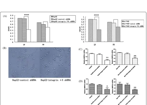
It has been mentioned above that adhesion and spreading is the first step of the model for tumor metastasis reported by Liotta et al., so we performed adhesion and spreading assays in vitro. These yielded results showing that the
adhesion ability reduced remarkably in the HCC cells with lower-level expression of integrin α6 (P< 0.0001) (Figure 4A). On spreading, the results showed that HepG2 cells with lower level of integrin α6 had a lower rate of spreading (2.4% in the HepG2-shRNA-integrin α6 and 94% in the HepG2) (Figure 4B). The qRT-PCR assay was used to detect the relative integrin MMP-2 and MMP-9 levels. Compared with control group, the rela-tive MMP-2 and MMP-9 mRNA reduced remarkably (P< 0.01) (Figure 4C, D). Thus, the down-regulation of MMP-2 and MMP-9 secretion, adhesion and spreading ability may cause the low invasion potential of HCC cells.
Figure 1HepG2 and Bel-7402 cells were infected with integrinα6 short hairpin RNA (shRNA) plasmids and control shRNA plasmids respectively.Stable transfectant clones were picked up by puromycin. Integrinα6 expression levels remarkably reduced.(A)Western blot assays yielded the expression level of integrinα6 on protein level in HepG2 and Bel-7402 cell lines.(B)The qRT-PCR assays showed the expression level of integrinα6 on mRNA level in HepG2 and Bel-7402 cell lines. *P< 0.05; ***P< 0.001 compared with control group.
Figure 3Cells were cultured in Transwell chambers with the upper part coated with laminin (LN) or fibronectin (FN) respectively for 48 hours.The invaded cells were numerated under a light microscope (×400).(A)HepG2 infected by control shRNA plasmids invaded LN.(B) HepG2 cells infected by control shRNA plasmids invaded FN.(C)HepG2 cells infected by integrinα6 shRNA plasmids invaded LN. HepG2 had a decreased invasion potential after the knockdown of integrinα6 with significant statistical difference (P< 0.0001). It also proved that LN can increase invasion potential of HepG2 (P< 0.05).
Integrinα6 reacting with LN induced the hyperactivation of PI3K/AKT and MAPK/ERK signal pathways
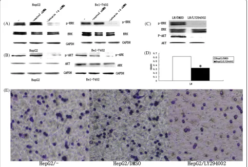
As shown in Figure 5, compared with the control group, the protein grayscale value of p-ERK, p-AKT was dra-matically reduced in HepG2 cells transfected with integ-rin α6 shRNA plasmids compared with control shRNA plasmids (Figure 5A, B). As revealed in Figure 5C, LY294002 decreased both the expression of p-ERK and p-AKT, but had no influence on the expression of ERK and AKT. The proliferation of HepG2 cells was remark-ably decreased after being inhibited by PI3K (P< 0.05) (Figure 5D). In Transwell assays, 38.33 ± 3.78 cells crossed LN in the HepG2/LY294002 group, while 64.0 ± 2.67 cells in the HepG2/DMSO group and 72.33 ± 3.11 cells in HepG2 group (P< 0.05) (Figure 5E) crossed LN. These results demonstrated that the reacting of integrin
α6 with LN induced the hyperactivation of PI3K/AKT and MAPK/ERK signal pathways.
Discussion
Metastatic recurrence is the main obstacle to the im-provement of treatment efficacy for HCC [13], and het-erodimeric transmembrane protein generated by integrin
α6 subunits is suggested to be associated with metastasis potential of HCC. In this study, we used LN, the import-ant structural component of ECM and integrin α6 lig-and, as a model molecule and applied several assays to determine the importance of integrinα6 in the metasta-sis of HCC cells. In order to make the results more ob-jective, the integrin α6 shRNA was used to silence the expression of integrin α6 of two HCC cell lines at RNA level. Western blot and qRT-PCR detection showed that over 75% of integrin α6 expression in HCC cells was through knockdown by shRNA.
Tumor metastasis is a multi-step process involving various molecules, pathways and organs. This process mainly includes the following steps before invasion to
form micrometastases in the metastatic site: the detach-ment of tumor cells from a primary tumor, invasion through the stromal tissues, intravasation into the blood/lymph vessels, arrest in capillary bed followed by extravasation and local crawling [14].
Based on the results of our assays, lower-level integ-rinα6 expression down-regulated the secretion of matrix metalloproteinases (MMPs), the adhesion force between HCC cells and LN, the spreading ability on the LN, as well as the invasion potential of HCC cells. p-ERK and p-AKT were reduced by shRNA targeting integrin α6 and PI3K inhibitor LY294002. LN could stimulate the MMPs secreted by HCC and thus was associated with a more metastatic phenotype of HCC cells [15]. The phos-phorylation level of ERK and AKT (p-ERK and p-AKT), which has been reported to be increased after combining integrinα6 with LN [16,17], is associated with a low sur-vival rate for HCC patients [17]. Furthermore, influen-cing factors increase the secretion of MMP-9 through the activation of the MAPK/ERK signal pathway, which is conducive with HCC metastasis [18]. The same results have been found in the PI3K/AKT signal by Chenet al. [19].
A model for tumor metastasis reported by Liottaet al. [20] in the early 1980s contributes in large part to the metastatic process. The model mentions that there are three stages for the complicated interactions between tumor cells and ECM: the adhesion of tumor cells to the ECM, secretion of proteolytic enzymes, such as MMPs, directly by the tumor cells or indirectly by the host cells as a response to the presence of tumor cells, and finally, cell movement by chemotaxis or haptotaxis. Although most integrin family members recognize more than one ECM protein, integrin α6 is a ligand with specificity for LN [21,22]. The signal pathways, MAPK/ERK and PI3K/ AKT have been proved to be necessary for HCC metas-tasis [19,23]. Thus, we can hypothesize that the combin-ation of integrin α6 with LN could influence the HCC metastasis by affecting cell adhesion and migration, and reduced expression level of integrin α6 may cause low potential of HCC metastasis through the activation of MAPK/ERK and PI3K/AKT signal pathways. There also exist cross-talk between the two signaling pathways acti-vated by LN [18,19]. Integrinα6 contributes to the regu-lation of interaction between HCC cells and LN through PI3K/AKT and MAPK/ERK signaling pathways which are shown to be essential for the metastasis of HCC [6].
Conclusions
Down-regulation of integrin α6 could decrease the po-tential of HCC metastasis through reducing the inter-action between HCC and LN. This study showed that over-expression of integrinα6 in HCC is a strong indi-cator for more aggressive tumors and poorer clinical
outcome. All the currently available therapies for HCC are mainly oriented towards removing or destroying the tumors, whereas few therapies aim at blocking the process of HCC metastasis [24]. This study would help us to understand the metastatic process of HCC better and develop targeted molecular therapy to improve pa-tients’survival.
Abbreviations
HCC:Hepatocellular carcinoma; shRNA: Short hairpin RNA; qRT-PCR: Real-time quantitative reverse transcription-polymerase chain reaction; MMPs: Matrix metalloproteinases; ECM: Extracellular matrix; LN: Laminin; Ct: Cycle threshold; DMSO: Dimethyl sulfoxide; FN: Fibronectin.
Competing interests
The authors declare that they have no competing interests.
Authors’contributions
GNL conducted experiments, prepared manuscript and analyzed data. TJLand SFQ performed experiments, revised manuscript and analyzed data. WL performed experiments. WRG performed experiments. XHZ erformed experiments. JKW designed experiments and gave final approval of manuscript. All authors read and approved the final manuscript.
Acknowledgement
This project was supported by the Department of Education, Liaoning Province, China (L2010288).
Author details
1The First Affiliated Hospital of Liaoning Medical College, Jinzhou 121001,
China.2Institute of Urology, Peking University First Hospital, Peking
University, Beijing 100034, China.
Received: 25 September 2013 Accepted: 14 November 2013 Published: 4 December 2013
References
1. Sengupta B, ASiddiqi S:Hepatocellular carcinoma: important biomarkers and their significance in molecular diagnostics and therapy.Curr Med Chem2012,19:3722–3729.
2. Zhu K, Dai Z, Pan Q, Wang Z, Yang G-H, Yu L, Ding Z-B, Shi G-M, Ke A-W, Yang X-R:Metadherin promotes hepatocellular carcinoma metastasis through induction of epithelial-mesenchymal transition.Clin Cancer Res 2011,17:7294–7302.
3. Desgrosellier JS, Cheresh DA:Integrins in cancer: biological implications and therapeutic opportunities.Nat Rev Cancer2010,10:9–22. 4. Fei Y, Wang J, Liu W, Zuo H, Qin J, Wang D, Zeng H, Liu Z:CD151
promotes cancer cell metastasis via integrins alpha3beta1 and alpha6beta1 in vitro.Mol Med Rep2012,6:1226–1230.
5. Stipp CS:Laminin-binding integrins and their tetraspanin partners as potential antimetastatic targets.Expert Rev Mol Med2010,12:e3. 6. Ke AW, Shi GM, Zhou J, Huang XY, Shi YH, Ding ZB, Wang XY, Devbhandari
RP, Fan J:CD151 amplifies signaling by integrinα6β1 to PI3K and induces the epithelial–mesenchymal transition in HCC Cells. Gastroenterology2011,140:1629–1641. e1615.
7. Choi YP, Kim BG, Gao M-Q, Kang S, Cho NH:Targeting ILK andβ4 integrin abrogates the invasive potential of ovarian cancer.Biochem Biophys Res Commun2012,427:642–648.
8. Fu B-H, Wu Z-Z, Qin J:Effects of integrinα6β1 on migration of hepatocellular carcinoma cells.Mol Biol Rep2011,38:3271–3276. 9. Carloni V, Mazzocca A, Pantaleo P, Cordella C, Laffi G, Gentilini P:The
integrin, alpha6beta1, is necessary for the matrix-dependent activation of FAK and MAP kinase and the migration of human hepatocarcinoma cells.Hepatology2001,34:42–49.
10. Owens DM, Romero MR, Gardner C, Watt FM:Suprabasal alpha6beta4 integrin expression in epidermis results in enhanced tumorigenesis and disruption of TGFbeta signaling.J Cell Sci2003,116:3783–3791. 11. Bergamini C, Sgarra C, Trerotoli P, Lupo L, Azzariti A, Antonaci S, Giannelli G:
different function ofα6β4 andα3β1 integrins.Hepatology2007, 46:1801–1809.
12. Lu T, Zhang Q, Zhou R:Laminin and its alpha 6 integrin receptor in the regulation of human hepatocellular carcinoma cell phenotypes. Zhonghua Zhong Liu Za Zhi2003,25:31–35.
13. Ding S-J, Li Y, Tan Y-X, Jiang M-R, Tian B, Liu Y-K, Shao X-X, Ye S-L, Wu J-R, Zeng R:From proteomic analysis to clinical significance overexpression of cytokeratin 19 correlates with hepatocellular carcinoma metastasis. Mol Cell Proteomics2004,3:73–81.
14. Geho DH, Bandle RW, Clair T, Liotta LA:Physiological mechanisms of tumor-cell invasion and migration.Physiology2005,20:194–200. 15. Giannelli G, Fransvea E, Bergamini C, Marinosci F, Antonaci S:Laminin-5
chains are expressed differentially in metastatic and nonmetastatic hepatocellular carcinoma.Clin Cancer Res2003,9:3684–3691.
16. Givant-Horwitz V, Davidson B, Reich R:Laminin-induced signaling in tumor cells.Cancer Lett2005,223:1–10.
17. Schmitz KJ, Wohlschlaeger J, Lang H, Sotiropoulos GC, Malago M, Steveling K, Reis H, Cicinnati VR, Schmid KW, Baba HA:Activation of the ERK and AKT signaling pathway predicts poor prognosis in hepatocellular carcinoma and ERK activation in cancer tissue is associated with hepatitis C virus infection.J Hepatol2008,48:83–90.
18. Weng C-J, Chau C-F, Hsieh Y-S, Yang S-F, Yen G-C:Lucidenic acid inhibits PMA-induced invasion of human hepatoma cells through inactivating MAPK/ERK signal transduction pathway and reducing binding activities of NF-κB and AP-1.Carcinogenesis2008,29:147–156.
19. Chen J, Wang Q, Fu X, Huang XH, Chen X, Cao L, Chen L, Tan H, Li W, Bi J: Involvement of PI3K/PTEN/AKT/mTOR pathway in invasion and metastasis in hepatocellular carcinoma: association with MMP‐9.Hepatol Res2009,39:177–186.
20. Pauli BU, Schwartz DE, Thonar EJ-M, Kuettner KE:Tumor invasion and host extracellular matrix.Cancer Metastasis Rev1983,2:129–152.
21. Humphries JD, Byron A, Humphries MJ:Integrin ligands at a glance.J Cell Sci2006,119:3901–3903.
22. Morgan MR, Humphries MJ, Bass MD:Synergistic control of cell adhesion by integrins and syndecans.Nat Rev Mol Cell Biol2007,8:957–969. 23. Huynh H, Nguyen TTT, Chow K-HP, Tan PH, Soo KC, Tran E:Over-expression
of the mitogen-activated protein kinase (MAPK) kinase (MEK)-MAPK in hepatocellular carcinoma: its role in tumor progression and apoptosis. BMC Gastroenterol2003,3:19.
24. Arosio D, Casagrande C, Manzoni L:Integrin-mediated drug delivery in cancer and cardiovascular diseases with peptide-functionalized nanoparticles.Curr Med Chem2012,19:3128–3151.
doi:10.1186/2047-783X-18-52
Cite this article as:Lvet al.:RNA interference targeting human integrin
α6 suppresses the metastasis potential of hepatocellular carcinoma cells.European Journal of Medical Research201318:52.
Submit your next manuscript to BioMed Central and take full advantage of:
• Convenient online submission
• Thorough peer review
• No space constraints or color figure charges
• Immediate publication on acceptance
• Inclusion in PubMed, CAS, Scopus and Google Scholar
• Research which is freely available for redistribution