S T U D Y P R O T O C O L
Open Access
Aortic valve replacement in elderly with
small aortic root and low body surface
area; the Perceval S valve and its impact in
effective orifice area
Panagiotis Dedeilias
1, Nikolaos G. Baikoussis
1*, Efstathia Prappa
2, Dimitrios Asvestas
2, Michalis Argiriou
1and Christos Charitos
1Abstract
Background:The aim of this study is to see how the sutureless, stentless, Perceval S aortic valves behave when implanted in elderly patients with small aortic root and the comparison with a second group of patients with similar characteristics where a conventional stented bioprosthesis was implanted. This is a prospective randomized institutional study.
Methods:Our material is composed from 25 patients who underwent aortic valve replacement with sutureless self-anchoring Perceval S valve implantation (LivaNova), compared with 25 patients with conventional stented
biological prosthesis implanted (soprano LivaNova group). The two groups of patients have similar demographic and medical characteristics with severe aortic stenosis. The study was conducted from January 2012 to June 2014. Preoperative, intraoperative and postoperative parameters were studied in order to investigate the utility of the Perceval S valves in this group of patients.
Results:The Perceval S valve implantation seems to be an interesting biological valve with good hemodynamic characteristics as compared with the typical biological prosthesis providing shorter ischemia time (40 ± 5.50 min vs 86 ± 15.86 min;p< 0.001), shorter extracorporeal circulation time (73.75 ± 8.12 min vs 120.36 ± 28.31 minp< 0.001), less operation time (149.38 ± 15.22 min vs 206.64 ± 42.85 min;p< 0.001) and better postoperative recovery. The postoperative gradients were 23.5 ± 19.20 mmHg vs 24.5 ± 19.90 mmHg respectively. The postoperative effective orifice area in these two groups were respectively 1.5 =/-0.19 cm2vs 1.1=/-0.5 cm2(p 0.002). Among the 25 patients of the Soprano stented valve, 3 (12 %) came back in 6 months with New York Heart Association (NYHA) 3. The PPM of these patients was the cause of readmission in the Hospital required diuresis and supplementary treatment.
Conclusions:Aortic valve replacement with Perceval aortic valves in geriatric patients with comorbidities and small aortic annulus seems to be an alternative, safe and“fast”intervention with excellent short and mid-term results which provides a better effective orifice area.
Keywords:Perceval S, Aortic valve, Stentless aortic valve, Sutureless valve, Self-expanding valve, Aortic valve stenosis, Heart valve surgery
* Correspondence:[email protected]
1Cardiothoracic and Vascular Surgery Department,“Evangelismos”General Hospital of Athens, 45-47 Ipsilantou Street, Kolonaki, Athens, Greece Full list of author information is available at the end of the article
Background
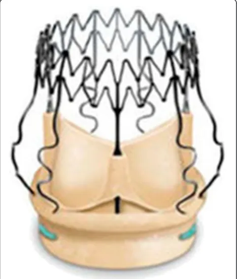
The sutureless Perceval bioprosthesis (LivaNova Biomedica Cardio Srl, Sallugia, Italy) was designed in order to obtain the hemodynamic benefits of the stentless valves without the increased difficulty in surgical implantation [1]. This valve is a bioprosthesis comprising a bovine pericardium tissue valve attached to a self-expanding anchoring device (Fig. 1), which has the dual role of supporting the biopros-thetic valve and offering fixation to the implantation site in the native aortic annulus [1]. As a result of the sutureless implant procedure, patients could benefit from reducing aortic cross-clamp time, with subsequent overall reduction of the surgical duration and reduction in related risks by avoiding passing the stitches through the calcified annulus and sutures knotting, with subsequent less risk of tearing the annulus and aortic wall or embolizing the systematic circulation [2]. In a small and calcified annulus it can be challenging to insert a stented valve and a significant re-sidual gradient is frequently observed afterwards. Stentless valves are designed in order to overcome some of the disad-vantages of the stented valves [1, 3, 4]. This device with its three button holes provides the correct positioning of the valve in the native aortic root (Fig. 2). In order to minimize or avoid the paravalvular leakage, the Perceval S valve is designed with an intra–annular and a supra–annular sealing collar (Fig. 3). This device is the ideal solution for
elderly patients who require a rapid procedure and for patients with small aortic root which require root enlarge-ment. As known, an aortic root enlargement (Nikcs-Nunez or Manougian technique) may be necessary in small annu-lus in order to avoid a “patients-prosthesis mismatch” [5]. This operation is challenging in elderly patients with comorbidities and heavily calcified aorta which a rapid intervention is necessary. The prosthetic implant is sup-ported by dedicated tools: crimping system, manometer and dilatation balloon (Fig. 4). Prior to its implantation the prosthesis diameter is reduced to a suitable size, using the Perceval S collapsing tool, and then loaded on the Perceval S special holder (Fig. 5). After in situ positioning the valve is released in two steps: first the inflow ring is released at the native aortic annulus level and then, when proper posi-tioning is verified, the complete prosthesis release is achieved (Fig. 6). After the implantation the Perceval S post-dilation balloon catheter is inflated inside the pros-thesis at the inflow level to improve apposition by modeling the inflow ring on the native annulus [1, 6, 7]. In vitro ac-celerated fatigue tests were performed under normal and hypertensive conditions. These tests demonstrated that the whole device remains functional up to 900 Million cycles (more than 20 year of normal equivalent life). These results exceed the minimal ISO and FDA requirements and suggest a wide safety margin of the Perceval S bioprosthesis [1, 7]. We report our experience with the Perceval S
Fig. 1The Perceval S aortic valve is a bioprosthesis comprising a bovine pericardium tissue valve attached to a self-expanding anchoring device
Fig. 2This device through its three button holes provides the correct positioning of the valve in the native aortic root
bioprosthesis implanted onto 25 elderly with small aortic annulus and low BSA and we compared this group with 25 similar patients receiving conventional stented bioprosth-esis. Our study is conducted in order to investigate two end points: effectiveness and performance so we will basically see its hemodynamic profile and its impact on effective ori-fice area other than the whole clinical outcome of the pa-tients in the early postoperative period (within a month following surgery).
Methods
In our study 25 sutureless self-anchoring, Perceval S, (LivaNova Biomedica Cardio Srl, Sallugia, Italy) valves are compared with 25 conventional biological stented pros-thesis (soprano- Sorin Group) implanted onto similar characteristic patients with severe aortic stenosis. The study was conducted from January 2012 to June 2014. Pa-tients were randomized divided into groups and they have previously consented for either method of surgical treat-ment. Randomization was done via a computerized assisted mathematic model. The inclusion criteria were defined as to see how the elderly patients with small aortic root and low BSA who are the real life difficult patients, will benefit from the use of a novel sutureless self-anchoring biological prosthesis. Limitations of this study were defined in a country with strict economic environ-ment. The mean EuroSCORE II was 9.5 ± 3.5 in the Perce-val group and 9.9 ± 3.6 in the conventional group. The
BSA in m2was 1.45 ± 1.2 and 1.78 ± 1.1. The rest patient’s characteristics are showing in the Table 1. The hemoglobin level was 33.3 g/L in the Perceval S group and 32.8 in the Soprano stented group preoperatively. Then, postoperatively, the hemoglobin was 28.6 in the Perceval S group and 28.8 in the second group. So, we did not find any statistically significate difference between the groups. All patients were treated with median full sternot-omy, routine cannulation to the extracorporeal circulation with Edwards aortic cannula at the distal part of the as-cending aorta and a two-stage venous cannula at the right atrium. Retrograde cardioplegia plus elective cardioplegia to the right coronary artery (ARC) was given in all pa-tients. There was just one initial dose of cardioplegia given to all patients accompanying by local cooling with ice slush. No systematic extra cooling was required in our patients. The cross clamp was applied as distal as possible and aortotomy was performed approximately 2 cm above the sino-tubular junction (STJ). For the cases of conven-tional stented valve implantation an extension of the aor-totomy towards the non-coronary sinus was performed. Fig. 4The prosthetic implant is supported by dedicated tools:
crimping system, manometer and dilatation balloon
Fig. 5Prior to its implantation the prosthesis diameter is reduced to a suitable size, using the Perceval S collapsing tool, and then loaded on the Perceval S special holder
Fig. 6Using the three guides it performs in situ positioning of the valve. It is released in two steps: first the inflow ring is released at the native aortic annulus level and then, when proper positioning is verified, the complete prosthesis release is achieved
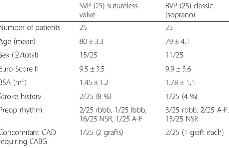
Table 1Preoperative patient’s characteristics and demographics
SVP (25) sutureless valve
BVP (25) classic (soprano)
Number of patients 25 25
Age (mean) 80 ± 3.3 79 ± 4.1
Sex (♀/total) 15/25 11/25
Euro Score II 9.5 ± 3.5 9.9 ± 3.6
BSA (m2) 1.45 ± 1.2 1.78 ± 1,1
Stroke history 2/25 (8 %) 1/25 (4 %)
Preop rhythm 2/25 rbbb, 1/25 lbbb, 16/25 NSR, 1/25 A-F
3/25 rbbb, 2/25 A-F, 15/25 NSR
Concomitant CAD requiring CABG
1/25 (2 grafts) 2/25 (1 graft each)
Pledged interrupted inverted stiches were met for the stented valve suturing. This is a prospective randomized study. From our study were excluded patients with previous cardiac surgery (redo operations), patients with aortic in-sufficiency, large aortic root (> or = 30 mm), sino-tubular junction periphery/height from the annulus > or = 1.3. Also, patients with BSA > or = 2 m2 and patients younger than 75 years old were excluded from our study.
Ethical approval for this clinical study was obtained by our Hospital (General Hospital of Athens), Scientific Committee.
Results
No structural prosthesis deterioration, valve thrombosis, or significant transvalvular aortic regurgitation occurred during the study period. There were no cases of tilting or migration once appropriately inserted during the en-tire study. This sutureless bioprosthesis appears to be ideal for patients with severe calcification of the aortic root and patients requiring concomitant procedures in whom a reduced bypass time is mandatory [1]. Cross clamp time and cardio-pulmonary bypass (CPB) time are reported at Table 2. All echo-cardiographic measure-ments were assessed by transthoracic echo. The annulus size was between 21 and 30 mm. We have implanted 18 perceval S valves size small, five medium and two large. In the conventional group we implanted 18 Soprano stented valves of the 21 mm and 4 Soprano stented valves of 20 mm. Postoperative EOA was 1.5 ± 0.3 cm2 in the Perceval group vs 1.1 ± 0.5 in the conventional group (p 0.002). The mean EOA index was 1.034 in the Perceval S group, while in the conventional group the EOA index was 0.617. This is the reason why three of
the patients with Soprano stented valve came back in 6 months with New York Heart Association (NYHA) 3. The PPM of these patients was the cause of readmission in the Hospital required diuresis and supplementary treatment. This is the most important result coming out from our study. Operation time, CPB time and cross clamp time were significantly lower in the Perceval group (p< 0.001) as we could see in Table 2. These data are very important in cardiac surgery procedure and es-pecially in elderly with comorbidities. The postoperative echo measurements were made within a month of period following surgery.
Conclusions
We studied the behavior of the Perceval S sutureless stent-less bioprosthesis in patients with small aortic annulus, small BSA and older than 75 years. It seems that this is the target group of this valve. This group of patients with co-morbidities and calcified aorta needs a rapid operation with minimal aortic manipulation. Age itself is not a contraindi-cation to conventional surgery but comorbidities such as low ejection fraction, renal dysfunction and calcified aorta are major risk factor for mortality and morbidity [1, 8]. Ac-cording the international bibliography [9], cross clamp time is an independent predictor of mortality and morbidity in low and high-risk cardiac patients. They found that pro-longed aortic cross clamp time significantly correlated with worse clinical outcomes. The spectrum of complications in-cluded in-hospital mortality, prolonged hospitalization, pro-longed ventilation, low cardiac output, higher requirements for blood transfusion and renal complications. In Perceval group patients, cross clamp time is significantly shorter
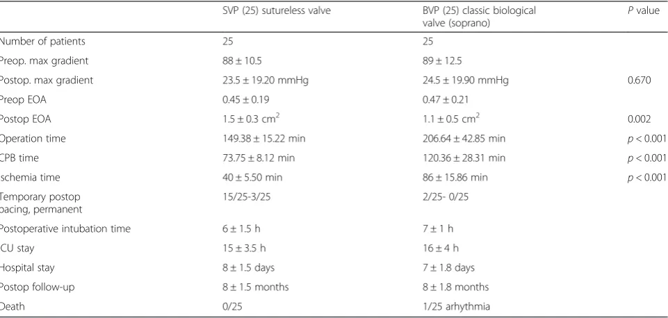
Table 2Peroperative date and results
SVP (25) sutureless valve BVP (25) classic biological valve (soprano)
Pvalue
Number of patients 25 25
Preop. max gradient 88 ± 10.5 89 ± 12.5
Postop. max gradient 23.5 ± 19.20 mmHg 24.5 ± 19.90 mmHg 0.670
Preop EOA 0.45 ± 0.19 0.47 ± 0.21
PostopΕΟΑ 1.5 ± 0.3 cm2 1.1 ± 0.5 cm2 0.002
Operation time 149.38 ± 15.22 min 206.64 ± 42.85 min p< 0.001
CPB time 73.75 ± 8.12 min 120.36 ± 28.31 min p< 0.001
Ischemia time 40 ± 5.50 min 86 ± 15.86 min p< 0.001
Temporary postop pacing, permanent
15/25-3/25 2/25- 0/25
Postoperative intubation time 6 ± 1.5 h 7 ± 1 h
ICU stay 15 ± 3.5 h 16 ± 4 h
Hospital stay 8 ± 1.5 days 7 ± 1.8 days
Postop follow-up 8 ± 1.5 months 8 ± 1.8 months
Death 0/25 1/25 arhythmia
than conventional group. This is an important advantage of this valve. Patients with low left ventricle ejection fraction (LVEF) are also candidates for this valve in order to implant rapidly an aortic valve without long ischemic time and con-sequently myocardial injuries. Due to the changing popula-tion demographics, the age of the patients presenting for AVR is also increasing [10]. A smaller-sized prosthetic valve may result in so-called patient-prosthesis mismatch (PPM). Therefore, different options have been proposed for pa-tients with small aortic root presenting for AVR [5, 10]. Aortic root enlargement in elderly patients with heavily calcified aorta and comorbidities is a challenging operation with prolonged cross clump time and possible complica-tions intra and postoperatively. In our opinion, old patients with renal dysfunction or history of cerebrovascular dis-eases may benefit from this kind of operation due to dimin-ished operation time and less aortic manipulation. This valve may enable a broader application of minimally inva-sive AVR. Further longer-term experience is needed to determine the potential clinical benefits and durability of the Perceval S self-anchoring valve [10]. In case of valve malposition, there is the possibility of removal and re-implantation according the literature [11]. The valve im-plantation is possible with partial“j”sternotomy at the third or fourth intercostal space [12] in order to minimize the chest wall trauma and the risk of chest instability or infec-tion. The studies and the international bibliography confirm the safety, efficacy, and ease of insertion of Perceval valves in elderly patients with small annulus [13–15]. As these valves do not need to be‘sutured’, shorter cross-clamp and CPB times are possible. Moreover, due to the absence of a sewing ring, these valves are also almost ‘stentless’, with a greater valve EOA for any given size. This may therefore re-sult in better hemodynamic even without the root enlargement [10]. According the literature [6], Perceval S valve could be implanted in elderly patients who require concomitant cardiac operation in order to minimise the op-eration time. Sutureless valves may be advantageous com-pared to transcatheter valve implantations as concomitant procedures other than percutaneous coronary artery angio-plasty are not always possible in the latter [6, 15]. Accord-ing our results, patients older than 75 years, with small aortic annulus and small BSA may benefit from a Perceval S aortic valve implantation. The limitation of our study is the small number of patients that were randomized into the two groups. However it is practically very difficult to study a large group of patients with the same characteristics in a single center. There is a large multicenter randomized trial going on and the results are expected with great interest. At the present time, the Perceval S prosthesis has been investi-gated in three clinical studies: 1. The “PERCEVAL TRIAL-Perceval S valve pilot trial-v10601”, 2. The “PERCEVAL Pivotal Trial – v10801”, 3. The “CAVALIER – Perceval S valve clinical trial for extended CE mark-TPS001”[7].
Competing interests
The authors declare that they have no competing interests.
Authors’contributions
PD study design, collection of the data, perform of interventions, final approval, NB: collection of bibliographic data, write the manuscript, participation in interventions, submission. EP: perform the peroperative echocardiography and participation of study design. DA: perform of echocardiography, participation of statistical analysis. MA: perform of some intervention, design of the study. CC: design of the study and supervision. All authors read and approved the final manuscript.
Author details
1Cardiothoracic and Vascular Surgery Department,“Evangelismos”General Hospital of Athens, 45-47 Ipsilantou Street, Kolonaki, Athens, Greece. 2
Department of Cardiology,“Evangelismos”General Hospital of Athens, Athens, Greece.
Received: 18 August 2015 Accepted: 3 April 2016
References
1. Folliguet TA, Laborde F, Zannis K, Gorayeb G, Haverich A, Shrestha M. Sutureless Perceval Aortic Valve Replacement: Results of Two European Centers. Ann Thorac Surg. 2012;93:1483–8.
2. Shrestha M, Foliiguet T, Meuris B, Dibie A, Bara C, Herregods MC, Meuris B, et al. Suturuless Perceval S aortic valve replacement: a multicenter, prospective pilot trial. J Heart Valve Dis. 2009;18:698–702.
3. Reposini A, Kolelnikov I, Bouchikhi R, Torre T, Passaretti B, Parodi O, et al. Single suture line placement of a pericardial stentless valve. J Thorac Cardiovasc Surg. 2005;130:1265–9.
4. David TE, Feindel CM, Bos J, Sun Z, Scully HE, Rakowski H. Aortic Valve replacement with a stentless porcine aortic valve. A six-year experience. J Cardiovasc Surg. 1994;108:1030–6.
5. Apostolakis E, Baikoussis NG, Papakonstantinou NA, Goudevenos J. Patient-prosthesis mismatch and strategies to prevent it during aortic valve replacement. Hellenic J Cardiol. 2011;52(1):41–51.
6. Shrestha M, Folliguet TA, Pfeiffer S, Meuris B, Carrel T, Bechtel M. Aortic Valve Replacement and Concomitant Procedures With the Perceval Valve: Results of European Trials. Ann Thorac Surg. 2014;98(4):1294–300. doi:10.1016/j.athoracsur.2014.05.033. Epub 2014 Aug 5.
7. Thierry C, Englberger L, Stalder M. Recent developments for surgical aortic valve replacement: The concept of sutureless valve technology. Switzerland: Clinic for Cardiovascular Surgery, University Hospital Berne. Volume 4. 2013. ISSN: 2075-9010.
8. Langanay T, Flécher E, Fouquet O, Ruggieri VG, De La Tour B, Félix C, et al. Aortic valve replacement in the elderly: the real life. Ann Thorac Surg. 2010;93:70–8. 9. Al-Sarraf N, Thalib L, Hughes A, Houlihan M, Tolan M, Young V, et al.
Cross-clamp time is a independent predictor of mortality and morbidity in low-and high–risk cardiac patients. Int J Surg. 2011;9:104–9.
10. Shrestha M, Maeding I, Höffler K, Koigeldiyev N, Marsch G, Siemeni T, et al. Valve replacement in geriatric patients with small aortic roots: are sutureless valves the future? Interact Cardiovasc Thorac Surg. 2013;17(5):778–82. 11. Santarpino G, Pfeiffer S, Concistrè G, Fischlein T. A supra-annular malposition
of the Perceval S sutureless aortic valve: the 'χ-movement' removal technique and subsequent reimplantation. Interact Cardiovasc Thorac Surg. 2012;15(2):280–1.
12. Santarpino G, Pfeiffer S, Concistrè G, Fischlein T. Perceval S aortic valve implantation in mini-invasive surgery: the simple sutureless solution. Interact Cardiovasc Thorac Surg. 2012;15(3):357–60.
13. Meuris B, Flameng WJ, Laborde F, Folliguet TA, Haverich A, Shrestha M. Five-year results of the pilot trial of a sutureless valve. J Thorac Cardiovasc Surg. 2015;150(1):84–8.
14. Zannis K, Joffre J, Czitrom D, Folliguet T, Noghin M, Lansac MN, et al. Aortic valve replacement with the perceval S bioprosthesis: single-center experience in 143 patients. J Heart Valve Dis. 2014;23(6):795–802. 15. Villa E, Messina A, Laborde F, Shrestha M, Troise G, Zannis K, et al. Challenge