subject of angiitis will be briefly addressed. A special effort will be made to relate renal biopsy findings to the immunologically mediated pathogenetic process which is thought to be operative in each case. Where it seems appropriate, a few comments will also be made on clinical and pathological correlations. The specific entities to be covered include: diffuse prolifer-ative glomerulonephritis; focal proliferprolifer-ative glomeru-lonephritis; membranous glomerulonephritis; a nti-basement membrane antibody disease; rapidly progressive glomerulonephritis ( crescentic disease); membranoproliferative glomerulonephritis; lipoid nephrosis (nil disease); focal, segmental, and global sclerosis; polyarteritis nodosa; hypersensitivity angi-itis; and Wegener granulomatosis. Few comments will be made about therapy because that subject is covered elsewhere in this issue. The review will be concluded by a discussion of the prognostic value of information gleaned from careful biopsy evaluation.
Two types of immunologic injury to the kidney have been clearly defined in experimental models and are thought to have distinct clinical counterparts.1 These are immune-complex mediated disease and antibasement membrane antibody disease. The former is the most common and will be considered first.
Immune-Complex Glomerulonephritis
The pathogenesis of several glomerulonephritic patterns to be discussed subsequently seems to be of
Correspondence and reprint requests to Dr. William F. Falls, Jr., Renal Section, VA Hospital, Richmond, VA 23249.
116
and is delivered to immunologically competent cells which begin making antibody in response to the chal-lenge; as antibody is released into the circulation, it combines with antigen to form complexes. Under certain circumstances of antigen excess, the com-plexes which are formed are small and soluble and are not phagocytized in the reticuloendothelial sys-tem; these activate the complement cascade via the classic pathway (C1 __, C4 __, C2 __, C3 ), and the entire aggregate of antigen, antibody, and complement components precipitates in the region of the glomeru-lar basement membrane. The terminal components of the complement system include substances which in-crease vascular permeability and are leukotactic. An increase in the permeability of the capillary wall leads to leakage of protein into Bowman space and the influx of leukocytes may cause proteolytic destruc-tion of pordestruc-tions of the glomerular capillary wall; in response to this assault, endothelial and mesangial cell proliferation occur. Thus, any antigen capable of stimulating an antibody response is a potential cause of immune-complex mediated injury. As we shall see, numerous inciting antigens have been identified, but the pattern of injury may vary considerably, probably depending on the nature of the antigen and the re-sponsiveness of the host.2
Our understanding of the nature of immunologic injury to the glomerular capillary has been enhanced greatly by the development of immunofluorescent staining techniques. In this process a quick-frozen renal biopsy section is treated with a fluoresce nt-tagged antibody against a specific class of human globulin, complement component, or fibrinogen.
Fig 1-Immunofluorescent microscopic preparation (anti-IgG) of a portion of a glomerulus from a patient with poststreptococcal glomerulonephritis. Note "lumpy-bumpy" pattern. (X 1500).
When this preparation is viewed with fluorescent mi-croscopy, a clear picture of the area of protein depo-sition is obtained. In the case of immune-complex mediated disease, a "lumpy-bumpy" pattern will be observed and is the hallmark of this type of injury.
Diffuse Proliferative Glomerulonephritis
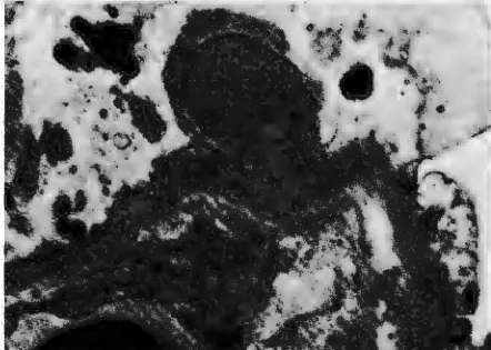
Figure 1 is a photomicrograph of the im-m unofluorescent staining for lgG in a patient with acute poststreptococcal glomerulonephritis. Note the coarse, granular deposition of the immuno-fluorescent material in the area of the capillary base-ment membrane and the mesangium. Figure 2 is the light microscopic correlate of this lesion and shows the characteristic picture of diffuse, proliferative glo-merulonephritis. The glomerulus is swollen and the capillary loops are occluded by proliferating mesan-gial and endothelial cells; there is marked hyper-cellularity and an influx of foreign inflammatory cells is noted. Figure 3 is a high-power view of an electron micrograph which demonstrates the classjc appear-ance and location of the electron-dense deposits which are thought to represent the deposited com-plexes. Note that the deposits are quite large, "hump-like," and located in a subepithelial position; the foot processes in the area of the deposit have become fused. This phenomenon of foot process fusion has been found to correlate closely with the presence of proteinuria.
The immunofluorescent and light microscopic picture described above is typical of the aggressive type of immune-complex mediated disease
character-Fig 2-Light microscopic preparation (H & E) of a portion of renal biopsy from a patient with diffuse proliferative glomerulone-phritis following streptococcal infection (X200).
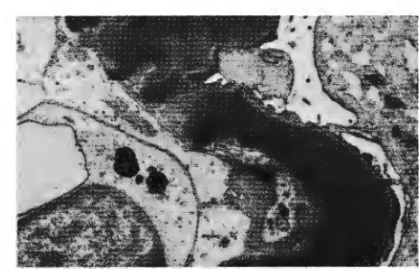
ized by poststreptococcal glomerulonephritis but is by no means specific to it. A similar light micro-scopic appearance may be seen in some patients with lupus erythematosus, bacterial endocarditis, nephritis associated with infected ventriculojugular shunts, cryoglobulinemic nephropathy, and other antigenic insults.2 Diffuse proliferative glomerulonephritis sec-ondary to lupus erythematosus can usually be distin-guished from poststreptococcal disease because the electron-dense deposits in lupus are usually located on the subendothelial surface of the basement mem-brane (Fig 4) and discrete viruslike particles may also be evident.2 The site of deposition in other diseases is
Fig 4-Electron microscopic preparation of a portion of a glomer-ulus from a patient with systemic I upus erythematosus and a diffuse proliferative lesion by light microscopy. Note large
sub-endothelial deposits (X 10,000).
variable, but in many cases with proliferative lesions they are subendothelial.
In most cases of diffuse proliferative glomerulo-nephritis associated with lupus, one can find serologic evidence of activation of the classic complement pathway (low serum C,q _, C2 _ , C4 _ , C3 ).2 Such changes are variable in other diseases with prolifera-tive lesions and in some cases of poststreptococcal disease there may also be evidence of activation of the alternate complement pathway. In this circumstance C3 is activated directly, bypassing the earlier com-ponents. A number of substances are known to
acti-Fig 5-Light microscopic preparation (H & E) of a portion of a renal biopsy from a patient with systemic lupus erythematosus,
showing a focal proliferative lesion (X 400).
Fig 6-Red blood cell cast in urine sediment from a patient with poststreptococcal glomerulonephritis (X 1,000).
vate C3 directly. These include endotoxin, properdin, and IgA-containing complexes.
The course of immune-complex mediated prolif-erative glomerulonephritis is highly variable, depend-ing on the disease process, and may well be related to the supply of antigen. In lupus erythematosus there is an inexhaustible supply of antigen in the form of various nucleoprotein derivatives including double-stranded DNA.3 As a consequence, lupus glomerulo-nephritis tends to be an ongoing, progressive disease. On the other hand, poststreptococcal disease tends to be self-limited, particularly in children,4 perhaps be-cause of the rapid eradication of antigen from the body.
Focal Proliferative Glomerulonephritis
Another histologic pattern of immune-complex mediated glomerular injury is demonstrated in Figure 5. This is the pattern of focal (among glomeruli) and segmental (within a glomerulus) glomerulonephritis. By immunofluorescence the deposited globulins tend to be localized more within the mesangial region than along the capillary walls, and IgA may frequently be found along with other immune globulins and plement components. Evidence of alternate com-plement pathway activity may also be seen and it has been suggested that lgA-containing complexes may set this pathway in motion.5
Focal proliferative lesions may be seen in a wide variety of disorders including some patients with lupus erythematosus, Schonlein-Henoch purpura, and Berger disease (IgA nephropathy).2
Fig 7-Light microscopic preparation (PAS) of a portion of a
glomerulus from a patient with membranous glomerulonephritis
(X 200).
cast. This structure is considered to be characteristic
of inflammatory or necrotizing disease of the
glomer-ulus. It is introduced at this time because, being the
most characteristic feature of the urine sediment in
patients with glomerulonephritis, it tends to correlate with the type of proliferative lesions which have just been described.
Membranous Glomerulonephritis
In each of the immune-complex mediated
dis-orders mentioned above, the light microscopic
pic-ture demonstrated evidence of cellular proliferation
Fig 8- Light microscopic preparation (H & E) from renal biopsy of a patient with rapidly progressive glomerulonephritis. Note
marked crescent formation by proliferating glomerular epithelial cells (X 80).
Fig 9-Immunofluorescent preparation (IgG) from a portion of biopsy of a patient with anti basement membrane antibody disease. Note linear pattern of fluorescence. (X 1500).
and inflammation. Such is ordinarily not the case in
membranous glomerulonephritis. Figure 7
demon-strates the typical light microscopic picture of this
lesion. The only recognizable abnormality is
thick-ening of the basement membrane. On
immuno-fluorescent and electron microscopic study this
mem-brane alteration is found to be secondary to finely
granular deposition of immunoprotein along the
sub-epithelial border of the basement membrane. In
addi-tion to IgG and IgM, complement components may
be deposited. Why this deposition is not associated
with the induction of an inflammatory reaction like
that described above is unclear. In animal models
similar lesions are associated with small,
weak-affin-ity antibodies6 and it is possible that this is also true
in human disease.
A membranous pattern may be seen in a number
of clinical states including: lupus erythematosus, drug
intoxication (heavy metals, tridione, penicillamine ),
solid tumors (carcinoma of the lung and colon), sickle cell disease, hepatitis B infection, and as an
idiopathic occurrence.2 Patients with membranous
nephropathy usually present with a nephrotic state
characterized by edema, hypoalbuminemia,
hyper-cholesterolemia, heavy proteinuria, and fat-filled
macrophages in the urine sediment.
Antibasement Membrane Antibody Disease
The second, well-established type of injury to the
glomerular capillary is that caused by circulating antibasement membrane antibody.1 In this disorder
brane reduplication in a patient with type I membranoproliferative glomerulonephritis. (X 14,000).
some antigenic component of the basement mem-brane. These abnormal antibodies enter the circula-tion and are carried to the kidney where they attach
to antigens on the basement membrane. This reaction
of antigen and antibody activates the complement cascade, probably via the classic pathway, and in-duces an inflammatory reaction in a manner analo-gous to that described earlier. Under certain cir-cumstances there may be cross-reactivity of the glomerular antibasement membrane antibody with
other basement membranes in the body, particularly
the lung. It is this sharing of antigenic determinants
that is thought to lead to lung hemorrhage and
glo-merulonephritis in Goodpasture syndrome.7
Rapidly Progressive Glomerulonephritis ( Crescentic Disease)
A histologic picture of marked proliferation of both visceral and parietal epithelial cells, leading to extensive crescent formation (Fig 8) associated with a rapidly progressive, downhill clinical course may be seen under a number of different clinical
circum-stances.8 It is classically seen in the kidneys of
pa-tients with Goodpasture syndrome and in this setting immunofluorescent staining invariably shows a linear
deposition of antibody (Fig 9). A crescentic pattern
may also be seen in patients with rapidly progressive
glomerulonephritis without lung hemorrhage. In this
circumstance only about 40% of the patients show an
immunofluorescent pattern which is indicative of
antibasement membrane antibody disease.2 The
re-mainder show a "lumpy-bumpy" fluorescent pattern indicative of an immune-complex pathogenesis. This
liferative glomerulonephritis type II. Note the homogeneous
elec-tron-dense deposit throughout the basement membrane. (X
15,000).
is confirmed by electron microscopy which demon-strates electron-dense deposits. The common de-nominator of the crescentic pattern seems to be dam-age to the glomerular basement membrane of such a magnitude that fibrinogen and other components leak into Bowman space where the coagulation
proc-ess is activated. This, in turn, stimulates proliferation
of epithelial cells and attracts an influx of
macro-phages.9 The picture of crescent formation and
im-mune-complex deposition may also be seen in diffuse
proliferative lupus and certain of the angiitic
proc-esses to be discussed later.
Glomerulonephritis in Which the Pathogenesis is Not Clearly Defined: Membranoproliferative (Mesan-giosclerotic Glomerulonephritis)
Great interest is currently being focused on
le-sions included under this heading1°; its classification
is in a state of almost daily flux. On light microscopy the glomeruli from patients with this type of lesion show a lobular pattern which is associated with an increase in both the cellular and acellular com-ponents of the mesangium. This type of lesion may be seen in a variety of disease states including lupus erythematosus and non-resolving poststreptococcal
glomerulonephritis. It is nonspecific in and of itself.
Typical membranoproliferative lesions have also been described in children and adolescents who usu-ally present with a nephrotic syndrome accompanied by varying numbers of red blood cells and red blood cell casts in the urine sediment. Early on, attention was drawn to the fact that many of these children had
Fig 12-Electron microscopic preparation of a portion of a renal biopsy from a patient with "nil disease." Note normal-appearing basement membrane, the absence of deposits, and the presence of fused foot processes (X 14,000).
activation of the alternate complement pathway as manifested by the presence of properdin in the glo-merular mesangium.2 Careful evaluation of biopsies with silver staining techniques revealed extensive reduplication of the basement membrane. Electron microscopic study confirmed this finding and sug-gested that the new basement membrane was laid down by proliferating mesangial cells burrowing be-neath the cytoplasm of the endothelial cells (Fig IO). Subsequent evaluation of a larger number of cases has revealed the presence of some lgG deposits and early-reactive complement components in the mesan-gium. These patients have been classified as having membranoproliferative glomerulonephritis, type I
Fig 13-Light microscopic representation (PAS) of renal biopsy section from a patient with focal, segmental sclerosis. Note the peripheral hyaline lesions in lower glomerulus while upper glomer-ulus is relatively spared. (X 200).
Fig 14-Light microscopic view of necrotic glomerulus (H & E) from a patient with Wegener granulomatosus. (X 500).
and are thought to have a variant of a classic im-mune-complex mediated disease. At present there is no clue as to the nature of the inciting antigen or antigens.
Membranoproliferative glomerulonephritis, type II, on the other hand, seems to be a more specific, homogeneous disease entity, albeit poorly under-stood. This disorder is also seen more frequently in younger individuals who usually present with a neph-rotic syndrome. The light-microscopic biopsy picture is indistinguishable from the lobular pattern with basement membrane reduplication seen in type I. A distinctive dense deposition involving all of the base-ment membranes is seen with electron microscopy (Fig 11 ),10 however, and positive immunofluorescent staining is seen only for complement. The staining is intense and seems to involve the same areas as the electron-dense deposits. These abnormal histologic changes accompany a blood chemical picture which is characterized by low Ca levels, normal early-react-ing complement components, and the presence of an abnormal circulating globulin component which is capable of activating Ca directly (Ca nephritic factor).
Type II membranoproliferative glomerular nephritis is felt to be mediated via alternate pathway complement activation alone. For reasons that are not clear, type II disease has been observed with some frequency in patients with partial lipodystrophy and glomerulonephritis. Both type I and type II may show recurrence in the transplanted kidney.
Lipoid Nephrosis of Childhood (Nil Disease)
may also be a manifestation of abnormal T-cell func-tion.11 Such conjecture is interesting but entirely un-proven.
Focal, Segmental, Global Sclerosis
Those young children with a nephrotic syn-drome who have neither a spontaneous remission nor a favorable response to steroid therapy probably have this disorder. In most cases the initial in-volvement is deep in the juxtamedullary glomeruli, and centrifugal progression occurs throughout the entire cortex. In the focal lesion, some glomeruli are involved by segmental areas of mesangial sclerosis or hyalinization which are easily noted on light micros-copy (Fig 13). Immunofluorescent studies may show spotty, nonspecific staining for IgG and complement, and electronmicroscopy demonstrates an increase in mesangial matrix material with an occasional mesan-gial deposit. In patients with global sclerosis the changes are similar but involve the entire glomerulus. The etiology of this disorder is uncertain, but an immune-complex mechanism is suggested by the finding of immunoglobulins in the sclerotic lesions and its occurrence in association with hepatitis B infection.12
Angiitis
The angiitic processes are probably immunologi-cally induced and for our purposes may be divided into three groups, depending on the size of vessels involved and the presence or absence of gran-ulomatous change.
Polyarteritis nodosa: Histologically this disorder is characterized by necrotizing, inflammatory lesions of medium-sized and large arteries. The hallmark of this process in the kidney is the presence of ischemic necrosis and infarction. Deposits of immune
globu-paniment of the renal lesions. This type of angiitis has been reported as a manifestation of hypersensitivity to a number of drugs. Hepatitis B infection has also been described in association with this disorder. 13
Granulomatus arteritis: Wegener
granulo-matosis, the prototype of this disturbance, is charac-terized by the presence of necrotizing granulomas in the respiratory tract and a necrotizing, proliferative lesion of the renal glomerulus (Fig 14). Immunoglob-ulins and fibrin are deposited in a nonspecific glo-merular pattern and crescents may be seen with fre-quency. Although thought to be of immunologic origin, the antigenic stimulus for this disease is un-known.14
Prognosis of Immunologically Mediated Renal Injury
The question, Why do a biopsy? frequently arises. Information from tissue examination should be of value in determining the proper diagnosis, prog-nosis, and treatment. We have pointed out the diag-nostic value in the histologic review above. The prog-nostic information to be gained may be substantial and is outlined below.
by improvement.
In summary, the histopathology of the more common glomerulonephritic and angiitic lesions which may be seen on renal biopsy has been re-viewed. Their diagnostic and prognostic implications have been discussed. Undoubtedly, the future will bring alterations in the presently outlined classifica-tions as our understanding of the underlying imm u-nologic pathogenetic mechanisms expands.
Acknowledgment: This work was funded by the Veterans Administration (MRIS 2737). The author is deeply indebted to Dr. Peter Schatzki of the Veterans Administration Hospital, Richmond, Virginia, and Dr. William J. S. Still of the Medical College of Virginia, Richmond, Virginia, for their assistance in the preparation of the pathologic material presented in the illustrations.
REFERENCES
I. McCLUSKEY RT, KLASSEN J: Immunologically mediated g lo-merular, tubular, and interstitial renal disease. N Engl J Med
288:564-570, 1973.
2. BRENNER BM, RECTOR FC JR (EDS): The Kidney. Philadelphia,
6. GERMUTH FG JR, RODRIGUEZ E: Immunopathology of the Renal G/omerulus. Immune Complex Deposit and Antibasement
Membrane Disease. Boston, Little Brown and Company, 1973.
7. WILSON CB, DIXON FJ: Anti-glomerular basement membrane
antibody-induced glomerulonephritis. Kidney Int 3:74-89,
1973..
8. BEIRNE OJ, WAGNILD JP, ZIMMERMAN SW, ET AL: Idiopathic
crescentic glomerulonephritis. Medicine 56:349-381, 1977.
9. ATKINS RC, HOLDSWORTH SR, GLASGOW EF, ET AL: The macrophage in human rapidly progressive glomerulonephritis. Lancet I :830-832, 1976.
10. JONES DB: Membranoproliferative glomerulonephritis. Arch
Pathol Lab Med 101:457-461, 1977.
11. SHERMAN RL, SUSIN M, WEKSLER MR, ET AL: Lipoid
neph-rosis in Hodgkin's disease. Am J Med 52:699-706, 1972.
12. JENIS EH, TEICHMAN S, BRIGGS WA, ET AL: Focal segmental glomerulosclerosis: Am J Med 57:695-705, 1974.
13. GOCKE DJ, Hsu K, MORGAN c, ET AL: Vasculitis in associa
-tion with Australia antigen. J Exp Med I 34:330S-336S, 1971.
14. WOLF SM, FAUCI AS, HORN RO, ET AL: Wegener's