Open Access
Research
Early correlation of microglial activation with enhanced tumor
necrosis factor-alpha and monocyte chemoattractant protein-1
expression specifically within the entorhinal cortex of triple
transgenic Alzheimer's disease mice
Michelle C Janelsins
2,3, Michael A Mastrangelo
3, Salvatore Oddo
4,
Frank M LaFerla
4, Howard J Federoff
1,2,3and William J Bowers*
1,3Address: 1Department of Neurology, University of Rochester School of Medicine and Dentistry, Rochester, New York 14642, USA, 2Department of Microbiology and Immunology, University of Rochester School of Medicine and Dentistry, Rochester, New York 14642, USA, 3Center for Aging and Developmental Biology, University of Rochester School of Medicine and Dentistry, Rochester, New York 14642, USA and 4Department of Neurobiology and Behavior, University of California, Irvine, California 92697, USA
Email: Michelle C Janelsins - [email protected]; Michael A Mastrangelo - [email protected]; Salvatore Oddo - [email protected]; Frank M LaFerla - [email protected]; Howard J Federoff - [email protected];
William J Bowers* - [email protected] * Corresponding author
NeuroinflammationAlzheimer's diseasebeta-amyloidpro-inflammatory moleculemicroglia3xTg-ADTNF-αMCP-1
Abstract
Background: Alzheimer's disease is a complex neurodegenerative disorder characterized pathologically by a temporal and spatial progression of beta-amyloid (Aβ) deposition, neurofibrillary tangle formation, and synaptic degeneration. Inflammatory processes have been implicated in initiating and/or propagating AD-associated pathology within the brain, as inflammatory cytokine expression and other markers of inflammation are pronounced in individuals with AD pathology. The current study examines whether inflammatory processes are evident early in the disease process in the 3xTg-AD mouse model and if regional differences in inflammatory profiles exist.
Methods: Coronal brain sections were used to identify Aβ in 2, 3, and 6-month 3xTg-AD and non-transgenic control mice. Quantitative real-time RT-PCR was performed on microdissected entorhinal cortex and hippocampus tissue of 2, 3, and 6-month 3xTg-AD and non-transgenic mice. Microglial/macrophage cell numbers were quantified using unbiased stereology in 3xTg-AD and non-transgenic entorhinal cortex and hippocampus containing sections.
Results: We observed human Aβ deposition at 3 months in 3xTg-AD mice which is enhanced by 6 months of age. Interestingly, we observed a 14.8-fold up-regulation of TNF-α and 10.8-fold up-regulation of MCP-1 in the entorhinal cortex of 3xTg-AD mice but no change was detected over time in the hippocampus or in either region of non-transgenic mice. Additionally, this increase correlated with a specific increase in F4/80-positive microglia and macrophages in 3xTg-AD entorhinal cortex.
Conclusion: Our data provide evidence for early induction of inflammatory processes in a model that develops amyloid and neurofibrillary tangle pathology. Additionally, our results link inflammatory processes within the entorhinal cortex, which represents one of the earliest AD-affected brain regions.
Published: 18 October 2005
Journal of Neuroinflammation 2005, 2:23 doi:10.1186/1742-2094-2-23
Received: 15 September 2005 Accepted: 18 October 2005 This article is available from: http://www.jneuroinflammation.com/content/2/1/23
© 2005 Janelsins et al; licensee BioMed Central Ltd.
Background
Alzheimer's disease (AD) is an age-related neurodegener-ative disorder associated with progressive functional decline, dementia and neuronal loss. Demographics make evident that the prevalence of AD will increase substan-tially over the coming decades. Patients inisubstan-tially exhibit an inability to assimilate new information and as the disease progresses, both declarative and nondeclarative memory become ever more profoundly impaired [1]. The pervasive societal and economic burden created by this debilitating disease should provide sufficient incentive for the devel-opment of new natural history-modifying therapeutic approaches. However, because the mechanistic underpin-nings of AD are incompletely understood, the clinical dis-ease spectrum broad, and the neuropathological features of its initiation and progression limited, the development of such potential disease modifying therapies has been relatively limited.
The pathological hallmarks of the AD brain include extra-cellular proteinaceous deposits (plaques), composed
largely of amyloid beta (Aβ) peptides, and intraneuronal
neurofibrillary tangles (NFTs), which are characterized by excessive phosphorylation of tau protein. Other AD-related histopathologic features include, but are not lim-ited to, astrogliosis, microglial activation, and reduction of synaptic integrity. These features appear to arise in a region- and time-dependent manner (reviewed in [2]). Amyloid pathology evolves in stages: early involvement is anatomically circumscribed to the basal neocortex, most often within poorly myelinated temporal areas; progres-sion involves adjacent neocortical areas, the hippocampal formation, perforant path inclusive of its coursing through the subiculum and termination within the molecular layers of the dentate gyrus, and; finally the process involves all cortical areas [3]. Neurofibrillary tan-gle pathology is also progressive: Initially involving pro-jection neurons with somata in the transentorhinal region, tangles then extend to the entorhinal region proper typically in the absence of amyloid deposition. Subsequent progression to the hippocampus and tempo-ral proneocortex, and then association neocortex, fol-lowed by superiolateral spread and ultimately extending to primary neocortical areas [4-6]. Moreover, individuals diagnosed with mild cognitive impairment, a forme fruste of AD, display decreased entorhinal and hippocampal volume, primarily associated with diminished neuron number as compared to non-cognitively impaired con-trols [7-10]. These data suggest that the entorhinal cortex and hippocampus are selectively vulnerable early during the disease process.
Gaining an enhanced understanding of why these brain regions are specifically susceptible to neurodegeneration in the context of AD and elucidating the mechanisms
underlying these disease processes has been the subject of intensive investigation over the past several decades. Attention has been focused upon synaptic dysfunction, due to the previously observed diminution of cholinergic synapse density and overall synapse numbers during early stages of AD [11,12]. Additionally, mouse models overex-pressing human amyloid precursor protein (APP), the
protein from which pathogenic Aβ peptides are
proteolyt-ically derived, exhibit decreased synaptic function ante-cedent to plaque deposition [13], thereby further implicating disrupted synaptic function in early stages of AD pathogenesis.
Inflammatory processes, marked by activated microglia and astrocytes in the post-mortem AD brain some of which co-localize to plaques and tangles, have long been hypothesized to contribute to AD pathogenesis [14]. The role that this response plays in the disease process, espe-cially during pre-symptomatic stages, is not well defined. There exist multiple means by which inflammatory proc-esses can affect neurons and potentially synaptic function in AD. Cytokines have been shown to be expressed in
response to Aβ generation and a subset of these molecules
processes as playing a role early during the disease proc-ess, and that regional differences exist that may elucidate why particular brain regions are more susceptible to AD-related disease mechanisms.
Materials and methods
Strains of mice
Triple transgenic (3xTg-AD) mice were created as previ-ously described [18,19]. Age-matched 2, 3, and 6 month-old male mice were used in all studies (n = 6 per experi-mental group for biochemical assays, n = 4 per experimen-tal group for quantitative stereological studies). Age-matched male C57BL/6 mice were used as non-transgenic controls in all experiments. All animal housing and proce-dures were performed in compliance with guidelines established by the University Committee of Animal Resources at the University of Rochester.
Quantitative real-time PCR analysis of pro-inflammatory molecules from brain-derived RNA
RNA was isolated from microdissected hippocampus- or entorhinal cortex-enriched tissue from 2, 3, and 6 month-old 3xTg-AD and non-transgenic mice with TRIzol solu-tion (Invitrogen, Carlsbad, CA). RNA was treated with RQ DNAse I (Promega, Madison, WI) to selectively degrade any contaminating genomic DNA, followed by phe-nol:chloroform extraction and ethanol precipitation. One microgram of total RNA was reverse transcribed using Applied Biosystems High-Capacity cDNA Archive Kit. An aliquot of cDNA (100 ng) was used to assess presence of 23 inflammatory targets per mouse, and was analyzed in a standard PE7900HT quantitative PCR reaction using a Taqman Assay on Demand primer probe sets in Microflu-idic cards (Applied Biosystems, Foster City, CA) and 100
µL MasterMix containing HotStart DNA polymerase
(Eurogentec, Belgium). 18s RNA served as the control to which all samples were normalized (Applied Biosystems, Foster City, CA). We further analyzed the data using the
∆∆CT method, normalizing the 3 and 6 month-old
AD and control mouse samples to the 2 month-old 3xTg-AD and non-transgenic samples, respectively.
Quantitative histochemical analysis of macrophages and microglia in brains of 3xTg-AD and non-transgenic mice Age-matched 3xTg-AD and non-transgenic mice were sac-rificed and processed with 4% paraformaldehyde (PFA)/ PB trans-cardiac perfusions; brains were removed and post-fixed overnight with 4% PFA/PB. Sequentially, brains were transferred to 20% sucrose in PBS overnight and then 30% sucrose where they remained until
section-ing. Brains were sectioned coronally (30 µm) on a sliding
microtome, and stored in cryoprotectant until used for immunohistochemistry.
Sections were washed four times for 3 min. each in PB to remove cyroprotectant. To quench endogenous peroxi-dase activity, sections were incubated for 25 min. with 3%
H2O2 (Sigma). Sections were mounted onto slides and
allowed to dry. Slides were incubated in 0.15 M PB + 0.4% Triton-X100 for 5 min. at room temperature (RT; 22°C) to permeabilize the tissue. Then slides were incubated with blocking solution containing 3% normal goat serum, 3% bovine serum albumin, and 0.4% Triton-X 100 in 0.15 M PB for 1 hr. Slides were incubated with rat monoclonal anti-F4/80 antibody (Serotec, 1:100) overnight in block-ing solution. Next, slides were washed eight times for 3 min. each with 0.15 M PB prior to incubation with Vectastain biotinylated goat anti-immunoglobulin (Vec-tor Labora(Vec-tories, Burlingame, CA) for 2 hrs. at RT. Exces-sive secondary antibody was washed in 0.15 M PB and incubated with A and B reagents (Vector Laboratories, Burlingame, CA) to conjugate HRP. Slides were developed using a DAB peroxidase kit, according to manufacturer's instructions for nickel enhancement (Vector Laboratories, Burlingame, CA).
Positively stained F4/80-expressing cells were visualized using an Olympus AX-70 microscope equipped with a motorized stage (Olympus, Melville, NY) and the MCID 6.0 Elite Imaging Software (Imaging Research, Inc.). Sec-tions were tiled under 4× magnification. Five equal sec-tions of entorhinal cortex and seven equal secsec-tions of hippocampus from each mouse (4 mice total) per time-point were analyzed. Fifty percent of the defined region of interest in the entorhinal cortex or hippocampus was assessed, under 60× magnification. The coordinates from which sections were chosen for the entorhinal cortex were 2.92 mm to 4.04 mm posterior from Bregma. The sections counted in the hippocampus were from 1.70 mm to 3.40 mm posterior from Bregma.
Qualitative immunohistochemical analysis of amyloid deposition in 3xTg-AD and non-transgenic mice
Sections were washed three times for 5 min. each, then twice for 30 min. in PBS to remove cryoprotectant. To quench endogenous peroxidase activity, sections were
incubated with 3% H2O2 and 3% methanol for 25 min.
according to manufacturer's protocol (Vector Laborato-ries, Burlingame, CA). Excessive secondary antibody was washed in PBS and developed using a DAB peroxidase kit, according to manufacturer's instructions for nickel enhancement (Vector Laboratories, Burlingame, CA) and mounted on slides.
Results
Temporal progression of intracellular Aβ accumulation in
3xTg-AD mice
Inflammatory processes have been intimately associated with classic AD pathology in the post-mortem human brain, where evidence of astrogliosis and activated micro-glia in the vicinity of amyloid plaques has been readily observed [20]. Implication of inflammatory mediators in early pathogenic events during pre-symptomatic stages of AD, however, has not been clearly defined at present due to limited availability of early-stage human clinical sam-ples and a lack of animal models that faithfully recapitu-late the human disorder. The recently characterized triple-transgenic AD mouse (3xTg-AD) presently represents the most advanced animal model available in that it harbors three AD-relevant genetic alterations, which result in spa-tial distribution and progression of amyloid and tau pathologies strikingly similar to human AD [18,19]. To clarify the role of inflammatory processes early during dis-ease progression, we initially assessed the age-dependent
accretion of human Aβ in the entorhinal cortex and
hip-pocampus of 3xTg-AD mice, as many posit accumulating
Aβ acts as a likely early trigger of AD-related inflammatory
processes [21]. The entorhinal cortex and hippocampus were the regions chosen because of the abundance of evi-dence implicating these regions in the earliest stages of disease [6,7,10,22]. Coronal sections from 2, 3, and 6-month old 3xTg-AD and non-transgenic mice were immu-nohistochemically stained with 6E10 antibody to assess
extent of intracellular and extracellular human Aβ
deposi-tion. Immunohistochemical analyses revealed
intracellu-lar human Aβ staining at the 3-month time-point whereas
Aβ is not detectable at 2 months of age. The number of Aβ
immuno-positive cells increased by 6 months of age and the intensity of individual cell staining was significantly enhanced in 3xTg-AD mice (Fig. 1A). Extracellular accu-mulations or amyloid plaque-like deposits were not observed at any of these early ages. Non-transgenic mice
did not exhibit Aβ staining, further confirming that the
6E10 antibody was specific for human Aβ in 3xTg-AD
mice (Fig. 1B).
Pro-inflammatory transcript profiling of 3xTg-AD and non-transgenic mouse entorhinal cortex and hippocampus
reveals temporal and spatial expression of TNF-α and
MCP-1
We predicted that if inflammation was involved at the ear-liest stages of the disease process, we would observe the
coordinate expression of immunomodulatory molecules
between 3 and 6 months of age, when intracellular Aβ
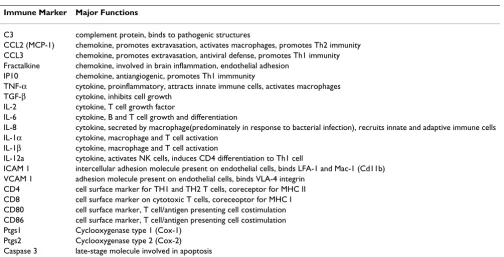
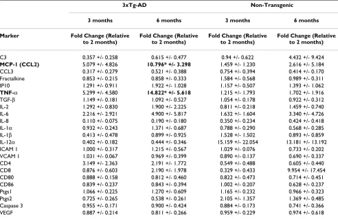
begins to accumulate in the entorhinal cortex and hippoc-ampus of 3xTg-AD mice. To this end, we selected a set of target inflammatory molecules that have been implicated in inflammatory responses within the central nervous sys-tem, including cytokines, chemokines, cell adhesion mol-ecules, T cell markers and immune-related enzymes (Table 1). Many of these markers have been implicated in late-stage AD and possess the potential to influence early pathogenic processes within the regions affected early in AD. Quantitative real-time RT-PCR was performed to determine levels of these targets in microdissected entorhinal cortex and hippocampus tissue of 2, 3, and 6 month-old 3xTg-AD mice. Age-matched non-transgenic mouse samples derived from identical regions were employed as controls (n = 6 per genotype per time point). Surprisingly, we detected a 14.8-fold up-regulation of
TNF-α, a pro-inflammatory modulator, and 10.8-fold
increase of the chemokine MCP-1 mRNA in the entorhi-nal cortex of 6 month-old 3xTg-AD mice versus the 2 month-old animals (Table 2). Levels of both pro-inflam-matory molecules are also slightly elevated in the 3-month 3xTg-AD entorhinal cortex, although not reaching statistical significance as compared to 2 month-old
coun-terparts. This trending increase of TNF-α and MCP-1
tran-script levels at 3 months of age correlates with the initial
appearance of human transgene-derived Aβ in 3xTg-AD
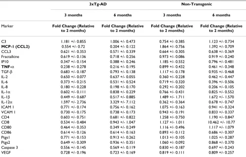
mice. Conversely, no detectable changes were observed in any of the assessed transcriptional targets in cDNA pools generated from hippocampal RNA samples at any of the time-points (Table 3), even though intracellular human
Aβ was readily detectable within this brain region (Fig.
1A). It is remarkable that the TNF-α and MCP-1 transcript
response is specific to cells resident to the entorhinal cor-tex, suggesting that aspects of the cellular environment may be responsible for differential inflammatory out-comes in these two disease-affected brain regions.
Increased microglial/macrophage numbers in the
entorhinal cortex correlates with enhanced TNF-α and
MCP-1 expression
Our finding that specific TNF-α and MCP-1 transcript
expression within the entorhinal cortex of 3xTg-AD mice indicates that a regional difference exists between the entorhinal cortex and hippocampus that would elaborate a time-dependent increase in the intensity of inflamma-tory processes. This difference appears to be independent
of solely intracellular human Aβ accumulation since
transgene expression is extant in both regions at 3 and 6 months. Because microglia and macrophages represent
likely candidate(s) of TNF-α and MCP-1 production
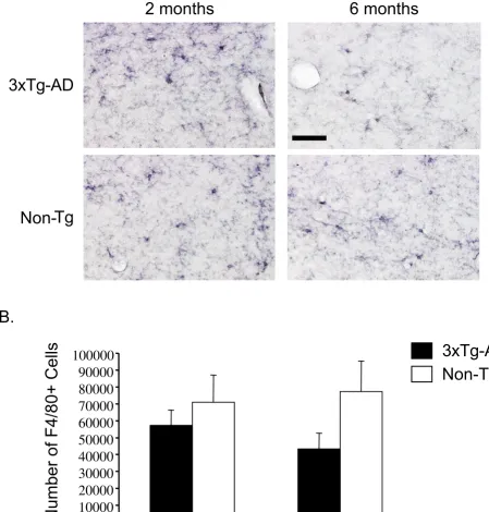
may explain regional differences of inflammatory cytokine expression profiles. Coronal sections containing the entorhinal cortex and hippocampus of 2 and 6-month 3xTg-AD and control mice were immunohistochemically stained with anti-F4/80 antibody to identify resident microglia and macrophages. Microscopic analysis
revealed that the microglial phenotype in the entorhinal cortex of 3xTg-AD mice was that of a highly activated state as shown by enhanced staining and cellular morphology at 6 months of age (Fig. 2A). Minimal differences in microglial activation state were apparent in identical regions in non-transgenic control animals (Fig. 2A). To
Intracellular Aβ appears at 3 months and is enhanced by 6 months in 3xTg-AD mice
Figure 1
Intracellular Aβ appears at 3 months and is enhanced by 6 months in 3xTg-AD mice. Coronal brain sections from 2, 3 and 6 month-old 3xTg-AD and non-transgenic mice were stained with human APP/Aβ-specific 6E10 antibody and devel-oped using DAB. Panel A illustrates that the brains of 2 month-old 3xTg-AD mice are pre-pathologic, while at 3 months, hAPP/ Aβ can be readily detected in both the entorhinal cortex and hippocampus of 3xTg-AD mice. By 6 months of age, 3xTg-AD mice exhibit further enhanced deposition of hAβ in both regions. Panel B identifies sections of entorhinal cortex and hippoc-ampus from non-transgenic mice, which are not immunohistochemically positive for endogenous mouse Aβ, therefore indicat-ing that the 6E10 antibody specifically detects transgene-driven expression of hAPP/Aβ in 3xTg-AD mice. The scale bars depict 50 µm. The insets represent 60× magnification.
A.
B.
Hippocampus
2 months
3 months
6 months
Entorhinal Cortex
3xTg-AD
2 months
3 months
6 months
Entorhinal Cortex
quantify cell number in this region, we performed unbi-ased stereology on 3xTg-AD and control entorhinal cortex sections. We detected a significant increase in the total number of F4/80-positive cells in the entorhinal cortex of 6 month-old 3xTg-AD mice as compared to 2 month-old counterparts (Fig. 2B). There was no change in the number of F4/80-positive cells in the entorhinal cortex of control mice, indicating that the increase in microglial cell number in transgenic mice was not due to normative aging. Assessment of F4/80-positive cells in the hippoc-ampus revealed no detectable alteration between 2 and 6 month-old 3xTg-AD and control mice in terms of micro-glial activation status (Fig. 3A). Quantification using unbiased stereology indicated no significant differences in F4/80-positive cell numbers between the 2 and 6-month time-points (Fig. 3B).
Discussion
Dissecting the role that inflammation plays early in AD is challenging, as AD is a complex chronic disorder with var-ying pathologic sequelae from which the underlvar-ying causative mechanisms are unknown. Activation of micro-glia and astrocytes, and the presence of many inflamma-tory mediators, including cytokines, chemokines and complement proteins have been only identified in the post-mortem AD brain in the vicinity of senile plaques and NFTs [20,25]. This observation leads one to question
if inflammation is involved early during the course of AD and, if so, how does it contribute to pathogenesis? Under-standing the earliest events is of utmost importance, as inflammation may represent a viable therapeutic target of AD. Interestingly, retrospective studies assessing the effects of non-steroidal anti-inflammatory drugs (NSAIDs) on nondemented individuals have shown decreased risk of developing AD when these individuals utilized NSAIDs for prolonged periods of time [26,27]. Our studies aimed to identify the earliest period during which inflammatory processes initiate in the 3xTg-AD mouse model [19]. Our results illustrate 3 main points: 1) Inflammatory processes precede significant extracellular amyloid plaque deposition in the 3xTg-AD brain,
sub-stantiated by increased TNF-α and MCP-1 transcript
lev-els, coincident temporally with the production of
intracellular Aβ accumulation. 2) The expression of these
molecules is spatially localized to the entorhinal cortex but not hippocampus at the early time-points assessed. 3) There is a marked increase in the number of microglia and macrophages in the entorhinal cortex that correlates with
when TNF-α and MCP-1 transcript levels are significantly
up-regulated.
In the late-stage AD brain, it has been shown that inflam-matory molecules are produced primarily by microglia and astrocytes as they respond to plaques and neuronal
Table 1: Proinflammatory markers investigated in the temporal and spatial progression of early AD pathogenesis. Immune cell molecules/inflammatory markers were assessed from RNA isolated from entorhinal cortex and hippocampus tissue of 2, 3, and 6 month-old 3xTg-AD and non-transgenic mice by qRT-PCR using Applied Biosystems Microfluidic Cards.
Immune Marker Major Functions
C3 complement protein, binds to pathogenic structures
CCL2 (MCP-1) chemokine, promotes extravasation, activates macrophages, promotes Th2 immunity CCL3 chemokine, promotes extravasation, antiviral defense, promotes Th1 immunity Fractalkine chemokine, involved in brain inflammation, endothelial adhesion
IP10 chemokine, antiangiogenic, promotes Th1 immmunity
TNF-α cytokine, proinflammatory, attracts innate immune cells, activates macrophages TGF-β cytokine, inhibits cell growth
IL-2 cytokine, T cell growth factor
IL-6 cytokine, B and T cell growth and differentiation
IL-8 cytokine, secreted by macrophage(predominately in response to bacterial infection), recruits innate and adaptive immune cells IL-1α cytokine, macrophage and T cell activation
IL-1β cytokine, macrophage and T cell activation
IL-12a cytokine, activates NK cells, induces CD4 differentiation to Th1 cell
ICAM 1 intercellular adhesion molecule present on endothelial cells, binds LFA-1 and Mac-1 (Cd11b) VCAM 1 adhesion molecule present on endothelial cells, binds VLA-4 integrin
CD4 cell surface marker for TH1 and TH2 T cells, coreceptor for MHC II CD8 cell surface marker on cytotoxic T cells, coreceoptor for MHC I CD80 cell surface marker, T cell/antigen presenting cell costimulation CD86 cell surface marker, T cell/antigen presenting cell costimulation Ptgs1 Cyclooxygenase type 1 (Cox-1)
damage [17]. Our finding of increased TNF-α and MCP-1 expression prior to significant plaque deposition in 3xTg-AD mice, which occurs extensively at 12 months [19], may represent a contributory role between inflammatory processes and early AD pathogenesis. Precisely how these molecules impart effects in 3xTg-AD mice at this early time-point is not certain; however, recent evidence has
suggested that TNF-α and MCP-1, as well as other
pro-inflammatory molecules may play a role in inhibiting
microglial phagocytosis of fibrillar Aβin vitro [28].
Like-wise, increased inflammatory responses and subsequent
secretion of cytokines, in particular, IL-1β, may play an
important role in tau phosphorylation in the 3xTg-AD model [29]. In this study, activation of microglia by LPS only affected the tau pathology via cdk5/p25 activation, but not the amyloid pathology, further highlighting the potential pathophysiological changes that can be induced
by inflammation in AD. Certainly, inflammatory media-tors have been implicated as being both protective and exacerbating, depending on the model system and the lev-els of cytokine present [30].
TNF-α can be expressed by astrocytes, microglia and
neu-rons in response to various stimuli in the CNS [17].
Initially, TNF-α is an innate mediator, promoting
chem-okine and cytchem-okine expression and extravasation of other immune cells. One possible mechanism that may
impli-cate TNF-α in contributing to AD pathogenesis is evidence
that it can increase Aβ peptide production [31].
Addition-ally, inflammatory molecule signaling may cause
increased cleavage of APP by the γ-secretase complex,
whereby TNF-α, IL-1β, and IFN-γ have been shown to
enhance production of Aβ peptides via a γ
-secretase-dependent mechanism in vitro. Moreover, antagonizing
Table 2: TNF-α and MCP-1 mRNA levels are selectively elevated in the entorhinal cortex of 3xTg-AD mice prior to overt amyloid plaque pathology. Total RNA was purified from microdissected entorhinal cortex from 2, 3, and 6 month-old 3xTg-AD and non-transgenic control mice. cDNA was generated and subjected to Applied Biosystems Microfluidic Card analysis, a high-throughput quantitative RT-PCR technology that facilitates the simultaneous quantitation of 23 inflammation-related transcriptional targets. Of the panel of transcripts analyzed, only TNF-α and MCP-1 transcript levels were significantly enhanced by 6 months of age specifically within the entorhinal cortex of 3xTg-AD mice (n = 6/group). These cytokine transcripts were unchanged in the entorhinal cortex of non-transgenic mice at all time-points analyzed. *p < 0.0005 when compared to the 2 month timepoint. Proinflammatory transcript expression in 3xTg-AD and non-transgenic mice in the entorhinal cortex
3xTg-AD Non-Transgenic
3 months 6 months 3 months 6 months
Marker Fold Change (Relative to 2 months)
Fold Change (Relative to 2 months)
Fold Change (Relative to 2 months)
Fold Change (Relative to 2 months)
C3 0.357 +/- 0.258 0.615 +/- 0.477 0.94 +/- 0.622 4.432 +/- 9.424
MCP-1 (CCL2) 5.079 +/- 4.826 10.796* +/- 3.298 1.459 +/- 1.230 2.616 +/- 5.184
CCL3 0.317 +/- 0.279 0.521 +/- 0.388 0.754 +/- 0.394 0.414 +/- 0.170
Fractalkine 0.853 +/- 0.215 0.858 +/- 0.333 1.584 +/- 0.568 0.989 +/- 0.311
IP10 1.291 +/- 0.911 1.922 +/- 1.028 1.157 +/- 0.507 1.393 +/- 1.062
TNF-α 5.299 +/- 4.580 14.822* +/- 5.618 1.215 +/- 1.793 1.702 +/- 1.916
TGF-β 1.149 +/- 0.181 1.092 +/- 0.527 1.054 +/- 0.178 0.922 +/- 0.312
IL-2 1.292 +/- 0.830 1.900 +/- 2.225 0.811 +/- 0.218 1.459 +/- 0.740
IL-6 2.216 +/- 2.921 4.900 +/- 5.817 1.632 +/- 1.604 3.340 +/- 4.726
IL-8 0.110 +/- 0.075 0.190 +/- 0.180 0.350 +/- 0.234 0.424 +/- 0.418
IL-1α 0.932 +/- 0.243 1.371 +/- 0.687 0.788 +/- 0.290 0.568 +/- 0.285
IL-1β 0.413 +/- 0.478 0.899 +/- 0.925 1.528 +/- 1.502 0.893 +/- 0.859
IL-12α 0.402 +/- 0.182 0.444 +/- 0.346 15.159 +/- 22.054 13.181 +/- 13.192
ICAM 1 1.000 +/- 0.317 1.215 +/- 0.567 1.029 +/- 0.076 0.733 +/- 0.202
VCAM 1 1.031 +/- 0.067 0.969 +/- 0.399 0.890 +/- 0.137 0.690 +/- 0.337
CD4 3.149 +/- 2.363 2.191 +/- 1.772 0.549 +/- 0.488 0.605 +/- 0.440
CD8 0.876 +/- 0.603 2.190 +/- 1.978 0.329 +/- 0.433 9.954 +/- 17.454
CD80 0.888 +/- 0.158 0.812 +/- 0.460 0.822 +/- 0.473 0.714 +/- 0.451
CD86 0.839 +/- 0.237 0.843 +/- 0.394 1.002 +/- 0.207 0.628 +/- 0.237
Ptgs1 1.066 +/- 0.225 1.270 +/- 0.609 1.165 +/- 0.232 0.966 +/- 0.323
Ptgs2 0.725 +/- 0.265 0.538 +/- 0.261 2.105 +/- 1.357 1.369 +/- 0.485
Caspase 3 0.955 +/- 0.171 0.900 +/- 0.424 0.884 +/- 0.173 0.741 +/- 0.366
VEGF 0.887 +/- 0.214 0.811 +/- 0.266 0.959 +/- 0.229 0.974 +/- 0.618
TNFR1 signaling can lead to diminished γ-secretase activ-ity [32]. Further evidence supporting pathogenic effects of
TNF-α-mediated signaling is TNFR1 and TRADD, a TNF
receptor adaptor protein that allows for NF-κB and JNK
activation, are both increased in AD tissue and APPswe mice. This increase is correlative with TUNEL-positive neurons in primary hippocampal cultures [33].
Collec-tively, these observations suggest TNF-α contributes to
aberrant APP processing and initiation of pro-apoptotic pathways.
MCP-1 is a chemokine that is expressed by microglia and astrocytes that facilitates extravasation of immune cells expressing its cognate receptor, CCR2, to cross the blood brain barrier and guides them to the site of damage. As
with TNF-α, the role of MCP-1 in AD pathophysiology is
uncertain. A recent study of APPswe/CCL2 (MCP-1)
bigenic mice showed increased diffuse Aβ deposition, as
compared to APPswe mice at 14 months of age. Since changes were not observed in APP processing, the authors
concluded that MCP-1 overexpression in APPswe mice
correlated with diminished clearance of Aβ [34]. Overall,
it is interesting that of the 23 immunomodulatory
mark-ers assessed in our study, TNF-α and MCP-1 were the only
two that changed significantly over time, possibly signify-ing their importance dursignify-ing nascent stages of AD patho-genesis. Perhaps, the other inflammatory targets are triggered at later stages of the disease in response to fur-ther neurodegenerative events.
Exogenously applied Aβ can trigger the expression of
cytokines in vitro and when injected directly into the
mouse brain [24,35]. However, the fascinating result in
our study is that expression of TNF-α and MCP-1 was
detected specifically within the entorhinal cortex and not the hippocampus, despite the fact that
immunocytochem-ically detectable intraneuronal Aβ increased over time in
both brain regions. Additionally, although not
statisti-cally significant, TNF-α and MCP-1 transcript levels were
elevated at 3 months of age in 3xTg-AD mouse entorhinal
Table 3: Inflammation-related transcript levels remain stable in the hippocampus of 2, 3, and 6 month-old 3xTg-AD and control mice. Total RNA was purified from microdissected hippocampus from 2, 3, and 6 month-old 3xTg-AD and non-transgenic control mice. cDNA was generated and subjected to Applied Biosystems Microfluidic Card analysis of 23 inflammation-related transcriptional targets. None of the transcriptional targets assessed exhibited altered expression at any of the assessed time-points. Proinflammatory transcript expression in 3xTg-AD and non-transgenic mice in the hippocampus
3xTg-AD Non-Transgenic
3 months 6 months 3 months 6 months
Marker Fold Change (Relative
to 2 months)
Fold Change (Relative to 2 months)
Fold Change (Relative to 2 months)
Fold Change (Relative to 2 months)
C3 1.181 +/- 0.855 1.006 +/- 0.473 0.754 +/- 0.385 1.123 +/- 0.734
MCP-1 (CCL2) 0.554 +/- 0.72 0.204 +/- 0.122 1.864 +/- 0.756 1.392 +/- 0.709
CCL3 0.621 +/- 0.353 0.571 +/- 0.339 0.664 +/- 0.305 0.638 +/- 0.369
Fractalkine 0.619 +/- 0.136 0.718 +/- 0.256 0.973 +/- 0.086 0.919 +/- 0.240
IP10 0.347 +/- 0.154 0.388 +/- 0.246 1.185 +/- 0.552 0.796 +/- 0.481
TNF-α 0.238 +/- 0.278 0.216 +/- 0.195 0.899 +/- 0.432 0.461 +/- 0.348
TGF-β 0.683 +/- 0.187 0.793 +/- 0.138 1.117 +/- 0.178 0.935 +/- 0.468
IL-2 0.650 +/- 0.077 0.637 +/- 0.055 0.360 +/- 0.228 0.462 +/- 0.447
IL-6 0.373 +/- 0.215 0.531 +/- 0.524 0.719 +/- 0.320 0.396 +/- 0.506
IL-8 0.180 +/- 0.228 0.198 +/- 0.170 0.292 +/- 0.202 0.206 +/- 0.105
IL-1α 0.602 +/- 0.111 0.838 +/- 0.229 0.766 +/- 0.431 0.825 +/- 0.552
IL-1β 0.449 +/- 0.687 0.517 +/- 0.885 1.489 +/- 1.711 1.672 +/- 1.570
IL-12α 1.597 +/- 2.736 3.329 +/- 7.112 0.362 +/- 0.364 0.678 +/- 0.747
ICAM 1 0.771 +/- 0.174 0.756 +/- 0.162 1.075 +/- 0.163 0.941 +/- 0.324
VCAM 1 0.730 +/- 0.175 0.738 +/- 0.173 0.943 +/- 0.191 0.833 +/- 0.337
CD4 0.603 +/- 0.751 0.681 +/- 0.822 1.258 +/- 0.750 1.190 +/- 0.847
CD8 0.534 +/- 0.484 0.943 +/- 1.047 1.127 +/- 1.011 10.462 +/- 10.77
CD80 0.464 +/- 0.353 0.349 +/- 0.249 1.116 +/- 0.496 1.117 +/- 1.079
CD86 0.614 +/- 0.126 0.614 +/- 0.163 0.893 +/- 0.112 0.686 +/- 0.307
Ptgs1 0.771 +/- 0.153 0.913 +/- 0.262 1.013 +/- 0.105 1.025 +/- 0.287
Ptgs2 0.649 +/- 0.309 0.936 +/- 0.351 1.060 +/- 0.092 0.868 +/- 0.370
Caspase 3 0.556 +/- 0.145 0.569 +/- 0.119 0.830 +/- 0.187 0.697 +/- 0.243
The 3xTg-AD entorhinal cortex harbors an increased number of macrophages/microglia at 6 months of age
Figure 2
The 3xTg-AD entorhinal cortex harbors an increased number of macrophages/microglia at 6 months of age. Coronal brain sections from 2 and 6 month-old 3xTg-AD and control mice were stained with anti-F4/80 antibody and devel-oped using DAB. (A) Qualitative image analysis reveals activation of F4/80-expressing macrophages and microglia specifically in the entorhinal cortex of 3xTg-AD mice at 6 months of age. (B) Unbiased quantitative stereologic analyses were performed on the entorhinal cortex to derive the total number of F4/80-positive cells. Error bars indicate standard deviation. N = 4 per gen-otype per time point. "*" indicates p < 0.008. The scale bar represents 50 µm.
A.
B.
3xTg-AD
2 months
6 months
Non-Tg
0
2000
4000
6000
8000
10000
12000
14000
16000
18000
2 months
6 months
Age
*
* p<0.008
3xTg-AD
Non-
Tg
The 3xTg-AD hippocampus does not have an increased number of macrophages/microglia at 6 months of age
Figure 3
The 3xTg-AD hippocampus does not have an increased number of macrophages/microglia at 6 months of age. Coronal brain sections from 2 and 6 month-old 3xTg-AD and control mice were stained with the anti-F4/80 antibody and developed using DAB. (A) Qualitative analyses shows little change in activation of microglia and macrophages in the hippocam-pus of 3xTg-AD or non-transgenic mice over time. (B) Unbiased stereology was performed on the hippocampal formation to determine total number of F4/80-positive cells. Error bars indicate standard deviation. N = 4 per genotype per time point. The scale bar represents 50 µm.
A.
3xTg-AD
2 months
6 months
B.
0
10000
20000
30000
40000
50000
60000
70000
80000
90000
100000
2 months
6 months
Age
Non-Tg
3xTg-AD
Number of F4/80+ Cells
cortex, and were increased to statistical significance by 6 months of age suggesting a state of chronic up-regulation and positive-feedback for the expression of both of these inflammatory molecules. Therefore, we are unable to con-clude, as detected by the methodology employed in this
study, that intracellular Aβ accumulation is the sole
con-tributing factor promoting TNF-α and MCP-1 transcript
expression specifically in the entorhinal cortex. It is possi-ble that these molecules are neuronally expressed within the entorhinal cortex because they are inherently more
sensitive to accumulating Aβ, as other neurons were
shown previously to express both of these molecules dur-ing times of stress [17]. Another possibility is that the entorhinal cortex elaborates inflammatory molecule
expression owing to the structure of Aβ elaborated. For
example, oligomeric Aβ is posited to be the more toxic
structural intermediate arising during Aβ fibrillogenesis,
and this form has been shown to readily induce cytokine
expression in vitro [36]. Regional differences in
intracellu-lar and extracelluintracellu-lar oligomeric Aβ profiles in vivo may,
therefore, account for the regional specificity of cytokine/ chemokine expression and microglial activation that we observed in the 3xTg-AD mice. Conversely, the regional
elaboration of TNF-α and MCP-1 may occur via an Aβ
-independent mechanism and caused by an environmen-tal stimulus, such as region-selective oxidative stress. Prac-tico and colleagues demonstrated that lipid peroxidation
in the APPswe brain can occur prior to Aβ deposition in
APPswe mice [37], suggesting that disruption of cellular membrane homeostasis could also contribute to inflam-matory molecule induction, and perhaps, regional differ-ences in lipid peroxidation profiles are responsible for our region-specific observations in 3xTg-AD mice.
The significant increase in the number of microglia and macrophages in the entorhinal cortex from 2 to 6 months of age in 3xTg-AD mice is coincident with the increase of
TNF-α and MCP-1. We believe this could be due to
micro-glial proliferation, activation of the resting resident popu-lation of brain microglia and macrophages and/or recruitment of peripheral macrophage-like cells (F4/80-positive) from outside the brain. Macrophages express CCR2 and thus, are capable of responding to a compro-mised entorhinal cortex via chemotaxis. Whether the
observed increase in F4/80+ cell number indicates a
home-ostatic or pathologic response is not clear. APPswe/CCL2 mice demonstrate enhanced microglial numbers that are
concurrent with increased extracellular Aβ deposition,
that the authors postulate is due to an inability to
effec-tively clear Aβ [34]. This may relate partially to the
increased ApoE levels observed in APPswe/CCL2 mice produced by microglia and macrophages. If a similar mechanism is at play in the 3xTg-AD mouse model, this finding suggests a pathogenic role for these cells in initiat-ing degeneration within the entorhinal cortex.
In summary, our results indicate a potential early role for inflammatory processes in the temporal and spatial
evolu-tion of AD pathogenesis. Because TNF-α and MCP-1 are
produced specifically within the entorhinal cortex where human AD has been shown to arise, these molecules are likely playing an instrumental role in disease perpetua-tion. This work provides insight into the involvement of
TNF-α and MCP-1 mediated inflammation in the
tempo-ral and spatial progression of early AD pathogenic events and may potentially herald new therapeutic targets. Our use of the 3xTg-AD model to assess these early events is unique, as all previous studies have examined inflamma-tory processes in the context of either amyloid or tau pathology, but not both. This transgenic mouse allows us to directly examine the dynamic interplay of
inflammation, Aβ pathology, and tau dysfunction.
Modu-lating TNF-α and MCP-1 function in future studies will
elucidate how these inflammatory mediators influence the severity and progression of AD-related pathology and synaptic dysfunction.
List of abbreviations
AD: Alzheimer's disease
APP: Amyloid precursor protein
APPswe: Amyloid precursor protein Swedish mutation
PS1: Presenilin 1
Aβ : Beta-amyloid
TNF-α : Tumor necrosis factor-alpha
MCP-1: monocyte chemoattractant protein-1
Tg: Transgenic
PBS: Phosphate-buffered saline
Competing interests
The author(s) declare that they have no competing interests.
Authors' contributions
Acknowledgements
The authors wish to thank Dr. Linda Callahan for immunohistochemistry, microscopy, and stereological advice. We also thank Landa Prifti for animal care and husbandry. This work was supported by NIH F31NS049995 to MCJ, NIH R01AG023593 to WJB and NIH R01AG020204 to HJF.
References
1. Selkoe DJ: Alzheimer's disease is a synaptic failure. Science
2002, 298:789-791.
2. Nestor PJ, Scheltens P, Hodges JR: Advances in the early detec-tion of Alzheimer's disease. Nat Med 2004, 10 Suppl:S34-41. 3. Braak H, Braak E: Neuropathological stageing of
Alzheimer-related changes. Acta Neuropathol (Berl) 1991, 82:239-259. 4. Haroutunian V, Purohit DP, Perl DP, Marin D, Khan K, Lantz M, Davis
KL, Mohs RC: Neurofibrillary tangles in nondemented elderly subjects and mild Alzheimer disease. Arch Neurol 1999,
56:713-718.
5. Haroutunian V, Perl DP, Purohit DP, Marin D, Khan K, Lantz M, Davis KL, Mohs RC: Regional distribution of neuritic plaques in the nondemented elderly and subjects with very mild Alzheimer disease. Arch Neurol 1998, 55:1185-1191.
6. Braak H, Braak E: Staging of Alzheimer's disease-related neu-rofibrillary changes. Neurobiol Aging 1995, 16:271-8; discussion 278-84.
7. Pennanen C, Kivipelto M, Tuomainen S, Hartikainen P, Hanninen T, Laakso MP, Hallikainen M, Vanhanen M, Nissinen A, Helkala EL, Vainio P, Vanninen R, Partanen K, Soininen H: Hippocampus and entorhinal cortex in mild cognitive impairment and early AD. Neurobiol Aging 2004, 25:303-310.
8. Kordower JH, Chu Y, Stebbins GT, DeKosky ST, Cochran EJ, Bennett D, Mufson EJ: Loss and atrophy of layer II entorhinal cortex neurons in elderly people with mild cognitive impairment.
Ann Neurol 2001, 49:202-213.
9. Gomez-Isla T, Price JL, McKeel DWJ, Morris JC, Growdon JH, Hyman BT: Profound loss of layer II entorhinal cortex neurons occurs in very mild Alzheimer's disease. J Neurosci 1996,
16:4491-4500.
10. deToledo-Morrell L, Stoub TR, Bulgakova M, Wilson RS, Bennett DA, Leurgans S, Wuu J, Turner DA: MRI-derived entorhinal volume is a good predictor of conversion from MCI to AD. Neurobiol Aging 2004, 25:1197-1203.
11. Burns A, Whitehouse P, Arendt T, Forsti H: Alzheimer's disease in senile dementia: loss of neurones in the basal forebrain. Whitehouse, P., Price, D., Struble, R., Clarke, A., Coyle, J. and Delong, M. Science (1982), 215, 1237-1239. Int J Geriatr Psychiatry 1997, 12:7-10.
12. Davies CA, Mann DM, Sumpter PQ, Yates PO: A quantitative mor-phometric analysis of the neuronal and synaptic content of the frontal and temporal cortex in patients with Alzheimer's disease. J Neurol Sci 1987, 78:151-164.
13. Hsia AY, Masliah E, McConlogue L, Yu GQ, Tatsuno G, Hu K, Kholodenko D, Malenka RC, Nicoll RA, Mucke L: Plaque-inde-pendent disruption of neural circuits in Alzheimer's disease mouse models. Proc Natl Acad Sci U S A 1999, 96:3228-3233. 14. McGeer EG, McGeer PL: Inflammatory processes in
Alzhe-imer's disease. Prog Neuropsychopharmacol Biol Psychiatry 2003,
27:741-749.
15. Li M, Pisalyaput K, Galvan M, Tenner AJ: Macrophage colony stim-ulatory factor and interferon-gamma trigger distinct mech-anisms for augmentation of beta-amyloid-induced microglia-mediated neurotoxicity. J Neurochem 2004, 91:623-633. 16. Strohmeyer R, Rogers J: Molecular and cellular mediators of
Alzheimer's disease inflammation. J Alzheimers Dis 2001,
3:131-157.
17. Hanisch UK: Microglia as a source and target of cytokines. Glia
2002, 40:140-155.
18. Oddo S, Caccamo A, Kitazawa M, Tseng BP, LaFerla FM: Amyloid deposition precedes tangle formation in a triple transgenic model of Alzheimer's disease. Neurobiol Aging 2003,
24:1063-1070.
19. Oddo S, Caccamo A, Shepherd JD, Murphy MP, Golde TE, Kayed R, Metherate R, Mattson MP, Akbari Y, LaFerla FM: Triple-transgenic model of Alzheimer's disease with plaques and tangles: intracellular Abeta and synaptic dysfunction. Neuron 2003,
39:409-421.
20. Vehmas AK, Kawas CH, Stewart WF, Troncoso JC: Immune reac-tive cells in senile plaques and cognireac-tive decline in Alzhe-imer's disease. Neurobiol Aging 2003, 24:321-331.
21. Strohmeyer R, Kovelowski CJ, Mastroeni D, Leonard B, Grover A, Rogers J: Microglial responses to amyloid beta peptide opsoni-zation and indomethacin treatment. J Neuroinflammation 2005,
2:18.
22. Braak H, Braak E: Staging of Alzheimer-related cortical destruction. Int Psychogeriatr 1997, 9 Suppl 1:257-61; discussion 269-72.
23. Streit WJ: Microglia and Alzheimer's disease pathogenesis. J Neurosci Res 2004, 77:1-8.
24. Tuppo EE, Arias HR: The role of inflammation in Alzheimer's disease. Int J Biochem Cell Biol 2005, 37:289-305.
25. Afagh A, Cummings BJ, Cribbs DH, Cotman CW, Tenner AJ: Local-ization and cell association of C1q in Alzheimer's disease brain. Exp Neurol 1996, 138:22-32.
26. in t' Veld BA, Ruitenberg A, Hofman A, Launer LJ, van Duijn CM, Sti-jnen T, Breteler MM, Stricker BH: Nonsteroidal antiinflamma-tory drugs and the risk of Alzheimer's disease. N Engl J Med
2001, 345:1515-1521.
27. Stewart WF, Kawas C, Corrada M, Metter EJ: Risk of Alzheimer's disease and duration of NSAID use. Neurology 1997,
48:626-632.
28. Koenigsknecht-Talboo J, Landreth GE: Microglial phagocytosis induced by fibrillar beta-amyloid and IgGs are differentially regulated by proinflammatory cytokines. J Neurosci 2005,
25:8240-8249.
29. Kitazawa M, Oddo S, Yamasaki TR, Green KN, LaFerla FM: Lipopol-ysaccharide-induced inflammation exacerbates tau pathol-ogy by a cyclin-dependent kinase 5-mediated pathway in a transgenic model of Alzheimer's disease. J Neurosci 2005,
25:8843-8853.
30. Zhao B, Schwartz JP: Involvement of cytokines in normal CNS development and neurological diseases: recent progress and perspectives. J Neurosci Res 1998, 52:7-16.
31. Blasko I, Veerhuis R, Stampfer-Kountchev M, Saurwein-Teissl M, Eike-lenboom P, Grubeck-Loebenstein B: Costimulatory effects of interferon-gamma and interleukin-1beta or tumor necrosis factor alpha on the synthesis of Abeta1-40 and Abeta1-42 by human astrocytes. Neurobiol Dis 2000, 7:682-689.
32. Liao YF, Wang BJ, Cheng HT, Kuo LH, Wolfe MS: Tumor necrosis factor-alpha, interleukin-1beta, and interferon-gamma stim-ulate gamma-secretase-mediated cleavage of amyloid pre-cursor protein through a JNK-dependent MAPK pathway. J Biol Chem 2004, 279:49523-49532.
33. Del Villar K, Miller CA: Down-regulation of DENN/MADD, a TNF receptor binding protein, correlates with neuronal cell death in Alzheimer's disease brain and hippocampal neurons. Proc Natl Acad Sci U S A 2004, 101:4210-4215.
34. Yamamoto M, Horiba M, Buescher JL, Huang D, Gendelman HE, Ran-sohoff RM, Ikezu T: Overexpression of monocyte chemotactic protein-1/CCL2 in beta-amyloid precursor protein trans-genic mice show accelerated diffuse beta-amyloid deposition. Am J Pathol 2005, 166:1475-1485.
35. Perry RT, Collins JS, Wiener H, Acton R, Go RC: The role of TNF and its receptors in Alzheimer's disease. Neurobiol Aging 2001,
22:873-883.
36. Lindberg C, Selenica ML, Westlind-Danielsson A, Schultzberg M:
beta-Amyloid Protein Structure Determines the Nature of Cytokine Release From Rat Microglia. J Mol Neurosci 2005,
27:1-12.
37. Pratico D, Uryu K, Leight S, Trojanoswki JQ, Lee VM: Increased lipid peroxidation precedes amyloid plaque formation in an animal model of Alzheimer amyloidosis. J Neurosci 2001,