R E S E A R C H
Open Access
Large-scale transcriptome sequencing
reveals novel expression patterns for key
sex-related genes in a sex-changing fish
Hui Liu
1*, Melissa S. Lamm
2,3, Kim Rutherford
1, Michael A. Black
4, John R. Godwin
2,3and Neil J. Gemmell
1Abstract
Background:Teleost fishes exhibit remarkably diverse and plastic sexual developmental patterns. One of the most astonishing is the rapid socially controlled female-to-male (protogynous) sex change observed in bluehead wrasses (Thalassoma bifasciatum). Such functional sex change is widespread in marine fishes, including species of commercial importance, yet its underlying molecular basis remains poorly explored.
Methods:RNA sequencing was performed to characterize the transcriptomic profiles and identify genes exhibiting sex-biased expression in the brain (forebrain and midbrain) and gonads of bluehead wrasses. Functional annotation and enrichment analysis were carried out for the sex-biased genes in the gonad to detect global differences in gene products and genetic pathways between males and females.
Results:Here we report the first transcriptomic analysis for a protogynous fish. Expression comparison between males and females reveals a large set of genes with sex-biased expression in the gonad, but relatively few such sex-biased genes in the brain. Functional annotation and enrichment analysis suggested that ovaries are mainly enriched for metabolic processes and testes for signal transduction, particularly receptors of neurotransmitters and steroid hormones. When compared to other species, many genes previously implicated in male sex determination and differentiation pathways showed conservation in their gonadal expression patterns in bluehead wrasses. However, some critical female-pathway genes (e.g.,rspo1andwnt4b) exhibited unanticipated expression patterns. In the brain, gene expression patterns suggest that local neurosteroid production and signaling likely contribute to the sex differences observed.
Conclusions:Expression patterns of key sex-related genes suggest that sex-changing fish predominantly use an evolutionarily conserved genetic toolkit, but that subtle variability in the standard sex-determination regulatory network likely contributes to sexual plasticity in these fish. This study not only provides the first molecular data on a system ideally suited to explore the molecular basis of sexual plasticity and tissue re-engineering, but also sheds some light on the evolution of diverse sex determination and differentiation systems.
Keywords:Sex-biased gene expression, Sexual dimorphism, Brain, Gonad, Transcriptome, RNA-seq, Protogynous sex change, Bluehead wrasse
* Correspondence:[email protected]
1Department of Anatomy, University of Otago, Dunedin, New Zealand
Full list of author information is available at the end of the article
© 2015 Liu et al.Open AccessThis article is distributed under the terms of the Creative Commons Attribution 4.0 International License (http://creativecommons.org/licenses/by/4.0/), which permits unrestricted use, distribution, and reproduction in any medium, provided you give appropriate credit to the original author(s) and the source, provide a link to the Creative Commons license, and indicate if changes were made. The Creative Commons Public Domain Dedication waiver (http://creativecommons.org/publicdomain/zero/1.0/) applies to the data made available in this article, unless otherwise stated.
with GSD systems, sex is determined during early develop-ment stages and individuals remain in the same sex for a lifetime (defined as gonochorism) [4]. However, this pri-mary sex differentiation guided by genetic signals can be interrupted or even reversed by temperature or endocrine-disrupting chemicals [6–11]. More extreme cases are found in fishes with ESD systems including sequential hermaph-roditism in which some adults in a social group undergo functional sex change in response to environmental stimuli (e.g., temperature or social cues) [12–16]. Revealing the mechanisms underlying such sexual plasticity may help us understand how sex is maintained and gain insights into the origin and evolution of sex-determination systems.
The genetic bases of sexual dimorphism have been in-tensively studied for decades in mammals and birds, but are less well characterized in teleost fishes [1–4, 17, 18]. So far, genetic studies on sex determination in fishes have examined either sex-specific genetic differences or sex-biased gene expression [19–23]. The search for sex-specific genetic markers has not met with much success because, unlike mammals and birds, fishes have rela-tively young sex chromosomes that are not usually
het-eromorphic [24–28] with exceptions including some
species of salmonids [29, 30], stickleback fishes [31], glass knife fishes [32], and half-smooth tongue sole, Cyno-glossus semilaevis[33]. Even in fishes with heterogenic sex-determination systems, sex differences are usually limited to a few loci or certain linkage groups [34–39]. No con-served sex-specific gene has been found in teleost fishes: six sex-determining genes have been reported to evolve separately in different fish lineages [22, 40]. In contrast, studies examining sex-biased gene expression in fishes have yielded many more genes, including some that play conserved roles in vertebrate sex differentiation. However, for most of these sex-biased genes, their detailed molecular functions in fishes remain to be clarified [23, 41–46]. Stud-ies to date have focused mainly on the expression patterns of a limited number of genes during primary sex differenti-ation stages in gonochoristic fishes [47–53]. Few studies have yet examined sex-biased gene expression in herm-aphroditic fishes, with the exception of genetic studies in protandrous black porgy, Acanthopagrus schlegelii
[19, 54], and transcriptomic studies in two other
social species exhibits two major color phases: females and smaller sneaker males in initial phase (IP) share the same color pattern, while the large males display a distinct terminal phase (TP) phenotype [57–59]. Natural social groups typically consist of one dominant TP male and nu-merous females as well as a few IP males. Following the loss of the dominant TP male from a social group, both large females and IP males can transform into TP males through sex or role change, although the latter is rarely re-ported [55, 59, 60]. In females, functional gonadal sex change takes about a week while behavioral sex change can begin within minutes to hours [60, 61]. Importantly, manipulation of the social environment can induce sex change in females, which makes the bluehead wrasse a useful model for investigating sexual plasticity.
Significant progress has been made in understanding the ecology and the neuroendocrine bases of sex change in this species, but detailed mechanisms still remain elu-sive, especially at the molecular level [55, 62]. According to the Animal Genome Size Database [63], the haploid DNA contents (C-value) of bluehead wrasse is 0.98 pico-gram (1 picopico-gram = 978 megabase pair). However, its genome and transcriptome sequences are not available yet. In this study, we took advantage of RNA sequencing technology and captured the transcriptomic profiles in the brain and gonads of TP male, female, and intersex bluehead wrasses. To identify genes exhibiting sex-biased expression in the brain (forebrain and midbrain) and gonads of the bluehead wrasse, we generated ade novo
transcriptome assembly for read mapping and compared gene expression patterns at the isoform level between con-trol females and TP males. We also conducted functional annotation and enrichment analysis on the genes showing sex-biased expression in the gonad to detect sex-biased genetic pathways that could contribute to gonadal sex differences in bluehead wrasses.
Methods Sample collection
from patch reefs off the coast of Key Largo in late May 2012. All fishes were euthanized with an overdose of MS-222 (Sigma) within 2 min of capture, and the brain and gonads were dissected immediately. These experiments were performed in accordance with guidelines established by the Institutional Animal Care and Use Committee at North Carolina State University (NCSU).
One gonadal lobe and the whole brain were preserved in RNAlater (Life Technologies, Inc.) on ice, followed by stor-age at−20 °C for less than 1 week and transfer to−80 °C until RNA extraction. The other gonadal lobe was fixed in 4 % paraformaldehyde/1X PBS overnight at 4 °C, followed by storage in 1X PBS before being fixed in paraffin for histological sectioning and HE (hematoxylin and eosin) staining (Histology Laboratory, College of Veterinary Medi-cine, NCSU) to determine the gonadal status [66]. Before RNA extraction, the hindbrain (corpus cerebelli, pons, and medulla) was removed from each brain. Only the forebrain/ midbrain was used for RNA sequencing, because the fore-brain and midfore-brain contain regions belonging to the social behavior network and mesolimbic reward system, two neural circuits that are involved in the regulation of social decision-making [67], and thus may be key integrators and drivers of socially induced sex change.
RNA extraction
The tissues were homogenized using TissueLyser II (QIA-GEN®) (Center for Neuroendocrinology, Department of Anatomy, University of Otago). Forebrain/midbrain and gonadal total RNA were extracted with TRI reagent (Invi-trogen) using chloroform (forebrain/midbrain) or bromo-chloropropane (gonads) as the phase separation reagent. Samples were then DNase-treated (TURBO DNA-free Kit, Ambion) and total RNA-cleaned (NucleoSpin RNA XS col-umns, Macherey-Nagel). RNA integrity was assessed on an Agilent 2100 Bioanalyzer. Sex-changing gonads consistently showed RNA profiles with a strong peak of low molecular weight RNA, which possibly corresponds to massive 5S RNA expression in atretic ovaries and masks the 18S and 28S rRNA peaks used for calculating RNA integrity num-bers (RIN). Such patterns were also observed in ovaries and intersex gonads of thicklip gray mullets, Chelon labrosus
[68], and ovaries of protandrous sharpsnout seabream,
Diplodus puntazzo [20]. Therefore, RIN values could not serve as useful measures of RNA integrity in these sex-changing gonads of bluehead wrasses. For brain RNA, samples with RIN values above 6.0 were used for RNA-seq. Total RNA concentration was measured by Qubit 2.0 Fluorometer (Qubit RNA HS Assay Kit, Life Technologies), and samples were diluted to 10 ng/μL.
RNA sequencing
Total RNA from 12 forebrain/midbrain and 12 gonadal samples (3 control females, 3 TP males, and 6 intersex fish),
500 ng per sample, were sent to the Otago Genomics and Bioinformatics Facility at the University of Otago under contract to New Zealand Genomics Limited for library con-struction and RNA sequencing. Twenty-four multiplexed libraries were prepared with the Illumina TruSeq Stranded mRNA Sample Prep Kit and 100-bp paired-end reads were generated using 8 flow cell lanes on the HiSeq 2000 plat-form. The insert size was designed to produce a small over-lap between paired reads.
Read pre-processing
Read quality was first assessed with FastQC (v0.10.1) [69]. Quality filtering was performed using Trimmomatic (v0.25) [70]: low quality reads were trimmed if average Phred quality scores were less than 20 within a 3-bp sliding win-dow and discarded if the length was below 40 bp after trim-ming (Trimmomatic parameters: SLIDINGWINDOW:3:20 MINILEN:40). Read pairs were processed with FLASH (v1.2.4) [71]. Overlapping read pairs were joined and used for assembly along with the non-merged read pairs.
De novotranscriptome assembly
Filtered short reads with high-quality scores were assem-bledde novowith Trinity [72] (r2014-03-23, default kmer 25, minimum contig length of 200 bp), an assembler de-veloped for efficient and robustde novoreconstruction of transcriptomes from RNA-seq data [20, 73, 74]. Since our libraries were made using the dUTP method [75], we specified the library type by setting the “strand-specific library type (—SS_lib_type)” as “RF.” We also used the “—jaccard_clip” option to reduce chimeric fusion of tran-scripts [76].
Quality checking
Assembled contigs were first searched against CEGMA (Core Eukaryotic Genes Mapping Approach, v2.5) KOGs (the eukaryotic orthologous groups) [77]. We then ran “TransDecoder” (v1.0) [76] to check the chimeric rate in our assembly (if two large open reading frames were found in one contig, it would be reported as chimeric). Full-length transcript analysis was carried out using the Trinity function “analyze_blastPlus_topHit_coverage.pl” with BLAST+ (BLASTN, E-value cut-off 10−50) against 17 blue-head wrasse expressed sequence tags (ESTs) and 19,712 Nile tilapia protein sequences (Ensembl release 75) [76, 78, 79]. Finally, we manually checked the sequences of all the candidate genes based on read mapping and visualization in Integrative Genomics Viewer (IGV, v2.3.40) [80].
Annotation
The assembly was searched against the UniProt (Swiss-Prot and TrEMBL) protein database [81] with BLAST+ (BLASTX, E-value cut-off 10−10, keeping the top hit) [78] for taxonomic distribution and bacterial contamination
10 , keeping the top hit) and mapped to the tilapia gen-ome downloaded from Ensembl (release 79, BLASTN, E-value cut-off 10−10) [78, 79]. Finally, putative open reading frames (ORFs) were searched in both annotated and unan-notated contigs using OrfPredictor (v2.3) [82].
Read mapping and differential expression analysis
The de novo transcriptome assembly served as a refer-ence for read mapping. Raw reads were aligned to the assembly with Bowtie (v0.12.9) [83] and transcript abun-dance estimation was calculated with RNA-seq by ex-pectation maximization (RSEM, v1.2.12) [84] using the align_and_estimate_abundance.pl script from the Trinity package [76]. RSEM expected counts for each contig (representing the isoform) were used for downstream differential expression analysis in R (v3.1.0) [85] using the DESeq package (v1.20.0) [86].
Comparisons between TP males and females were con-ducted separately for the brain and gonadal samples (3 samples for 2 conditions each) using the DESeq function nbinomTest [86]. Principal component analysis (PCA) [87] and the heatmap.2 function in the gplots package [88] were used to visualize global similarities and differences among either the brain or gonadal samples. Contigs with very low expression in either gonad or brain (average expected counts of mapped reads fewer than 1 per sample) were excluded prior to differential expression analysis to im-prove the statistical power [89]. All samples (including intersex samples) were used for estimating dispersions.
p value adjustment was performed using the false dis-covery rate controlling procedure [90]. Contigs with an adjustedpvalue less than 0.05 and a fold change larger than 2 were reported as significantly differentially expressed between sexes in the gonad, while contigs with an adjusted
pvalue less than 0.05 were reported as significantly differ-entially expressed between sexes in the brain.
Gene ontology and pathway analysis
Contigs showing significantly sex-biased expression in the gonad were searched against the Ensembl zebrafish pro-tein database (BLASTX, E-value cut-off 10−10). Matched zebrafish protein IDs were converted to unique Ensembl zebrafish gene IDs via BioMart [91]. These gene IDs were
0.05 after adjustment using the Benjamini and Hochberg (BH) procedure are indicated by stars.
Results and discussion De novotranscriptome assembly
The Illumina HiSeq 2000 sequencing produced more than two billion 100-bp paired-end reads (1,106,170,692 read pairs). The raw sequence data in FASTQ format have been submitted to the National Centre for Biotechnology Infor-mation (NCBI) Sequence Read Archive (SRA) database and are accessible under accession number SRP06302. After trimming, 1,586,678,582 (71.7 %) high-quality reads were retained for the transcriptome assembly.
The de novo assembled transcriptome using Trinity [72] resulted in 230,626 contigs with a N50 of 1,146 bp and minimum and maximum contig lengths of 201 and 27,427 bp, respectively. There are 77,632 contigs having a length of 500 bp or more. Short contigs (<500 bp) were retained for annotation and mapping because many neuropeptides have a short protein sequence.
Assembly quality was assessed by three means: the rep-resentation of core eukaryotic genes, predicted chimeric rate, and full-length recovery of the bluehead wrasse expressed sequence tags (ESTs) and Nile tilapia protein se-quences (Ensembl release 75) [79]. All CEGMA KOGs (the eukaryotic orthologous groups) [77] were present in this assembly (98 % complete, 100 % partial). The pre-dicted chimeric rate [76] was 3.2 %. All of the bluehead ESTs were recovered (BLASTN, E-value ≤10−50): 14 ESTs with >90 % recovery and 3 ESTs with 70–80 % recovery. Fifty-eight percent of Nile tilapia protein sequences had a match in the bluehead wrasse transcriptome assembly with alignment coverage above 90 % (BLASTX, E-value≤10−10).
Transcriptome annotation
expectations that our assembly comprises mainly brain and gonadal coding RNAs.
Searching against the Ensembl protein databases [79] (BLASTX, E-value≤10−10, keeping the top hit), we found 16–17 % of the assembled contigs had a significant BLASTX match (E-value≤10−10, keeping the top hit). Of 26,763 input Nile tilapia protein sequences, 20,182 se-quences were found in our assembly (Table 1). Similar re-sults have been reported in two other non-model fish transcriptomes [20, 95].
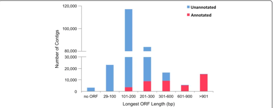
Surprisingly, a large portion (82 %) of the contigs had no significant BLASTX match to any known protein se-quence. These sequences were searched against the Ensembl zebrafish ncRNA database [79] (BLASTN, E-value≤10−5, 8819 input ncRNA sequences), but only 93 contigs had a match, including processed or antisense transcripts with no protein product, miRNA, miscRNA, snoRNA etc. Putative ORFs were searched in both an-notated and unanan-notated contigs using OrfPredictor [82]. The length distribution of the longest ORFs is shown in Fig. 2. Briefly, over 98 % of the contigs con-tained ORFs, but most (69 %) were smaller than
300 bp. Almost all of the unannotated contigs (>99 %) had an ORF smaller than 600 bp. These contigs may represent novel protein-coding transcripts, fragmented UTRs, pre-mature mRNA sequences with retained in-trons, or polyadenylated non-coding RNAs of potential biological importance. At present, however, it is still challenging to provide complete annotations for ade novo
assembled transcriptome, especially for a non-model teleost fish for which few genomic resources are available. Multiple BLAST searches provide a powerful means for automated annotations but are limited by available sequences, sequence similarity among homologues, and alignment sensitivity. Future genome sequencing and more information on alternatively spliced isoforms and non-coding RNAs will improve our current annotation. It will be useful to revisit these data as more gene sequences become available.
Differential gene expression between female and TP male
All of the contigs were kept for read mapping, and the RSEM [84] expected value table of contigs (represent-ing isoforms) was used for expression analysis. This al-lows detection of isoform-specific expression patterns and clustering of the contigs based on both annota-tions and expression patterns.
In total, 889,652,430 raw read pairs (from control females and TP males) were mapped back to the ref-erence transcriptome assembly [83]. In this paper, we focused on the sex-biased gene expression; thus, we only report the comparison between females and TP males.
Fig. 1Taxonomic distribution of top BLASTX hits in UniProt protein database. Numbers of unique hits of protein sequences in each group are shown in theparentheses
Table 1Annotation of thede novotranscriptome assembly
Species Nile tilapia Zebrafish Medaka Zebrafish
Ensembl database type Protein Protein Protein ncRNA
Contigs with hits 38,606 37,149 36,695 168
Unique hits 20,182 20,496 17,994 149
Input sequences 26,763 43,153 24,674 8319
Cut-off E-value 10−10 10−10 10−10 10−5
Global gene expression patterns in the brain and gonad
Comparisons between TP males and females were con-ducted separately for the brain and gonadal samples (3 samples for 2 conditions each) using the DESeq
func-tion nbinomTest [86]. p value adjustment was
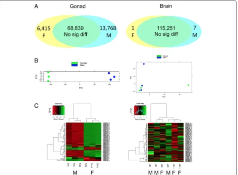
per-formed using Benjamini and Hochberg (BH) procedure [90]. Principal component analysis (PCA) plots [87] and heatmaps [88] both showed that sex differences in gene expression were much more pronounced between the ovary and testis than those between male and female brains (Fig. 3b, c). Consistently, a large number of contigs showed sex-biased expression in the gonad (fold change≥2 and BH adjustedpvalue≤0.05), while only eight contigs showed sex-biased expression in the brain (BH adjustedpvalue≤0.05). Similar global expression patterns were also reported in zebrafish [44, 96] and sharpsnout seabream [20], which may reflect the functional and regulatory differences between the brain and gonads.
A large set of genes showed significant sex-biased expression in the gonad
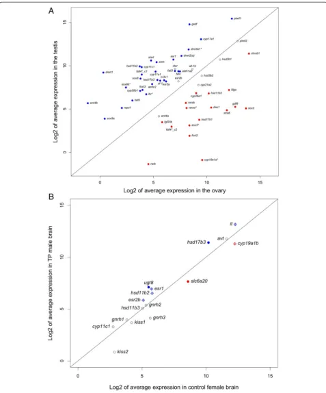
Expression analysis revealed a large set of transcripts differ-entially expressed between ovary and testis of the bluehead wrasse (fold change ≥2 and BH adjusted p value ≤0.05). Contigs showing male-biased expression were twice as abundant as those showing female-biased expression, al-though contigs with the highest expression in the gonad were female-biased (Fig. 3a, c). Of the 13,768 male-biased contigs, 6769 (49 %) contigs had a significant BLASTX match in the UniProt protein databases (E-value ≤10−10), including 6279 hits in the Ensembl zebrafish protein data-base. Of the 6415 female-biased contigs, 4246 (66 %) had a significant BLASTX match in the UniProt protein data-bases (E-value≤10−10), including 4074 hits in the Ensembl zebrafish protein database.
Enriched gene ontology terms and pathways in testis and ovary
Contigs showing sex-biased expression in the gonad were mapped to the Ensembl zebrafish protein data-base and further converted to their equivalent Ensembl zebrafish gene IDs (4824 male-biased, 3373 female-biased) via BioMart [91]. These gene IDs (Additional file 1) were searched against the DAVID (v6.7) [92] zebrafish database to detect which GO terms [93] and KEGG pathways [94] were enriched in the testis and ovary of bluehead wrasses, respectively. As a result, 4080 (male-biased) and 2989 (female-biased) DAVID
IDs were reported, of which 30–50 % were assigned
with GO terms and about 20 % were mapped to KEGG pathways.
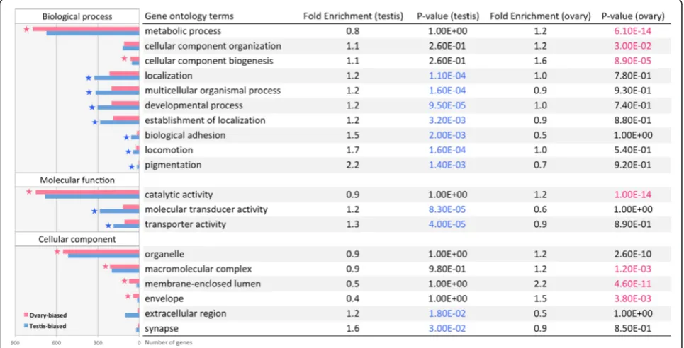
Significantly enriched GO terms (level 1) in the ovary and testis are shown in Fig. 4. In general, the ovary was enriched for metabolic process, while the testis was enriched for signal transduction and receptor activity. Similarly, the pathway enrichment analysis also found that ovaries are enriched for RNA and protein metabol-ism, while testes are enriched for signal transduction (Fig. 5).
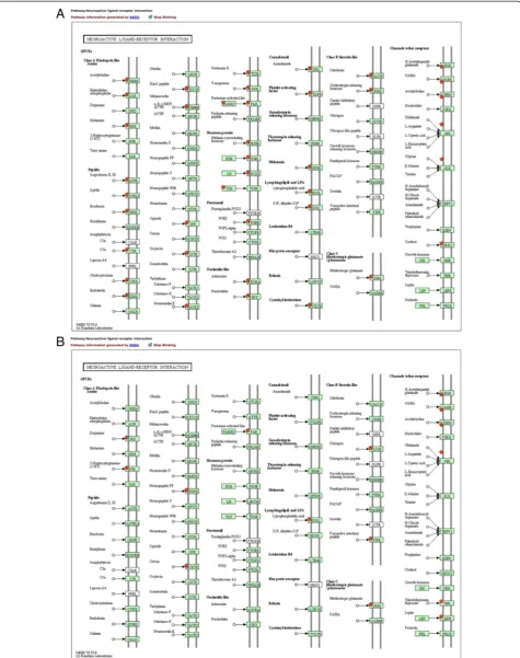
Interestingly, the top pathway enriched in the testis was “neuroactive ligand-receptor interaction” (Fig. 4), which includes receptors for many neuropeptides (Fig. 6a, b). Within this pathway, receptors of norepinephrine, epi-nephrine, melatonin, oxytocin/isotocin (IT), and vasopres-sion/vasotocin (AVT) were significantly over-expressed in the testis (Fig. 6a) while receptors of dopamine, serotonin (5-HT), and neuropeptide FF were significantly over-expressed in the ovary (Fig. 6b). Some of these neuropep-tides have been suggested to play important roles at the onset of protogynous sex change [55, 56, 62]. Briefly, nor-epinephrine and vasotocin have a promoting effect on
gonadal or behavioral sex change, while dopamine and serotonin have an inhibitory effect [97–100]. The function of isotocin and melatonin in sex-changing fishes are largely unknown due to limited information. Nevertheless, the interesting point here is that these neuropeptides were thought to act on the brain and regulate gonadal sex change indirectly through the hypothalamic-pituitary-gonadal (HPG) axis [55, 101, 102]. However, their re-ceptors are widely expressed in the gonad. Some recent studies suggest these neuropeptides can act directly on the gonad, in addition to their classical actions through the HPG axis. For example, vasotocin and melatonin are reported to regulate oocyte maturation in catfish [103] and carp [104], whereas vasotocin and catecholamines (dopamine, norepinephrine, and epinephrine) are shown to modulate ovarian steroidogenesis in catfish in a bi-phasic manner [103, 105]. Further studies on expression and function of these neuropeptides in both the brain and gonads of sex-changing fishes are warranted.
Another pathway enriched in the testis is related to steroid hormone biosynthesis (Fig. 4). Steroid hormones are known to play a critical role in sex differentiation across vertebrates [4]. Within this pathway, only three genes (cyp19a1a,hsd11b3,hsd17b1) showed significantly female-biased expression in the gonad while 11 genes showed significantly male-biased expression, including
cyp11c1,hsd11b2,hsd17b3,cyp11a1, andcyp17a1(Figs. 7 and 8a). These results are generally consistent with our current knowledge of sexually dimorphic levels of ster-oid hormones in fish.
In teleost fishes, 17β-estradiol (E2) and 11-ketotestos-terone (11-KT) function as the major estrogen and andro-gen, respectively [4]. Testosterone (T) serum levels can also be high in males and females, but T can be converted into either E2 by aromatase (cyp19a1a in the gonad) or 11-KT by 11β-hydroxylase (cyp11b or cyp11c1 in zebra-fish) and 11β-hydroxysteroid dehydrogenase 2 (hsd11b2) [4, 106, 107]. In protogynous species, steroid hormones
Fig. 3Gene expression patterns in the brain (right) and gonads (left).aNumbers of differentially expressed contigs between TP males (M) and females (F).bPCA plots of brain and gonadal samples (green: female,blue: TP male).cHeatmaps showing the expression of top 100 contigs in brain and gonads (ordered by average normalized read counts across the row;red: lower expression,green: higher expression;M: TP male,F: female)
Fig. 4Top GO terms enriched in the ovary (pink) and testis (blue). Enriched GO terms with modified Fisher exact testpvalue below 0.05 and fold enrichment above 1.2 are shown here.Starsindicate GO terms with BH adjustedpvalue below 0.05
Fig. 6The neuroactive ligand-receptor interaction pathway was significantly enriched in the testis (a) but not in the ovary (b).Red starsindicate the DE genes significantly up-regulated (fold change above 2 and BH adjustedpvalue below 0.05) in the testis (a) or the ovary (b)
play a central role in controlling sex change: high plasma E2 levels prevent females from changing into males, whereas blocking E2production or injecting 11-KT in fe-males can induce sex change [108]. As expected in our study,cyp19a1aexpression was only detected in the ovary,
whereas cyp11c1 and hsd11b2 expression were
significantly higher in the testis (Fig. 8a, Table 2, and Additional file 2). The brain aromatase gene cyp19a1b
was also detected in the gonad of rainbow trout [109], but our data showed almost no expression ofcyp19a1b
in bluehead wrasse gonads. In addition, the genes that encode androgen receptors (ar1 and ar2) showed no sex-biased expression, while the estrogen receptor genes (esr1,esr2a, andesr2b) had higher expression in the testis, although the male-biased expression of esr2b was not statistically significant (Fig. 8a, Table 2, and Additional file 2). Such male-biased expression of estrogen receptors has been shown during late sexual differentiation for Nile tilapia (70dah) [49] and rainbow trout (60-110dpf) [110]. A recent study on rainbow trout also showed significantly elevated testicular expression of esr1a and esr2a during the final stage of spermiation, whileesr1bandesr2bwere expressed at early stages of testicular development [111]. In the same study, androgen implants up-regulated tes-ticularesr1a,esr2a, andesr2bexpression but down-reg-ulated cyp19a1a expression, whereas estrogens reduced testicular cyp19a1a expression but increased the expres-sion ofcyp19a1b and esr1b. These findings all suggest a potential role for estrogens and their receptors in teleost testicular development.
Interestingly, cyp11c1 and hsd11b2 are also involved in cortisol (or glucocorticoid, GC) production: Cyp11c1 converts 11-deoxycortisol to cortisol while Hsd11b2 converts cortisol to its inactive form cortisone, and Hsd11b3 (or Hsd11b1-like) could convert cortisone to cortisol [107, 112]. Cortisol treatment has been re-ported to cause masculinization of genetic females of medaka [113, 114], Japanese flounder [2, 115], southern
flounder [116], and pejerrey [117]. In Japanese flounder, cortisol was suggested to cause female-to-male sex re-versal by suppressing cyp19a1aexpression [20, 115]. In zebrafish [118] and pejerrey [112], cortisol treatment of larvae elevated hsd11b2expression, while cortisol also enhancedin vitro11-KT synthesis in pejerrey testes. Sexu-ally dimorphic expression of cyp11c1, hsd11b2, hsd11b3, andnr3c1(nuclear receptor subfamily 3, group C, member 1 or glucocorticoid receptor) found in the gonad of blue-head wrasse (Fig. 8a, Table 2, and Additional file 2) sug-gests that local cortisol production could be important for gonadal sex differences. Moreover, cortisol treatment can induce protogynous sex change in three-spot wrasse [119], but a peak in serum cortisol levels appears to be a key event during gonadal sex change in both protandrous and protogynous species [120, 121]. The specific role of cortisol in regulating gonadal sex change remains to be clarified.
Expression patterns of genes involved in sex determination/ differentiation
The processes of sex determination and differentiation can be viewed as a battle for primacy between a male regula-tory gene network (e.g.,dmrt1,sf-1,amh,sox9) and female genetic pathways involvingfoxl2and Rspo1/Wnt/β-catenin signaling [122, 123]. Despite the diverse regulatory mecha-nisms, expression patterns of these genes are generally consistent across taxa [1, 40, 122, 124]. In our study, male-pathway genes all showed significantly higher expression in the testis (e.g.,dmrt1,sf-1,amh,amhr2,sox9a/b,sox8, and
gsdf). In contrast, a few genes involved in the female-pathway (e.g.,rspo1,wnt4b) showed unexpected expression patterns (Fig. 8a, Table 2, and Additional file 2).
First, two paralogues of forkhead box L2 genes (re-ferred to as foxl2and foxl3) were detected in the gonad of the bluehead wrasse. Foxl2 and foxl3 probably origi-nated from an ancient genome duplication event; they are present ubiquitously in fish lineages but foxl3 was repeatedly lost in the tetrapods [125]. Foxl2 is critical for
Fig. 8Expression patterns of candidate genes in the gonad (a) and brain (b).aExpression patterns of 56 sex-related genes in the gonad of bluehead wrasses. Genes with BH adjustedpvalue below 0.05 are shown insolid circles(blue: male-biased,red: female-biased).bExpression patterns of 16 genes of interest in TP male and female forebrain/midbrain of bluehead wrasses. Genes (hsd17b3,ugt8,slc6a20) with BH adjusted pvalue below 0.05 are shown insolid circles. Genes (it,cyp19a1b,esr1,esr2b, andhsd11b2) with pre-adjustedpvalues below 0.05 prior to BH correction are shown inopen circleswith across. Genes (avt,hsd11b3,cyp11c1,gnrh1,gnrh2,gnrh3,kiss1, andkiss2) withpvalues above 0.05 before and after BH correction are shown inopen circles. Genes showing male- or female-biased expression are colored inblueorred, respectively
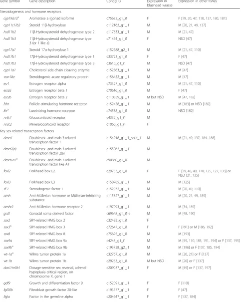
hsd17b3 17β-Hydroxysteroid dehydrogenase type 3 c3610_g1_i1 M NSD [47]
cyp11a1 Cholesterol side-chain cleaving enzyme c152363_g1_i1 M M [47]
star-like Steroidogenic acute regulatory protein c156452_g1_i1 M M [47]
esr1 Estrogen receptor alpha c73327_g1_i1 M M [21,47,110]
esr2a Estrogen receptor beta 1 c70616_g1_i1 M F [47]
esr2b Estrogen receptor beta 2 c110359_g1_i1 M but NSD M [47,182]
fshr Follicle-stimulating hormone receptor c152458_g1_i1 M M [183] or NSD [182]
lhra Luteinizing hormone receptor c74538_g1_i1 M NSD [182]
nr3c1 Glucocorticoid receptor c4332_g1_i1 M
nr3c2 Mineralocorticoid receptor c1060_g1_i1 F
Key sex-related transcription factors
dmrt1 Doublesex- and mab-3-related transcription factor 1
c154918_g1_i1_split_1 M M [21,49,137,184–188]
dmrt2(a) Doublesex- and mab-3-related transcription factor 2(a)
c155062_g1_i1 M
dmrt1a1a Doublesex- and mab-3-related transcription factor like A1
c90860_g1_i1 M
foxl2 Forkhead box L2 c29733_g1_i1 F F [19,46,49,110,125,127,133] or NSD [21,135]
foxl3 Forkhead box L3 c158785_g1_i1 M M [125]
sf-1 Steroidogenic factor-1 c152032_g1_i1 M M [20,49,110]
amh Anti-Müllerian hormone or Müllerian-inhibiting substance
c115827_g1_i1 M M [20,21,49,189]
amhr2 Anti-Müllerian hormone receptor 2 c197093_g1_i1 M M [34,189]
gsdf Gonadal soma derived factor c69648_g1_i1-a M M [48,190]
sox2 SRY-related HMG box 2 c32495_g1_i1 F
sox3a SRY-related HMG box 3 c72647_g1_i1 F F [191] or M [186,192]
sox8 SRY-related HMG box 8 c75695_g1_i1 M M [193]
sox9a SRY-related HMG box 9a c4248_g1_i1 M M [49,110,185,191,194] or F [137,195]
sox9ba SRY-related HMG box 9b c193758_g2_i1 M M [196] or F [137,185,194]
wt-1aa Wilms tumor protein 1a c32767_g1_i1 M M [20,21] or F [137]
wt-1b Wilms tumor protein 1b c29263_g1_i1 M but NSD M [20] or F [137]
dax1/nr0b1 Dosage-sensitive sex reversal, adrenal hypoplasia critical region, on chromosome X, gene 1
c209037_g1_i1 F M [49] or F [137,197]
gdf9 Growth and differentiation factor 9 c152091_g1_i1 F F [110]
fgf20b Fibroblast growth factor 20-like c193577_g1_i1 F F [47]
Table 2Genes showing sex-biased expression in the gonad(Continued)
Rspo1/Wnt4/β-catenin pathway
wnt4a Wingless-type MMTV integration site family, member 4a
c167432_g1_i1 F but NSD F [19,137] or M [141]
wnt4b Wingless-type MMTV integration site family, member 4b
c203717_g1_i1 M NSD [141] or M [137]
rspo1 R-spondin-1 (precursor) c155259_g1_i1 M F [142] or M [137]
ctnnb1 Catenin (cadherin-associated protein), beta 1
c70814_g1_i1 F F [20,21,137]
fstl3 Follistatin-like 3 c85803_g1_i1 M
fstl4a_c1 Follistatin-like 4_contig1 c6910_g1_i1 M
fstl4a_c2 Follistatin-like 4_contig2 c76818_g1_i1 F
fstl5 Follistatin-like 5 c110224_g1_i1 M
Retinoid acid signaling pathway
aldh1a2 Aldehyde dehydrogenase 1 family, member A2
c158408_g1_i1 M M [145]
cyp26a1 Cytochrome P450, family 26, subfamily a, polypeptide 1
c29815_g1_i1 F F [21,145]
cyp26b1 Cytochrome P450, family 26, subfamily b, polypeptide 1
c117560_g1_i1 M M [21] or NSD [145]
raraa Retinoid acid receptor alpha a c199432_g1_i1 F
rarab Retinoid acid receptor alpha b c153705_g1_i1 F
rarb Retinoid acid receptor beta c209577_g1_i1 F
stra6 Stimulated by retinoic acid gene 6 c153800_g1_i1 F F but NSD [21]
Epigenetic regulatory factors
piwi-like1 P-element induced wimpy testis (piwi) like 1
c1516_g1_i1 M M [21]
dnmt1 DNA methyltransferase 1 c30017_g1_i1 F
dnmt3aa DNA methyltransferase 3aa c755_g1_i1 M
dnmt3aba_c1 DNA methyltransferase 3ab_contig 1 c193863_g1_i1 F
dnmt3aba_c2 DNA methyltransferase 3ab_contig 2 c157308_g1_i1 M
dnmt3b DNA methyltransferase 3b c71358_g1_i1 M
dnmt3 DNA methyltransferase 3 c194062_g1_i1 F
dnmt4 DNA methyltransferase 4 c161106_g1_i1 M
hdac2a Histone deacetylase 2 c75925_g1_i1 F
hdac7 Histone deacetylase 7 c193969_g1_i1 F
hdac8 Histone deacetylase 8 c71086_g1_i1 M
hdac10 Histone deacetylase 10 c37022_g1_i1 F
hdac11a_c1 Histone deacetylase 11_contig 1 c152723_g1_i1 M
hdac11a_c2 Histone deacetylase 11_contig 2 c115912_g1_i1 F
Ep300aa Histone acetyltransferase—E1A binding protein 300a
c193959_g1_i1 F
Ep300ba Histone acetyltransferase—E1A binding protein 300b
c112702_g1_i1 F
KAT2b Histone acetyltransferase—K(lysine) acetyltransferase 2b
c152736_g1_i1 M
KAT7a Histone acetyltransferase—K(lysine) acetyltransferase 7
c72324_g1_i1 M
Mmale-biased,Ffemale-biased,NSDnot significantly different a
Indicates genes that have more than one contigs: only the longest contig of each gene is shown in this table
In recent years, Dmrt1 (doublesex and mab-3 related transcription factor 1) has received much attention due to its conserved role in vertebrate testicular differentiation and maintenance [127, 129–132].Dmrt1expression was signifi-cantly higher in the testis than in the ovary of the bluehead wrasse (Fig. 8a, Table 2, and Additional file 2). Dmrt1 and Foxl2 have been proposed to have antagonistic effects on
cyp19a1aexpression to control gonadal sex fate [124, 127]. This hypothesis has been supported by studies in tilapia where knockout of cyp19a1a or foxl2 expression caused gonadal sex reversal in females while dmrt1 and cyp11b2
(11β-hydroxylase) were co-expressed in follicular cells sur-rounding the degenerating oocytes [127]. Moreover, foxl2
expression decreased while dmrt1 expression increased during female-to-male sex change in honeycomb grouper [133]. However, such shifts in foxl2and dmrt1 expression did not occur until the late transitioning stage, which was downstream of declining E2levels [134].Foxl2 showed no strong sexually dimorphic expression in the gonad of prot-ogynous three-spotted wrasses, and its expression even increased during aromatase-inhibitor-induced sex change [135]. Thus, the roles offoxl2 anddmrt1 may be species-specific in sex-changing fishes. Further manipulative studies will be especially useful for elucidating the precise functions of these key genes in sex-changing fishes.
Most genes involved in the ovary-specific Rspo1/Wnt/
β-catenin signaling pathway showed sexually dimorphic expression in the gonad of bluehead wrasses (Fig. 8a, Table 2, and Additional file 2). However, some of these genes displayed an expression pattern that was opposite to our expectations based on studies from mammalian models [17, 136]. For example, ctnnb1 (β-catenin) was highly expressed in both the ovary and testis of bluehead wrasse, but its expression was significantly female-biased. In contrast,rspo1(R-spondin-1) and wnt4b(wingless-type MMTV integration site family, member 4b) were expressed at much lower levels in the bluehead wrasse gonads, but they both showed significantly male-biased expression. Similar sexually dimorphic expression patterns of
ctnnb1, rspo1, and wnt4b were also reported in east cichlid fishes [137]. Fst (follistatin) is downstream to Wnt4 signaling [17, 136, 138]. We detected a few fst-likegenes in the bluehead wrasse gonad:fstl3andfstl5
across vertebrate taxa, while other genes involved in Rspo1/ Wnt/β-catenin signaling pathway may participate in both ovarian and testicular development in fishes. More manipu-lative studies are needed to better characterize the roles of these genes in teleost fishes and to test whether the male-biased expression ofrspo1 andwnt4b is involved in prot-ogynous sex change.
The RA (retinoid acid) signaling pathway is important in ovarian differentiation because RA controls the sex-specific timing of meiosis initiation [144–146]. RA level is regulated by Aldh1a (retinal dehydrogenase) and Cyp26 enzymes: Aldh1a2 increases RA level and initiates meiosis, while Cyp26a1 and Cyp26b1 decrease RA level and pre-vent germ cells from entering into meiosis [145]. Our study revealed higher expression ofaldh1a2andcyp26b1
but lower expression ofcyp26a1 and genes encoding RA receptors (raraa,rarab, andrarb) in the testis of bluehead wrasses (Fig. 8a, Table 2, and Additional file 2). These pat-terns are consistent with findings in Nile tilapia [145] and mice [147]. In addition, Cyp26b1 prevents stra8 (stimu-lated by retinoic acid gene 8) expression in mouse testes [147, 148]. Stra8 is lost in teleost fishes [149], but we found stra6, the receptor for retinol-binding protein 4 [150], in bluehead wrasse gonads. Its expression is much lower in the testis than in the ovary, which is consistent with high expression of cyp26b1 in the testis (Fig. 8a, Table 2, and Additional file 2). Interestingly, studies in mice suggest that dmrt1 expression is essential to main-tain male-sex fate because it can protect the testis from transdifferentiation into ovary by RA signaling [131, 132]. Another study in mice also supports the hypothesis that Sox9 and Sf-1 up-regulate cyp26b1to maintain the male fate of germ cells in testes, while Foxl2 acts to antagonize
cyp26b1 expression in ovaries [151]. Taken together, the RA signaling pathway may play a key role in regulating gonadal sex change in hermaphroditic fishes and warrants further investigation.
expression in the gonad (Table 2, and Additional file 2). However, because the epigenetic mechanisms underlying sex differentiation are still poorly understood, we cannot infer any detailed functions of these genes from their ex-pression patterns. Future studies are needed to reveal their molecular functions in sex differentiation and sex change.
Few sex-biased genes detected in the forebrain/midbrain
The brain represents a key site where environment stimuli and internal signals are integrated to regulate vertebrate physiology and behavior. Sex differences in the brain have been a major and growing focus in neuroscience [2, 3]. In mammals, sex differences in the brain are likely established by both organizational effects of sex steroid hormones and cellular autonomous regulation based on sex chromosomes [2]. Teleost brains, however, appear to show less sex bias in brain structure or gene expression [20, 45, 96]. Thus, the sex differences observed in teleost brains may be due pri-marily to the activational influences of steroid hormones [158], which may also explain the brain sexual lability of teleost fishes.
In our study, expression analysis using the DESeq package revealed seven up-regulated contigs and one down-regulated contig in the TP male bluehead wrasse forebrain/midbrain (Additional file 3). Only four of these contigs had a significant BLASTX match in the UniProt protein databases (E-value ≤10−10): 17β-hydroxysteriod dehydrogenase (hsd17b3), UDP glycosyltransferase 8 (ugt8), solute carrier family 6 (proline IMINO trans-porter) member 20 (slc6a20; also BLASTs to zebrafish
slc6a19b), and a novel gene with unknown function. Lar-ger numbers of sex-biased genes have been reported in the brains of other fishes (e.g., zebrafish [96], seabream [20], and black-faced blenny [159]). We conducted dif-ferential expression analysis at the isoform (represented by contigs) level with the most conservative software
(DESeq) and stringent cut-offs (BH adjusted p value
below 0.05) in order to reduce false positives [160]. We also included six intersex samples from the same experi-mental group in dispersion estimation and read count normalization (see “Methods” section). Such stringent analyses are likely to detect fewer but more reliable sex-biased contigs.
17β-hydroxysteroid dehydrogenase (Hsd17b3), the en-zyme converting androstenedione to testosterone, was sig-nificantly up-regulated at the transcriptional level in the forebrain/midbrain of TP males (Fig. 8b and Additional file 3). Analyses in zebrafish have also shown male-biased expression of hsd17b3at the whole-brain level [96], sug-gesting conserved sex differences in local testosterone pro-duction in the brain. In teleosts, testosterone in the brain can be converted to E2by the brain isoform of aromatase (cyp19a1b) or to 11-KT by Cyp11b and Hsd11b2 [161]. Although not significantly different after BH correction,
cyp19a1b showed a 1.9-fold up-regulation in female brains, whilehsd11b2was 1.7-fold higher in TP male brains (Fig. 8b and Additional file 3), suggesting a potentially higher E2synthesis in female brains and 11-KT synthesis in TP male brains. Also, not significant after BH correction but likely biologically relevant, estrogen receptor 1 (esr1) andesr2bwere up-regulated in TP male brains compared to female brains (Fig. 8b and Additional file 3). These pat-terns suggest that local neurosteroid production and signal-ing likely contribute to sex differences in the brain [161, 162] and are consistent with the previously documented in-fluences of estrogen on behavioral sex change in bluehead wrasses [163].
The significance of the sexually dimorphic patterns of expression for other genes uncovered in the brain of the bluehead wrasse is unclear. Ugt8 was significantly up-regulated in the forebrain/midbrain of TP males, while
slc6a20showed an opposite pattern (Fig. 8b and Additional file 3). Wong et al. [96] also foundugt8to be up-regulated at the whole-brain level in male zebrafish. In mammals, UGT8 synthesizes galactocerebrosides, a major component of the myelin sheath surrounding nerves [164, 165]. Knocking outugt8 in mice reduces myelin thickness and nerve conduction, resulting in tremor and motor weakness [166, 167]. SLC6A20 transports proline and other imino acids and N-methylated amino acids across cell mem-branes [168]. Proline has been implicated in neuromodula-tion [169] and has been shown to modulate glutaminergic neurotransmission in mammals [170, 171]. There is cur-rently no information on distribution or function of UGT8 and SLC6A20 in teleost brains. Sex differences inugt8and
slc6a20expression within the forebrain/midbrain of blue-head wrasses may translate into differences in neurotrans-mission and behavior, but these possibilities require more research to address.
We did not find significant differences in the expres-sion of a number of key neuropeptide genes (Fig. 8b and Additional file 3), including arginine vasotocin (avt), isotocin (it), gonadotropin-releasing hormone (gnrh), and kisspeptin (kiss), that are known to be in-volved in socio-sexual behavior and/or reproduction in teleost fishes [172–175] and implicated in the regulation of socially induced sex change (reviewed in [55, 62]).Avtand
itmRNAs are highly expressed in both TP male and female brains, but only itshowed male-biased expression in our dataset, although this sex difference in it expression was not statistically significant after BH correction (Fig. 8b and Additional file 3).AvtmRNA expression was shown to be male-biased in the magnocellular preoptic area of bluehead wrasses [176] and to increase with behavioral sex change [64], but such differences may be masked due to the lower neuroanatomical resolution of whole-forebrain/midbrain sampling. The role of isotocin in sex change is less studied. However, it was shown that the number of
and the molecular mechanisms underlying the protogynous and protandrous sex change common to teleosts remain to be fully elucidated. In this study, we took advantage of high-throughput sequencing technology to generate the first high-quality transcriptome for a protogynous fish, the bluehead wrasse. This resource will make future compara-tive and experimental analyses of protogynous sex change possible. We also identified a large number of genes that exhibit sexually dimorphic expression in the gonad and several sex-biased genes in the forebrain/midbrain of blue-head wrasses. These genes include most known vertebrate sex-related genes as well as numerous novel genes that cur-rently lack annotation but may well have important bio-logical roles in sex differentiation and/or sex change. In addition, we find that most candidate genes implicated or known to be involved in sex determination and differenti-ation in other vertebrate systems showed conserved expres-sion patterns in the bluehead wrasse with a few exceptions. This suggests that some subtle variability in the stand-ard sex-determination regulatory network, although having evolved from a conserved toolkit, could be re-sponsible for the sexual plasticity in these fishes. Over-all, this study provides not only key data on the molecular basis of sexual dimorphism in the brain and gonad of bluehead wrasse, but also valuable resources for investigating the molecular pathways that underpin this extraordinary example of sexual plasticity in response to environmental influences. Further examination of the gene expression dynamics across the process of protogyn-ous sex change will uncover the genetic cascade that pro-gressively re-engineers a female into a male.
Availability of supporting data
All sequencing data have been uploaded to NCBI Sequence Read Archive under accession number SRP06302.
Additional files
Additional file 1: Table S1.Ensembl zebrafish gene IDs of sex-biased contigs in the gonad for GO and pathway enrichment analysis in DAVID. Additional file 2: Table S2.Results of differential expression analysis for the gonad (blue: male-biased contigs, red: female-biased contigs).
Acknowledgements
We would like to thank Brian Haas for his help with thede novotranscriptome assembly and Alice Dannis, Thomas Buckley, Margaret Ryan, and Lei Ma for their helpful input on the RNA-seq data processing and analysis. We are grateful to Erica Todd for her valuable comments on drafts of the manuscript. Financial support for this study has been provided by the Marsden Fund [UOO1308], a University of Otago Research Grant, and funds from the Department of Anatomy at the University of Otago, W.M. Keck Center for Behavioral Biology at North Carolina State University, and Department of Biological Sciences at North Carolina State University. HL is supported by the University of Otago PhD scholarship.
Author details
1Department of Anatomy, University of Otago, Dunedin, New Zealand. 2Department of Biological Sciences, North Carolina State University, Raleigh,
NC, USA.3W.M. Keck Center for Behavioral Biology, North Carolina State University, Raleigh, NC, USA.4Department of Biochemistry, University of Otago, Dunedin, New Zealand.
Received: 11 September 2015 Accepted: 9 November 2015
References
1. Gamble T, Zarkower D. Sex determination. Curr Biol. 2012;22:R257–62. 2. Jazin E, Cahill L. Sex differences in molecular neuroscience: from fruit flies to
humans. Nat Rev Neurosci. 2010;11:9–17.
3. McCarthy MM, Arnold AP, Ball GF, Blaustein JD, De Vries GJ. Sex differences in the brain: the not so inconvenient truth. J Neurosci. 2012;32:2241–7. 4. Devlin RH, Nagahama Y. Sex determination and sex differentiation in fish:
an overview of genetic, physiological, and en vironmental influences. Aquaculture. 2002;208:191–364.
5. Barske LA, Capel B. Blurring the edges in vertebrate sex determination. Curr Opin Genet Dev. 2008;18:499–505.
6. Paul-Prasanth B, Bhandari RK, Kobayashi T, Horiguchi R, Kobayashi Y, Nakamoto M, et al. Estrogen oversees the maintenance of the female genetic program in terminally differentiated gonochorists. Sci Rep. 2013;3:2862.
7. Kobayashi H, Iwamatsu T. Sex reversal in the medaka Oryzias latipes by brief exposure of early embryos to estradiol-17beta. Zool Sci. 2005;22:1163–7. 8. Sato T, Endo T, Yamahira K, Hamaguchi S, Sakaizumi M. Induction of
female-to-male sex reversal by high temperature treatment in Medaka, Oryzias latipes. Zool Sci. 2005;22:985–8.
9. Kobayashi T, Kajiura-Kobayashi H, Nagahama Y. Induction of XY sex reversal by estrogen involves altered gene expression in a teleost, tilapia. Cytogenet Genome Res. 2003;101:289–94.
10. Kwon JY, McAndrew BJ, Penman DJ. Treatment with an aromatase inhibitor suppresses high-temperature feminization of genetic male (YY) Nile tilapia. J of Fish Bio. 2002;60:625–36.
11. Bhandari RK, Nakamura M, Kobayashi T, Nagahama Y. Suppression of steroidogenic enzyme expression during androgen-induced sex reversal in Nile tilapia (Oreochromis niloticus). Gen Comp Endocrinol. 2006;145:20–4. 12. Nakamura M, Kobayashi Y, Miura S, Alam MA, Bhandari RK. Sex change in
coral reef fish. Fish Physiol Biochem. 2005;31:117–22.
14. Munday PL, Buston PM, Warner RR. Diversity and flexibility of sex-change strategies in animals. Trends Ecol Evol. 2006;21:89–95.
15. Avise JC, Mank JE. Evolutionary perspectives on hermaphroditism in fishes. Sex Dev. 2009;3:152–63.
16. Godwin J. Social determination of sex in reef fishes. Semin Cell Dev Biol. 2009;20:264–70.
17. Ungewitter EK, Yao HHC. How to make a gonad: cellular mechanisms governing formation of the testes and ovaries. Sex Dev. 2013;7:7–20. 18. Herpin A, Adolfi MC, Nicol B, Hinzmann M, Schmidt C, Klughammer J, et al.
Divergent expression regulation of gonad development genes in medaka shows incomplete conservation of the downstream regulatory network of vertebrate sex determination. Mol Biol Evol. 2013;30:2328–46.
19. Wu G-C, Tomy S, Lee M-F, Lee Y-H, Yueh W-S, Lin C-J, et al. Sex differentiation and sex change in the protandrous black porgy, Acanthopagrus schlegeli. Gen Comp Endocrinol. 2010;167:417–21. 20. Manousaki T, Tsakogiannis A, Lagnel J, Sarropoulou E, Xiang JZ, Papandroulakis
N, et al. The sex-specific transcriptome of the hermaphrodite sparid sharpsnout seabream (Diplodus puntazzo). BMC Genomics. 2014;15:655.
21. Ravi P, Jiang J, Liew WC, Orban L. Small-scale transcriptomics reveals differences among gonadal stages in Asian seabass (Lates calcarifer). Reprod Biol Endocrinol. 2014;12:5.
22. Kikuchi K, Hamaguchi S. Novel sex-determining genes in fish and sex chromosome evolution. Dev Dyn. 2013;242:339–53.
23. Sun F, Liu S, Gao X, Jiang Y, Perera D, Wang X, et al. Male-biased genes in catfish as revealed by RNA-seq analysis of the testis transcriptome. PLoS ONE. 2013;8:e68452.
24. Parise-Maltempi PP, da Silva EL, Rens W, Dearden F, O’Brien PCM, Trifonov V, et al. Comparative analysis of sex chromosomes in Leporinus species (Teleostei, Characiformes) using chromosome painting. BMC Genet. 2013;14:60. 25. Takehana Y et al. Chapter 15—frequent turnover of sex chromosomes in
the medaka fishes. In: Naruse K, editor. Medaka : a model for organogenesis, human disease, and evolution. Berlin: Springer; 2011. p. 229–40.
26. Charlesworth D, Charlesworth B, Marais G. Steps in the evolution of heteromorphic sex chromosomes. Heredity. 2005;95:118–28. 27. Tanaka K, Takehana Y, Naruse K, Hamaguchi S, Sakaizumi M. Evidence for
different origins of sex chromosomes in closely related Oryzias fishes: substitution of the master sex-determining gene. Genetics. 2007;177:2075–81. 28. Arkhipchuk VV. Role of chromosomal and genome mutations in the
evolution of bony fishes. Hydrobiol J. 1995;31:55–65.
29. Iturra P, Lam N, La Fuente De M, Vergara N, Medrano JF. Characterization of sex chromosomes in rainbow trout and coho salmon using fluorescence in situ hybridization (FISH). Genetica. 2001;111:125–31.
30. Phillips RB, Park LK, Naish KA. Assignment of Chinook salmon (Oncorhynchus tshawytscha) linkage groups to specific chromosomes reveals a karyotype with multiple rearrangements of the chromosome arms of rainbow trout (Oncorhynchus mykiss). G3 (Bethesda). 2013;3:2289–95. 31. Ross JA, Urton JR, Boland J, Shapiro MD, Peichel CL. Turnover of sex
chromosomes in the stickleback fishes (Gasterosteidae). PLoS Genet. 2009;5: e1000391.
32. Henning F, Moysés CB, Calcagnotto D, Meyer A, de Almeida-Toledo LF. Independent fusions and recent origins of sex chromosomes in the evolution and diversification of glass knife fishes (Eigenmannia). Heredity. 2011;106:391–400. 33. Chen S, Zhang G, Shao C, Huang Q, Liu G, Zhang P, et al. Whole-genome
sequence of a flatfish provides insights into ZW sex chromosome evolution and adaptation to a benthic lifestyle. Nat Genet. 2014;46:253–60.
34. Kamiya T, Kai W, Tasumi S, Oka A, Matsunaga T, Mizuno N, et al. A trans-species missense SNP in Amhr2 is associated with sex determination in the tiger pufferfish, Takifugu rubripes (fugu). PLoS Genet. 2012;8:e1002798.
35. Yano A, Guyomard R, Nicol B, Jouanno E, Quillet E, Klopp C, et al. An immune-related gene evolved into the master sex-determining gene in rainbow trout, Oncorhynchus mykiss. Curr Biol. 2012;22:1423–8.
36. Liu F, Sun F, Li J, Xia JH, Lin G, Tu RJ, et al. A microsatellite-based linkage map of salt tolerant tilapia (Oreochromis mossambicus x Oreochromis spp.) and mapping of sex-determining loci. BMC Genomics. 2013;14:58. 37. Bradley KM, Breyer JP, Melville DB, Broman KW, Knapik EW, Smith JR. An
SNP-based linkage map for zebrafish reveals sex determination loci. G3 (Bethesda). 2011;1:3–9.
38. Shirak A, Seroussi E, Cnaani A, Howe AE, Domokhovsky R, Zilberman N, et al. Amh and Dmrta2 genes map to tilapia (Oreochromis spp.) linkage group 23 within quantitative trait locus regions for sex determination. Genetics. 2006; 174:1573–81.
39. Hattori RS, Murai Y, Oura M, Masuda S, Majhi SK, Sakamoto T, et al. A Y-linked anti-Mullerian hormone duplication takes over a critical role in sex determination. Proc Natl Acad Sci U S A. 2012;109:2955–9. 40. Mei J, Gui J-F. Genetic basis and biotechnological manipulation of sexual
dimorphism and sex determination in fish. Sci China Life Sci. 2015;58:124–36. 41. Penman DJ, Piferrer F. Fish gonadogenesis. Part I: genetic and environmental
mechanisms of sex determination. Rew Fish Sci. 2008;16:16–34. 42. Piferrer F, Guiguen Y. Fish gonadogenesis. Part II: molecular biology and
genomics of sex differentiation. Rew Fish Sci. 2008;16:35–55.
43. Siegfried KR. In search of determinants: gene expression during gonadal sex differentiation. J Fish Biol. 2010;76:1879–902.
44. Small CM, Carney GE, Mo Q, Vannucci M, Jones AG. A microarray analysis of sex- and gonad-biased gene expression in the zebrafish: Evidence for masculinization of the transcriptome. BMC Genomics. 2009;10:579. 45. Sreenivasan R, Cai M, Bartfai R, Wang X, Christoffels A, Orban L. Transcriptomic
analyses reveal novel genes with sexually dimorphic expression in the zebrafish gonad and brain. PLoS ONE. 2008;3:e1791.
46. Nakamoto M, Matsuda M, Wang DS, Nagahama Y, Shibata N. Molecular cloning and analysis of gonadal expression of Foxl2 in the medaka, Oryzias latipes. Biochem Biophys Res Commun. 2006;344:353–61.
47. Tao W, Yuan J, Zhou L, Sun L, Sun Y, Yang S, et al. Characterization of gonadal transcriptomes from Nile tilapia (Oreochromis niloticus) reveals differentially expressed genes. PLoS ONE. 2013;8:e63604.
48. Shibata Y, Paul-Prasanth B, Suzuki A, Usami T, Nakamoto M, Matsuda M, et al. Expression of gonadal soma derived factor (GSDF) is spatially and temporally correlated with early testicular differentiation in medaka. Gene Expr Patterns. 2010;10:283–9.
49. Ijiri S, Kaneko H, Kobayashi T, Wang D-S, Sakai F, Paul-Prasanth B, et al. Sexual dimorphic expression of genes in gonads during early differentiation of a teleost fish, the Nile tilapia Oreochromis niloticus. Biol Reprod. 2008;78: 333–41.
50. Kobayashi T, Kajiura-Kobayashi H, Guan GJ, Nagahama Y. Sexual dimorphic expression of DMRT1 and Sox9a during gonadal differentiation and hormone-induced sex reversal in the teleost fish Nile tilapia (Oreochromis niloticus). Dev Dyn. 2008;237:297–306.
51. Vizziano D, Randuineau G, Baron D, Cauty C, Guiguen Y. Characterization of early molecular sex differentiation in rainbow trout, Oncorhynchus mykiss. Dev Dyn. 2007;236:2198–206.
52. Baron D, Guiguen Y. Gene expression during gonadal sex differentiation in rainbow trout (Oncorhynchus mykiss): from candidate genes studies to high throughout genomic approach. Fish Physiol Biochem. 2003;28:119–23. 53. Nicol B, Guiguen Y. Expression profiling of Wnt signaling genes during
gonadal differentiation and gametogenesis in rainbow trout. Sex Dev. 2011; 5:318–29.
54. Wu G-C, Chang C-F. The switch of secondary sex determination in protandrous black porgy, Acanthopagrus schlegeli. Fish Physiol Biochem. 2013;39:33–8.
55. Godwin J. Neuroendocrinology of sexual plasticity in teleost fishes. Front Neuroendocrinol. 2010;31:203–16.
56. Larson ET. Neuroendocrine regulation in sex-changing fishes. In: Norris DO, editor. Hormones and Reproduction of Vertebrates. Waltham: Academic; 2010. p. 149–68.
57. Feddern HA. The spawning, growth, and general behavior of the bluehead wrasse, Thalassoma bifasciatum (Pisces: Labridae). Bull Mar Sci. 1965;896–941. 58. Warner RR, Robertson DR. Sexual patterns in the labroid fishes of the
western Caribbean I: the wrasses. Smithson Contrib Zool. 1978;254:1–27. 59. Warner RR. Mating behavior and hermaphroditism in coral reef fishes. Am
Sci. 1984;72:128–36.
60. Warner RR, Swearer SE. Social control of sex change in the bluehead wrasse, Thalassoma bifasciatum (Pisces: Labridae). Biol Bull. 1991;181:199–204. 61. Godwin J, Crews D, Warner RR. Behavioural sex change in the absence of
gonads in a coral reef fish. Proc Biol Sci. 1996;263:1683–8. 62. Lamm MS, Liu H, Gemmell NJ, Godwin JR. The need for speed:
neuroendocrine regulation of socially-controlled sex change. Integr Comp Biol. 2015;55:307–22.
63. Gregory TR. Animal Genome Size Database. 2015. Available online at: http:// www.genomesize.com. Accessed 06 Nov 2015.
64. Semsar K, Godwin J. Social influences on the arginine vasotocin system are independent of gonads in a sex-changing fish. J Neurosci. 2003;23:4386–93. 65. Semsar K, Godwin J. Multiple mechanisms of phenotype development in
the bluehead wrasse. Horm Behav. 2004;45:345–53.
sequence data. Bioinformatics. 2014;30:2114–20.
71. MagočT, Salzberg SL. FLASH: fast length adjustment of short reads to improve genome assemblies. Bioinformatics. 2011;27:2957–63.
72. Grabherr MG, Haas BJ, Yassour M, Levin JZ, Thompson DA, Amit I, et al. Trinity: reconstructing a full-length transcriptome without a genome from RNA-Seq data. Nat Biotechnol. 2011;29:644.
73. Singhal S.De novotranscriptomic analyses for non-model organisms: an evaluation of methods across a multi-species data set. Mol Ecol Resour. 2013;13:403–16.
74. Li B, Fillmore N, Bai Y, Collins M, Thomson JA, Stewart R, et al. Evaluation of de novotranscriptome assemblies from RNA-Seq data. Genome Biol. 2013; 15:553.
75. Levin JZ, Yassour M, Adiconis X, Nusbaum C, Thompson DA, Friedman N, et al. Comprehensive comparative analysis of strand-specific RNA sequencing methods. Nat Methods. 2010;7:709–15.
76. Haas BJ, Papanicolaou A, Yassour M, Grabherr M, Blood PD, Bowden J, et al. De novotranscript sequence reconstruction from RNA-seq using the Trinity platform for reference generation and analysis. Nat Protoc. 2013;8:1494–512. 77. Parra G, Bradnam K, Korf I. CEGMA: a pipeline to accurately annotate core
genes in eukaryotic genomes. Bioinformatics. 2007;23:1061–7.
78. Camacho C, Coulouris G, Avagyan V, Ma N, Papadopoulos J, Bealer K, et al. BLAST+: architecture and applications. BMC bioinformatics. 2009;10:421. 79. Flicek P, Amode MR, Barrell D, Beal K, Billis K, Brent S, et al. Ensembl 2014.
Nucleic Acids Res. 2014;42:D749–55.
80. Thorvaldsdóttir H, Robinson JT, Mesirov JP. Integrative Genomics Viewer (IGV): high-performance genomics data visualization and exploration. Brief Bioinform. 2013;14:178–92.
81. UniProt Consortium. UniProt: a hub for protein information. Nucleic Acids Res. 2015;43:D204–12.
82. Min XJ, Butler G, Storms R, Tsang A. OrfPredictor: predicting protein-coding regions in EST-derived sequences. Nucleic Acids Res. 2005;33:W677–80. 83. Langmead B: Aligning short sequencing reads with Bowtie.Curr Protoc
Bioinformatics2010, Chapter 11:Unit 11.7.doi: 10.1002/0471250953.bi1107s32 84. Li B, Dewey CN. RSEM: accurate transcript quantification from RNA-Seq data
with or without a reference genome. BMC bioinformatics. 2011;12:323. 85. R Core Team. R: A language and environment for statistical computing. R
Foundation for Statistical Computing, Vienna, Austria; 2014.
86. Anders S, Huber W. Differential expression analysis for sequence count data. Genome Biol. 2010;11:R106.
87. Jolliffe IT. Principal component analysis. 2nd ed. Berlin: Springer; 2002. 88. Warnes GR, Bolker B, Bonebakker L, Gentleman R, Huber W, Liaw A, et al.
gplots: Various R programming tools for plotting data. R package version. 2009;2:4.
89. Richard Bourgon RGWH. Independent filtering increases detection power for high-throughput experiments. Proc Natl Acad Sci U S A. 2010;107:9546–51. 90. Benjamini Y, Hochberg Y. Controlling the false discovery rate—a practical
and powerful approach to multiple testing. J R Stat Soc Series B Stat Methodol. 1995;57:289–300.
91. Smedley D, Haider S, Durinck S, Pandini L, Provero P, Allen J, et al. The BioMart community portal: an innovative alternative to large, centralized data repositories. Nucleic Acids Res. 2015;43:W589–98.
92. Huang DW, Sherman BT, Lempicki RA. Systematic and integrative analysis of large gene lists using DAVID bioinformatics resources. Nat Protoc. 2009;4: 44–57.
93. Ashburner M, Ball CA, Blake JA, Botstein D, Butler H, Cherry JM, et al. Gene ontology: tool for the unification of biology. Nat Genet. 2000;25:25–9.
receptor gene expression link to social status and aggression in sex-dependent patterns. J Neuroendocrinol. 2015;27:142–57.
101. Zohar Y, Muñoz-Cueto JA, Elizur A, Kah O. Neuroendocrinology of reproduction in teleost fish. Gen Comp Endocrinol. 2010;165:438–55.
102. Maruska KP, Fernald RD. Social regulation of gene expression in the hypothalamic-pituitary-gonadal axis. Physiology (Bethesda). 2011;26: 412–23.
103. Joy KP, Chaube R. Vasotocin—a new player in the control of oocyte maturation and ovulation in fish. Gen Comp Endocrinol. 2015.
104. Maitra SK, Chattoraj A, Mukherjee S, Moniruzzaman M. Melatonin: a potent candidate in the regulation of fish oocyte growth and maturation. Gen Comp Endocrinol. 2013;181:215–22.
105. Joy KP, Singh V, Chaube R. Anin vitrostudy on catecholamine modulation of ovarian steroidogenic activity in the catfish Heteropneustes fossilis. Gen Comp Endocrinol. 2014;196:91–9.
106. Guiguen Y, Fostier A, Piferrer F, Chang C-F. Ovarian aromatase and estrogens: a pivotal role for gonadal sex differentiation and sex change in fish. Gen Comp Endocrinol. 2010;165:352–66.
107. Tokarz J, Möller G, de Angelis MH, Adamski J. Zebrafish and steroids: what do we know and what do we need to know? J Steroid Biochem Mol Biol. 2013;137:165–73.
108. Higa M, Ogasawara K, Sakaguchi A, Nagahama Y, Nakamura M. Role of steriod hormones in sex change of protogynous wrasse. Fish Physiol Biochem. 2003; 28:149–50.
109. von Schalburg KR, Yasuike M, Davidson WS, Koop BF. Regulation, expression and characterization of aromatase (cyp19b1) transcripts in ovary and testis of rainbow trout (Oncorhynchus mykiss). Comp Biochem Physiol B Biochem Mol Biol. 2010;155:118–25.
110. Baron D, Houlgatte R, Fostier A, Guiguen Y. Expression profiling of candidate genes during ovary-to-testis trans-differentiation in rainbow trout masculinized by androgens. Gen Comp Endocrinol. 2008;156:369–78.
111. Delalande C, Goupil A-S, Lareyre J-J, Le Gac F. Differential expression patterns of three aromatase genes and of four estrogen receptors genes in the testes of trout (Oncorhynchus mykiss). Mol Reprod Dev. 2015;82:694–708. 112. Fernandino JI, Hattori RS, Kishii A, Strüssmann CA, Somoza GM. The cortisol
and androgen pathways cross talk in high temperature-induced masculinization: the 11β-hydroxysteroid dehydrogenase as a key enzyme. Endocrinol. 2012;153:6003–11.
113. Hayashi Y, Kobira H, Yamaguchi T, Shiraishi E, Yazawa T, Hirai T, et al. High temperature causes masculinization of genetically female medaka by elevation of cortisol. Mol Reprod Dev. 2010;77:679–86.
114. Kitano T, Hayashi Y, Shiraishi E, Kamei Y. Estrogen rescues masculinization of genetically female medaka by exposure to cortisol or high temperature. Mol Reprod Dev. 2012;79:719–26.
115. Yamaguchi T, Yoshinaga N, Yazawa T, Gen K, Kitano T. Cortisol is involved in temperature-dependent sex determination in the Japanese flounder. Endocrinol. 2010;151:3900–8.
116. Mankiewicz JL, Godwin J, Holler BL, Turner PM, Murashige R, Shamey R, et al. Masculinizing effect of background color and cortisol in a flatfish with environmental sex-determination. Integr Comp Biol. 2013;53:755–65. 117. Yamamoto Y, Hattori RS, Kitahara A, Kimura H, Yamashita M, Strussmann CA.
Thermal and endocrine regulation of gonadal apoptosis during sex differentiation in pejerrey Odontesthes bonariensis. Sex Dev. 2013;7:316–24. 118. Tokarz J, Norton W, Möller G, de Angelis MH, Adamski J. Zebrafish 20β

![Fig. 7 Postulated pathways of steroidogenesis in the gonad (adapted from [118, 178, 179])](https://thumb-us.123doks.com/thumbv2/123dok_us/8979236.1888604/10.595.60.539.89.235/fig-postulated-pathways-steroidogenesis-gonad-adapted.webp)