BIROn - Birkbeck Institutional Research Online
Dimeloe, S. and Rice, L.V. and Chen, H. and Cheadle, C. and Raynes, J.
and Pfeffer, P. and Lavender, P. and Richards, D.F. and Nyon, Mun and
McDonnell, J.M. and Kemper, C. and Gooptu, Bibekbrata and Hawrylowicz,
C. (2019) Vitamin D (1,25(OH. The Journal of Steroid Biochemistry and
Molecular Biology 189 , pp. 1-9. ISSN 1879-1220.
Downloaded from:
Usage Guidelines:
Please refer to usage guidelines at
or alternatively
Contents lists available atScienceDirect
Journal of Steroid Biochemistry and Molecular Biology
journal homepage:www.elsevier.com/locate/jsbmb
Vitamin D (1,25(OH)
2
D3) induces
α
-1-antitrypsin synthesis by CD4
+
T cells,
which is required for 1,25(OH)
2
D3-driven IL-10
Sarah Dimeloe
a,1, Louise V. Rice
a,1, Hebe Chen
a, Charlotte Cheadle
a, John Raynes
b, Paul Pfe
ff
er
a,
Paul Lavender
a, David F. Richards
a, Mun Peak Nyon
c, James M. McDonnell
a, Claudia Kemper
d,
Bibek Gooptu
a,c,e, Catherine M. Hawrylowicz
a,⁎aMRC and Asthma UK Centre for Allergic Mechanisms of Asthma, King’s College London, Guy’s Hospital, London, SE1 9RT, United Kingdom bImmunology and Infection Department, London School of Hygiene and Tropical Medicine, London, WC1E 7HT, United Kingdom
cInstitute of Structural and Molecular Biology/Crystallography, Department of Biological Sciences, Birkbeck College, University of London, Malet Street, London, WC1E
7HX, United Kingdom
dMRC Centre for Transplantation, Division of Transplantation Immunology and Mucosal Biology, King’s College London, Guy’s Hospital, London, SE1 9RT, United
Kingdom
eNIHR Leicester BRC-Respiratory and Leicester Institute of Structural & Chemical Biology, Glenfield Hospital, Groby Road, Leicester, LE3 9QP, United Kingdom
A R T I C L E I N F O
Keywords:
Immune regulation IL-10
α-1-Antitrypsin Complement C3a
A B S T R A C T
Studies to identify novel immune-regulatory functions of active vitamin D (1,25(OH)2D3) in human CD4+T cells revealed that 1,25(OH)2D3 potently induced expression of the geneSERPINA1, encoding the anti-protease α-1-antitrypsin. We confirmedα-1-antitrypsin protein expression by 1,25(OH)2D3-treated CD4+T cells, but not in CD8+T cells or monocytes.α-1-Antitrypsin promotes anti-inflammatory IL-10 synthesis in other immune cell populations. We therefore investigated its immune-regulatory effects in CD4+T cells. Plasma-derivedα -1-an-titrypsin drove IL-10 synthesis by CD4+T cells, which was not dependent on anti-protease activity, but appeared to require a serum-binding factor, since this could not be achieved with recombinant protein.α-1-Antitrypsin is reported to bind complement components, which regulate T cell function. A role for this interaction was therefore probed. Plasma-derived, but not recombinant α-1-antitrypsin contained C3a. Surface Plasmon Resonance and Microscale Thermophoresis demonstratedα-1-antitrypsin binding to C3a. Addition of C3a to CD4+T cells cultured with recombinantα-1-antitrypsin restored induction of IL-10, whereas neutralisation of C3a abrogated IL-10 induced by plasma-derivedα-1-antitrypsin. To interrogate an endogenous role for theα -1-antitrypsin-C3a axis in 1,25(OH)2D3-driven CD4+T cell IL-10 synthesis, we treated cells from healthy or α-1-antitrypsin-deficient individuals (which transcribeSERPINA1but do not secrete protein) with 1,25(OH)2D3. A significant correlation was identified betweenSERPINA1andIL10gene expression in healthy donor CD4+T cells, which was absent in cells fromα-1-antitrypsin-deficient individuals. Therefore,α-1-antitrypsin is required for 1,25(OH)2D3-induced IL-10 expression in CD4+T cells, interacting with C3a to drive IL-10 expression.
1. Introduction
Vitamin D is a potent anti-inflammatory mediator, with its active form (1,25-dihydroxyvitamin D, 1,25(OH)2D3) having a range of im-mune-regulatory functions including direct induction of anti-microbial functions and the generation of tolerogenic antigen presenting cells and regulatory T cells [1]. Effects of 1,25(OH)2D3 on CD4+‘helper’T cells, which co-ordinate the immune response have been a particular interest of our laboratory and previous work has identified 1,25(OH)2
D3-mediated induction of the anti-inflammatory cytokine IL-10, the im-mune-suppressive ligand CD200 and the regulatory T cell hallmark transcription factor FoxP3 [2–5]. The induction of inhibitory antigens such as CTLA4,first described by Jeffery et al. [6], occur rapidly in culture and likely represent direct transcriptional regulation by 1,25(OH)2D3. In contrast, evidence exists that 1,25(OH)2D3 controls synthesis of the anti-inflammatory cytokine IL-10, with positive corre-lations seen in vivo [4,7]. However the capacity of 1,25(OH)2D3 to induce IL-10 synthesis by CD4+T cells occurs comparatively slowly in
https://doi.org/10.1016/j.jsbmb.2019.01.014
Received 1 October 2018; Received in revised form 18 December 2018; Accepted 24 January 2019
⁎Corresponding author at: Asthma UK Centre for Allergic Mechanisms of Asthma, Peter Gorer Department of Immunobiology, School of Immunology and Microbial
Sciences, 5th Floor Tower Wing, Guy’s Hospital, King’s College London, London, SE1 9RT, United Kingdom. E-mail address:[email protected](C.M. Hawrylowicz).
1These authors contributed equally to this work.
Available online 25 January 2019
0960-0760/ © 2019 Published by Elsevier Ltd.
culture [4], and it seems probable that this may represent an indirect effect, requiring signals from additional cells and/or 1,25(OH)2 D3-in-duced mediators.
Alpha-1-antitrypsin (α-1-antitrypsin) is a key inhibitor of neutrophil elastase with particular relevance in the airways. Variant alleles ofα -1-antitrypsin predispose to conformational change in the protein, poly-merisation and retention within the endoplasmic reticulum, and a concomitant deficiency of protein. Its key anti-protease role in the airway is evidenced by the fact that α-1-antitrypsin-deficiency re-presents the only established heritable cause of chronic obstructive pulmonary disease (COPD) caused by excessive elastolytic activity and tissue damage [8]. However, nativeα-1-antitrypsin additionally has independent anti-inflammatory properties, with reported effects in vitro on neutrophil chemotaxis, monocyte and dendritic cell maturation and cytokine profiles [9]. Consistent with this, in animal models of autoimmunity and transplantation, humanα-1-antitrypsin is protective, with evidence for induction of immune-regulatory pathways [10,11]. Associations exist between reduced α-1-antitrypsin expression and/or function and a number of human diseases of immune dysregulation [10]. As yet these properties ofα-1-antitrypsin are not fully understood, but the cytokine IL-10 may play a key role. Human innate immune cells produce IL-10 in response toα-1-antitrypsin in vitro, COPD progression inα-1-antitrypsin deficiency associates with IL-10 polymorphisms and α-1-antitrypsin replacement therapy increases circulating IL-10 in those individuals [12].
This study investigated whether vitamin D (1,25(OH)2D3) controls gene and protein expression forα-1-antitrypsin in human peripheral T cells. Subsequent studies investigated whether this represents a direct action ofα-1-antitrypsin, or whether additional co-factors are required, as well as whether α-1-antitrypsin may be required for the immune regulatory functions, specifically IL-10 induction, of 1,25(OH)2D3.
2. Materials and methods
2.1. Patient details
Peripheral blood was obtained from healthy volunteers or in-dividuals with diagnosed α-1-antitrypsin deficiency (PiZZ) attending the London Alpha-1 Antitrypsin Deficiency Service, UCL/The Royal Free Hospital. All volunteers signed a consent form and all studies were fully approved by the NRES research committee (London) (REC re-ference 14/LO/1699 for healthy donors and 13/LO/1085 forα -1-an-titrypsin-deficient individuals).
For the data inFig. 5volunteer demographics were: Healthy sub-jects (aged 25–62, 5:4 female:male ratio); α-1-antitrypsin-deficient (PiZZ) individuals (25–74 years, 3:7 female:male ratio). PiZZ in-dividuals had not receivedα-1-antitrypsin replacement therapy.
2.2. Cell purification and culture
Cell purification from peripheral blood was performed as previously described [4]. Purified T cells (1 × 10⁶cells/ml, in AIM-V serum-free medium, or RMPI medium plus 10% foetal calf serum (FCS); both Life Technologies Ltd.–indicated in thefigure legend) were stimulated with 1μg/ml plate-bound anti-CD3 (OKT-3, in-house) and 2μg/ml plate-bound anti-CD28 (CD28.2, BD Biosciences, San Diego, CA) and 50U/ml IL-2 (Eurocetus, Harefield, UK). They were treated withα-1-antitrypsin (at concentrations indicated), C3a (100 ng/ml, Complement Tech-nology, Inc., Tyler, TX) or a titration of 1,25(OH)2D3 (10−9–10−7M, Enzo Life Sciences, USA). α-1-Antitrypsin protein samples were ob-tained commercially (Sigma-Aldrich, UK) or purified in-house from human plasma or anE. colirecombinant system. Other sources of gly-cosylated recombinant protein were not used, since the functional ex-periments withE. coligenerated protein identified glycosylation not to be required. In-house preparations were as described previously [13,14] but with a modification to the plasma protein purification in
which the initial chromatography step was performed using an alpha-select (GE Heathcare, US) column rather than thiol exchange. Ad-ditionally in some cultures, neutralising anti-C3a (5μg/ml, clone 2991, gift from Professor Joerg Koehl, Lubeck University, Germany) was in-cluded. Supernatants were harvested at time points indicated for ana-lysis of cytokines andα-1-antitrypsin. Cell pellets were harvested at 14 for qPCR analysis of SERPINA1 gene expression. Purified monocytes (1 × 10⁶cells/ml, in AIM-V serum-free medium, or RMPI medium plus 10% foetal calf serum; both Life Technologies Ltd) were unstimulated or stimulated with lipopolysaccharide (LPS, 500 ng/ml, Sigma-Aldrich, UK) for 48 h in the absence or presence of 1,25(OH)2D3 as indicated. Supernatants were harvested at 48 h forα-1-antitrypsin analysis.
2.3. Cytokine analysis
Cytokine concentrations in cell culture supernatants were assessed using the cytometric bead array (CBA)flex-set, according to the man-ufacturer’s instructions and using AIM-V serum-free medium as a di-luent for standards (1 in 5 dilution of reagents, validated). Samples were assayed using the BDFortessa or FACSCaliburflow cytometer (BD, UK). Data were analysed using FlowJo (version 9.2, TreeStar Inc) and GraphPad Prism (version 5 for Mac OS X, GraphPad Software Inc.). The lower limit of detection for all cytokines was 1.5 pg/ml.
2.4. qPCR
qPCR (real time RT-PCR) was performed as previously described [4], in triplicate, using an Applied Biosystems 7900 HT system and a
FAM-labelled Assay-on-Demand reagent set for SERPINA1 :
Hs01097800_m1. Real time RT-PCR reactions were multiplexed using VIC labelled 18 s primers and probes (Hs99999901_s1) as an en-dogenous control and analyzed using SDS software version 2.1 (Applied Biosystems), according to the 2-(ΔΔCt) method.
2.5. Alpha-1-antitrypsin ELISA
Theα-1-antitrypsin ELISA employed a commercial polyclonal rabbit anti-humanα-1-antitrypsin primary antibody (DAKO, Cambridgeshire, UK) and a biotinylated affinity-purified rabbit anti-human α -1-anti-trypsin secondary antibody. This was purified from rabbit antiserum (Dade Behring, Marburg, Germany) using antitrypsin coupled to acti-vated CH-Sepharose 4B beads (Amersham Bioscience, Pittsburgh, USA) and conjugated with biotin using Pierce EZ-link NHS-LC Biotin®(Pierce, Rockford, Illinois, USA).α-1-Antitrypsin standards were prepared in RPMI medium plus 10% foetal calf serum, by serial 1:2 dilutions from the top standard (200 ng/ml; prepared from human plasma-purifiedα -1-antitrypsin, Sigma-Aldrich). ExtrAvidin® alkaline phosphatase en-zyme (Sigma-Aldrich) and phospho-nitrophenylphosphate substrate (Sigma-Aldrich) were used as a detection system.α-1-Antitrypsin levels were measured using an Anthos HTII Plate reader (Anthos, UK) at an absorbance at 405 nm and quantified using GraphPad Prism software (version 5 for Mac OS X, GraphPad Software Inc.). The lower limit of detection for this assay was 0.32 ng/ml.
2.6. Chymotrypsin activity assay
1–5 nM α-1 antitrypsin was pre-incubated with 1μM α -chymo-trypsin from bovine pancreas (Sigma, UK) made up to a total volume of 100μl with reaction buffer (0.03MNa2HPO4, 160 mM NaCl, 0.1% PEG4K, pH7.4). The substrate for chymotrypsin, 0.1 mM N-succinyl-Ala-Ala-Pro-Phe P-nitroanilide (Sigma, UK) was added and absorbance (405 nm) was read on the CARY UV spectrophotometer (Agilant Technologies, USA) at 0 min (T0) and after 10 min (T10) incubation at room temperature. Readings are plotted as normalised T10-T0 405 nm absorbance againstα-1-antitrypsin concentration.
S. Dimeloe et al. Journal of Steroid Biochemistry and Molecular Biology 189 (2019) 1–9
2.7. Surface plasmon resonance
SPR assays were conducted with a Biacore T200 instrument (GE Healthcare). Specific binding surfaces were prepared by coupling pro-tein diluted in sodium acetate buffer, pH 4.5, to the dextran matrix of CM5 sensor chips using a Biacore Amine Coupling Kit. Biotinylated-C3a (Complement Technology Inc., Texas, US) was produced using a pro-tocol designed to haved no more than one biotin molecule per molecule of C3a, by reacting protein with biotinamidohexanoic acid 3-sulfo-N-hydroxysuccinimide ester sodium salt (Sigma-Aldrich) at a 3:1 M ratio. When commercially producedα-1-antitrypsin was used it was dissolved to 10 mg/ml in phosphate buffered saline (PBS) andfiltered using a 0.45μm filter; size-exclusion chromatography (performed by Marie Pang) was used to remove anyα-1-antitrypsin aggregates. Assays re-ported here typically used immobilization levels of 800–1500 re-sonance units. Samples of serially dilutedα-1-antitrypsin were injected, and all measurements were performed at a continuous flow rate of 10μl/minute in sterile Dulbecco’s PBS buffer. Regeneration of ligand-bound surfaces after binding of each potential ligand sample was achieved using a single 60 s injection of 2.0 M KCl. Non-specific binding was assessed by performing sample injections over a sensor surface that underwent identical activation and blocking but with no ligand im-mobilised. Nonspecific binding was subsequently subtracted from the specific reaction prior to analysis. Data were analysed using the Biaevaluation 3.1 analysis package (GE Healthcare).
2.8. Microscale thermophoresis
MST was performed using the Monolith NT.115 instrument (NanoTemper-Technologies, Munich, Germany), with LED settings at 70% and MST power at 20%. NT-647 labeled AAT (NanoTemper-Technologies, Munich, Germany) at 0.5μM was incubated for 30 min with C3a at concentrations between 6 nM and 205μM before loading in standard MST capillary tubes for binding measurements.
2.9. Statistics
Results are presented either as individual results of independent experiments or summarised as mean ± standard error of the mean
deviation (SEM) for normally distributed data or median ±
interquartile range (IQR) for non-normal data as indicated in thefigure legend. Summarised paired data were statistically tested by paired or unpairedt-test or repeated measures ANOVA for normally distributed data, or using the Wilcoxon, Mann–WhitneyUor Friedman test for non-normal data as indicated in specificfigure legends. All p values were corrected for multiple comparisons using the Bonferroni test or Dunns test respectively. Differences were considered significant at the 95% confidence level. All statistical analyses were carried out using Graphpad Prism version 5.0 for Macintosh OS X.
3. Results
3.1. 1,25(OH)2D3 inducesα-1-antitrypsin expression by human CD4+T
cells
Preliminary studies to identify novel, unidentified immune-reg-ulatory functions of 1,25(OH)2D3 in human CD4+ T cells, and to identify potential mediators of 1,25(OH)2D3-induced anti-in-flammatory IL-10 expression, identifiedSERPINA1, encodingα
-1-anti-trypsin, as potently upregulated. qPCR analysis confirmed
1,25(OH)2D3-upregulation ofSERPINA1(Fig. 1a) and ELISA confirmed α-1-antitrypsin protein secretion by these cells (Fig. 1b), although not by 1,25(OH)2D3-treated CD8+T cells studied under the same experi-mental conditions (Fig. 1c). CD14+ monocytes produced α -1-anti-trypsin constitutively and increased their secretion to similar levels to CD4+T cells when stimulated with LPS–in agreement with previous
reports [15]. However, monocytes did not respond to 1,25(OH)2D3 for further upregulation. (Fig. 1d). We have therefore identified that 1,25(OH)2D3 promotes α-1-antitrypsin synthesis by human CD4+ T cells–a novel cellular sourceof this protein.
3.2. Human plasma-derivedα-1-antitrypsin increases CD4+ T cell IL-10
production
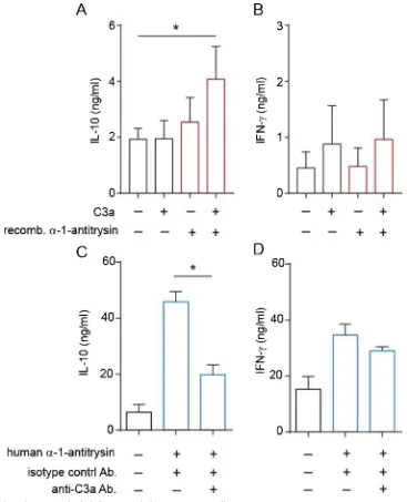
α-1-Antitrypsin is reported to have potent immune-regulatory ef-fects in vivo, as well as on total peripheral blood mononuclear cells (PBMC) and innate immune cell populations [9]. We were therefore interested to define its effects on human CD4+T cells, which we have identified produce this protein when stimulated in the presence of 1,25(OH)2D3 (Fig. 1). Initially, we assessed cytokine profiles of CD4+T cells stimulated in the presence ofα-1-antitrypsin purified from human plasma, using concentrations of α-1-antitrypsin similar to circulating levels (in vitro doses of 10–1000μg/ml are equivalent to 0.18–18μM; circulating levels are 20–48μM). These experiments revealed that human plasma-derivedα-1-antitrypsin significantly increased CD4+T cell IL-10 secretion (Fig. 2a). Enhanced secretion of IFN-γ(Fig. 2b), but not TNF-α(Fig. 2c) or IL-13 (Fig. 2d) were also observed. CD4+T cells responded toα-1-antitrypsin for induction of IL-10 in a dose-dependent manner from 10 to 1000 μg/ml with significant effects observed at 500μg/ml and maximal effects observed at 1 mg/ml α-1-antitrypsin (Supplementary Fig. 1). Commercial preparations of human plasma-derivedα-1-antitrypsin are known to contain non-native conformations of the protein that are inactive as protease inhibitors [16]. We therefore repeated our experiments, comparing human plasma α-1-antitrypsin with a recombinantα-1-antitrypsin preparation, as well as a control for exogenous protein addition (at the same concentration). These experi-ments interestingly revealed that human plasma-derived, but not re-combinantα-1-antitrypsin induced IL-10 (Fig. 2E) and IFN-γ(Fig. 2F) secretion, whereas the protein control had little effect on cytokine le-vels. A chymotrypsin activity assay confirmed that human plasma-de-rivedα-1-antitrypsin had very little anti-protease activity compared to the recombinant preparation (Fig. 2G). Taken together, these data suggest that the effects ofα-1-antitrypsin on cytokine secretion do not require serine protease inhibitory activity, but may require a plasma-derived molecule binding to α-1-antitrypsin –not present in the re-combinant preparation. We next explored this possibility.
3.3. α-1-Antitrypsin directly binds to complement C3a
physiologically relevant given the high circulating levels of α -1-anti-trypsin of around 20–48μM [19].
3.4. α-1-Antitrypsin and C3a interact to promote CD4+T cell IL-10
secretion
To establish the role for C3a in α-1-antitrypsin-induced IL-10 in human CD4+T cells, we undertook two complementary experimental approaches. Firstly, we added exogenous C3a to cultures containing recombinantα-1-antitrypsin. This resulted in significant IL-10 produc-tion (Fig. 4a), whilst not increasing IFN-γlevels (Fig. 4b). Secondly, C3a was neutralized with a specific antibody in cultures of CD4+
T cells stimulated in the presence of plasma- derived α-1-antitrypsin. These experiments demonstrated that inclusion of an C3a specific anti-body, but not an isotype control antibody significantly reduced IL-10 levels in (Fig. 4c), whilst again not affecting IFN-γ secretion. These experiments indicate that α-1-antitrypsin and C3a act in concert to drive IL-10 secretion by CD4+T cells.
3.5. α-1-Antitrypsin is required for 1,25(OH)2D3-induced IL-10
[image:5.595.89.511.57.467.2]This work has identified that 1,25(OH)2D3-treatment promotesα -1-antitrypsin expression by CD4+T cells, and furthermore thatα -1-an-titrypsin, at circulating levels, acts together with its binding partner, C3a, to promote IL-10 expression. We therefore next probed whether the α-1-antitrypsin-C3a axis may play an endogenous role in 1,25(OH)2D3-driven IL-10 expression by CD4+T cells. Despite being a well-established effect of 1,25(OH)2D3, with evidence for in vivo re-levance, this remains poorly understood and the kinetics indicate a likely indirect mechanism. To do so, we compared effects of 1,25(OH)2D3 onSERPINA1andIL10expression in CD4+T cells from healthy volunteer donors with those from age-matched patients ex-hibiting a genetic mutation in theSERPINA1 gene (PiZZ genotype), which can transcribe theSERPINA1gene, but cannot secrete the mu-tant, polymerised protein. As expected,SERPINA1mRNA levels were similarly induced by 1,25(OH)2D3 treatment in healthy control and PiZZ genotype CD4+T cells (Fig. 5a). However, consistent with their
Fig. 1. 1,25(OH)2D3 drivesα-1-antitrypsin expression by human peripheral CD4+T cells, but not CD8+ T cells or monocytes.
(a&b) CD4+T cells, or (c) CD8+T cells (1 × 10⁶in 1 ml), in RPMI 10%FCS, were stimulated with anti-CD3 (1μg/ml) and IL-2 (50 u/ml) for two rounds of seven days, in presence of 1,25(OH)2D3 as indicated (a: n = 9, b: n = 4, c: n = 4). (d) Human purified peripheral CD14+monocytes (1 × 10⁶in 1 ml), in RPMI 10%FCS, were stimulated with LPS (500 ng/ml) for 48 h in presence of 10−7M 1,25(OH)
2D3 as indicated (n = 4).α-1-Antitrypsin expression was assessed by (a) qPCR for SERPINA1 mRNA and (b–d) ELISA. mRNA quantification by qPCR was normalised to 18S transcription and is shown as a relative quantity (RQ) compared to expression under control conditions in vitro. Data are summarised as mean ± SEM. (a) p value derives from Friedman’s non-parametric repeated measures test. * p < 0.050 as assessed by Dunn’s paired post-test for multiple comparisons. (b) p values derive from a repeated measures ANOVA.
S. Dimeloe et al. Journal of Steroid Biochemistry and Molecular Biology 189 (2019) 1–9
genotype, reduced levels ofα-1-antitrypsin protein synthesis by PiZZ CD4+T cells compared to control individuals are routinely detected in our laboratory (Supplementary Fig. 3). Nevertheless, when the capacity of 1,25(OH)2D3 to enhance gene expression of IL10by CD4+T cell cultures was compared in these two cohorts, PiZZ CD4+T cells were found to have significantly lower abundance ofIL10mRNA, which was indeed not upregulated compared to untreated cells (Fig. 5b). Addi-tional analyses of the relationship betweenSERPINA1andIL10gene
expression in 1,25(OH)2D3-treated CD4+T cells identified a significant correlation was observed in cultures from healthy donors (r = 0.75; p = 0.02; n = 9), which was not observed in theα-1-antitrypsin-defi -cient patient cohort (r = 0.50; p = 0.14; n = 10) (Fig. 5c–d).
Taken together these data indicate that 1,25(OH)2D3 increasesα -1-antitrypsin expression by CD4+ T cells, which then acts in turn to
promote anti-inflammatory IL-10 expression by these cells.
[image:6.595.90.510.54.386.2]Additionally, we identify a novel interaction betweenα-1-antitrypsin
Fig. 2. Human plasma-derived, but not recombinantα-1-antitrypsin promotes production of IL-10 and IFN-γby human CD4+T cells, which is independent
of anti-protease activity.
(a–f) CD4+T cells (1 × 10⁶in 1 ml) were stimulated with anti-CD3 (1μg/ml) and IL-2 (50 u/ml) for 48 h without or with the indicatedα-1-antitrypsin preparation or protein control. (a–d) Humanα-1-antitrypsin was commercially obtained (e–f) humanα-1-antitrypsin was lab-purified (see Methods for further details). Cytokine levels were assessed by CBA and are summarised as mean ± SEM. p values derive from (a–d, n = 8) pairedt-tests and (e–f, n = 4) one-way ANOVA. * p < 0.05 and *** p < 0.001 as assessed by pairedt-test (a–d) or Bonferroni’s paired post-test for multiple comparisons (e–f). (g) 0–5 nMα-1 antitrypsin was pre-incubated with chymotrypsin before the addition of 0.1 mM substrateN-succinyl-Ala-Ala-Pro-Phe P-nitroanilide and the inhibitory capacity of eachα-1-antitrypsin preparation assed by the 405 nm absorbance. Data is presented as 405 nM absorbance T10-T0 normalised to 1 againstα-1-antitrypsin concentration.
Fig. 3.α-1-Antitrypsin and C3a directly interact.
[image:6.595.90.508.587.703.2]and complement C3a, which is critical for IL-10 induction. The model is summarised in graphical form in Supplementary Fig. 4.
4. Discussion
This study identifies a novel interaction in the human immune system, between two ubiquitous mediators, α-1-antitrypsin and C3a, which directly bind to each other, and at physiological concentrations interact to drive anti-inflammatory IL-10 expression by CD4+T cells. Additionally, we show that 1,25(OH)2D3 is a key upstream regulator in this axis, stimulating α-1-antitrypsin production by human CD4+ T cells–a novel cellular source of this protein.
A number of similarities exist in the anti-inflammatory function of α-1-antitrypsin and vitamin D. Deficiency of bothα-1-antitrypsin and vitamin D are associated with inflammation and autoimmunity [20,21]. Additionally, the airway is an environment where the immune-mod-ulatory effects of bothα-1-antitrypsin and 1,25(OH)2D3 appear to be of
[image:7.595.114.481.54.507.2]key importance. Beyond its critical anti-elastase function, clearly evi-denced by the development of COPD in deficient individuals, α -1-an-titrypsin deficiency is also implicated in control of airway inflammation since asthma symptoms are also reported inα-1-antitrypsin deficiency [22–24]. Vitamin D deficiency is also implicated in airway immune homeostasis, as in respiratory diseases, including asthma, epidemio-logic associations exist between vitamin D insufficiency and worse asthma control. Notably, the phenotype of vitamin D deficiency in se-vere asthma, and in particular treatment refractory asthma is associated with a neutrophilic profile, an enhanced pro-inflammatory Th17 phe-notype and impaired induction of anti-inflammatory IL-10 [25]. Neu-trophils are also proposed to play a prominent role in lung disease as-sociated with α-1-antitrypsin deficiency, and our unpublished data demonstrate a significantly increased frequency of pro-inflammatory Th17 in these individuals (manuscript in preparation). As we already discuss in the introduction to this paper, DeMeo et al. [12] report that COPD progression inα-1-antitrypsin deficiency associated with IL-10
Fig. 4. C3a andα-1-antitrypsin cooperatively induce IL-10 in human CD4+ T cells.
CD4+T cells were cultured for 48 h in serum-free medium, in the absence or presence of (a–b) recombinantα-1-antitrypsin (100μg/ml) and/or C3a (100 ng/ml) as indicated or (c–d) commercial human plasma-derivedα-1-antitrypsin (500μg/ml) with or without a C3a neutralising or isotype control (anti-C3a/IgG1, 5μg/ml) antibody. Cytokine secretion was assessed by CBA and presented as mean ± SEM. p values derived from a one-way ANOVA. * p < 0.05, ** p < 0.01 as assessed by Bonferroni’s paired post-test for multiple comparisons (n = 4).
S. Dimeloe et al. Journal of Steroid Biochemistry and Molecular Biology 189 (2019) 1–9
polymorphisms andα-1 antitrypsin replacement therapy increases cir-culating IL-10 in these individuals. Hence a number of clear parallels exist. The present study indicates that the similarity betweenα -1-an-titrypsin and vitamin D deficiency may be explained by a biological link between these two mediators–specifically that 1,25(OH)2D3 regulates α-1-antitrypsin abundance and thereby its immune-modulatory prop-erties (see model proposed in Supplementary Fig. 4). Further studies are now warranted to interrogate the importance of this axis in tissue en-vironments and in the context of inflammation.
Previous reports have demonstrated anti-inflammatory actions ofα -1-antitrypsin in vitro and in experimental models in vivo [9], with early papers for example reporting thatα-1-antitrypsin therapy induces an-tigen-specific immune tolerance during islet allograft transplantation in mice [11,26]. The authors implicated a role for α-1-antitrypsin in maintaining dendritic cells in an immature state and in the induction of regulatory T cells (Treg), as assessed by increased mRNA for foxp3, cytotoxic T lymphocyte antigen-4, TGF-beta, IL-10, and IL-1 receptor antagonist. These functions closely mirror those described for vitamin D (1,25(OH)2D3) on immune cells [4,6,27]. In the present study we fo-cused on the capacity ofα-1-antitrypsin to enhance IL-10 synthesis by T cells since a major interest of the lab has been the control of IL-10 synthesis by 1,25(OH)2D3 [4,5]. A limitation of the current study is that we did not comprehensively address whether Foxp3 expression was also enhanced in these experiments, in part due to our earlier work showing that 1,25(OH)2D3 increases the frequency of distinct IL-10 or Foxp3 expressing CD4+T cells, with little or no co-expression [3]. However, we did observe a reduced expression ofFOXP3mRNA in CD4+T cells
obtained from PiZZ compared to control donors (data not shown: n = 5 per group; p = 0.0079). Furthermore, Mueller et al. [28] demonstrated that a single intramuscular administration of recombinant adeno-asso-ciated virus serotype 1 alpha-1 antitrypsin vector into alpha-1 deficient patients resulted in an active Treg response (approx. 10% of CD3+cells were Foxp3 up to 5-years post administration, which was not seen in normal control muscle), suggesting future directions for our work.
The present study was able to compare responsiveness to 1,25(OH)2D3 in healthy control subjects versus those with a hereditary deficiency ofα-1-antitrypsin protein, characterised by the PiZZ geno-type, who possess around only 15% of normal wildtype circulatingα -1-antitrypsin protein. Whilst CD4+ T cells from healthy subjects de-monstrated a significant positive correlation between the capacity of 1,25(OH)2D3 to increase gene expression for IL10 and SERPINA1, this association was absent in the α-1-antitrypsin deficient donors. This comparison of the response to 1,25(OH)2D3 inα-1-antitrypsin deficient versus control subjects strengthen evidence implicating 1,25(OH)2D3 as an upstream regulator of this anti-inflammatory axis in these cells.
There appear to be a growing number of underlying mechanisms by which α-1-antitrypsin exerts immunomodulatory activity, which re-main poorly understood (reviewed in 9). However, the anti-protease function ofα-1-antitrypsin is not consistently reported to be required, and precedence exists for functions of α-1-antitrypsin that require binding to other serum components [9]. For example Bergin et al. [29] described the capacity ofα-1-antitrypsin to regulate chemotaxis in-duced by soluble immune complexes and IL-8 by human neutrophils. Specifically, α-1-antitrypsin decreased the chemotactic response of
Fig. 5.α-1-Antitrypsin is required for 1,25(OH)2D3-induced IL-10 expression by
CD4+T cells
.
CXCR1 signaling by binding IL-8 and inhibiting receptor engagement. In a very different example, additional protease-independent activity of α-1-antitrypsin was suggested by studies describing the induction byα -1-antitrypsin of angiopoietin-like protein 4 in human blood monocytes and lung microvascular endothelial cells [30]. This function of α -1-antitrypsin required binding to the fatty acids linoleic acid and oleic acid. Linoleic acid was also reported to enhance the capacity ofα -1-antitrypsin to inhibit LPS-induced IL-1βsynthesis by human neutrophils [31]. Thus our demonstration that induction of IL-10 in CD4+T cells by α-1-antitrypsin is dependent on binding to the complement component C3a, adds to this growing body of evidence for protease-independent functions ofα-1-antitrypsin.
C3a is shown to be critical to one of the major anti-inflammatory functions of α-1-antitrypsin. In addition to its key innate immune function, the complement system is now recognized to be involved in both the generation and regulation of adaptive immunity. C3a and C5a are essential for induction of inflammatory adaptive immune responses, signaling via their surface G-protein coupled receptors [17]. The com-plement system also promotes immune tolerance and drives IL-10, for example via ligation of CD46 [32]. The anti-inflammatory role we have identified for C3a in promoting IL-10 synthesis initially appears in contrast to its well-known pro-inflammatory effects, as highlighted by the impaired immunity associated with C3 deficiency [33]. However it is consistent with recent, apparently opposing observations of immune dysregulation,failure of immune tolerance mechanisms and allograft rejection in C3 and C3aR deficient animal models [34,35] and with observations in humans thatpurified CD4+T cells from C3 deficient individuals demonstrate defective IL-10 responses in vitro [36]. Indeed, complement has been described as a “double-edged sword” in the regulation of immunity [37]. The dependency upon C3a for the anti-inflammatory effects of α-1-antitrypsin in vivo in disease models re-mains to be assessed using relevant transgenic systems.
Our study identifies 1,25(OH)2D3 as an upstream regulator ofα -1-antitrypsin expression in human T cells. The major source of circulating α-1-antitrypsin is hepatocytes, although other cell types synthesise this protein. This includes monocyte-derived macrophages and dendritic cells, alveolar macrophages, bronchial epithelial cells and neutrophils, which may significantly contribute to localα-1-antitrypsin levels in the context of an immune response. However, to date we have found no evidence for regulation by 1,25(OH)2D3 ofα-1-antitrypsin gene and/or protein levels in monocytes, respiratory epithelial cells or primary he-patocytes (data not shown). Our interpretation of these data is that this
function of 1,25(OH)2D3 may therefore reflect an
im-munoregulatory–specific activity of 1,25(OH)2D3 in human T cells, adding to the growing list of such functions attributed to 1,25(OH)2D3 [1,38].
Growing recognition of vitamin D deficiency and insufficiency in recent years and its association with a wide range of immune-mediated pathologies, has meant that pharmacological vitamin D supplementa-tion (with precursors of 1,25(OH)2D3) is now increasingly employed [1,38]. It is known to be safe, well-tolerated, relatively inexpensive and with a half-life of weeks. A number of clinical studies of vitamin D supplementation, with highly differing design and primary outcomes, have been performed. Notably, systematic review of these trials to date, indicate that vitamin D is beneficial in asthma [27]. Understanding the mechanisms whereby vitamin D acts to promote respiratory health are therefore likely to be important in improving clinical trial design. To date, the capacity of vitamin D to promote antimicrobial pathways, a common cause of asthma exacerbations, together with induction and/ or maintenance of immune tolerance pathways are proposed [25]. In addition we, and others, have proposed that vitamin D beneficially modulates immune and clinical response to glucocorticoids. In early studies we demonstrated that 1,25(OH)2D3 restored the impaired steroid-induced IL-10 response in steroid refractory asthma patients both in vitro and in vivo [5], and clinical studies now provide evidence for modest steroid-sparing effects on vitamin D in asthma patients
(reviewed in [25]). A very recent report of α-1-antitrypsin infusion therapy for steroid-resistant graft-versus-host disease suggested thatα -1-antitrypsin was safe and potentially effective in treating these pa-tients, and was associated with an increased ratio of regulatory to ef-fector T cells following treatment [39], further demonstrating parallels with known effects of vitamin D.
α-1-antitrypsin has been used successfully in animal models of various autoimmune diseases as well as transplantation [9], and this has contributed to the growing interest in the therapeutic application of α-1-antitrypsin as an anti-inflammatory mediator, beyond its applica-tion in AAT deficiency. In addiapplica-tion to the above example in graft-versus-host disease, clinical studies of the safety and efficacy ofα -1-antitrypsin treatment have been reported in conditions as diverse as type 1 diabetes [40], lung transplantation and acute myocardial in-farction ([41]; see alsoclincialtrials.gov).
In summary, we have identified a novel, vitamin D (1,25(OH)2D3) controlled regulatory axis in the immune system, mediated through the interaction ofα-1-antitrypsin and the complement anaphylatoxin C3a to stimulate IL-10 synthesis by CD4+T cells. Our data highlight a novel potential therapeutic application for vitamin D in modulation of this axis, and suggest new roles for α-1-antitrypsin in the control of in-flammatory disease. There is wide experience of the use of both vitamin D andα-1-antitrypsin as therapeutic agents in the US and Europe and both have very good safety profiles. Future trials offer the capacity to directly examine evidence for the role ofα-1-antitrypsin in vitamin D-mediated immune regulation.
Author contributions
Experimental work was largely carried out by SD and LR. HC, CC, PEP, MPN, DFR, and JR also contributed to experiments. PL supported analyses of transcriptomic datasets providing pilot observations upon which the experiments were conceived. JM and LR designed and per-formed all binding experiments. CK provided expertise on complement biology and identified that C3a alone regulates IL-10 in human CD4+
T cells [42]. The manuscript was prepared by SD, LR, BG and CMH, and all studies were conceived by SD, LR, BG, CK and CMH.
Acknowledgements
SD and LR were recipients of Medical Research Council (MRC) funded PhD Studentships through the MRC & Asthma UK Centre for Allergic Mechanisms of Asthma. CK was supported by an MRC Research Grant (Grant no. G1002165) and the Medical Research Council Centre for Transplantation, KCL. CH with LR, HC and CC, as well as MPN were supported by grants from the Alpha-1 Foundation. CMH, CK and PEP are supported by the Department of Health, National Institute for Health Research comprehensive Biomedical Research Centre award to Guy’s & St. Thomas’NHS Foundation Trust in partnership with King’s College London and King’s College Hospital NHS Foundation Trust. We gratefully acknowledge the work undertaken to provide reagents by Marie Pang (KCL, London), Doris Quay (Birkbeck College, London) and Professor Joerg Koehl (Lubeck University, Germany) and the assistance of Professor David Lomas (UCL) and the Cambridge Institute of Medical Research (CIMR, University of Cambridge) for use of laboratory facil-ities. Microscale thermophoresis assays were conducted with the kind assistance of Dr Tina Daviter (ISMB Biophysics facility), Dr Mark Pfuhl (Randall Division, KCL) and Dr James Wilkinson (Nanotemper Technologies). We acknowledge support from our research nurse, Kheem Jones.
Appendix A. Supplementary data
Supplementary material related to this article can be found, in the online version, at doi:https://doi.org/10.1016/j.jsbmb.2019.01.014.
S. Dimeloe et al. Journal of Steroid Biochemistry and Molecular Biology 189 (2019) 1–9
References
[1] A.S. Vanherwegen, C. Gysemans, C. Mathieu, Regulation of immune function by vitamin d and its use in diseases of immunity, Endocrinol. Metab. Clin. North Am. 46 (4) (2017) 1061–1094.
[2] S. Dimeloe, D.F. Richards, Z.L. Urry, A. Gupta, V. Stratigou, S. Farooque, et al., 1alpha,25-dihydroxyvitamin D3 promotes CD200 expression by human peripheral and airway-resident T cells, Thorax 67 (7) (2012) 574–581.
[3] Z. Urry, E.S. Chambers, E. Xystrakis, S. Dimeloe, D.F. Richards, L. Gabrysova, et al., The role of 1alpha,25-dihydroxyvitamin D3 and cytokines in the promotion of distinct Foxp3+ and IL-10+ CD4+ T cells, Eur. J. Immunol. 42 (10) (2012) 2697–2708.
[4] Z. Urry, E. Xystrakis, D.F. Richards, J. McDonald, Z. Sattar, D.J. Cousins, et al., Ligation of TLR9 induced on human IL-10-secreting Tregs by 1alpha,25-dihydrox-yvitamin D3 abrogates regulatory function, J. Clin. Invest. 119 (2) (2009) 387–398. [5] E. Xystrakis, S. Kusumakar, S. Boswell, E. Peek, Z. Urry, D.F. Richards, et al.,
Reversing the defective induction of IL-10-secreting regulatory T cells in gluco-corticoid-resistant asthma patients, J. Clin. Invest. 116 (1) (2006) 146–155. [6] L.E. Jeffery, F. Burke, M. Mura, Y. Zheng, O.S. Qureshi, M. Hewison, et al.,
1,25-Dihydroxyvitamin D3 and IL-2 combine to inhibit T cell production of inflammatory cytokines and promote development of regulatory T cells expressing CTLA-4 and FoxP3, J. Immunol. 183 (9) (2009) 5458–5467.
[7] A. Gupta, A. Sjoukes, D. Richards, W. Banya, C. Hawrylowicz, A. Bush, et al., Relationship between serum vitamin D, disease severity, and airway remodeling in children with asthma, Am. J. Respir. Crit. Care Med. 184 (12) (2011) 1342–1349. [8] S.J. Marciniak, D.A. Lomas, What can naturally occurring mutations tell us about
the pathogenesis of COPD? Thorax 64 (4) (2009) 359–364.
[9] S. Janciauskiene, T. Welte, Well-known and less well-known functions of alpha-1 antitrypsin. Its role in chronic obstructive pulmonary disease and other disease developments, Ann. Am. Thorac. Soc. 13 (Suppl. 4) (2016) S280–8.
[10] E.C. Lewis, Expanding the clinical indications for alpha(1)-antitrypsin therapy, Mol. Med. 18 (2012) 957–970.
[11] E.C. Lewis, M. Mizrahi, M. Toledano, N. Defelice, J.L. Wright, A. Churg, et al., alpha1-Antitrypsin monotherapy induces immune tolerance during islet allograft transplantation in mice, Proc. Natl. Acad. Sci. U. S. A. 105 (42) (2008) 16236–16241.
[12] D.L. Demeo, E.J. Campbell, A.F. Barker, M.L. Brantly, E. Eden, N.G. McElvaney, et al., IL10 polymorphisms are associated with airflow obstruction in severe alpha1-antitrypsin deficiency, Am. J. Respir. Cell Mol. Biol. 38 (1) (2008) 114–120. [13] D.A. Lomas, D.L. Evans, S.R. Stone, W.S. Chang, R.W. Carrell, Effect of the Z
mu-tation on the physical and inhibitory properties of alpha 1-antitrypsin, Biochemistry 32 (2) (1993) 500–508.
[14] H. Parfrey, R. Mahadeva, N.A. Ravenhill, A. Zhou, T.R. Dafforn, R.C. Foreman, et al., Targeting a surface cavity of alpha 1-antitrypsin to prevent conformational disease, J. Biol. Chem. 278 (35) (2003) 33060–33066.
[15] C. Barbey-Morel, D.H. Perlmutter, Effect of pseudomonas elastase on human mononuclear phagocyte alpha 1-antitrypsin expression, Pediatr. Res. 29 (2) (1991) 133–140.
[16] D.A. Lomas, P.R. Elliott, R.W. Carrell, Commercial plasma alpha1-antitrypsin (Prolastin) contains a conformationally inactive, latent component, Eur. Respir. J. 10 (3) (1997) 672–675.
[17] S. Freeley, C. Kemper, G. Le Friec, The "ins and outs" of complement-driven immune responses, Immunol. Rev. 274 (1) (2016) 16–32.
[18] M.P. Dierich, B. Landen, M. Schmitt, Complement receptor analogous factors in human serum: I. Isolation of a molecule inhibitory for complement dependent ro-sette formation, its identification as alpha 1-antitrypsin and its functional char-acterization, Immunobiology 156 (1–2) (1979) 153–167.
[19] J.A. Bornhorst, D.N. Greene, E.R. Ashwood, D.G. Grenache, alpha1-Antitrypsin phenotypes and associated serum protein concentrations in a large clinical popu-lation, Ches 143 (4) (2013) 1000–1008.
[20] F. de Serres, I. Blanco, Role of alpha-1 antitrypsin in human health and disease, J. Intern. Med. 276 (4) (2014) 311–335.
[21] W. Dankers, E.M. Colin, J.P. van Hamburg, E. Lubberts, Vitamin D in auto-immunity: molecular mechanisms and therapeutic potential, Front. Immunol. 7 (2016) 697.
[22] C.M. Greene, S.J. Marciniak, J. Teckman, I. Ferrarotti, M.L. Brantly, D.A. Lomas, et al., alpha1-Antitrypsin deficiency, Nat. Rev. Dis. Primers 2 (2016) 16051. [23] A. Gramegna, S. Aliberti, M. Confalonieri, A. Corsico, L. Richeldi, C. Vancheri, et al.,
Alpha-1 antitrypsin deficiency as a common treatable mechanism in chronic re-spiratory disorders and for conditions different from pulmonary emphysema? A commentary on the new European Respiratory Society statement, Multidiscip. Respir. Med. 13 (2018) 39.
[24] E. Eden, J. Hammel, F.N. Rouhani, M.L. Brantly, A.F. Barker, A.S. Buist, et al., Asthma features in severe alpha1-antitrypsin deficiency: experience of the National Heart, Lung, and Blood Institute Registry, Chest 123 (3) (2003) 765–771. [25] P.E. Pfeffer, C.M. Hawrylowicz, Vitamin D in asthma: mechanisms of action and
considerations for clinical trials, Chest 153 (5) (2018) 1229–1239. [26] E.C. Lewis, L. Shapiro, O.J. Bowers, C.A. Dinarello, Alpha1-antitrypsin
mono-therapy prolongs islet allograft survival in mice, Proc. Natl. Acad. Sci. U. S. A. 102 (34) (2005) 12153–12158.
[27] E.H. Mann, E.S. Chambers, P.E. Pfeffer, C.M. Hawrylowicz, Immunoregulatory mechanisms of vitamin D relevant to respiratory health and asthma, Ann. N. Y. Acad. Sci. 1317 (2014) 57–69.
[28] C. Mueller, G. Gernoux, A.M. Gruntman, F. Borel, E.P. Reeves, R. Calcedo, et al., 5 year expression and neutrophil defect repair after gene therapy in Alpha-1 anti-trypsin deficiency, Mol. Ther. 25 (6) (2017) 1387–1394.
[29] D.A. Bergin, E.P. Reeves, P. Meleady, M. Henry, O.J. McElvaney, T.P. Carroll, et al., alpha-1 Antitrypsin regulates human neutrophil chemotaxis induced by soluble immune complexes and IL-8, J. Clin. Invest. 120 (12) (2010) 4236–4250. [30] E. Frenzel, S. Wrenger, B. Brugger, S. Salipalli, S. Immenschuh, N. Aggarwal, et al.,
alpha1-Antitrypsin combines with plasma fatty acids and induces angiopoietin-like protein 4 expression, J. Immunol. 195 (8) (2015) 3605–3616.
[31] N. Aggarwal, E. Korenbaum, R. Mahadeva, S. Immenschuh, V. Grau, C.A. Dinarello, et al., alpha-Linoleic acid enhances the capacity of alpha-1 antitrypsin to inhibit lipopolysaccharide induced IL-1beta in human blood neutrophils, Mol Med. 22 (2016) 680–693.
[32] C. Kemper, A.C. Chan, J.M. Green, K.A. Brett, K.M. Murphy, J.P. Atkinson, Activation of human CD4+ cells with CD3 and CD46 induces a T-regulatory cell 1 phenotype, Nature 421 (6921) (2003) 388–392.
[33] M.R. Daha, Role of complement in innate immunity and infections, Crit. Rev. Immunol. 30 (1) (2010) 47–52.
[34] C. Hammerberg, S.K. Katiyar, M.C. Carroll, K.D. Cooper, Activated complement component 3 (C3) is required for ultraviolet induction of immunosuppression and antigenic tolerance, J. Exp. Med. 187 (7) (1998) 1133–1138.
[35] P. Baruah, E. Simpson, I.E. Dumitriu, K. Derbyshire, D. Coe, C. Addey, et al., Mice lacking C1q or C3 show accelerated rejection of minor H disparate skin grafts and resistance to induction of tolerance, Eur. J. Immunol. 40 (6) (2010) 1758–1767. [36] J. Kildsgaard, T.J. Hollmann, K.W. Matthews, K. Bian, F. Murad, R.A. Wetsel,
Cutting edge: targeted disruption of the C3a receptor gene demonstrates a novel protective anti-inflammatory role for C3a in endotoxin-shock, J. Immunol. 165 (10) (2000) 5406–5409.
[37] P. Vignesh, A. Rawat, M. Sharma, S. Singh, Complement in autoimmune diseases, Clin. Chim. Acta 465 (2017) 123–130.
[38] S. Christakos, M. Hewison, D.G. Gardner, C.L. Wagner, I.N. Sergeev, E. Rutten, et al., Vitamin D: beyond bone, Ann. N. Y. Acad. Sci. 1287 (2013) 45–58. [39] J.M. Magenau, S.C. Goldstein, D. Peltier, R.J. Soiffer, T. Braun, A. Pawarode, et al.,
alpha1-Antitrypsin infusion for treatment of steroid-resistant acute graft-versus-host disease, Blood 131 (12) (2018) 1372–1379.
[40] P.A. Gottlieb, A.K. Alkanani, A.W. Michels, E.C. Lewis, L. Shapiro, C.A. Dinarello, et al., alpha1-Antitrypsin therapy downregulates toll-like receptor-induced IL-1beta responses in monocytes and myeloid dendritic cells and may improve islet function in recently diagnosed patients with type 1 diabetes, J. Clin. Endocrinol. Metab. 99 (8) (2014) E1418–26.
[41] A. Abbate, B.W. Van Tassell, S. Christopher, N.A. Abouzaki, C. Sonnino, C. Oddi, et al., Effects of prolastin C (plasma-derived alpha-1 antitrypsin) on the acute in-flammatory response in patients with ST-segment elevation myocardial infarction (from the VCU-alpha 1-RT pilot study), Am. J. Cardiol. 115 (1) (2015) 8–12. [42] A. Ghannam, J.L. Fauquert, C. Thomas, C. Kemper, C. Drouet, Human complement