ADAMTS-1: a metalloproteinase-disintegrin
essential for normal growth, fertility, and organ
morphology and function
Takayuki Shindo, … , Yoshio Yazaki, Kouji Matsushima
J Clin Invest. 2000;
105(10)
:1345-1352.
https://doi.org/10.1172/JCI8635
.
A disintegrin and metalloproteinase (ADAM) represents a protein family possessing both
metalloproteinase and disintegrin domains. ADAMTS-1, an ADAM family member cloned
from cachexigenic colon adenocarcinoma, is unusual in that it contains thrombospondin
type I motifs and anchors to the extracellular matrix. To elucidate the biological role of
ADAMTS-1, we developed ADAMTS-1–null mice by gene targeting. Targeted disruption of
the mouse ADAMTS-1 gene resulted in growth retardation with adipose tissue
malformation. Impaired female fertilization accompanied by histological changes in the
uterus and ovaries also resulted. Furthermore, ADAMTS-1
–/–mice demonstrated enlarged
renal calices with fibrotic changes from the ureteropelvic junction through the ureter, and
abnormal adrenal medullary architecture without capillary formation. ADAMTS-1 thus
appears necessary for normal growth, fertility, and organ morphology and function.
Moreover, the resemblance of the renal phenotype to human ureteropelvic junction
obstruction may provide a clue to the pathogenesis of this common congenital disease.
Article
Introduction
Proteolytic modification of cell-surface proteins and extracellular matrices is pivotal for a diverse array of biological and pathological processes, including embryogenesis, wound healing, and cancer metasta-sis. A disintegrin and metalloproteinase (ADAM) rep-resents a protein family possessing both metallopro-teinase and disintegrin domains. Recently, the ADAM protein family has emerged as a key partici-pant in these processes (1, 2). More than 20 proteins have been identified as members of the ADAM fami-ly. Fertilin-α/β, the first ADAM described, has been implicated in integrin-mediated sperm-egg binding (3, 4). Subsequently, meltrin-α was shown to be involved in muscle fusion (5). TNF-α–converting enzyme, which cleaves the membrane-anchored pre-cursor of TNF-α, thus upregulating production of TNF-α, has recently proved to be a member of the
ADAM family (6–9). In the fruit fly Drosophila, an ADAM family gene called Kuzbanian has been demonstrated to play a role in lateral inhibition dur-ing neurogenesis by coddur-ing for cleavage of the extra-cellular domain of the transmembrane receptor Notch (10, 11). Despite accumulating findings about the biological role of these ADAM proteins, the respective functions of the growing number of ADAM family members remain largely unknown.
ADAMTS-1 is a new member protein of the ADAM family, which was originally identified by differential display analysis as a gene highly expressed in the murine colon 26 cachexigenic tumor (12). In vivo expression of the gene is induced in the kidney and heart of mice treated with LPS, suggesting a possible role of ADAMTS-1 in the inflammatory reaction (12). Whereas typical ADAMs are membrane-anchored pro-teins that have a transmembrane region in the
car-ADAMTS-1: a metalloproteinase-disintegrin essential
for normal growth, fertility, and organ
morphology and function
Takayuki Shindo,
1Hiroki Kurihara,
1,2Kouji Kuno,
2,3Hitoshi Yokoyama,
4Takashi Wada,
5Yukiko Kurihara,
1Tomihiko Imai,
1Yuhui Wang,
1Masafumi Ogata,
3Hiroaki Nishimatsu,
6Nobuo Moriyama,
6Yoshio Oh-hashi,
1Hiroyuki Morita,
1Takatoshi Ishikawa,
7Ryozo Nagai,
1Yoshio Yazaki,
8and Kouji Matsushima
2,91Department of Cardiovascular Medicine, Graduate School of Medicine, University of Tokyo, Tokyo, Japan
2Core Research for Evolutional Science and Technology (CREST), Japan Science and Technology Corporation, Tokyo, Japan 3Department of Molecular Pharmacology, Cancer Research Institute,
4Division of Blood Purification, and
5First Department of Internal Medicine, School of Medicine, Kanazawa University, Kanazawa, Japan 6Department of Urology, and
7Department of Pathology, Graduate School of Medicine, University of Tokyo, Tokyo, Japan 8The Hospital International Medical Center of Japan, Tokyo, Japan
9Department of Molecular Preventive Medicine, Graduate School of Medicine, University of Tokyo, Tokyo, Japan
Address correspondence to: Hiroki Kurihara, Department of Cardiovascular Medicine, Graduate School of Medicine, University of Tokyo, 7-3-1 Hongo, Bunkyo-ku, Tokyo 113-8655, Japan. Phone: 81-3-5800-6519; Fax: 81-3-5800-8824; E-mail: [email protected].
Takayuki Shindo, Hiroki Kurihara, and Kouji Kuno contributed equally to this work. Received for publication October 7, 1999, and accepted in revised form March 17, 2000.
A disintegrin and metalloproteinase (ADAM) represents a protein family possessing both metallo-proteinase and disintegrin domains. ADAMTS-1, an ADAM family member cloned from cachexigenic colon adenocarcinoma, is unusual in that it contains thrombospondin type I motifs and anchors to the extracellular matrix. To elucidate the biological role of ADAMTS-1, we developed ADAMTS-1–null mice by gene targeting. Targeted disruption of the mouse ADAMTS-1gene resulted in growth retar-dation with adipose tissue malformation. Impaired female fertilization accompanied by histological changes in the uterus and ovaries also resulted. Furthermore, ADAMTS-1–/–mice demonstrated
enlarged renal calices with fibrotic changes from the ureteropelvic junction through the ureter, and abnormal adrenal medullary architecture without capillary formation. ADAMTS-1 thus appears nec-essary for normal growth, fertility, and organ morphology and function. Moreover, the resemblance of the renal phenotype to human ureteropelvic junction obstruction may provide a clue to the patho-genesis of this common congenital disease.
boxyl-terminal region, ADAMTS-1 does not contain a transmembrane domain, but possesses three throm-bospondin type I motifs at its carboxyl terminus, which are conserved motifs in both thrombospondin 1 and 2 (12, 13). After secretion, ADAMTS-1 is incor-porated into the extracellular matrix. Site-directed mutagenesis revealed that these thrombospondin type I motifs, together with the carboxyl-terminal spacing region, are responsible for anchoring to the extracellular matrix (14). Recent studies using α 2-macroglobulin as a substrate demonstrated that the metalloproteinase domain of ADAMTS-1 is func-tional (15). Thus, ADAMTS-1 is an active ADAM pro-tease that is closely associated with the extracellular matrix, although its physiological substrate or sub-strates remain unclear.
Recent reports have identified several proteins whose structures are highly similar to ADAMTS-1. Procolla-gen I N-proteinase and aggrecanase-1/ADAMTS-4, which are involved in collagen maturation and pro-teoglycan degradation, respectively, possess throm-bospondin type I motifs as well as disintegrin and metalloproteinase domains (16–18). METH-1, a human orthologue of ADAMTS-1, and METH-2 have been shown to inhibit FGF-2–induced vascularization in the cornea pocket assay, and to inhibit VEGF-induced angiogenesis in the chorioallantoic mem-brane assay (19). Furthermore, GON-1, a metallopro-teinase that controls gonadogenesis by remodeling basement membranes in Caenorhabditis elegans, also demonstrates a structural similarity to ADAMTS-1 (20). Thus, these proteins appear to constitute a sub-family of ADAM (21), and the widely conserved struc-ture among organisms ranging from C. elegans to humans leads us to suppose that their veiled biologi-cal functions are fundamental to life. To uncover the physiological and pathophysiological roles of ADAMTS-1 and gain insight into the biology of the ADAMTS protein family, we chose a gene-targeting strategy in mice. Here we show the multiple roles for ADAMTS-1 in normal growth, fertility, and organ structure and function.
Methods
Generation of ADAMTS-1–null mice. A 12.0-kb fragment spanning exons 1–7 of the mouse ADAMTS-1gene was subcloned into pBluescript (Stratagene, La Jolla, California, USA). The targeting vector was construct-ed by replacing the 1.6-kb EcoRI-BglII fragment encompassing exons 2–4 with the neomycin-resist-ance gene and flanking the thymidine kinase gene. This plasmid was linearized with NotI and introduced into 129/Sv–derived SM-1 embryonic stem (ES) cells by electroporation. The ES cells were selected in medi-um containing G418 and ganciclovir. Homologous recombinants were identified by PCR and Southern blot analysis. Two independently targeted ES cell clones were injected into C57BL/6 blastocysts to gen-erate chimeric mice. Male chimeras were then
cross-bred with C57BL/6 females, and germline transmis-sion was obtained from the two independent ES clones. Littermates obtained by breeding heterozy-gotes with the genetic background of the 129/Sv × C57BL/6 hybrid were used for phenotypic analysis.
RT-PCR. Total RNA was prepared from kidneys using RNAzol (BIOTEX), and RT-PCR was performed on the resultant cDNA samples. Primer sets were chosen as shown in Figure 1a, top; and sequences are as follows: (a) Sense primer 5′-CGCTTCCATAACAATGCTGC-3′and antisense primer5′-CCTCAGGATGTGGAACTGCG-3′were chosen within exon 1.
(b) Sense primer 5′-CGCAGTTCCACATCCTGAGG-3′was chosen within exon 1, and antisense primer 5′ -GCTG-GACACAAATCGCTTCT-3′was chosen within exon 2.
(c) Sense primer 5′-CAGGAAGCATAAGGAAGAAG-3′was chosen within exon 2, and antisense primer 5′ -GGCTTGTCCATCAAACATTC-3′was chosen within exon 5. (d) Sense primer 5′-CAGGAAGCATAAGGAAGAAG-3′was chosen within exon 2, and antisense primer 5′ -GCACAGT-GCTTAGCATCATC-3′was chosen within exon 4.
(e) Sense primer 5′-TTTTCAGAGTCTGGCAGAAG-3′ and antisense primer 5′ -TGAGATGAGTGATCACCATG-3′were chosen within exon 9. Thirty cycles (95°C for 1 minute, 62°C for 1 minute, 72°C for 1 minute) were used to amplify the products, which were then subjected to electrophoresis.
Histological examination. Specimens of kidney, adrenal gland, uterus, and ovary were fixed in 10% phosphate-buffered formalin (pH 7.4), embedded in paraffin, and cut into 4-µm sections. The sections were stained with hematoxylin and eosin or Mallory-azan and examined under light microscopy.
Uterus and ovaries were taken after 24 hours of treat-ment (subcutaneously) with 0.1 µg 17-β-estradiol.
Immunohistochemistry. Paraffin-embedded kidney tis-sue sections were dewaxed with Histo-Clear (National Diagnostics, Tokyo, Japan) and rehydrated with grad-ed concentrations of ethanol. After blocking with avidin, biotin, and 1% skim milk, tissue sections were incubated with polyclonal antibodies against either the metalloproteinase domain of the mouse ADAMTS-1 protein or normal rabbit IgG overnight at 4°C. The sec-tions were rinsed and then incubated for 30 minutes with biotinylated swine anti-rabbit Ig (1:400 dilution) (DAKO, Copenhagen, Denmark). The tissue sections were then rinsed and incubated for 30 minutes with alkaline phosphatase–labeled streptavidin (1:70 dilu-tion). The slides were rinsed again in PBS and reacted with alkaline phosphatase substrate solution (Vector Laboratories, Burlingame, California, USA) containing 1 mM levamisole for 30 minutes at room temperature. Finally, the sections were rinsed and counterstained with methyl green.
acid and lead citrate, and examined with a Hitachi H-600 electron microscope (Hitachi Co., Tokyo, Japan).
Intravenous pyelography. After inducing anesthesia with intraperitoneal pentobarbital, 0.1 mL of radiopaque agent (meglumine sodium amidotrizoate) diluted to 1:1 with sterile saline was injected into the tail vein of each mouse. X-ray films were taken at 5, 10, 15, and 30 minutes after injection.
Statistical analysis. Quantitative values were expressed as mean ± SEM. The Student’s t test was used to determine significant differences. A value of P < 0.05 was considered significant.
Results
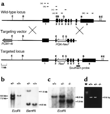
Generation of ADAMTS-1–null mice. A targeting DNA construct was designed to replace exons 2–4, which encode most of the metalloproteinase domain (12, 13), with the neomycin-resistance gene (Figure 1a). The tar-geting vector was introduced into 129/Sv–derived ES cells by electroporation, and targeted ES clones (Figure 1b) were then injected into C57BL/6 blastocysts. As confirmed by Southern blotting, six chimeras derived from two independent ES clones showed germline transmission of the targeted allele (Figure 1c). Among the offspring of the clones, ADAMTS-1+/–heterozygotes
were normal in appearance and were fertile. By cross-breeding heterozygotes, we obtained live ADAMTS-1–/–
homozygotes. Using all conceivable RT-PCR primer sets as indicated in Figure 1a (top), we could not detect amplified bands from ADAMTS-1–/–templates;
there-fore, even truncated ADAMTS-1mRNA was not effec-tively transcribed in ADAMTS-1–/– mice. Figure 1d
shows the result of RT-PCR using primer set (d).
Growth retardation in ADAMTS-1–null mice. ADAMTS-1–/– mice were smaller than their wild-type
litter-mates, although their proportions were correct (Fig-ure 2a). The reduction in body weight of ADAMTS-1–/–
mice was already significant at birth, and was accen-tuated thereafter (Figure 2b): at 9 months the average body weight of ADAMTS-1–/–mice was only about
70% that of their wild-type or heterozygous litter-mates (Figure 2c). ADAMTS-1–/–mice were
exception-ally lean, and the volume of epididymal fat was sig-nificantly smaller in ADAMTS-1–/–mice (Figure 2, d
and e). Histological examination did not detect apparent changes in ADAMTS-1–/– adipose tissues
except for the relatively small size of individual adipocytes (data not shown). Serum levels of nutri-tional indices were not significantly different between
ADAMTS-1–/–and ADAMTS-1+/+males (respective
val-ues: total cholesterol, 92.0 ± 6.7 mg/dL vs. 100.3 ± 12.0 mg/dL, n = 7 in each; glucose, 174.7 ± 22.7 mg/dL vs. 168.1 ± 16.1 mg/dL, n = 7 in each; total pro-tein, 4.5 ± 0.2 g/dL, n = 10 vs. 4.6 ± 0.4 g/dL, n = 9).
[image:4.612.60.292.53.294.2]Urologic abnormalities in ADAMTS-1–null mice. The most prominent morphological feature of ADAMTS-1–/–mice was detected in the kidney. In 4-week-old
Figure 1
[image:4.612.308.538.422.624.2]Targeted disruption of the mouse ADAMTS-1gene. (a) Genomic locus, targeting vector, and predicted targeted locus. Black boxes represent exons 1–9. The probe for Southern analysis is indicated. B, BamHI; E, EcoRI; G, BglII. (bandc) Southern analysis of genomic DNA from targeted ES cells (b) and the offspring from a heterozy-gote intercross (c). The 7.0-kb EcoRI fragment and the 11.5-kb BamHI fragment denote the homologous recombinant allele. (d) RT-PCR of total RNA extracted from the kidney of each genotype. No ADAMTS-1expression is detected in ADAMTS-1–/–mice.
Figure 2
Growth retardation in ADAMTS-1–/–mice. (a) Appearance of 1-week-old ADAMTS-1+/+(left) and ADAMTS-1–/–(right) littermates. (b) Comparison of growth after birth between ADAMTS-1+/+(n = 10) and
ADAMTS-1–/–(n = 9) littermates. AP < 0.05 vs. wild-type littermates. (c) Comparison of body weight among 9-month-old ADAMTS-1+/+,
ADAMTS-1–/– mice, the caliceal space was greatly
enlarged, and the corticomedullary tissue was severe-ly reduced, forming a thin rim surrounding the enlarged calyx (Figure 3, a–d). In mice of both C57BL/6J and 129/Sv backgrounds, this defect was observed bilaterally with complete penetrance. The papillae of wild-type mice protruded beyond the hilum and filled most of the pelvic space, whereas the papillae of ADAMTS-1–/–mice were poorly developed
or virtually absent (Figure 3, a–d). Electron microscopy showed degeneration of the collecting
duct epithelial cells and interstitial fibrosis (data not shown). Slight but significant enlargement of the cal-iceal space with hypoplastic or atrophic papillae were first observed at around 1 week of age, suggesting that the renal phenotype appears after birth and develops progressively thereafter. As for renal func-tion, we could detect no evidence of severe renal fail-ure in ADAMTS-1–/– mice. Serum creatinine (Cr),
blood urea nitrogen (BUN), and creatinine clearance (CCr) were not significantly different between
[image:5.612.225.537.52.575.2]ADAMTS-1–/–and ADAMTS-1+/+male mice (respective
Figure 3
Comparison of kidneys between ADAMTS-1+/+(a,c,e,g) and ADAMTS-1–/–(b,d,f,h,
i) mice at 4 weeks. (aandb) Inner aspects of the kidneys. Arrow, renal papilla; arrow-head, fibrous tissue surrounding the ureteropelvic junction. (c and d) Hema-toxylin and eosin–stained sections of the whole kidney. CS, caliceal space. (eandf) Mallory-azan–stained sections through the renal hilum; the ureteropelvic junction of ADAMTS-1–/–mice is rich in fibrous tissue (arrows). P, renal papilla. (g–i) Electron microscopy findings at the ureteropelvic junction in ADAMTS-1–/–mice and controls. (h) Excessive collagen fibers (arrows) between smooth muscle cells (Sm) at the ureteropelvic junction of ADAMTS-1–/– mice. (i) Thickening of the collagenous seg-ment between the epithelial cell (Epi) layer and smooth muscle cell layer (12–30 µm), and proliferation of fibroblasts at the ureteropelvic junction of ADAMTS-1–/– mice. (g) Collagenous segment between the epithelial cell layer and smooth muscle cell layer (6–12 µm) from the ureteropelvic junc-tion of normal mice. (j–m) Electron microscopy ureteral findings in ADAMTS-1–/–mice (k–m) and controls (j). (k) Thick-ening of the collagenous segment (arrows) between the epithelial cell layer and smooth muscle cell layer. (l) High magnification showing the accumulation of excessive col-lagen fibers in ADAMTS-1–/– mice. (m) Deformity and fissures (arrowheads) in smooth muscle cells in ADAMTS-1–/–mice. Bars in eand frepresent 20 µm; bars in g–m
values: Cr, 0.078 ± 0.006 mg/dL, n = 11 vs. 0.103 ± 0.022 mg/dL, n = 17; BUN, 25.6 ± 1.6 mg/dL, n = 16 vs. 26.4 ± 1.8 mg/dL, n = 12; CCr, 0.64 ± 0.09 mL/min,
n = 6 vs. 0.70 ± 0.09 mL/min, n = 8). In contrast, the ability to concentrate urine was significantly com-promised in ADAMTS-1–/–mice on a 0.7% NaCl diet
compared with ADAMTS-1+/+mice, and urine volume
was increased (2.37 ± 0.27 mL in –/– mice, n = 4 vs. 1.61 ± 0.12 mL in +/+ mice,n = 8; P < 0.05), whereas the specific gravity was decreased (1.054 ± 0.003, in –/– mice, n = 4 vs. 1.081 ± 0.004, in +/+ mice, n = 8; P
< 0.001). In accordance with the increased urine vol-ume, urinary concentrations of Na+(–/–, 90.1 ± 3.2 mEq/mL, n = 10 vs. +/+, 128.3 ± 4.3 mEq/mL, n = 10;
P < 0.0001), K+(–/–, 249.2 ± 10.5 mEq/mL, n = 10 vs. +/+, 377.7 ± 15.7 mEq/mL, n = 10; P < 0.0001) and Cl– (–/–, 126.9 ± 5.3 mEq/mL, n = 10 vs. +/+, 195.6 ± 7.7 mEq/mL, n = 10; P < 0.0001) were significantly lower in ADAMTS-1–/– mice than in ADAMTS-1+/+ mice.
After 2 weeks on a high-salt diet containing 8% NaCl, urine volume of ADAMTS-1–/–mice was increased by
about two-fold over that of ADAMTS-1+/+mice (–/–,
11.72 ± 2.27 mL, n = 6 vs. +/+, 6.19 ± 0.78 mL, n = 6; P
< 0.01). These anatomical and functional abnormal-ities are consistent with a state of hydronephrosis resulting from partial urinary obstruction (22).
The kidneys of ADAMTS-1–/–mice also exhibited
retraction of the ureteropelvic junction inside the hilum, which was surrounded by fibrous tissue (Fig-ure 3, a, b, e, and f). Electron microscopy revealed thickening of the collagenous segment between the epithelial cell layer and the smooth muscle cell layer in ADAMTS-1–/–mice (Figure 3, g–i). We further
stud-ied the histology of the ureter, and detected funda-mentally the same type of abnormalities as seen in the ureteropelvic junction. As shown in Figure 3, k–l, accumulations of excessive collagen fibers were also found between the epithelial and smooth muscle cell layers in the ureteral specimens of ADAMTS-1–/–
mice. Deformity and fissures were also detected in the smooth muscle cells in the ureter (Figure 3m). Thus, fibrotic changes diffusely involve the whole ureter in ADAMTS-1–/–mice.
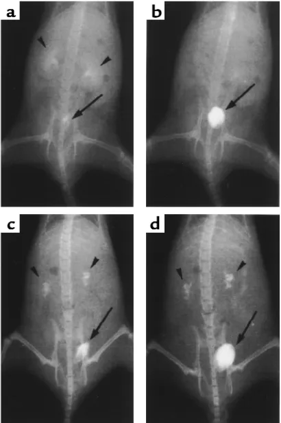
Therefore, we performed intravenous pyelography to assess changes in urinary flow and to determine the extent to which the lesions could cause urinary obstruction. In wild-type mice, nephrograms and cys-tograms observed 5 minutes after injection of con-trast medium showed both the calyces and pelvis to be smooth and pyramidal in shape (Figure 4a). By 30 minutes, the nephrograms had completely disap-peared (Figure 4b). In ADAMTS-1–/–mice, the calyces
were enlarged and distorted in shape, with a narrow pelvic space (Figure 4c). Washout of the contrast medium from the calyces was still incomplete after 30 minutes (Figure 4d).
Furthermore, ADAMTS-1 immunoreactivity was detected in the collecting ducts and (more intensely) in the urinary epithelium at the ureteropelvic junction
(Figure 5). Taken together, the anatomical, radi-ographic, and immunohistochemical findings suggest that obstruction at the ureteropelvic junction may cause progressive caliceal enlargement.
Adrenal abnormalities in ADAMTS-1–null mice.
ADAMTS-1–/– mice also showed adrenal glandular
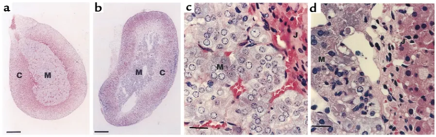
abnormalities. The adrenal medulla is normally com-posed of closely packed secretory cells supported by a fine reticular network containing numerous capillar-ies (Figure 6, a and c). In ADAMTS-1–/–mice, this
struc-ture was severely disrupted and contained numerous cavities (Figure 6, b and d). Remarkably, few capillar-ies containing blood cells were observed in the adrenal medulla of ADAMTS-1–/–mice (Figure 6, b and d). In
contrast, there were no detectable abnormalities in the adrenal cortex of ADAMTS-1–/–mice. The urine levels
of catecholamine metabolites were not different between ADAMTS-1–/–mice and ADAMTS-1+/+ mice
(respective values: metanephrine, 0.101 ± 0.021
[image:6.612.319.524.323.631.2]µg/day, n = 4 vs. 0.097 ± 0.014 µg/day, n = 4; normetanephrine: 0.707 ± 0.101 µg/day, n = 4 vs. 0.518
Figure 4
Intravenous pyelography (a–d). X-ray films were taken 5 minutes (a
± 0.051 µg/day, n = 4), suggesting that the function of the adrenal medulla may not be seriously affected.
Abnormalities in female genital organs and impaired fer-tility in ADAMTS-1–null mice. Finally, ADAMTS-1–/–
female mice showed abnormalities in the morpholo-gy of uterus and ovaries. The uterus of ADAMTS-1–/–
females had a thick and convoluted shape (Figure 7b). In uterine sections of ADAMTS-1–/– females,
extensive abnormal cystic formations were detected (Figure 7d). In addition, sections of ovary showed fewer numbers of mature follicles in ADAMTS-1–/–
females (Figure 7f) than in ADAMTS-1+/+females.
In accordance with the anatomical changes, fertili-ty was impaired in ADAMTS-1–/– females. When
mated with males, ADAMTS-1–/–female mice
charac-teristically experienced plug formation that was not followed by pregnancy. After mating with ADAMTS-1–/–males, only 13% of ADAMTS-1–/–females became
pregnant after plug formation (5 pregnancies out of 40 detected plugs). On the other hand, more than 90% of ADAMTS-1+/– and ADAMTS-1+/+ females
became pregnant after plug formation. Even when
ADAMTS-1–/–females became pregnant after
breed-ing, the average number of pups was very small (2.5 ± 0.3, n = 20 vs. 10.4 ± 0.3 in +/+ mice, n = 20; P < 0.001). The number of implantation sites at 10 days after
coitus was also reduced in ADAMTS-1–/–females (5.0
± 0.8, n = 4 vs. 11.6 ± 0.7 in ADAMTS-1+/+mice, n = 5;
P < 0.01). These data show that development of folli-cles, implantation, and intrauterine development may be affected in ADAMTS-1–/– female mice.
Impaired fertility was observed only in females;
ADAMTS-1–/– males sired both wild-type and
het-erozygous females normally.
Discussion
Data from ADAMTS-1–null mice obtained in this study show that ADAMTS-1 is necessary for normal growth and the structure and function of the kidneys, adrenal glands, and female reproductive organs. The lack of severe renal failure with azotemia or major metabolic disturbance suggests that the ADAMTS-1 gene is pleiotropic, and that ADAMTS-1 has multiple and nonredundant functions throughout the body.
ADAMTS-1 is a member of the ADAM protein fam-ily that is involved in proteolytic modification of cell-surface proteins and extracellular matrices. The unique structure of ADAMTS-1, characterized by the presence of thrombospondin type I motifs, is shared by other newly identified proteins in mammals and in
[image:7.612.233.538.56.182.2]C. elegans, which constitute the ADAMTS subfamily that may perform well-conserved biological functions Figure 5
[image:7.612.74.523.542.684.2]Immunohistochemistry of a mouse kidney sec-tion using an anti–ADAMTS-1 antibody (a) and a nonimmune control (b). J, urinary epithelium at the ureteropelvic junction. The pink staining shows the location of ADAMTS-1 expression in the urinary epitheli-um at the ureteropelvic junction.
Figure 6
(21). ADAMTS-1 is anchored to the extracellular matrix by an interaction between its carboxyl-termi-nal spacing region together with its thrombospondin type I motifs, and sulfated glycosaminoglycans such as heparan sulfate (14). ADAMTS-1 may, therefore, serve as a local factor processing as-yet unknown sub-strates by protease activity (15). It is notable that among the tissues we examined, the urinary epitheli-um at the ureteropelvic junction expressed the high-est levels of ADAMTS-1 protein. The renal abnormal-ities involving the calyces and corticomedullary structure in ADAMTS-1–/–mice might be secondary to
the structural abnormalities of the urinary tract. Thus ADAMTS-1 produced by the urinary epithelium may contribute to normal development or remodeling of the underlying fibrous tissue in the ureteropelvic junction and ureter.
Interestingly, mice lacking angiotensinogen, angiotensin-converting enzyme, or angiotensin type I receptor have a very similar renal phenotype (23–26). In these mice, hypoplastic ureteral smooth muscle and impaired ureteropelvic peristalsis are thought to cause functional obstruction of the urinary tract (23 and S. Okubo, personal communication). In contrast,
ADAMTS-1–/–mice exhibit accumulation of excessive
fibrous tissue and deformity of smooth muscle cells at the ureteropelvic junction and ureter, which could lead to both organic and functional obstruction. Elec-tron microscopy showing the accumulation of colla-gen fibers suggests that processing of collacolla-gen and related matrix substances may be impaired in
ADAMTS-1–/–mice. In this respect, it is noteworthy
that ADAMTS-1 shares unique structural features with procollagen I N-proteinase and aggrecanase-1, other ADAM proteins that contain four throm-bospondin type I motifs (16–18). This supports the notion that ADAMTS-1 may process collagen or relat-ed substrates (e.g., proteoglycans) that are important for the organization of organ structures.
The mechanisms responsible for growth retardation, female infertility with histological changes in the uterus and ovaries, and disrupted adrenomedullary architecture remain unknown. It is difficult to infer the molecular substrate or substrates of ADAMTS-1 that are common to the organs affected in knockout mice because the characteristics of histological changes seem to be different among organs. The discovery of physio-logically relevant substrates of ADAMTS-1 would be expected to pave the way for further understanding of growth, fertility, and organ morphogenesis.
Recently, METH-1, the human orthologue of ADAMTS-1, was shown to suppress FGF-2–induced vascularization in the cornea pocket assay and to inhibit VEGF-induced angiogenesis in the chorioal-lantoic membrane assay (19). Consistent with this inhibitory effect on angiogenesis, METH-1 inhibits endothelial cell proliferation, but not fibroblast or smooth muscle growth (19). On the other hand, the lack of adrenomedullary capillary network formation
in ADAMTS-1–/–mice suggests that ADAMTS-1 may be
necessary for the capillary formation of the adrenal medulla. It is presently unclear how these contradic-tory findings could be reconciled. It is possible that the effect of ADAMTS-1 on angiogenesis may be different among tissues, developmental stages, and pathophys-iological conditions. It is also possible that the lack of a capillary network in the adrenal medulla of
ADAMTS-1–/– mice is not the direct effect of the
ADAMTS-1null mutation. Further studies are neces-sary to clarify the role of ADAMTS-1 in angiogenesis and its molecular mechanism, which potentially involves extracellular matrices.
This study also has significant clinical implications. The renal phenotype of ADAMTS-1–/–mice resembles
human ureteropelvic junction obstruction, which is the most common cause of congenital hydronephrosis and is recognized in nearly 1 in 500 live births (27). This dis-ease is characterized by abnormal collagen and smooth muscle components at the ureteropelvic junction that contribute to mechanical or functional obstruction of urinary flow (27, 28). Interestingly, the structural abnormalities in ADAMTS-1–/–mice are responsible for
[image:8.612.308.535.341.652.2]the same dysfunctions described in cases of congenital
Figure 7
Comparison of uterus and ovary between ADAMTS-1+/+(a,c,e) and
ureteropelvic junction obstruction and primary obstructive megaureter in humans (29, 30). Thus, the
ADAMTS-1–null mouse may represent a useful new dis-ease model for clarification of the pathogenesis of con-genital ureteropelvic junction obstruction.
Acknowledgments
We thank R.E. Hammer for SM-1 ES cells, P. Mene-ton for the Neo-tk plasmids, H. Iwasa for technical assistance, and W.F. Goldman for critical reading of the manuscript. Y. Kurihara is a Research Fellow of the Organization for Pharmaceutical Safety and Research, Japan. This work was supported by a Grant-in-Aid for Scientific Research from the Min-istry of Education, Science and Culture, Japan (to H. Kurihara, Kouji Kuno, and H. Yokoyama) and by grants from the Cardiovascular Research Founda-tion, Tanabe Medical Frontier Conference, Suzuken Memorial Foundation, the Ryoichi Naito Founda-tion for Medical Research, and Tokyo Biochemical Research Foundation (to H. Kurihara).
1. Wolfsberg, T.G., Primakoff, P., Myles, D.G., and White, J.M. 1995. ADAM, a novel family of membrane proteins containing A Disintegrin And Metalloprotease domain: multipotential functions in cell-cell and cell-matrix interactions. J. Cell Biol.131:275–278.
2. Blobel, C.P. 1997. Metalloprotease-disintegrins: links to cell adhesion and cleavage of TNF alpha and Notch. Cell.90:589–592.
3. Blobel, C.P., et al. 1992. A potential fusion peptide and an integrin lig-and domain in a protein active in sperm-egg fusion. Nature. 356:248–252.
4. Almeida, E.A., et al. 1995. Mouse egg integrin alpha 6 beta 1 functions as a sperm receptor. Cell.81:1095–1104.
5. Yagami-Hiromasa, T., et al. 1995. A metalloprotease-disintegrin partici-pating in myoblast fusion. Nature.377:652–656.
6. Moss, M.L., et al. 1997. Cloning of a disintegrin metalloproteinase that processes precursor tumour-necrosis factor-alpha. Nature.385:733–736. 7. Black, R.A., et al. 1997. A metalloproteinase disintegrin that releases
tumour-necrosis factor-alpha from cells. Nature.385:729–733. 8. Peschon, J.J., et al. 1998. An essential role for ectodomain shedding in
mammalian development. Science. 282:1281–1284.
9. Buxbaum, J.D., et al. 1998. Evidence that tumor necrosis factor alpha converting enzyme is involved in regulated alpha-secretase cleavage of the Alzheimer amyloid protein precursor. J. Biol. Chem.273:27765–27767. 10. Rooke, J., Pan, D., Xu, T., and Rubin, G.M. 1996. KUZ, a conserved met-alloprotease-disintegrin protein with two roles in Drosophila neuroge-nesis. Science.273:1227–1231.
11. Pan, D., and Rubin, G.M. 1997. Kuzbanian controls proteolytic process-ing of Notch and mediates lateral inhibition durprocess-ing Drosophila and ver-tebrate neurogenesis. Cell.90:271–280.
12. Kuno, K., et al. 1997. Molecular cloning of a gene encoding a new type of metalloproteinase-disintegrin family protein with thrombospondin motifs as an inflammation associated gene. J. Biol. Chem.272:556–562. 13. Kuno, K., Iizasa, H., Ohno, S., and Matsushima, K. 1997. The exon/intron organization and chromosomal mapping of the mouse ADAMTS-1 gene encoding an ADAM family protein with TSP motifs. Genomics.46:466–471.
14. Kuno, K., and Matsushima, K. 1998. ADAMTS-1 protein anchors at the extracellular matrix through the thrombospondin type I motifs and its spacing region. J. Biol. Chem.273:13912–13917.
15. Kuno, K., Terashima, Y., and Matsushima, K. 1999. ADAMTS-1 is an active metalloproteinase associated with the extracellular matrix. J. Biol. Chem.274:18821–18826.
16. Colige, A., et al. 1997. cDNA cloning and expression of bovine procolla-gen I N-proteinase: a new member of the superfamily of zinc-metallo-proteinases with binding sites for cells and other matrix components. Proc. Natl. Acad. Sci. USA.94:2374–2379.
17. Prockop, D.J., Sieron, A.L., and Li, S.W. 1998. Procollagen N-proteinase and procollagen C-proteinase. Two unusual metalloproteinases that are essential for procollagen processing probably have important roles in development and cell signaling. Matrix Biol.16:399–408.
18. Tortorella, M.D., et al. 1999. Purification and cloning of aggrecanase-1: a member of the ADAMTS family of proteins. Science.284:1664–1666. 19. Vazquez, F., et al. 1999. METH-1, a human ortholog of ADAMTS-1, and METH-2 are members of a new family of proteins with angio-inhibito-ry activity.J. Biol. Chem.274:23349–23357.
20. Blelloch, R., and Kimble, J. 1999. Control of organ shape by a secreted metalloprotease in the nematode Caenorhabditis elegans. Nature. 399:586–590.
21. Tang, B.L., and Hong, W. 1999. ADAMTS: a novel family of proteases with an ADAM protease domain and thrombospondin 1 repeats. FEBS Lett.445:223–225.
22. Sheehan, M.L., and Davis, J.C. 1959. Experimental hydronephrosis. Arch. Pathol.68:185–225.
23. Niimura, F., et al. 1995. Gene targeting in mice reveals a requirement for angiotensin in the development and maintenance of kidney morpholo-gy and growth factor regulation. J. Clin. Invest.96:2947–2954. 24. Okubo, S., et al. 1998. Angiotensinogen gene null-mutant mice lack
homeostatic regulation of glomerular filtration and tubular reabsorp-tion. Kidney Int.53:617–625.
25. Krege, J.H., et al. 1995. Male-female differences in fertility and blood pressure in ACE-deficient mice. Nature.375:146–148.
26. Miyazaki, Y., et al. 1998. Angiotensin induces the urinary peristaltic machinery during the perinatal period. J. Clin. Invest.102:1489–1497. 27. Koff, S.A., and Wise, H.A., II. 1996. Anomalies of the kidney. In Adult and pediatric urology. 3rd edition. J.Y. Gillenwater, J.T. Grayhack, S.S. Howards, and J.W. Duckett, editors. Mosby Inc. St. Louis, Missouri, USA. 2171–2196.
28. Murakumo, M., et al. 1997. Structural changes of collagen components and diminution of nerves in congenital ureteropelvic junction obstruc-tion. J. Urol.157:1963–1968.
29. Hanna, M.K., Jeffs, R.D., Sturgess, J.M., and Barkin, M. 1976. Ureter-al structure and ultrastructure. Part II. CongenitUreter-al ureteropelvic junction obstruction and primary obstructive megaureter. J. Urol. 116:725–730.