129
ACCELERATING EMPHYSEMA DIAGNOSIS ON LUNG CT
IMAGES USING EMPHYSEMA PRE-DETECTION METHOD
1KHAIRUL MUZZAMMIL BIN SAIPULLAH, 2DEOK-HWAN KIM, 3NURUL ATIQAH ISMAIL
1Lecturer, 3Student, Faculty of Electronic Engineering and Computer Engineering, Universiti Teknikal
Malaysia Melaka, Malaysia
2
Prof., Department Dept. of Electronic Engineering, Inha University
E-mail: [email protected] , [email protected] , 3
[email protected]
ABSTRACT
In this paper, we propose a simple but effective algorithm to increase the speed of Emphysema region classification. Emphysema region classification method based on CT image consumes a lot of time because of the large number of sub-regions due to the large size of CT image. Some of the sub-regions contain no Emphysema and the classification of these regions is worthless. To speed up the classification process, we create an algorithm to select Emphysema region candidates and only use these candidates in the Emphysema region classification instead of all of the sub-regions. First, the lung region is detected. Then we threshold the lung region and only select the dark pixels because Emphysema only appeared in the dark area of the CT image. Then the thresholded pixels are clustered into a region that called the Emphysema pre-detected region or Emphysema region candidate. This region is then divided into sub-region for the Emphysema region classification. The experimental result shows that Emphysema region classification using pre-detected Emphysema region decreases the size of lung region which will result in about 84.51% of time reduction in Emphysema region classification
Keywords: Computer Aided Diagnosis, Emphysema, CT Image
1. INTRODUCTION
Computed tomography (CT) scans are usually applied to examine the pathological change of the tissues inside the body. However, for examining the pathological change of the tissues, CT scans generate a large number of images. Thus, radiologists are exhausted to diagnose pathological changes using a lot of CT images. Recently, a number of computer-aided detection (CAD) systems [1] [2] have been developed so as to help the radiologists to diagnose diseases. Using CAD systems to detect lung diseases such as emphysema, lung cancer, etc., is one of the important fields in the medical image processing nowadays [3] [4].
As for Emphysema detection, standard method detects the Emphysema region by classifying sub-regions in the CT image whether it contains Emphysema or not [4]. This operation consumes a lot of time because there are a lot of sub-region need to be classified. Some of the sub-region may not contain any Emphysema and the classification of these sub-regions is useless. To decrease the classification time of Emphysema region, we need
to remove some sub-regions that have zero percentage of having Emphysema.
With the characteristics of the CT images, pathological change of the tissues in the CT image is usually with some local texture and brightness characteristics. Emphysema can be easily detected in lung CT image just by its color intensity. Radiologists pre-determine Emphysema region using the brightness in the lung region. Fig. 1 shows a CT lung image with emphysema. As we can observe from the image, we can find that the emphysema region is darker than the normal region and its surface is smooth. Using this brightness information of lung region, we can filter out regions that have no possibility of have Emphysema.
130 algorithm to select Emphysema region candidates and only use these candidates in the Emphysema region classification instead of all of the sub-regions. First, the lung region is detected. Then the lung region is thresholded to create region that only contain dark pixels. Then the thresholded pixels are clustered into a region that called the Emphysema pre-detected region or Emphysema region candidate. This region is then divided into sub-region for the Emphysema sub-region classification.
Fig.1: An Example Of Emphysema Region
The remainder of this paper is organized as follows: in Section 2, related work to the current research is presented; in Section 3, the main idea of the proposed pre-detection of Emphysema region algorithm is then presented; then the experimental results are described in Section 4 followed finally by the conclusion in Section 5.
2. RELATED WORKS
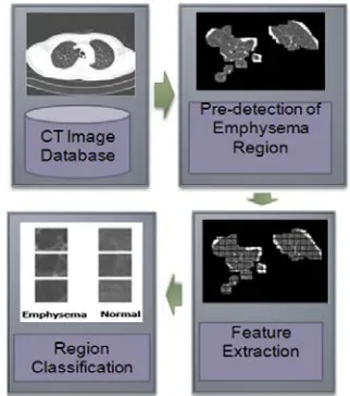
[image:2.612.339.488.157.328.2]Liang et al. [3] and Peng et al. [5] have developed texture descriptors for classifying abnormal regions of lung CT images as shown in Fig. 2. To locate the lung region from the CT image of lung, the contrast of the input image is enhanced using gamma correction. Then the binary image is obtained using the Otsu method [6]. Using morphology and region growing method, the noise in the image and lung vessels themselves are excluded to obtain the lung region without the vessels.
For the feature extraction, the texture feature of the subregion separated from the lung image is extracted using texture descriptors. This operation consumes a lot of time because there are a lot of region need to be classified. Some of the sub-region may not contain any Emphysema and the classification of these sub-regions is useless. To decrease the time consumption of Emphysema
[image:2.612.120.270.216.355.2]region, we need to remove some sub-regions that have zero percentage of having Emphysema. Lastly the resulting features are used to determine whether it contains emphysema or not in the region classification step.
Fig.2: Emphysema Region Classification Scheme
3. THE PROPOSED METHOD
The algorithm of the Emphysema region pre-detection is shown in Fig. 4. To locate the lung region from the CT image of lung, the contrast of the input image is enhanced using gamma correction. Then the binary image is obtaind using the Otsu method. Using morphology and region growing method, the noise in the image and lung vessels themselves are excluded to obtain the lung region without the vessels. This binary image of the lung region without vessels is than masked with the original image to get the gray image of lung region.
[image:2.612.336.497.517.699.2]131 The next step is to threshold the gray image of lung region using the following formula:
{
1 ,if ( , ) ( , )0 , otherwise l x y T
t x y = ≤ (1)
where t(x, y) is the thresholded image, l(x, y) is the gray image of the lung region and T is the threshold value.
From the distribution of the pixels in the lung region without vessels as shown in Fig. 5, we can see that the pixels of Emphysema region are distributed at the starting of the histogram (in the circle). Because of that, it is adequate to set T to a value corresponding to 22% of the range between the highest and the lowest pixel intensity value in the lung region without vesscels as shown by the line in Fig. 5. The formula to calculate T is as follows:
( )
T =α Hv−Lv (2)
where Hv and Lv are the highest and the lowest pixel intensity value in the lung region without region image, respectively. Based on our experiments, Emphysema is impossible to be having pixel value larger than T.
Fig.4:The Algorithm Of Emphysema Region Pre-Detection.
The next step is the clustering of these threholded pixels. As we can see in Fig. 4, the thresholded pixels are not in group and are distributed around the lung region. So, we need to cluster these pixels into larger group in order to create a region of them. The first step is to divide the thresholded lung region image into 8x8 pixels of blocks. Then the clustering algorithm is executed to each block.
The pseudo code of the clustering algorithm is shown in Algorithm 1. For each window of 8x8 pixels in the lung region image, we count the number of the thresholded pixels t(x, y). And if the number of t(x, y) occupy in that 8x8 region is more than 10% of the pixels in 8x8 window (above 7 pixels), the whole pixels in the 8x8 window is clustered into one group. Applying this process to the thresholded image, we then obtain the image as shown in Fig.4 under the subtitle clustering.
Fig.5: The Average Histogram Of Lung Region Without Vessels.
The next step is to filter the clustered region from the lung region. This clustered region will be the pre-detected Emphysema region. The resulting image is shown in the last image in Fig. 4. Using this pre-detected Emphysema region, the Emphysema region classification can be executed. Because the size of lung region becomes smaller than the original lung region image, the time consumption of the classification is reduced.
Algorithm 1: Thresholded pixels clustering algorithm.
for each 8x8 window
count the number n of thresholded pixels in the region
132 4. EXPERIMENTAL RESULT
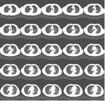
[image:4.612.316.528.261.627.2]For the experimental studies, the proposed method is executed on 25 lung CT images of one Emphysema patient that gathered from Inha University Hospital. The CT image setting for the images is the lung setting. The original images of the lung region are shown in Fig. 6. For each image in Fig. 6, the image is preprocess to find the lung region, threshold and clustered, in order to detect the pre-detected Emphysema region. Some of the images contain small amount of Emphysema while some of the images contain large Emphysema region. For standard Emphysema region classification method, the lung image in Fig.5 is divided into small subregion e.g., 30x30 pixels of subregion size. But using our proposed method, only a few parts of the lung region that might contain the Emphysema is used in the Emphysema region classification. Therefore, the time consumption of the Emphysema region classification can be reduced significantly.
Fig.6: The Lung CT Image Of An Emphysema Patient
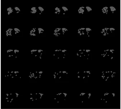
After executing the proposed algorithm to the whole images, a lot of parts and region can be excluded. From the images shown in Fig. 7, we can estimate that about 50% or more of lung region can be reduces for a lung that contain a lot of Emphysema. For lung that contain small amount of Emphysema, about 80% of the lung region can be removed.
The time consumption using pre-detection and without using pre-detection is also recorded in the experiment. The result can be seen in Table 1. As we can see, our proposed method only consumes a
little time more than that of standard method. The average time consumption increase is 0.026ms or with an increase of 6.7% of time consumption. With this amount of increase, the time increase can almost be neglected. From the table, we also can see that some of CT images produce no time differences. This means that the time consumption of our proposed method is the same as the time consumption of method without using pre-detection for some images. This case may occur on images that have little Emphysema.
Table 1: Time Consumption Of Lung Region Detection
Table 2 shows the lung region size reduction of the proposed method compared to original lung region. The formula to calculate the size reduction is as follows:
size of reduced lung region
SR(%) 1
size of original lung region
= − (3)
CT image
Without Pre-detection(s)
With Pre-detection(s)
[image:4.612.92.306.355.562.2]133
Table 2: Size Reduction Of Lung Region
CT image
Size reduction (%)
CT image
Size reduction (%) 1 68.18 14 88.05 2 67.75 15 89.23 3 70.92 16 93.57 4 67.99 17 91.31 5 73.32 18 93.27 6 78.15 19 91.59 7 78.49 20 91.29 8 82.52 21 92.41 9 81.16 22 94.94 10 82.49 23 90.76 11 86.17 24 92.56 12 83.52 25 94.69 13 88.44 Mean 84.51
From the table we can see that the proposed method reduces a significant amount of lung region with an average of 84.51% of the original lung region. The subimage that will be generated for the Emphysema region classification also will be reduced with the same ratio.
Fig.7: The Pre-Detected Emphysema Region
We can estimate the time that we can save in the Emphysema region classification just by scrutinizing the size reduction ratio. The time consumption for the Emphysema region classification is depended on the number of the subimage that need to be classified. Because our proposed method reduces the number of the subimage, it also will reduce the Emphysema region classification time consumption with the same ratio.
5. CONCLUSION AND FUTURE WORKS
In this paper, we proposed a simple but effective algorithm to increase the speed of Emphysema region classification. First, the lung region is detected. Then the lung region is thresholded to create region that only contains dark pixels. Then the thresholded pixels are clustered into a region that called the Emphysema pre-detected region or Emphysema region candidate. This region is then divided into sub-region for the Emphysema region classification. The proposed algorithm is simple so only small amount of time is consumed. With only this small time overhead, the Emphysema region classification can be reduce more than 50% due to the smaller size of the lung region that need to be classified.
For the future works, a proper Emphysema region classification experiment will be conducted to measure the exact time consumption difference between the proposed method and traditional method. A lot of new texture feature can be used for the Emphysema region classification such as Gray Level Local Binary Patterns (GLLBP) [7] and Local Circular Difference Phase Patterns (LCDPP) [8] or 2D Local Fourier Transform [9]. The classification accuracy experiment will also be conducted to evaluate whether the proposed method removes important region or not.
ACKNOWLEDGMENT:
This paper is supported by UniversitiTeknikal Malaysia Melaka under PJP/2011/FKEKK(44B)/ S00979.
REFRENCES:
[1] H. Zhang, J. E. Fritts, and S. A. Goldman, “A Fast Texture Feature Extraction Method for Region-based Image Segmentation”, Image and video comm.. and proc., vol. 5685 (2), pp. 957-968, 2005.
[2] A. M. R. Schilham, B. V. Ginneken, and M. Loog, “A Computer-Aided Diagnosis System for Detection of Lung Nodules in Chest Radiographs with an Evaluation on a Public Database”, Medical Image Analysis, vol. 10, pp. 247-258, 2006.
[image:5.612.89.299.403.593.2]134 [4] J. Dehmeshki, X. Ye, X. Y. Lin, M. Valdivieso,
and H. Amin. “Automated Detection of Lung Nodules in CT Images Using Shape-based Genetic Algorithm”, Computerized Medical Imaging and Graphics, vol. 31, pp. 408–417, 2007.
[5] Shao-Hu Peng, Hyun-Soo Kim and Deok-Hwan Kim, “Speeded Up Feature Extraction for CT image Based on Integral Image Technique”, IFMIA, pp. 319-324, 2009.
[6] N. Otsu, “A Threshold Selection Method from Gray-Level Histogram”,. IEEE Trans.on Sys. Man Cybern. vol. SMC-9, No.1, pp. 62-66, 1979
[7] Shao-Hu Peng, Khairul-Muzzammil Saipullah, Deok-Hwan Kim," Quantitative Image Analysis of Chest CT Using Gray Level Local Binary Pattern", International Conference on Convergence Content, pp. 129, Dec 2009
[8] Khairul Muzzammil Saipullah, Shao-Hu Peng, Hyun-Soo Kim, Deok-Hwan Kim, “Texture Classification By Implementing Blur, Scale and Grey Shift Insensitive Texture Descriptor Based on Local Fourier Transform”, Proc. IWAIT, pp. 74, Jan 2010.