0095-1137/09/$12.00
doi:10.1128/JCM.01478-09
Copyright © 2009, American Society for Microbiology. All Rights Reserved.
Development and Application of a Universal Hemoplasma Screening
Assay Based on the SYBR Green PCR Principle
䌤
Barbara Willi,
1,3Marina L. Meli,
1Ruedi Lu
¨thy,
4Hanspeter Honegger,
5Nicole Wengi,
1Ludwig E. Hoelzle,
2Claudia E. Reusch,
3Hans Lutz,
1and Regina Hofmann-Lehmann
1*
Clinical Laboratory,
1Institute of Veterinary Bacteriology,
2and Clinic for Small Animal Internal Medicine,
3University of Zurich,
Zurich, Switzerland; Swiss Aids Care International, Zurich, Switzerland
4; and Clinic of Medical Oncology,
Triemli Hospital, Zurich, Switzerland
5Received 31 July 2009/Returned for modification 3 September 2009/Accepted 2 October 2009
Hemotropic mycoplasmas (hemoplasmas) are the causative agents of infectious anemia in several
mamma-lian species. Their zoonotic potential has recently been substantiated by the identification of a feline
hemo-plasma isolate in an immunocompromised human patient. Although species-specific diagnostic molecular
methods have been developed, their application as screening tools is limited due to the species diversity of
hemoplasmas. The goals of this study were to develop a universal hemoplasma screening assay with broad
specificity based on the SYBR green PCR principle, to compare the assay with hemoplasma-specific TaqMan
PCR, and to analyze potential tick vectors and human blood samples to address the zoonotic potential. The
newly developed PCR assay based on the 16S rRNA gene amplified feline, canine, bovine, porcine, camelid, and
murine hemoplasmas, as well as
Mycoplasma penetrans
and
Mycoplasma pneumoniae
. The lower detection limit
for feline and canine hemoplasmas was 1 to 10 copies/PCR. The assay exhibited 98.2% diagnostic sensitivity
and 92.1% diagnostic specificity for feline hemoplasmas. All 1,950
Ixodes
ticks were PCR negative, suggesting
that
Ixodes
ticks are not relevant vectors for the above-mentioned hemoplasma species in Switzerland. None of
the 414 blood samples derived from anemic or immunocompromised human patients revealed a clear positive
result. The SYBR green PCR assay described here is a suitable tool to screen for known and
so-far-undiscov-ered hemoplasma species. Positive results should be confirmed by specific TaqMan PCR or sequencing.
Hemotropic mycoplasmas, also known as hemoplasmas, are
small, pleomorphic, cell wall-free bacteria that have been
de-tected in the blood of various mammalian species (17).
Orig-inally classified as
Haemobartonella
and
Eperythrozoon
species
within the order
Rickettsiales
, these organisms have recently been
reclassified within the genus
Mycoplasma
(17, 19, 21, 27).
Hemotropic mycoplasmas are clinically relevant as causative
agents of acute, life-threatening hemolytic anemia in infected
animals. Some animals, however, develop only mild clinical signs
or remain asymptomatic. Many cofactors, such as gender, age,
immune status, or coinfection with other pathogenic agents, have
been proposed to be involved in the development of disease
(10, 16, 25, 32). It is thought that animals become chronic,
asymptomatic carriers after infection, although clearance of
the infectious agents from the host blood has been reported
(26, 32, 33).
To date, hemoplasmas have been documented in numerous
mammalian species (Table 1). The close relationship between
the feline hemoplasma “
Candidatus
Mycoplasma turicensis”
and rodent hemoplasmas and the similarity of feline and
ca-nine hemoplasmas suggest potential interspecies transmission
of these agents (24, 33). This is especially important since
hemotropic mycoplasmas are thought to be transmitted by
blood-sucking arthropods such as ticks, fleas, and lice, given
that these agents have been hypothesized to exhibit zoonotic
potential. Some authors have described organisms with
mor-phological similarities to hemotropic mycoplasmas in the blood
of human patients (3, 7, 14, 22, 37). Furthermore, a recently
published report demonstrating the molecular detection of a
feline hemoplasma species in an immunocompromised human
patient further substantiated the zoonotic potential of these
agents (6).
No in vitro cultivation system has been established to
culti-vate these organisms outside their hosts, and diagnosis of
in-fection relies mainly upon the molecular detection of
hemo-plasmal genes in blood or tissue samples. Specific conventional
and quantitative real-time TaqMan PCR systems have been
established to diagnose hemotropic mycoplasmas (5, 11, 13, 18,
20, 25, 32). These assays provided the initial insight into the
epidemiology and pathogenesis of hemoplasmas; however,
conventional PCR is laborious and prone to carryover of PCR
amplicons. Real-time TaqMan PCR assays, on the other hand,
allow quantification, have minimal risk of amplicon carryover
(being closed-tube systems), and are highly specific due to the
use of a third labeled oligonucleotide. However, because of
their high specificity, these assays are unlikely to detect novel
hemoplasma species.
Real-time SYBR green PCR assays combine the advantages
of conventional and real-time PCR methods. They employ two
primers and a dye (SYBR green) in a closed-tube system. The
SYBR green principle allows for quantification, and its
speci-ficity is less restrictive than that of TaqMan PCR assays.
Fur-thermore, melting curves that differentiate between various
PCR amplicons can be generated after PCR amplification.
The goals of this study were to develop a universal
hemo-* Corresponding author. Mailing address: Clinical Laboratory,
Vet-suisse-Faculty, University of Zurich, Winterthurerstrasse 260, 8057
Zurich, Switzerland. Phone: 41 (44) 635 83 22. Fax: 41 (44) 635 89 23.
E-mail: [email protected].
䌤
Published ahead of print on 14 October 2009.
4049
on May 16, 2020 by guest
http://jcm.asm.org/
plasma screening assay based on the SYBR green PCR
prin-ciple, to compare the assay to specific TaqMan PCR assays,
and to screen potential tick vectors and blood samples from
anemic or immunocompromised human patients to address
zoo-notic potential and elucidate the possible occurrence of human
hemotropic mycoplasmas by means of molecular methods.
MATERIALS AND METHODS
Samples.For optimization of the SYBR green PCR assay and comparison
with specific TaqMan PCR assays, nucleic acid (NA) samples from 99 felids (42 uninfected, 15 singly infected, and 42 coinfected with feline hemoplasmas) were included. The samples were obtained from 35 specific-pathogen-free cats (4), 19 Swiss pet cats (32), and 45 African lions (Panthera leo) (35). The pet cats and lions had been analyzed for the presence of feline hemoplasmas by specific real-time TaqMan PCR assays (32, 35). The lions were included in the study because they are commonly coinfected with feline hemoplasmas (35).
To address the zoonotic potential of hemoplasmas, NA samples from a total of 1,950Ixodesticks collected from the vegetation in the area around Zurich, Switzerland, by the cloth-dragging method were included (34). The arthropods were mechanically disrupted with sterile scalpel blades and homogenized in a Mixer Mill MM 300 device (Retsch GmbH, Haan, Germany), and NA was extracted with a MagNA Pure LC TNA isolation kit (Roche Diagnostics, Rot-kreuz, Switzerland) as described previously (34). After NA extraction, the sam-ples were subjected to an 18S rRNA gene real-time PCR assay as described previously (34) to confirm the presence of amplifiable NA and exclude PCR inhibition.
Furthermore, EDTA-anticoagulated blood samples from 414 anonymous hu-man patients were used, including 200 blood samples collected from immuno-compromised patients from Zimbabwe infected with human immunodeficiency virus (HIV) and 214 blood samples from immunocompromised or anemic pa-tients from Switzerland. NA was purified from 100l of human blood using the MagNaPure LC TNA isolation external lysis protocol (Roche Diagnostics) with a final elution volume of 100l. To monitor cross-contamination, negative controls consisting of 100l of phosphate-buffered saline were prepared con-currently with each batch of 15 samples.
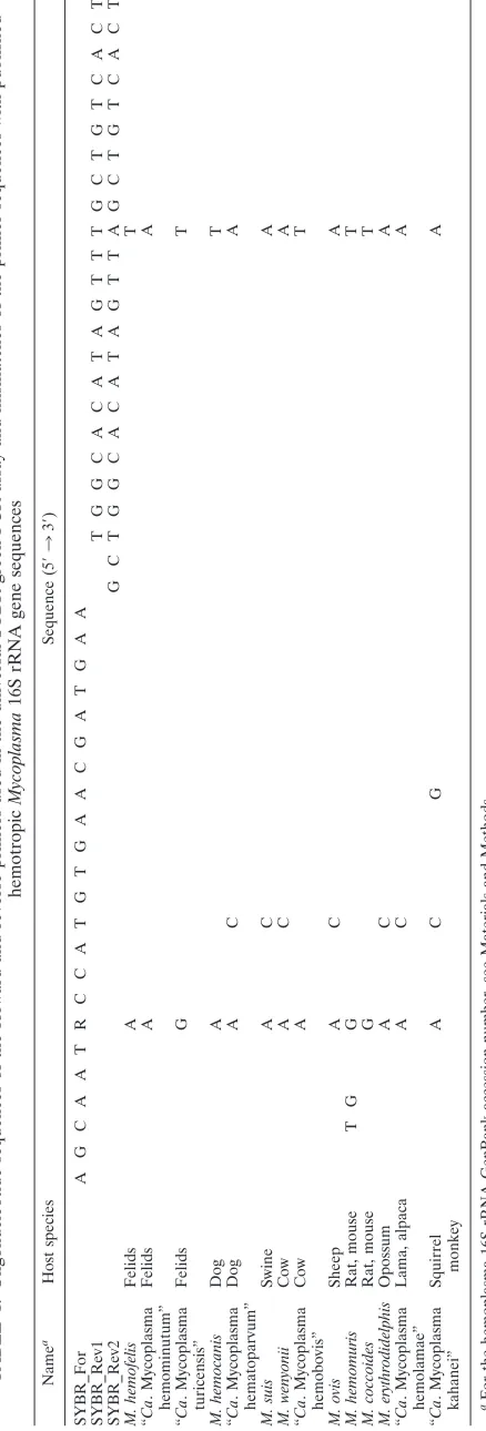
SYBR green real-time PCR primer design.To design primers for a universal
hemoplasma SYBR green PCR assay, the 16S rRNA genes of the following hemo-tropicMycoplasmaspecies were retrieved from GenBank and aligned using the GCG Wisconsin Package (Accelrys GmbH, Munich, Germany) and ClustalW (29): M. haemofelis(accession no. DQ157160), “CandidatusMycoplasma haemominu-tum” (DQ157149), “CandidatusMycoplasma turicensis” (DQ464421),M. haemoca-nis(EF416568), “CandidatusMycoplasma haematoparvum” (EF416569),M. suis (AY492086), M.wenyonii (DQ641256), “Candidatus Mycoplasma haemobovis” (EF616468), M. ovis (AF338268), M. haemomuris (U82963), M. coccoides (AY171918),M. erythrodidelphis (AF178676), “Candidatus Mycoplasma haemo-lamae” (AF306346), and “CandidatusMycoplasma kahanei” (AF338269). One for-ward and two reverse primers were designed using Primer Express software v2.0 (Applied Biosystems, Rotkreuz, Switzerland) (Table 1). The two reverse primers were used as a 1:1 mixture in the SYBR green PCR.
SYBR green real-time PCR primer optimization.For SYBR green PCR
op-timization, a primer matrix containing forward or reverse primer concentration combinations of 50 nM, 300 nM, and 900 nM was assessed using plasmids containing the nearly full-length 16S rRNA genes ofM. haemofelis, “Candidatus Mycoplasma haemominutum,” and “CandidatusMycoplasma turicensis” as tar-get templates (32, 33) or no-template controls (NTC). The three tartar-get templates (positive controls) were chosen because each represents a member of the three distinct phylogenetic clusters of hemoplasmas (17). The reaction mixture was composed of 12.5l of 2⫻SYBR green PCR master mix (Applied Biosystems), 50 to 900 nM concentrations of the forward and reverse primers, and 5l of NA template brought to a total volume of 25l with water. Assays were performed using an ABI Prism 7700 sequence detection system (Applied Biosystems). The SYBR green PCR protocol comprised 50°C for 2 min and 95°C for 10 min, followed by 40 cycles of 95°C for 15 s and 60°C for 1 min. After the PCR run, dissociation was performed with the following thermal profile: 95°C for 15 s, 60°C for 20 s, an increase from 60°C to 95°C for 20 min, and finally 95°C for 15 s. The minimum primer concentration demonstrating the maximum difference in fluo-rescence intensity (⌬Rn) was determined.⌬Rn was calculated by the ABI Prism 7700 sequence detection system as described previously (2).
SYBR green real-time PCR master mix optimization.Using the optimized
primer concentration and the PCR cycle conditions mentioned above, the SYBR green PCR master mix (Applied Biosystems) was compared to two additional
TABLE
1.
Oligonucleotide
sequences
of
the
forward
and
reverse
primers
used
in
the
universal
SYBR
green
PCR
assay
and
mismatches
of
the
primer
sequence
s
with
published
hemotropic
Mycoplasma
16S
rRNA
gene
sequences
Name a Host species Sequence (5 ⬘ 3 3 ⬘ ) SYBR_For A G C A A T R C C A T G T G A A C G A T G A A SYBR_Rev1 TGGCACATAGTTTGCTGTCACTT SYBR_Rev2 GCTGGCACATAGTTA GCTGTCACT M. hemofelis Felids A T “ Ca . Mycoplasma hemominutum” Felids A A “ Ca . Mycoplasma turicensis” Felids G T M. hemocanis Dog A T “ Ca . Mycoplasma hematoparvum” Dog A C A M. suis Swine A C A M. wenyonii Cow A C A “ Ca . Mycoplasma hemobovis” Cow A T M. ovis Sheep A C A M. hemomuris Rat, mouse T G G T M. coccoides Rat, mouse G T M. erythrodidelphis Opossum A C A “ Ca . Mycoplasma hemolamae” Lama, alpaca A C A “ Ca . Mycoplasma kahanei” Squirrel monkey AC G A aFor the hemoplasma 16S rRNA GenBank accession number, see Materials and Methods.on May 16, 2020 by guest
http://jcm.asm.org/
[image:2.585.55.274.82.727.2]SYBR green PCR master mixes: Power SYBR green PCR master mix (Applied Biosystems) and QuantiTect SYBR green master mix (Qiagen, Hombrechtikon, Switzerland). For this purpose, cats singly infected (n⫽8) or coinfected (n⫽3) with the three feline hemoplasmas and hemoplasma-uninfected cats (n⫽11), based on the results of specific TaqMan PCR, were selected from the above-mentioned samples and run with the three different master mixes. In each PCR run, positive controls and NTC were included. The diagnostic accuracy was calculated (diagnostic accuracy [%]⫽number of correctly classified samples/ number of tested samples) (23), and the master mix demonstrating maximal diagnostic accuracy was selected for further analyses.
SYBR green real-time PCR specificity and sensitivity.Using the optimized
primer concentration and master mix, NA samples from the following bacteria were used to determine the specificity of the SYBR green PCR assay:M. haemofelis, “Candidatus Mycoplasma haemominutum,” “Candidatus Myco-plasma turicensis,”M. haemocanis, “CandidatusMycoplasma haematoparvum,” M. suis,M. wenyonii, “CandidatusMycoplasma haemobovis,” “Candidatus My-coplasma haemolamae,”M. coccoides,M. pneumoniae,M. penetrans,M. equigeni-talium,M. argini,M. agalactiae,Chlamydophila felis,Pasteurella multocida, and Cytauxoon felis.
To determine the sensitivity of the assay for canine and feline hemoplasmas, recently published linearized plasmid standards containing the cloned 16S rRNA genes ofM. haemofelis, “CandidatusMycoplasma haemominutum,” “Candidatus Mycoplasma turicensis,” and “CandidatusMycoplasma haematoparvum” were used (30, 32, 33). The amplification efficiency was calculated asR⫽101/⫺slope⫺ 1 (15).
Comparison of the SYBR green PCR assay with specific real-time TaqMan
assays.For comparison with the universal SYBR green PCR assay, specific
real-time TaqMan PCR assays for the detection ofM. haemofelis, “Candidatus Mycoplasma haemominutum,” and “CandidatusMycoplasma turicensis” were performed as previously described (32, 33). Of the above-mentioned NA sam-ples, 93 were used for comparison. They comprised samples from 38 uninfected cats, 15 singly infected cats, and 40 cats coinfected with feline hemoplasmas, as assessed by the specific TaqMan PCR assays.
Statistical evaluation.For primer concentration optimization, the⌬Rns of the
two primer groups (group 1, primer combinations containing 50 nM; group 2, primer combinations containing 300 nM or 900 nM) were compared using the nonparametric Mann-Whitney U test. Differences with aPvalue of⬍0.05 were considered significant.
RESULTS
SYBR green PCR assay primer design.
Forward and reverse
primers were designed based on published hemotropic
Myco-plasma
16S rRNA gene sequences (Table 1). Sequence
align-ment revealed up to two mismatches in the forward or reverse
primer sequence when aligned with those of known
hemo-plasma species (Table 1). No mismatches were located near
the 3
⬘
ends of the primer sequences.
SYBR green PCR assay optimization.
For all three feline
hemoplasmas, all primer combinations containing
concentra-tions of 50 nM resulted in significantly lower
⌬
Rn values than
for the remaining primer combinations containing only 300
and/or 900 nM (
M. haemofelis
,
P
⫽
0.016; “
Candidatus
Myco-plasma haemominutum,”
P
⫽
0.016; and “
Candidatus
Myco-plasma turicensis,”
P
⫽
0.016). Among the four giving high
⌬
Rn values, the minimum primer concentration was selected;
thus, final primer concentrations of 300 and 300 nM were used
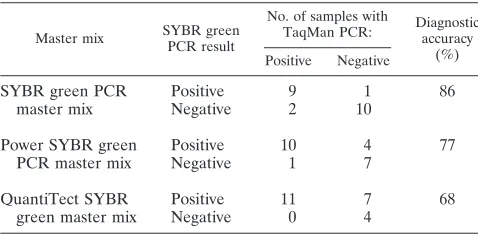
for further testing. Three master mixes were then evaluated,
and the diagnostic accuracy was assessed (Table 2). Based on
the highest diagnostic accuracy of the SYBR green PCR
mas-ter mix (86%), it was used for further testing.
SYBR green PCR specificity and sensitivity.
All 10
hemo-tropic mycoplasmas tested were amplified using the SYBR
green PCR assay.
M. pneumoniae
and
M. penetrans
, two
my-coplasmas that are closely related to the hemotropic
Myco-plasma
group, were also amplified, whereas the remaining
agents listed in Materials and Methods revealed threshold
cycle (
C
T) values in the range of those for the NTC.
The lower detection limit for the tested feline and canine
standards ranged from 1 to 10 copies/PCR. Amplification
ef-ficiencies were calculated using the same threshold and
base-line for all four standard curves (threshold, 0.283; basebase-line, 3 to
10). Amplification efficiencies were
ⱖ
92.0%.
Melting curve analysis of SYBR green PCR products.
In
animals infected with a single hemoplasma species, the melting
temperature (
T
m) was distinctly different among the three
fe-line, two canine, and two bovine hemoplasmas (Table 3 and
Fig. 1). In hemoplasma-coinfected animals, species
differenti-ation was not possible due to
T
mvariability among the
ampli-fication products.
Nonspecific product formation.
When nonspecific product
formation was found, it was commonly observed in the NTC
rather than in the uninfected samples. The
C
Tvalues ranged
from 34.4 to 39.9, corresponding to
⬍
10 copies/PCR. Because
the
T
mreported for primer dimers (about 75°C) (2) is in the
range of the
T
ms for the different hemoplasmas (Table 3),
differentiation of primer dimers and specific product formation
by melting curve analysis was not possible.
Comparison of the universal SYBR green PCR assay with
specific TaqMan PCR assays.
NA samples extracted from 93
[image:3.585.302.541.86.199.2]felines were used for comparison. Using SYBR green PCR, 54
out of 55 infected and 35 out of 38 uninfected samples were
correctly identified (Table 4). The SYBR green assay exhibited
98.2% diagnostic sensitivity and 92.1% diagnostic specificity
compared to feline hemoplasma-specific TaqMan PCR assays.
TABLE 2. Diagnostic accuracy of the three tested SYBR green
PCR master mixes compared to feline hemoplasma-specific
TaqMan PCR
Master mix SYBR green PCR result
No. of samples with
TaqMan PCR: Diagnosticaccuracy
(%) Positive Negative
SYBR green PCR
master mix
Positive
9
1
86
Negative
2
10
Power SYBR green
PCR master mix
Positive
10
4
77
Negative
1
7
QuantiTect SYBR
green master mix
Positive
11
7
68
Negative
0
4
TABLE 3.
T
ms of the tested hemotropic
Mycoplasma
species as
assessed by melting curve analyses
Species Tm(°C)
a
M. hemofelis
...74.5–76.0
“Candidatus
Mycoplasma hemominutum”...73.0–74.5
“Candidatus
Mycoplasma turicensis”...76.0–77.5
M. hemocanis
...
75.0
“Candidatus
Mycoplasma hematoparvum” ...73.0–74.0
M. suis...
76.5
M. wenyonii...
76.5
“Candidatus
Mycoplasma hemobovis”...
74.0
M. coccoides
...
74.5
“Candidatus
Mycoplasma hemolamae” ...73.5–74.0
aWhen multiple samples were analyzed, the melting temperature range is
specified.
on May 16, 2020 by guest
http://jcm.asm.org/
[image:3.585.43.282.98.215.2]The one “
Candidatus
Mycoplasma haemominutum”-infected
sample that was not detected by the SYBR green assay was
obtained from a Swiss pet cat. By quantitative real-time
Taq-Man PCR, this animal had a hemoplasma blood load of 1,960
copies/ml of blood. The three hemoplasma-uninfected samples
that displayed a positive result in SYBR green PCR but a
negative result in specific TaqMan PCR assays were obtained
from two specific-pathogen-free cats and one African lion.
Ticks.
In order to assess whether hemoplasmas could be
found in the most common tick in Switzerland,
Ixodes ricinus
,
and pose a potential zoonotic risk, 1,950 unfed
Ixodes
ticks
were analyzed. All NA samples extracted from ticks tested 18S
rRNA PCR positive (
C
Tvalues of
⬍
27), confirming the
pres-ence of amplifiable NA and the abspres-ence of relevant PCR
inhibition. In SYBR green PCR, all 1,950 samples revealed
PCR-negative results for hemoplasma species.
Human blood samples.
None of the 414 human blood
sam-ples revealed clear positive results. Five blood samsam-ples from
immunocompromised patients from Switzerland exhibited
C
Tvalues that were slightly below 40 (range from 39.2 to 39.9,
corresponding to
⬍
1 copy/PCR); however,
C
Tvalues in this
range were also observed for some NTC. The high
C
Tvalues
did not allow for further analysis, e.g., by sequencing.
DISCUSSION
The universal SYBR green PCR assay described here was
designed as a quantitative, closed-tube method for inexpensive
and rapid screening of samples for hemotropic
Mycoplasma
species. The assay was shown to amplify all 10 hemoplasma
species tested, including feline, canine, bovine, porcine,
cam-elid, and murine hemoplasmas. To the best of our knowledge,
no other hemoplasma PCR assay published thus far has been
able to amplify that many different hemoplasma species. For
feline hemoplasmas, the assay exhibited 98% diagnostic
sensi-tivity. Because high assay sensitivity is a prerequisite for the
detection of infections at low prevalence, our assay represents
an excellent hemoplasma screening method. Recently, the
number of reported hemoplasma species in mammals has
steadily increased, and the universal SYBR green PCR assay,
with its broad specificity, represents an important tool to
sim-plify and boost the search for other, thus-far-unknown
hemo-plasma species.
The recent identification of the feline hemoplasma
M.
hae-mofelis
in an immunocompromised HIV-positive patient in
Brazil represents the first report of human hemoplasma
infec-tion based on molecular methods (6). This discovery supports
the hypothesis of the zoonotic potential of these agents and
underscores the importance of searching for hemoplasma
spe-cies in humans using assays with broad specificity. Through
testing of human blood samples from Switzerland and
Zimba-bwe, patients living in different climate zones and social
envi-ronments were included in the present study. All human
pa-tients were either anemic or immunocompromised and therefore
were assumed to be at risk of hemoplasma infection.
Further-more, a remarkably high hemoplasma prevalence was recently
reported in cats in South Africa and dogs in Sudan (12, 28, 36),
and hemoplasmas were detected in lions and in ticks collected
from lions in Tanzania (8, 35). Nonetheless, none of the human
blood samples tested here were PCR positive.
Melting curve analysis is commonly used to differentiate
amplicons after SYBR green PCR. The
T
mdepends on both
[image:4.585.132.451.70.171.2]the PCR amplicon size and the GC content. In the present
FIG. 1. Melting curve analysis after hemoplasma SYBR green PCR of samples from animals infected singly with a feline, canine, murine, or
porcine hemoplasma species. Each curve represents one PCR amplicon and depicts the change in fluorescence during a continuous increase in
temperature. The temperature demonstrating the peak change in fluorescence represents the
T
m.
TABLE 4. Comparison of the results obtained by universal
hemoplasma SYBR green PCR and feline
hemoplasma-specific TaqMan PCR
TaqMan PCR-positive hemoplasma species
Total no. of samplesa
No. SYBR green PCRb:
Positive Negative
M. hemofelis
5
5
0
“Candidatus
Mycoplasma
hemominutum”
8
7
1
“Candidatus
Mycoplasma turicensis”
2
2
0
M. hemofelis-“Candidatus
Mycoplasma hemominutum”
5
5
0
“Candidatus
Mycoplasma
hemominutum”-“Candidatus
Mycoplasma turicensis”
10
10
0
M. hemofelis-“Candidatus
Mycoplasma
hemominutum”-“Candidatus
Mycoplasma
turicensis”
25
25
0
Total
Positive
55
54
1
Negative
38
3
35
Total no.
93
57
36
a
Number of samples uninfected, singly infected, or coinfected with feline hemoplasmas, as assessed by specific TaqMan PCR assays.
b
Results based on melting curve analysis.
on May 16, 2020 by guest
http://jcm.asm.org/
[image:4.585.42.282.482.699.2]study, melting curve analysis revealed different
T
mvalues for
the three feline, two canine, and two bovine hemoplasmas. The
T
mvalue was unpredictable, however, in samples containing
two or more hemoplasma species, suggesting that species
dif-ferentiation in coinfected animals is not feasible. Similar
re-sults have recently been reported for other SYBR green PCR
assays (9). Since coinfection with different hemoplasma species
is very common (20, 32, 35, 36), species-specific TaqMan PCR
assays or sequencing of PCR products remains a prerequisite
for specification of the hemoplasma species present in
coin-fected animals.
In some instances, low-level nonspecific product formation
was observed. According to the manufacturer’s instructions (2),
the weakly positive signals probably represent primer dimer
formation or elongation of primers nonspecifically bound to
genomic DNA. Because the
T
ms for the tested hemoplasma
species ranged from 73.0°C to 77.5°C, discrimination of primer
dimer formation (with a reported
T
mof 75°C) and specific
product formation by melting curve analysis was not feasible.
All 1,950
Ixodes
ticks investigated in the present study tested
negative by SYBR green PCR, suggesting that these ticks do
not play a role in the transmission of the hemoplasma species
detected by our assay in Switzerland. This extends our recent
results showing that
Ixodes
ticks are not a relevant vector for
feline hemoplasmas in Switzerland (34). Since
Ixodes ricinus
represents the most common tick species in Switzerland (1,
31), vector-borne transmission of hemoplasmas via ticks from
animals to humans in Switzerland seems unlikely.
In conclusion, the present study demonstrates that the
SYBR green PCR assay described here is suitable to screen for
known and so-far-undiscovered hemoplasma species. Positive
results should be confirmed by species-specific TaqMan PCR
assays or sequencing of the PCR products. Despite the recent
molecular detection of
M. haemofelis
infection in an
HIV-positive patient, in this study no hemoplasma infections were
detected in blood samples derived from anemic or
immuno-compromised humans. The question of whether undiscovered
hemoplasma species could play a role in human health should
be addressed in subsequent studies. The assay described here
will be an important tool in the pursuit of this issue.
ACKNOWLEDGMENTS
We thank V. Cattori, R. Tandon, C. Brunner, B. Weibel, T. Meili
Prodan, and E. Go
¨nczi for helpful contributions. Laboratory work was
performed using the logistics of the Center for Clinical Studies at the
Vetsuisse Faculty of the University of Zurich, Switzerland.
This study was supported by the UBS AG. B. Willi was supported by
the Roche Research Foundation, Basel. R.H.-L. is the recipient of a
professorship from the Swiss National Science Foundation (grants
PP00B-102866/1 and PP00B-119136/1).
REFERENCES
1.Aeschlimann, A.1972. Ixodes ricinus, Limmeus, 1758 (Ixodoidea: Ixodidae).
Preliminary study of the biology of the species in Switzerland. Acta Trop. 29:321–340.
2.Applied Biosystems.2006. SYBR® Green PCR master mix and RT-PCR
reagents, protocol. Applied Biosystems, Foster City, CA.
3.Archer, G. L., P. H. Coleman, R. M. Cole, R. J. Duma, and C. L. Johnston,
Jr.1980. Hemotropic bacteria. N. Engl. J. Med.302:1151–1152.
4.Brunner, C., T. Kanellos, M. L. Meli, D. J. Sutton, R. Gisler, M. A.
Gomes-Keller, R. Hofmann-Lehmann, and H. Lutz.2006. Antibody induction after
combined application of an adjuvanted recombinant FeLV vaccine and a multivalent modified live virus vaccine with a chlamydial component.
Vac-cine24:1838–1846.
5.Criado-Fornelio, A., A. Martinez-Marcos, A. Buling-Sarana, and J. C.
Barba-Carretero. 2003. Presence ofMycoplasma haemofelis,Mycoplasma
haemominutumand piroplasmids in cats from southern Europe: a molecular study. Vet. Microbiol.93:307–317.
6.dos Santos, A. P., R. P. dos Santos, A. W. Biondo, J. M. Dora, L. Z. Goldani,
S. T. de Oliveira, A. M. de Sa Guimaraes, J. Timenetsky, H. A. de Morais,
F. H. Gonzalez, and J. B. Messick.2008. Hemoplasma infection in
HIV-positive patient, Brazil. Emerg. Infect. Dis.14:1922–1924.
7.Duarte, M. I., M. S. Oliveira, M. A. Shikanai-Yasuda, O. N. Mariano, C. F.
Takakura, C. Pagliari, and C. E. Corbett.1992.Haemobartonella-like
mi-croorganism infection in AIDS patients: ultrastructural pathology. J. Infect.
Dis.165:976–977.
8.Fyumagwa, R. D., S. Simmler, B. Willi, M. L. Meli, A. Sutter, R. Hoare, G.
Dasen, R. Hofmann-Lehmann, and H. Lutz.2007. Molecular detection of
hemotropic mycoplasmas inRhipicephalus sanguineustick species collected on lions (Panthera leo) from Ngorongoro Crater, Tanzania. S. Afr. J. Wildl.
Res.38:117–122.
9.Giglio, S., P. T. Monis, and C. P. Saint.2003. Demonstration of preferential
binding of SYBR Green I to specific DNA fragments in real-time multiplex PCR. Nucleic Acids Res.31:e136.
10.Grindem, C. B., W. T. Corbett, and M. T. Tomkins.1990. Risk factors for
Haemobartonella felis infection in cats. J. Am. Vet. Med. Assoc.196:96–99.
11.Hoelzle, L. E., M. Helbling, K. Hoelzle, M. Ritzmann, K. Heinritzi, and
M. M. Wittenbrink.2007. First LightCycler real-time PCR assay for the
quantitative detection ofMycoplasma suisin clinical samples. J. Microbiol. Methods70:346–354.
12.Inokuma, H., M. Oyamada, B. Davoust, M. Boni, J. Dereure, B. Bucheton, A.
Hammad, M. Watanabe, K. Itamoto, M. Okuda, and P. Brouqui.2006.
Epidemiological survey ofEhrlichia canisand related species infection in dogs in eastern Sudan. Ann. N. Y. Acad. Sci.1078:461–463.
13.Jensen, W. A., M. R. Lappin, S. Kamkar, and W. J. Reagan.2001. Use of a
polymerase chain reaction assay to detect and differentiate two strains of Haemobartonella felisin naturally infected cats. Am. J. Vet. Res.62:604–608.
14.Kallick, C. A., S. Levin, K. T. Reddi, and W. L. Landau.1972. Systemic lupus
erythematosus associated with haemobartonella-like organisms. Nat. New Biol.236:145–146.
15.Klein, D., P. Janda, R. Steinborn, M. Muller, B. Salmons, and W. H.
Gunzburg.1999. Proviral load determination of different feline
immunode-ficiency virus isolates using real-time polymerase chain reaction: influence of mismatches on quantification. Electrophoresis20:291–299.
16.Luria, B. J., J. K. Levy, M. R. Lappin, E. B. Breitschwerdt, A. M. Legendre,
J. A. Hernandez, S. P. Gorman, and I. T. Lee.2004. Prevalence of infectious
diseases in feral cats in Northern Florida. J. Feline Med. Surg.6:287–296.
17.Messick, J. B.2003. New perspectives about hemotrophic mycoplasma
(for-merly,HaemobartonellaandEperythrozoonspecies) infections in dogs and cats. Vet. Clin. N. Am. Small Anim. Pract.33:1453–1465.
18.Messick, J. B., L. M. Berent, and S. K. Cooper.1998. Development and
evaluation of a PCR-based assay for detection ofHaemobartonella felisin cats and differentiation ofH. felisfrom related bacteria by restriction frag-ment length polymorphism analysis. J. Clin. Microbiol.36:462–466.
19.Neimark, H., K. E. Johansson, Y. Rikihisa, and J. G. Tully.2002. Revision
of haemotrophic Mycoplasma species names. Int. J. Syst. Evol. Microbiol. 52:683.
20.Peters, I. R., C. R. Helps, B. Willi, R. Hofmann-Lehmann, and S. Tasker.
2008. The prevalence of three species of feline haemoplasmas in samples submitted to a diagnostics service as determined by three novel real-time duplex PCR assays. Vet. Microbiol.126:142–150.
21.Rikihisa, Y., M. Kawahara, B. Wen, G. Kociba, P. Fuerst, F. Kawamori, C.
Suto, S. Shibata, and M. Futohashi.1997. Western immunoblot analysis of
Haemobartonella murisand comparison of 16S rRNA gene sequences ofH. muris,H. felis, andEperythrozoon suis. J. Clin. Microbiol.35:823–829.
22.Ristic, M., and J. P. Kreier.1979. Hemotropic bacteria. N. Engl. J. Med.
301:937–939.
23.Stockham, S. L., and A. M. Scott.2008. Fundamentals of veterinary clinical
pathology, 2nd ed. Iowa State University Press, Ames, IA.
24.Sykes, J. E., N. L. Bailiff, L. M. Ball, O. Foreman, J. W. George, and M. M.
Fry.2004. Identification of a novel hemotropic mycoplasma in a splenecto-mized dog with hemic neoplasia. J. Am. Vet. Med. Assoc.224:1946–1951, 1930–1931.
25.Tasker, S., S. H. Binns, M. J. Day, T. J. Gruffydd-Jones, D. A. Harbour, C. R.
Helps, W. A. Jensen, C. S. Olver, and M. R. Lappin.2003. Use of a PCR
assay to assess the prevalence and risk factors forMycoplasma haemofelisand ‘CandidatusMycoplasma haemominutum’ in cats in the United Kingdom. Vet. Rec.152:193–198.
26.Tasker, S., S. M. Caney, M. J. Day, R. S. Dean, C. R. Helps, T. G. Knowles,
P. J. Lait, M. D. Pinches, and T. J. Gruffydd-Jones.2006. Effect of chronic
FIV infection, and efficacy of marbofloxacin treatment, onMycoplasma hae-mofelisinfection. Vet. Microbiol.117:169–179.
27.Tasker, S., C. R. Helps, C. J. Belford, R. J. Birtles, M. J. Day, A. H. Sparkes,
T. J. Gruffydd-Jones, and D. A. Harbour.2001. 16S rDNA comparison
demonstrates near identity between an United KingdomHaemobartonella felisstrain and the American California strain. Vet. Microbiol.81:73–78.
on May 16, 2020 by guest
http://jcm.asm.org/
28.Tasker, S., C. R. Helps, M. J. Day, D. A. Harbour, S. E. Shaw, S. Harrus, G. Baneth, R. G. Lobetti, R. Malik, J. P. Beaufils, C. R. Belford, and T. J.
Gruffydd-Jones.2003. Phylogenetic analysis of hemoplasma species: an
in-ternational study. J. Clin. Microbiol.41:3877–3880.
29.Thompson, J. D., D. G. Higgins, and T. J. Gibson.1994. CLUSTAL W:
improving the sensitivity of progressive multiple sequence alignment through sequence weighting, position-specific gap penalties and weight matrix choice. Nucleic Acids Res.22:4673–4680.
30.Wengi, N., B. Willi, F. S. Boretti, V. Cattori, B. Riond, M. L. Meli, C. E.
Reusch, H. Lutz, and R. Hofmann-Lehmann.2008. Real-time PCR-based
prevalence study, infection follow-up and molecular characterization of ca-nine hemotropic mycoplasmas. Vet. Microbiol.126:132–141.
31.Wicki, R., P. Sauter, C. Mettler, A. Natsch, T. Enzler, N. Pusterla, P.
Kuhnert, G. Egli, M. Bernasconi, R. Lienhard, H. Lutz, and C. M. Leuteneg-ger.2000. Swiss Army survey in Switzerland to determine the prevalence of Francisella tularensis, members of theEhrlichia phagocytophilagenogroup, Borrelia burgdorferisensu lato, and tick-borne encephalitis virus in ticks. Eur. J. Clin. Microbiol. Infect. Dis.19:427–432.
32.Willi, B., F. S. Boretti, C. Baumgartner, S. Tasker, B. Wenger, V. Cattori,
M. L. Meli, C. E. Reusch, H. Lutz, and R. Hofmann-Lehmann.2006.
Prev-alence, risk factor analysis, and follow-up of infections caused by three feline hemoplasma species in cats in Switzerland. J. Clin. Microbiol.44:961–969.
33.Willi, B., F. S. Boretti, V. Cattori, S. Tasker, M. L. Meli, C. Reusch, H. Lutz,
and R. Hofmann-Lehmann.2005. Identification, molecular characterization,
and experimental transmission of a new hemoplasma isolate from a cat with hemolytic anemia in Switzerland. J. Clin. Microbiol.43:2581–2585.
34.Willi, B., F. S. Boretti, M. L. Meli, M. V. Bernasconi, S. Casati, D. Hegglin,
M. Puorger, H. Neimark, V. Cattori, N. Wengi, C. E. Reusch, H. Lutz, and
R. Hofmann-Lehmann.2007. Real-time PCR investigation of potential
vec-tors, reservoirs, and shedding patterns of feline hemotropic mycoplasmas. Appl. Environ. Microbiol.73:3798–3802.
35.Willi, B., C. Filoni, J. L. Catao-Dias, V. Cattori, M. L. Meli, A. Vargas, F.
Martinez, M. E. Roelke, M. P. Ryser-Degiorgis, C. M. Leutenegger, H. Lutz,
and R. Hofmann-Lehmann.2007. Worldwide occurrence of feline
hemo-plasma infections in wild felid species. J. Clin. Microbiol.45:1159–1166.
36.Willi, B., S. Tasker, F. S. Boretti, M. G. Doherr, V. Cattori, M. L. Meli, R. G.
Lobetti, R. Malik, C. E. Reusch, H. Lutz, and R. Hofmann-Lehmann.2006.
Phylogenetic analysis of “CandidatusMycoplasma turicensis” isolates from pet cats in the United Kingdom, Australia, and South Africa, with analysis of risk factors for infection. J. Clin. Microbiol.44:4430–4435.
37.Yang, D., X. Tai, Y. Qiu, and S. Yun.2000. Prevalence of Eperythrozoon spp.
infection and congenital eperythrozoonosis in humans in Inner Mongolia, China. Epidemiol. Infect.125:421–426.