0095-1137/11/$12.00 doi:10.1128/JCM.01033-11
Copyright © 2011, American Society for Microbiology. All Rights Reserved.
Novel One-Step Method for Detection and Isolation of
Active-Toxin-Producing
Clostridium difficile
Strains
Directly from Stool Samples
䌤
Charles Darkoh,
1,3Herbert L. DuPont,
1,2,3,4,5* and Heidi B. Kaplan
1,2The University of Texas Graduate School of Biomedical Sciences,1The University of Texas Medical School at Houston,2
The University of Texas School of Public Health-Center for Infectious Diseases,3St. Luke’s Episcopal Hospital,4and
Baylor College of Medicine,5Houston, Texas 77030
Received 19 May 2011/Returned for modification 23 June 2011/Accepted 27 September 2011
The alarming emergence of hypervirulent strains of Clostridium difficilewith increased toxin production, severity of disease, morbidity, and mortality emphasizes the need for a culture method that permits simulta-neous isolation and detection of virulent strains. TheC. difficiletoxins A and B are critical virulence factors, and strains can either be toxin-producing (virulent) or non-toxin-producing (nonvirulent). Strains that are isolated from human infections generally produce either toxin A or toxin B or both. The methods currently available for culturingC. difficiledo not differentiate strains that produce active toxins from strains that do not produce toxins or produce inactive toxins. As a result, the identification and isolation of toxin-producing strains from stool is currently a two-step process. First, the stool is plated on a selective medium, and then suspected colonies are analyzed for toxin production or the presence of the toxin genes. We describe here a novel selective and differential culture method, the Cdifftox plate assay, which combines in a single step the specific isolation ofC. difficilestrains and the detection of active toxin. This assay was developed based on our recent finding that the A and B toxins ofC. difficilecleave chromogenic substrates that have stereochemical characteristics similar to their natural substrate, UDP-glucose. The Cdifftox plate assay is shown here to be extremely accurate (99.8% effective) in detecting toxin-producing strains through the analysis of 528C. difficile
isolates selected from 50 tissue culture cytotoxicity assay-positive clinical stool samples. The Cdifftox plate assay advances and improves the culture approach such that onlyC. difficilestrains will grow on this agar, and virulent strains producing active toxins can be differentiated from nonvirulent strains, which do not produce active toxins. This new method reduces the time and effort required to isolate and confirm toxin-producingC.
difficilestrains.
Clostridium difficileis the leading definable cause of
nosoco-mial diarrhea worldwide due to its virulence, multidrug resis-tance, spore-forming ability, and environmental persistence (34, 45). The number of cases ofC. difficileinfection (CDI) in the United States is estimated to exceed 250,000 per year (46, 48), with the total cost of treatment being between 1 and 3.2 billion U.S. dollars annually (27, 36). Morbidity and mortality resulting from CDI in recent years have increased significantly as a result of changes in the virulence of the causative strains and antibiotic usage patterns (34, 36, 38, 48). This bacterium has been identified as the causative organism associated with 10 to 25% of the cases of antibiotic-associated diarrhea, 50 to 75% of the cases of antibiotic-associated colitis, and 90 to 100% of the cases of pseudomembranous colitis (1, 8).
The toxin-producing strains ofC. difficilepossess a 19.6-kb pathogenicity locus that contains the tcdA and tcdB genes, which encode two notable proteins: toxins A (308 kDa) and B (269 kDa), respectively. These toxins are both critical virulence factors in the pathogenesis ofC. difficile(26, 33, 44). Interest-ingly, strains producing either toxin A or toxin B or both are
virulent. Both toxins have the same enzymatic cleavage activity (4, 23, 24) and are cytotoxic to some cultured cells; however, toxin B is 100- to 1,000-fold more potent than toxin A in most cell lines (22, 43, 44). Both toxin A and B possess an N-termi-nally located glucosyltransferase domain that monoglucosyl-ates low-molecular-weight GTPases of the Rho family (RhoA, RhoB, RhoC, Rac, and Cdc42) in the host cytosol using cel-lular uridine diphosphoglucose (UDP-glucose) as the glucose donor (22, 23). This monoglucosylation interrupts the normal function of the Rho GTPases, leading to a variety of effects on intoxicated cells, such as apoptosis, cell rounding, actin cyto-skeleton dysregulation, disruption of tight junctions, loosening of the epithelial barrier, and altered cellular signaling (10, 11, 19, 22, 23). Cellular intoxication by the toxins also induces the release of various immunomodulatory mediators from epithe-lial cells, phagocytes, and mast cells resulting in inflammation and accumulation of neutrophils (37, 41).
Currently, two general approaches are used in the clinical identification of CDI. In one case, stool samples are examined directly by various methods including PCR to detect toxin-encoding genes, by enzyme-linked immunosorbent assay (ELISA) to detect toxin production, and/or the tissue culture cytotoxicity assay to detect toxin activity. Alternatively or in addition, C. difficile strains can be isolated on commercially available selective media such as cycloserine-cefoxitin fructose agar (CCFA) (6, 12), and the same methods are performed to
* Corresponding author. Mailing address: The University of Texas School of Public Health-Center for Infectious Diseases, 1200 Herman Pressler, Suite 733, Houston, TX 77030. Phone: (713) 500-9366. Fax: (713) 500-9359. E-mail: [email protected].
䌤Published ahead of print on 5 October 2011.
4219
on May 16, 2020 by guest
http://jcm.asm.org/
confirm the CDI. The isolation of viable strains is advanta-geous because the assay methods are more sensitive when performed on pure cultures and the strains are available for further analysis such as antibiotic susceptibility testing, which is critical in antibiotic dose administration. Although PCR assays are gaining popularity for the diagnosis of CDI due to their high sensitivity in detecting the toxin-encoding genes directly from stool or isolated colonies, PCR detection of the toxin genes does not ensure that the genes are expressed and active toxins are produced. ELISAs are available from many manu-facturers for the detection of both toxins A and B. Reports from several comparative studies of different ELISA kits con-clude that the sensitivity and specificity are slightly lower than the cytotoxicity assays (9, 21, 25, 28, 42). The tissue culture cytotoxicity method is not as sensitive as culture isolation com-bined with PCR or ELISA (3, 12), although it is considered by some laboratories as the gold standard. A culture method that permits simultaneous isolation and detection of virulent C.
difficilestrains would be ideal for efficient detection of CDIs.
We report here, the development of a one-step selective and differential agar-based assay, the Cdifftox plate assay (CDPA), which enables detection and isolation ofC. difficileproducing active toxins in 24 to 48 h directly from stool samples. This simple plate assay eliminates the need for additional toxin-confirmatory tests. The CDPA assay was developed based on our recent finding that the A and B toxins ofC. difficilecleave chromogenic substrates that have stereochemical characteris-tics similar to their natural substrate, UDP-glucose. The CDPA is a major improvement in the analysis of CDI and is expected to improve patient care by reducing the time and effort required to isolate and identify active toxin-producingC.
difficilestrains.
MATERIALS AND METHODS
Bacterial strains and growth conditions.TheC. difficilestrains ATCC 43255 (tcdA⫹tcdB⫹), ATCC BAA-1382 (tcdA⫹tcdB⫹), ATCC 700057 (⌬tcdA tcdB⫹), and the hypervirulent strain, ATCC BAA-1805 (tcdA⫹tcdB⫹), which all produce active toxins were purchased from the American Type Culture Collection (Ma-nassas, VA). Brain heart infusion (BHI) medium was purchased from Becton Dickinson (Cockeysville, MD). The substrate, X-Gal (5-bromo-4-chloro-3-indo-lyl--D-galactopyranoside), was purchased from Biosynth International (Itasca, IL). The liquid or plate cultures were incubated anaerobically in an atmosphere of 10% H2, 5% CO2, and 85% N2at 37°C in a controlled atmosphere anaerobic chamber (Plas-Labs, Inc., Lansing, MI).
Sample storage conditions.The clinical isolates were either stored short term (⬍1 month) in chopped meat broth (BD Diagnostics, Franklin Lakes, NJ) at room temperature or long term in 15% glycerol stocks at⫺80°C. The culture supernatants were stored at 4°C for a maximum of 2 weeks with no loss of toxin activity.
Clinical stool samples. The clinical stool samples were obtained from an ongoing study approved by the Institutional Review Board of The University of Texas Health Science Center at Houston and St. Luke’s Episcopal Hospital (Houston, TX). All of the participating patients or their legal guardians provided written informed consent upon admission to the hospital. The 80 stool samples evaluated consisted of 60 samples that tested positive for the tissue culture cytotoxicity assay and 20 samples that tested negative in the same assay, as determined by the Medical Microbiology Laboratory at the St. Luke’s Episcopal Hospital.
Cdifftox plate assay medium.This agar-based culture medium was developed to be selective for growth ofC. difficileand differential in that it simultaneously identifies colonies producing active toxins A and/or B, while inhibiting the growth of non-C. difficilecolonies. The following components were chosen to compose this new Cdifftox plate assay (CDPA) medium (per liter): BHI (6 g), peptic digest of animal tissue (6 g), pancreatic digest of gelatin (14.5 g), NaCl (5 g), dextrose (3 g), anhydrous Na2HPO4(2.5 g), sodium taurocholate (0.1%) (Sigma-Aldrich,
St. Louis, MO),D-cycloserine (300 mg) and cefoxitin (8.5 mg) (Fisher Scientific, Pittsburgh, PA), X-Gal (150 mg), 0.5 mM glycine, 4-methylphenol (0.025%), defibrinated horse blood (6%), and agar (12 g). Alternatively, BHI (6 g), peptic digest of animal tissue (6 g), pancreatic digest of gelatin (14.5 g), NaCl (5 g), dextrose (3 g), and anhydrous Na2HPO4(2.5 g) can be replaced with 37 g of BBL BHI medium (BD Diagnostic Systems).
CDPA.For the CDPA, each stool sample was streaked directly onto two CDPA plates using a sterile loop. The plates were incubated anaerobically for 24 to 72 h at 37°C, until colonies appeared, and then the plates were exposed to air at room temperature for at least 30 min to allow the chromogenic reaction to complete. The assay was initially tested using the well-characterized toxin-over-producingC. difficilestrain ATCC 43255 (tcdA⫹tcdB⫹) and the hypervirulent strain ATCC BAA-1805 (tcdA⫹tcdB⫹), along with two strains that produce less toxins, ATCC BAA-1382 (tcdA⫹tcdB⫹) and ATCC 700057 (⌬tcdA tcdB⫹). The presumptive active toxin-producingC. difficile colonies appeared blue, while non-toxin producers remained pale white. The blue colonies were phenotypically classified as Tox⫹(presumablytcdA⫹and/ortcdB⫹), whereas the pale white colonies were denoted Tox⫺(presumably⌬tcdAand⌬tcdBmutants or mutants with genetic alterations that affect toxin production or activity).
Isolation and PCR identification of toxin-producingC. difficilestrains.A total of 528 single colonies consisting of 486 Tox⫹and 42 Tox⫺colonies were selected from the CDPA plates for further analysis. These colonies represented a total of 10 to 12 independent isolates from each stool sample. When possible eight Tox⫹ and two Tox⫺colonies were selected from each sample. The presence of the toxin-encoding genes (tcdAortcdB) in the genomes of the presumptive Tox⫹and Tox⫺isolates was examined by PCR amplification of a portion of each of these genes. A portion of the 16S rRNA genes was also amplified. These reactions were performed by first isolating genomic DNA from 1 ml of an overnight culture of each isolated colony using a DNeasy blood and tissue kit (Qiagen, German-town, MD). Amplification was performed using a Phire Hot Start DNA Poly-merase II kit (Finnzymes, Woburn, MA). The following primers were used: toxin A (F-5⬘-TGATGCTAATAATGAATCTAAAATGGTAAC-3⬘; R-5⬘-ACCACC AGCTGCAGCCATA-3⬘), toxin B (F-5⬘-GTGTAGCAATGAAAGTCCAAGT TTACGC-3⬘; R-5⬘-CACTTAGCTCTTTGATTGCTGCACCT-3⬘), and 16S rRNA (F-5⬘-ACACGGTCCAAACTCCTACG-3⬘and R-5⬘-AGGCGAGTTTC AGCCTACAA-3⬘). The DNA was amplified with an initial denaturation of 98°C for 30 s, followed by 36 cycles of 98°C for 10 s, 60°C for 10 s, and 72°C for 10 s, with a final extension of 72°C for 1 min.
Toxin assays. (i) Preparation of culture supernatants from the Toxⴙand Toxⴚclinical isolates.Single colonies (486 Tox⫹and 42 Tox⫺) were inoculated into 10 ml of BHI medium and incubated anaerobically at 37°C for 72 h resulting in an optical density at 600 nm of about 1.3 to 1.4. After centrifugation at 10,000
⫻gfor 10 min to remove the cells, the culture supernatants were collected and stored at 4°C until use.
(ii) ELISA assay.The presence of toxins A and/or B in the culture superna-tants from the isolates was evaluated using a WampoleC. difficileTOX A/B II kit (TechLab, Blacksburg, VA). This was performed according to the protocol provided by the manufacturer.
(iii) Cdifftox activity assay.The activities of toxin A and/or toxin B produced by theC. difficileisolates were measured by using the Cdifftox activity assay (2), recently developed in our laboratory. Briefly, this assay quantitatively measures the activity of toxins A and B based on the inherent properties of these toxins to cleavep-nitrophenyl--D-glucopyranoside, a chromogenic substrate with stereo-chemical characteristics similar to the natural substrate of the toxins, UDP-glucose. The assay was performed on 250l of each sample supernatant fluid, to which 50l of the substrate reagent (30 mMp-nitrophenyl--D-glucopyranoside, 50 mM Tris-HCl [pH 7.4], 50 mM NaCl, and 100M MnCl2) was added in a Costar sterile polystyrene 96-well plate (Corning, Inc., NY). Cleavage of the substrate was monitored by measuring the absorbance at 410 nm with a Spectra Max Plus 384 spectrophotometer (Molecular Devices, Sunnyvale, CA). A molar extinction coefficient forp-nitrophenol ofε⫽17,700 M⫺1
cm⫺1
was used in the calculations (40).
Evaluation of non-Clostridium difficilebacteria on the CDPA.The growth of non-Clostridium difficilebacteria on the CDPA medium under the standard conditions used for the stool samples was examined. The following strains were tested (asterisks indicate Gram-positive strains):Alcaligenes faecalis,Bacteroides fragilis,Bacteroides thetaiotaomicron,Bacteroides uniformis,Bifidobacterium ado-lescentis*,Campylobacter jejuni,Clostridium perfringens*,Enterobacter cloacae,
Enterococcus faecalis*, enteropathogenicEscherichia coli, enterotoxigenic Esch-erichia coliH10407,Eubacterium lentum*,Lactobacillus*spp.,Parabacteroides distasonis,Peptostreptococcus anaerobius*, Plesiomonas shigelloides,Prevotella melaninogenica,Proteus mirabilis,Proteus vulgaris,Salmonella entericaserovar Typhimurium,Shigella flexneri,Shigella sonnei,Staphylococcus aureus*,Vibrio
on May 16, 2020 by guest
http://jcm.asm.org/
alginolyticus,Vibrio parahaemolyticus, andYersinia enterocolitica. Single colonies of these bacteria were selected from agar plates and cultured in 10 ml of appropriate broth medium. Pure broth cultures of each of these strains were spread directly on the CDPA plates using a sterile loop and incubated as de-scribed above.
Growth ofC. difficile on cycloserine-cefoxitin fructose agar and BHI-agar medium.The stool samples were also cultured on CCFA medium (6, 12), which was prepared as follows per liter: proteose peptone 2 (40 g), anhydrous Na2HPO4(5 g), anhydrous KH2PO4(1 g), NaCl (2 g), anhydrous MgSO4(0.1 g), fructose (6 g), neutral red (0.003%),D-cycloserine (500 mg), cefoxitin (15.5 mg), and agar (15 g). To test for the growth of other anaerobes present in the stool, the samples were also cultured on BHI-agar plates without antibiotics. All plates were incubated for 24 to 72 h under anaerobic conditions at 37°C.
RESULTS
Examination of known active toxin-producing C. difficile
strains using the CDPA.The Cdifftox plate assay (CDPA) uses a novel selective and differential agar-based culture medium to specifically allow the growth ofC. difficileand simultaneously identify colonies producing active toxins A and/or B, while inhibiting the growth of non-C. difficilecolonies. The assay was initially tested using the well-characterized
toxin-overproduc-ingC. difficilestrain ATCC 43255 (tcdA⫹tcdB⫹) and the
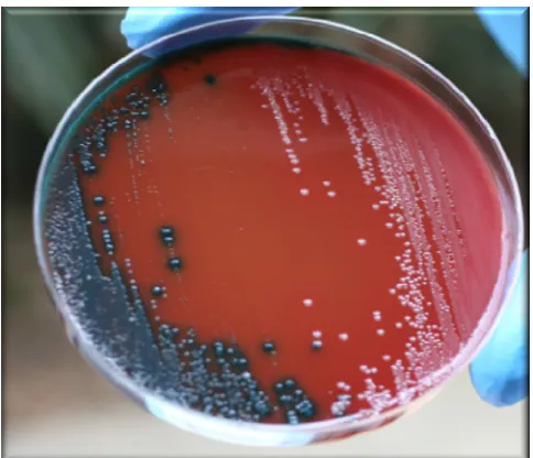
hy-pervirulent strain ATCC BAA-1805 (tcdA⫹tcdB⫹), along with two strains that produce less toxins, ATCC BAA-1382 (tcdA⫹ tcdB⫹) and ATCC 700057 (⌬tcdA tcdB⫹). After 24 h of incu-bation, colonies of these strains that produced high levels of active toxins appeared blue (Tox⫹), whereas those that pro-duced undetectable toxin levels remained pale white (Tox⫺), similar to the colonies shown in Fig. 1. By 48 h, all of the colonies were blue, indicating they produced active toxins.
Examination of clinicalC. difficileisolates from stool sam-ples using the CDPA.To evaluate the ability of the new CDPA to detectC. difficilestrains producing active toxins from clinical
stool samples, 60 tissue culture cytotoxicity assay-positive clin-ical stool samples collected at the St. Luke’s Episcopal Hospi-tal (Houston, TX) were tested. A loopful of each stool sample was spread directly onto the CDPA plates and incubated an-aerobically at 37°C for 24 to 72 h. Viable bacterial colonies were successfully isolated from 50 of the 60 tissue culture cytotoxicity assay-positive stool samples analyzed. The percent-age of colonies from the 50 stool samples that appeared Tox⫹ on the CDPA plates is shown in Table 1. After 24 h of anaer-obic incubation, Tox⫹colonies were detected in 44 (88%) of the 50 CDPA culture-positive stool samples. By 48 h of incu-bation, Tox⫹colonies were detected in all (100%) of the 50 CDPA culture-positive stool samples. It is noteworthy that none of the stool samples required more than 48 h of incuba-tion for the Tox⫹colonies to be detected. Tox⫺colonies se-lected after 72 h of incubation and transferred onto new CDPA plates remained Tox⫺, suggesting that no further incubation was necessary after the initial 48 h of incubation to confirm Tox⫺strains.
To further evaluate the CDPA, 20 cytotoxicity assay-nega-tive stool samples were tested on the CDPA plates, and no colonies grew. To confirm that the 30 CDPA culture-negative stool samples, which included 10 cytotoxicity assay-positive and 20 cytotoxicity assay-negative stool samples, did not contain toxins, an ELISA was performed on each of the 30 samples. All of the ELISA results were negative, confirming that no detect-able toxins (whole toxins or portions thereof), were present in the 30 CDPA culture-negative stool samples. These data indi-cate that the CDPA is accurate in discriminating stool samples that do not contain viable C. difficile strains. Furthermore, these results demonstrate that the 10 cytotoxicity assay-positive stool samples that were CDPA culture-negative and ELISA negative should be considered false positives.
PCR amplification of C. difficile toxin-encoding genes. To confirm that the Tox⫹colonies isolated using the CDPA pos-sessed the genes in their genomes that encode the toxins, a total of 528 single bacterial colonies comprised of 486 Tox⫹ and 42 Tox⫺independent clinical isolates from the 50 CDPA culture-positive stool samples were examined by PCR ampli-fication. (Figure 2 shows a representative number of these results.) All 528 isolates tested were positive for the conserved region of theC. difficile16S rRNA gene (Fig. 2). Of the 486 Tox⫹isolates evaluated, 485 (99.8%) were positive for either
[image:3.585.42.284.68.276.2]tcdAand/ortcdB. Of the 42 Tox⫺isolates evaluated, 31 (74%) were positive for eithertcdAand/ortcdB, whereas 11 (26%) were negative for bothtcdAandtcdB. These data indicate that
TABLE 1. Detection of Tox⫹colonies from 50 cytotoxicity assay-positive samples on the Cdifftox agar plates at different timesa
% Tox⫹colonies No. of stool samples
24 h 48 h 72 h
76–100 0 31 47
51–75 1 14 0
26–50 5 4 2
1–25 38 1 1
0 6 0 0
a
Clinical stool samples were streaked directly onto the CDPA plates, incu-bated anaerobically for 24, 48, and 72 h, and examined after 30 min of exposure to air.
FIG. 1. Differentiation of active toxin-producing and non-toxin producing strains ofC. difficileby the CDPA. Two stool samples were spread directly onto different parts of the plate, incubated anaerobi-cally at 37°C for 48 h, and exposed to air at room temperature for more than 30 min. Blue colonies on the left are toxin-producingC. difficile (Tox⫹); pale white colonies on the right are non-toxin-producingC. difficile(Tox⫺).
on May 16, 2020 by guest
http://jcm.asm.org/
all of the Tox⫹and Tox⫺isolates that were able to grow on the CDPA plates from the stool samples wereC. difficile. Further-more, almost 100% (99.8%) of the Tox⫹strains encode the genes for synthesis of eitherC. difficiletoxin A and/or toxin B. Interestingly, 74% of the Tox⫺strains encoded one or both of the toxin genes in their genomes.
Analysis of toxin production and activity by the Toxⴙand Toxⴚstrains.The CDPA differentiatesC. difficilestrains pro-ducing active toxins from strains propro-ducing either inactive or no toxins, based on their ability to cleave a chromogenic
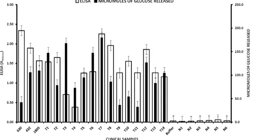
sub-strate. To ensure that the Tox⫹C. difficilecells were able to secrete active toxins, the presence and activity of toxins in the culture supernatants of Tox⫹ and Tox⫺isolates were evalu-ated. Toxin detection using the ELISA was performed on cul-ture supernatants from three isolates of each stool sample. Toxin activity was also tested on all of these isolates using the Cdifftox activity assay, recently developed in our laboratory (2). The results (Fig. 3 shows a subset of these results) reveal that all of the CPDA Tox⫹culture supernatants from the clinical isolates that were positive fortcdAand/ortcdBby PCR am-plification also tested positive for the presence of toxin pro-teins by ELISA, and tested positive for toxin activity by the Cdifftox activity assay. Remarkably, all of the colonies deter-mined by the Cdifftox plate assay to be Tox⫺, whether they encodedtcdAortcdBin their genomes or not, were negative for the presence and activity of the toxins in both the ELISA and Cdifftox activity assays. These data support the results that all but one of the 486 Tox⫹colonies selected on the CDPA were active toxin-producing (toxigenic)C. difficileand the 42 Tox⫺ colonies selected were non-toxin-producing (nontoxi-genic)C. difficile.
[image:4.585.41.283.67.151.2]Selectivity of the Cdifftox agar medium. To evaluate the specificity of the CDPA, non-Clostridium difficilebacteria were tested for growth on the CDPA agar under the same culture conditions as the clinical stool samples. No viable colonies were observed when pure cultures of the following strains were streaked on the CDPA plates:Alcaligenes faecalis,Bacteroides
FIG. 2. PCR analysis of representative Tox⫹ and Tox⫺ strains. Genomic DNA was isolated from the colonies and used as a template in PCRs with primers specific for the genes that encode toxin A (tcdA) and toxin B (tcdB) and a conserved region of theC. difficilerRNA (16S rRNA) gene. M, 1-kb marker (New England Biolabs, Ipswich, MA); lanes 1 to 3,tcdAamplicons; lanes 4 to 6,tcdBamplicons; lanes 7 to 9, 16S rRNA amplicons. The PCR products were electrophoresed through a 1% agarose gel, and the DNA was detected digitally upon exposure of the ethidium bromide-treated gel to UV light.
FIG. 3. Analysis ofC. difficiletoxin production in culture supernatants of representative Tox⫹and Tox⫺clinical isolates and defined ATCC strains. Toxin detection was performed by ELISA and the Cdifftox activity assay as described in Materials and Methods. Of the ATCC strains, 1805 represents strain BAA-1805 (tcdA⫹tcdB⫹[NAP1]), 432 represents strain 43255 (tcdA⫹tcdB⫹), and 630 is a historical strain BAA-1382 (tcdA⫹ tcdB⫹). Of the clinical isolates, T1 to T14 represent Tox⫹strains, and N1 to N6 are Tox⫺strains (⌬tcdAand⌬tcdBmutants). Error bars represent the standard deviation from two replicate experiments.
on May 16, 2020 by guest
http://jcm.asm.org/
[image:4.585.43.551.396.676.2]fragilis,Bacteroides thetaiotaomicron,Bacteroides uniformis,
Bi-fidobacterium adolescentis, Campylobacter jejuni, Clostridium
perfringens,Enterobacter cloacae,Enterococcus faecalis,
entero-pathogenic Escherichia coli, enterotoxigenic Escherichia coli
H10407, Eubacterium lentum, Lactobacillus spp.,
Parabacte-roides distasonis, Peptostreptococcus anaerobius, Plesiomonas
shigelloides, Prevotella melaninogenica, Proteus mirabilis,
Pro-teus vulgaris, Salmonella enterica serovar Typhimurium,
Shi-gella flexneri,Shigella sonnei, Staphylococcus aureus,Vibrio
al-ginolyticus,Vibrio parahaemolyticus, andYersinia enterocolitica.
These data suggest that the CDPA discriminates non-C. diffi-cilebacteria.
To more directly compare bacterial growth on the CDPA medium with CCFA medium (6), which is anotherC. difficile
selective medium, and BHI-agar, a nonselective medium, all 60 cytotoxic-positive stool samples were spread directly onto the prereduced plates of CCFA- and BHI-agar media and incu-bated anaerobically at 37°C for 24 to 72 h. The CCFA- and BHI-agar media allowed the growth of colonies from the same 50 stool samples that were CDPA culture positive. Overall, ca. 15% more bacterial colonies that appeared morphologically different grew on the CCFA-agar plates, and ca. 40% more bacterial colonies grew on the BHI-agar plates compared to the CDPA-agar plates. It appears that the additional colonies observed on the CCFA-agar and BHI-agar plates represent
non-C. difficileanaerobic bacteria in the stool samples.
DISCUSSION
Strains of C. difficile are broadly classified as being either toxin producing or non-toxin producing. It has been estab-lished that only the toxin-producing strains cause disease and that toxins A and B play critical roles in the pathogenesis ofC.
difficile(26, 30–33). The alarming emergence of hypervirulent
strains ofC. difficilewith increased toxin production, severity of disease, and mortality (7, 13, 14, 29) emphasizes the need for a sensitive diagnostic method that can simultaneously isolate and identify toxin-producing strains. The current available cul-ture methods do not differentiate toxin-producing and non-toxin-producing strains of C. difficile. The CDPA described here advances and improves the culture approach by combin-ing the isolation ofC. difficile strains on a selective medium with the detection of active toxins in a single step, such that
only C. difficile strains will grown on this agar and virulent
strains producing active toxins can be differentiated from non-virulent strains that do not produce active toxins and as a result do not cause disease. This new assay drastically reduces the time and effort required to isolate and confirm an infection resulting from toxin-producingC. difficilestrains.
This new CDPA was evaluated by testing 60 tissue culture cytotoxicity-positive and 20 cytotoxicity-negative clinical stool samples. After 24 h of incubation, the assay detected toxin-producing C. difficile colonies in 88% (44/50) of the CDPA culture-positive samples tested (Table 1). Toxin-producingC.
difficilecolonies were detected from 100% of the 50 CDPA
culture-positive stool samples evaluated within 48 h of incuba-tion. No viable bacterial colonies grew on the CDPA plates when the 20 cytotoxicity assay-negative, ELISA-negative stool samples and the 10 cytotoxicity assay-positive, ELISA-negative
stool samples were tested. These results reveal the high accu-racy and specificity of this method.
The CDPA identifies toxin-producingC. difficilecolonies by their ability to cleave a chromogenic substrate, X-Gal, into a distinct insoluble blue product that precipitates around the toxin-producing cells. This substrate cleavage by the toxins was confirmed by the examination of 528 independentC. difficile
colonies isolated from 50 stool samples from different patients suffering from C. difficileinfection. Although, non-toxin-pro-ducing strains ofC. difficilecan also grow on the CDPA plates, they are differentiated by their inability to cleave the substrate and appear as white colonies. Furthermore, none of the non-C.
difficilebacterial pathogens tested could grow on the CDPA
plates under the culture conditions used to growC. difficile. The CDPA plate medium was more selective than the CCFA agar (6), in that the CCFA agar allowed the growth of more colonies from the 50 culture-positive, cytotoxicity-positive stool samples.
Surprisingly, 10 stool samples evaluated determined to be positive by the tissue culture cytotoxicity assay did not result in colony growth on any of the culture media utilized (selective and nonselective) under anaerobic conditions. Furthermore, the ELISA that utilizes antibodies specific for toxins A and B did not detect any toxin in all of the 10 cytotoxic-positive samples. These results suggest that the tissue culture cytotox-icity assay misdiagnosed 10 (16%) of the 60 samples as toxin positive. This could have been a result of mishandling of the samples, absence of viable bacteria in the 10 samples, or mis-interpretation of the initial cytotoxicity assay results. As a re-sult, we agree with others (3, 12) who suggest that it is neces-sary to culture a toxin-producingC. difficilebacterium from the stool to confirm a diagnosis of an active infection or coloniza-tion.
It is important to note that whereas almost all Tox⫹colonies (99.8%) on the CDPA plates encoded the toxin genes, 74% of the Tox⫺colonies also encoded thetcdAortcdBgenes in their genomes. This suggests that the genomes of someC. difficile
strains may encode the toxin genes but do not secrete detect-able amount of toxins. It is possible that these active proteins are not produced as a consequence of mutations in the toxin coding sequences. It is also possible that these toxins are not produced due to alternations in the regulatory elements nec-essary for transcription, translation, or secretion. Alternatively, the bacterial cells may not have been exposed to the necessary conditions to activate toxin gene expression (15–17, 20, 35). Factors that have been suggested to influence toxin production are cell density, exposure to antibiotics, phage lysogeny, growth medium composition, and nutrient limitation (5, 15, 18, 39, 47). Furthermore,C. difficilecells in stool samples may exist as either vegetative cells at different growth stages or as spores. Perhaps, variations in cell physiology may explain why some colonies became Tox⫹later than others. In any case, our re-sults indicate that this heterogeneity did not lead to false neg-ative interpretations of any of the samples analyzed by the Cdifftox plate assay.
To our knowledge, the use of the glucosyltransferase activ-ities of the A and B toxins to identify toxin-producing C.
difficileis unique and has not been reported in the literature or
used commercially. The Cdifftox plate assay represents a novel
on May 16, 2020 by guest
http://jcm.asm.org/
detection method with potentially improved sensitivity and ef-ficiency compared to current diagnostic methods.
ACKNOWLEDGMENTS
We thank Zhi-Dong Jiang (The University of Texas Health Science Center, School of Public Health) for her assistance in obtaining the clinical stool samples used in the study. We also acknowledge Nicole M. Poole (The University of Texas Medical School at Houston) and Hoonmo L. Koo (Baylor College of Medicine, Houston) for their help in data collection and analysis.
This study was supported in part by The University of Texas Health Science Center–School of Public Health and The Molecular Basis of Infectious Diseases Training grant (T32-2T32AI055449-06) of the University of Texas Health Science Center, Houston, TX.
REFERENCES
1.Bartlett, J. G.2002. Clinical practice: antibiotic-associated diarrhea. N. Engl. J. Med.346:334–339.
2.Darkoh, C., H. B. Kaplan, and H. L. Dupont.2011. Harnessing the gluco-syltransferase activities ofClostridium difficilefor functional studies of toxins A and B. J. Clin. Microbiol.49:2933–2941.
3.Delmee, M.2001. Laboratory diagnosis ofClostridium difficiledisease. Clin. Microbiol. Infect.7:411–416.
4.Dillon, S. T., et al.1995. Involvement of Ras-related Rho proteins in the mechanisms of action ofClostridium difficiletoxin A and toxin B. Infect. Immun.63:1421–1426.
5.Dineen, S. S., A. C. Villapakkam, J. T. Nordman, and A. L. Sonenshein.
2007. Repression ofClostridium difficiletoxin gene expression by CodY. Mol. Microbiol.66:206–219.
6.Dowell, V. R., Jr.1975. Methods for isolation of anaerobes in the clinical laboratory. Am. J. Med. Technol.41:402–410.
7.Eggertson, L., and B. Sibbald.2004. Hospitals battling outbreaks of Clos-tridium difficile. CMAJ171:19–21.
8.Elliott, B., B. J. Chang, C. L. Golledge, and T. V. Riley.2007.Clostridium difficile-associated diarrhoea. Intern. Med. J.37:561–568.
9.Fedorko, D. P., et al.1999. Evaluation of two rapid assays for detection of
Clostridium difficiletoxin A in stool specimens. J. Clin. Microbiol.37:3044– 3047.
10.Feltis, B. A., et al. 2000.Clostridium difficiletoxins A and B can alter epithelial permeability and promote bacterial paracellular migration through HT-29 enterocytes. Shock14:629–634.
11.Genth, H., S. C. Dreger, J. Huelsenbeck, and I. Just.2008.Clostridium difficiletoxins: more than mere inhibitors of Rho proteins. Int. J. Biochem. Cell Biol.40:592–597.
12.George, W. L., V. L. Sutter, D. Citron, and S. M. Finegold.1979. Selective and differential medium for isolation ofClostridium difficile. J. Clin. Micro-biol.9:214–219.
13.Goorhuis, A., et al.2008. Emergence ofClostridium difficileinfection due to a new hypervirulent strain, polymerase chain reaction ribotype 078. Clin. Infect. Dis.47:1162–1170.
14.Goorhuis, A., et al.2008.Clostridium difficilePCR ribotype 078: an emerging strain in humans and in pigs? J. Clin. Microbiol.46:1157. (Author Reply,
46:1158.)
15.Govind, R., G. Vediyappan, R. D. Rolfe, B. Dupuy, and J. A. Fralick.2009. Bacteriophage-mediated toxin gene regulation inClostridium difficile. J. Vi-rol.83:12037–12045.
16.Hammond, G. A., and J. L. Johnson.1995. The toxigenic element of Clos-tridium difficilestrain VPI 10463. Microb. Pathog.19:203–213.
17.Hammond, G. A., D. M. Lyerly, and J. L. Johnson.1997. Transcriptional analysis of the toxigenic element ofClostridium difficile. Microb. Pathog.
22:143–154.
18.Haslam, S. C., et al.1986. Growth ofClostridium difficileand production of toxins A and B in complex and defined media. J. Med. Microbiol.21:293– 297.
19.Hofmann, F., C. Busch, U. Prepens, I. Just, and K. Aktories.1997. Local-ization of the glucosyltransferase activity ofClostridium difficiletoxin B to the N-terminal part of the holotoxin. J. Biol. Chem.272:11074–11078. 20.Hundsberger, T., et al.1997. Transcription analysis of the genestcdA-Eof
the pathogenicity locus ofClostridium difficile. Eur. J. Biochem.244:735–742. 21.Johnson, S., et al.2001. Fatal pseudomembranous colitis associated with a
variantClostridium difficilestrain not detected by toxin A immunoassay. Ann. Intern. Med.135:434–438.
22.Just, I., and R. Gerhard.2004. LargeClostridialcytotoxins. Rev. Physiol. Biochem. Pharmacol.152:23–47.
23.Just, I., et al.1995. Glucosylation of Rho proteins byClostridium difficile
toxin B. Nature375:500–503.
24.Just, I., et al.1995. The enterotoxin fromClostridium difficile(ToxA) mono-glucosylates the Rho proteins. J. Biol. Chem.270:13932–13936.
25.Kader, H. A., D. A. Piccoli, A. F. Jawad, K. L. McGowan, and E. S. Maller.
1998. Single toxin detection is inadequate to diagnoseClostridium difficile
diarrhea in pediatric patients. Gastroenterology115:1329–1334.
26.Kuehne, S. A., et al.2010. The role of toxin A and toxin B inClostridium difficileinfection. Nature467:711–713.
27.Kyne, L., M. B. Hamel, R. Polavaram, and C. P. Kelly.2002. Health care costs and mortality associated with nosocomial diarrhea due toClostridium difficile.Clin. Infect. Dis.34:346–353.
28.Limaye, A. P., D. K. Turgeon, B. T. Cookson, and T. R. Fritsche.2000. Pseudomembranous colitis caused by a toxin A(⫺) B(⫹) strain of Clostrid-ium difficile. J. Clin. Microbiol.38:1696–1697.
29.Loo, V. G., et al.2005. A predominantly clonal multi-institutional outbreak ofClostridium difficile-associated diarrhea with high morbidity and mortality. N. Engl. J. Med.353:2442–2449.
30.Lyerly, D. M., H. C. Krivan, and T. D. Wilkins.1988.Clostridium difficile: its disease and toxins. Clin. Microbiol. Rev.1:1–18.
31.Lyerly, D. M., D. E. Lockwood, S. H. Richardson, and T. D. Wilkins.1982. Biological activities of toxins A and B ofClostridium difficile. Infect. Immun.
35:1147–1150.
32.Lyerly, D. M., C. J. Phelps, J. Toth, and T. D. Wilkins.1986. Characteriza-tion of toxins A and B ofClostridium difficilewith monoclonal antibodies. Infect. Immun.54:70–76.
33.Lyerly, D. M., K. E. Saum, D. K. MacDonald, and T. D. Wilkins.1985. Effects ofClostridium difficiletoxins given intragastrically to animals. Infect. Immun.47:349–352.
34.McDonald, L. C., et al.2005. An epidemic, toxin gene-variant strain of
Clostridium difficile. N. Engl. J. Med.353:2433–2441.
35.Moncrief, J. S., L. A. Barroso, and T. D. Wilkins.1997. Positive regulation of
Clostridium difficiletoxins. Infect. Immun.65:1105–1108.
36.O’Brien, J. A., B. J. Lahue, J. J. Caro, and D. M. Davidson.2007. The emerging infectious challenge ofClostridium difficile-associated disease in Massachusetts hospitals: clinical and economic consequences. Infect. Con-trol Hosp. Epidemiol.28:1219–1227.
37.Pothoulakis, C.2000. Effects ofClostridium difficiletoxins on epithelial cell barrier. Ann. N. Y. Acad. Sci.915:347–356.
38.Redelings, M. D., F. Sorvillo, and L. Mascola.2007. Increase inClostridium difficile-related mortality rates, United States, 1999–2004. Emerg. Infect. Dis.
13:1417–1419.
39.Rolfe, R. D., and S. M. Finegold.1979. Purification and characterization of
Clostridium difficiletoxin. Infect. Immun.25:191–201.
40.Shikita, M., J. W. Fahey, T. R. Golden, W. D. Holtzclaw, and P. Talalay.
1999. An unusual case of “uncompetitive activation” by ascorbic acid: puri-fication and kinetic properties of a myrosinase fromRaphanus sativus seed-lings. Biochem. J.341(Pt. 3):725–732.
41.Thelestam, M., and E. Chaves-Olarte.2000. Cytotoxic effects of the Clos-tridium difficiletoxins. Curr. Top. Microbiol. Immunol.250:85–96. 42.Ticehurst, J. R., et al.2006. Effective detection of toxigenic Clostridium
difficileby a two-step algorithm including tests for antigen and cytotoxin. J. Clin. Microbiol.44:1145–1149.
43.von Eichel-Streiber, C., P. Boquet, M. Sauerborn, and M. Thelestam.1996. Large clostridial cytotoxins: a family of glycosyltransferases modifying small GTP-binding proteins. Trends Microbiol.4:375–382.
44.Voth, D. E., and J. D. Ballard.2005.Clostridium difficiletoxins: mechanism of action and role in disease. Clin. Microbiol. Rev.18:247–263.
45.Warny, M., et al.2005. Toxin production by an emerging strain of Clostrid-ium difficileassociated with outbreaks of severe disease in North America and Europe. Lancet366:1079–1084.
46.Wilkins, T. D., and D. M. Lyerly.2003.Clostridium difficiletesting: after 20 years, still challenging. J. Clin. Microbiol.41:531–534.
47.Yamakawa, K., S. Kamiya, X. Q. Meng, T. Karasawa, and S. Nakamura.
1994. Toxin production byClostridium difficilein a defined medium with limited amino acids. J. Med. Microbiol.41:319–323.
48.Zilberberg, M. D., A. F. Shorr, and M. H. Kollef.2008. Increase in adult
Clostridium difficile-related hospitalizations and case-fatality rate, United States, 2000–2005. Emerg. Infect. Dis.14:929–931.