0095-1137/11/$12.00
doi:10.1128/JCM.00914-10
Copyright © 2011, American Society for Microbiology. All Rights Reserved.
Vancomycin Susceptibility Trends and Prevalence of Heterogeneous
Vancomycin-Intermediate
Staphylococcus aureus
in Clinical
Methicillin-Resistant
S. aureus
Isolates
䌤
†
Adam M. Pitz,
1Fang Yu,
2Elizabeth D. Hermsen,
1,3,4Mark E. Rupp,
4Paul D. Fey,
4,5and Keith M. Olsen
1*
Department of Pharmacy Practice, College of Pharmacy, University of Nebraska Medical Center, Omaha, Nebraska
1;
Department of Biostatistics, College of Public Health, University of Nebraska Medical Center, Omaha, Nebraska
2;
Department of Pharmaceutical and Nutrition Care, Nebraska Medical Center, Omaha, Nebraska
3; Department of
Internal Medicine, College of Medicine, University of Nebraska Medical Center, Omaha, Nebraska
4;
and Department of Pathology and Microbiology, College of Medicine, University of
Nebraska Medical Center, Omaha, Nebraska
5Received 6 May 2010/Returned for modification 27 July 2010/Accepted 11 October 2010
Due to the rise in methicillin-resistant
Staphylococcus aureus
(MRSA) infections and widespread use of
vanco-mycin, MRSA isolates with reduced susceptibility to vancomycin are emerging (i.e., MIC creep). However, the
prevalence of heterogeneous vancomycin-intermediate
S. aureus
(hVISA) is unknown due to the difficulty in
detect-ing this phenotype. Recently, Etest glycopeptide resistance detection (GRD) strips have been developed to detect
hVISA. This study assessed vancomycin susceptibility in MRSA isolates and determined the prevalence of hVISA
by Etest GRD and population analysis profile-area under the curve ratio (PAP-AUC). The genetic backgrounds of
167 MRSA isolates collected from 2000 to 2008 were identified by pulsed-field gel electrophoresis. Vancomycin MICs
were determined using Etest and two broth microdilution assays, MicroScan and Sensititre. Etest GRD was
performed on all isolates, and those exhibiting a hVISA phenotype were further tested by PAP-AUC. The
vanco-mycin MIC modes remained consistent at 1
g/ml, as assessed by Sensititre and MicroScan. Etest reported a
significant increase (mode MIC
ⴝ
1.5
g/ml) in the MIC between 2000 and 2008 (
P
< 0.01); however, this increase
did not reflect a
>
2-fold change. In addition, the slight MIC increase did not increase linearly from 2000 to 2008,
suggesting biological fluctuation, and is inconsistent with the concept of MIC creep. Etest GRD identified six hVISA
isolates, two of which were confirmed to be hVISA by PAP-AUC. In conclusion, reduced vancomycin susceptibility
was not detected in our hospital over a 9-year period using three different MIC methodologies, and the hVISA
incidence was 1.2%, as determined by Etest GRD and PAP-AUC.
Vancomycin remains the antibiotic of choice to treat many
methicillin-resistant
Staphylococcus aureus
(MRSA) infections.
However, due to the dramatic rise in MRSA infections and
widespread use of vancomycin, which is known to have
mar-ginal tissue penetration and slow bactericidal activity, MRSA
strains with reduced susceptibility to vancomycin are emerging
(10, 30, 44, 46). Although most of these strains have a
vanco-mycin MIC within the susceptible range, according to the
Clin-ical and Laboratory Standards Institute (CLSI), some reports
have shown a generalized increase in vancomycin MIC over
time (also known as MIC creep) (38, 41–43, 52).
Associated with this issue is the presence of heterogeneous
vancomycin-intermediate
S. aureus
(hVISA). These organisms
are described as being susceptible to vancomycin but contain a
subpopulation that possesses a thicker cell wall and expresses
resistance to vancomycin (5, 6, 49). With these characteristics,
hVISA is considered the precursor of
vancomycin-intermedi-ate
S. aureus
(51). Infections caused by hVISA are a growing
concern in hospitals, resulting in prolonged bacteremia,
endo-carditis, and osteomyelitis and ultimately leading to
vancomy-cin treatment failure (4, 9, 16, 37). Contributing to this
prob-lem of hVISA is the fact that current diagnostic methods often
fail to detect this resistance phenotype, and additional
screen-ing is required to detect hVISA (15, 24). The “gold standard”
for identifying hVISA is the population analysis profile-area
under the curve ratio (PAP-AUC), but this method is
time-consuming, labor-intensive, and costly for clinical laboratories.
Additional methods have been used to detect hVISA, but these
are not standardized and have low predictability in hVISA
detection (14). A new modified Etest, called Etest
glycopep-tide resistant detection (GRD), has recently been developed
and demonstrated to have high sensitivity and specificity in
detecting hVISA (24, 55).
The purpose of this study was to determine if the MIC to
vancomycin had increased significantly among clinical MRSA
isolates that were collected over a 9-year period and to
deter-mine the prevalence of hVISA within this population using the
Etest GRD methodology and vancomycin population analysis
profile.
MATERIALS AND METHODS
Bacterial strains.A random selection of 167 clinical MRSA bloodstream isolates collected from 2000 to 2008 at a 689-bed academic medical center was
* Corresponding author. Mailing address: Department of Pharmacy
Practice, College of Pharmacy, 986045 Nebraska Medical Center,
Omaha, NE 68198-6045. Phone: (402) 559-9016. Fax: (402) 559-5673.
E-mail: [email protected].
† Supplemental material for this article may be found at http://jcm
.asm.org/.
䌤
Published ahead of print on 20 October 2010.
269
on May 16, 2020 by guest
http://jcm.asm.org/
used in this study. All isolates were processed at the time of collection and stored at⫺80°C until they were tested.S. aureusMu3 (ATCC 700698; hVISA) and a non-methicillin-resistantS. aureusstrain (ATCC 29213) were used as reference organisms for the antimicrobial susceptibility tests and PAP-AUC analysis.S. aureusstrains NRS382 (USA100), NRS383 (USA200), NRS384 (USA300-0114), and NRS123 (USA400) were acquired from the Network on Antimicrobial Resistance inStaphylococcus aureus(NARSA).
PFGE.To determine the epidemiological relatedness of the MRSA isolates, pulsed-field gel electrophoresis (PFGE) was performed using SmaI DNA diges-tion as previously described (32). All gels were analyzed using BioNumerics software (Applied Maths, Austin, TX) and normalized with NCTC 8325 (19). Seventeen to 20 unique isolates, as assessed by PFGE, were selected from each year (2000 to 2008) for vancomycin susceptibility testing and evaluation for the presence of hVISA. Some isolates that were indistinguishable by PFGE and that appeared in multiple years were included in the analysis.S. aureus isolates NRS382, NRS383, NRS384, and NRS123 were used as standard controls for PFGE types USA100, USA200, USA300, and USA400, respectively.
Antimicrobial susceptibility tests. Vancomycin MICs were measured and compared by Etest (AB bioMe´rieux, Solna, Sweden) and two broth microdilution tests, Sensititre (TREK Diagnostic Systems, Cleveland, OH) and MicroScan (Siemens Healthcare Diagnostics, Deerfield, IL). Each of the susceptibility tests utilized a 0.5 McFarland standard inoculum in sterile water and was performed according to the manufacturers’ instructions. Results from the broth microdilu-tion tests were read manually.
Etest GRD.Etest GRD was performed on all isolates according to the man-ufacturer’s instructions (AB bioMe´rieux). Briefly, a bacterial suspension of a 0.5 McFarland standard inoculum in sterile water was spread on a Mueller-Hinton agar plate with 5% sheep blood (Remel, Lenexa, KS). Next, a GRD strip consisting of a double-sided gradient with vancomycin and teicoplanin was ap-plied to the plate. The plate was incubated at 37°C, and the elliptical zones of the Etest GRD strip were read at 24 and 48 h. Isolates defined to be positive for hVISA had a GRD value ofⱖ8g/ml for either vancomycin or teicoplanin and a standard vancomycin Etest MIC of⬍4g/ml. Two or more microcolonies present in the zone of inhibition atⱖ8g/ml also were an indicator for a positive GRD test (55).
PAP-AUC.PAP-AUC was done as described by Wootton et al. (53). Briefly, overnight cultures grown in Trypticase soy broth (BD Diagnostics, Sparks, MD) on an orbital shaker at 37°C and 250 rpm were serially diluted onto brain heart infusion agar (BD Diagnostics) plates containing 0, 0.5, 1, 1.5, 2, 4, and 8g/ml of vancomycin. The numbers of CFU were counted after 48 h of incubation at 37°C. The number of CFU/ml was plotted against the aforementioned vancomy-cin concentrations using GraphPad Prism software (San Diego, CA). The AUC was computed by the trapezoidal method for each test isolate andS. aureusMu3. A ratio was calculated by dividing the AUC of the test isolate by the AUC of Mu3; a ratio of betweenⱖ0.9 and⬍1.3 was considered positive for hVISA.
Statistical analysis.Statistical software (SAS, version 9.2; SAS Institute, Inc., Cary, NC) was utilized to analyze the MICs over time for each antimicrobial susceptibility test and to compare the MICs among the different tests for each year. The summary statistics for the MICs from the different tests for each year were presented by the geometric mean values. For comparison, the MIC values were sorted into four categories:ⱕ0.75, 1, 1.5, andⱖ2. The generalized linear mixed models (GLIMMIX) procedure was used to fit a multinomial model with fixed effects for the type of test, year, and the interaction of the two and a random effect for different bacterial isolates nested within each year. APvalue of⬍0.05 was considered significant for all statistical tests.
RESULTS
PFGE profiles of MRSA isolates.
PFGE was utilized to
iden-tify the genetic backgrounds of 167 MRSA isolates, and the
pulsed-field profiles were compared to those of standard
USA100 to USA400 strains (see Fig. S1 in the supplemental
material). Forty-four (26%), 1 (0.6%), 12 (7%), and 8 (5%) of
the isolates had profiles that were within two band differences
of those for USA100, USA200, USA300, and USA400,
respec-tively. The remaining 102 (61%) isolates were distinguishable
from the USA100 to USA400 strains. Fifty-four percent of the
isolates appeared more than once between 2000 and 2008,
including one USA100-like isolate that was present in 7 of the
9 years.
Vancomycin MICs from Sensititre, MicroScan, and Etest.
The vancomycin geometric mean MICs from the three
differ-ent antimicrobial susceptibility tests ranged from 1.00 to 1.31
g/ml for Sensititre, 0.96 to 1.08
g/ml for MicroScan, and 1.12
to 1.53
g/ml for Etest (Fig. 1). The Sensititre MIC results
fluctuated the most from year to year, yet there were no
sig-nificant differences between the years due to the large
variabil-ity among the isolates from each year. MicroScan displayed the
lowest MICs and remained stable throughout the study. When
the MIC values from Etest were compared, there was a
signif-icant increase between 2000 and 2008 (
P
⬍
0.01); however, this
was not a gradual increase over time (Fig. 1). There was a
significant increase in the MIC between 2000 and 2001 (
P
⫽
0.04), but the MIC did not significantly change for the next 2
years. In 2003 and 2004, the MIC significantly dropped (
P
⫽
0.01) and did not rise again until 2007 (
P
⫽
0.02).
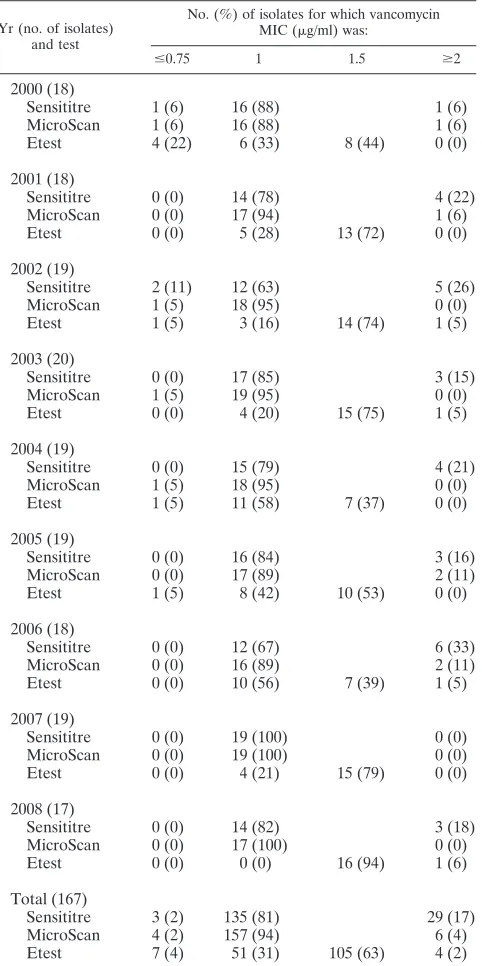
The vancomycin MIC modes remained fixed throughout the
study period, with overall modes of 1
g/ml for Sensititre and
MicroScan and 1.5
g/ml for Etest (Table 1). Utilizing the
Sensititre and MicroScan methodologies, 81% and 94% of the
isolates, respectively, had a MIC of 1
g/ml, while using Etest,
31% and 63% of the isolates had MICs of 1
g/ml and 1.5
g/ml, respectively. The overall frequency of MRSA isolates
for which the MIC was
ⱖ
2
g/ml varied from 17% by Sensititre
to 4% by MicroScan and 2% by Etest. Etest results were
significantly elevated compared with those of MicroScan for
every year, and this was also true when the Etest MICs were
compared to the Sensititre MICs for 5 of the 9 years (Fig. 1;
P
⬍
0.05). For 2002 to 2004 and 2006, the Sensititre results
were statistically higher than the MicroScan results (Fig. 1;
P
⬍
0.05).
Etest GRD for detecting hVISA.
Of the 167 isolates tested by
Etest GRD, 6 were positive for hVISA by having a teicoplanin
MIC value of
ⱖ
8
g/ml and a standard Etest vancomycin MIC
of
⬍
4
g/ml (Table 2). No consistent relationship existed for
the year that the isolate was obtained or the clonal pattern and
the presence of hVISA.
FIG. 1. Vancomycin MICs of MRSA, displayed as geometric
means
⫾
standard errors of the means, were measured by Sensititre,
MicroScan, and Etest for isolates recovered from 2000 to 2008. Etest
reported significantly higher MICs than MicroScan (indicated by A)
and Sensititre (indicated by B). Sensititre results were also significantly
higher than MicroScan results for certain years (indicated by C) (
P
⬍
0.05).
on May 16, 2020 by guest
http://jcm.asm.org/
PAP-AUC for detecting hVISA.
PAP-AUC was performed to
confirm the positive results from the GRD Etest, and two
isolates were identified to be hVISA using this analysis (Fig. 2).
The two hVISA isolates from 2000 and 2003 had PAP-AUC
ratios of 0.91 and 1.23, respectively. Both hVISAs had
vanco-mycin MICs of 1.5 to 2
g/ml according to Sensititre and Etest,
while MicroScan reported a 1-
g/ml MIC for both isolates
(Table 2).
DISCUSSION
Reduced susceptibility to vancomycin in
S. aureus
has been
a major medical concern for over a decade. Several studies
have reported elevated vancomycin MICs in MRSA isolates
where the MICs are at the upper end of the susceptibility range
(18, 27, 38, 40–43, 50, 52). However, not all medical institutions
are reporting an increase in vancomycin MICs (1, 11, 20, 33).
One study measured vancomycin MICs in MRSA isolates
col-lected from 2002 to 2006 from nine different U.S. medical
centers using a reference broth microdilution assay with a
broad range of precise incremental dilutions (39). Their results
showed that they did not detect a reduction in vancomycin
susceptibility at any of the test sites, which are similar to our
results using three separate antimicrobial susceptibility tests.
Although the MIC by Etest did increase from 2000 to 2008, it
fluctuated over time and was clinically insignificant to support
a consistent decline in vancomycin susceptibility. More
specif-ically, the majority of the isolates in this study had a
vancomy-TABLE 1. Annual frequencies and percentages for
each test for vancomycin
Yr (no. of isolates) and test
No. (%) of isolates for which vancomycin MIC (g/ml) was:
ⱕ0.75 1 1.5 ⱖ2
2000 (18)
Sensititre
1 (6)
16 (88)
1 (6)
MicroScan
1 (6)
16 (88)
1 (6)
Etest
4 (22)
6 (33)
8 (44)
0 (0)
2001 (18)
Sensititre
0 (0)
14 (78)
4 (22)
MicroScan
0 (0)
17 (94)
1 (6)
Etest
0 (0)
5 (28)
13 (72)
0 (0)
2002 (19)
Sensititre
2 (11)
12 (63)
5 (26)
MicroScan
1 (5)
18 (95)
0 (0)
Etest
1 (5)
3 (16)
14 (74)
1 (5)
2003 (20)
Sensititre
0 (0)
17 (85)
3 (15)
MicroScan
1 (5)
19 (95)
0 (0)
Etest
0 (0)
4 (20)
15 (75)
1 (5)
2004 (19)
Sensititre
0 (0)
15 (79)
4 (21)
MicroScan
1 (5)
18 (95)
0 (0)
Etest
1 (5)
11 (58)
7 (37)
0 (0)
2005 (19)
Sensititre
0 (0)
16 (84)
3 (16)
MicroScan
0 (0)
17 (89)
2 (11)
Etest
1 (5)
8 (42)
10 (53)
0 (0)
2006 (18)
Sensititre
0 (0)
12 (67)
6 (33)
MicroScan
0 (0)
16 (89)
2 (11)
Etest
0 (0)
10 (56)
7 (39)
1 (5)
2007 (19)
Sensititre
0 (0)
19 (100)
0 (0)
MicroScan
0 (0)
19 (100)
0 (0)
Etest
0 (0)
4 (21)
15 (79)
0 (0)
2008 (17)
Sensititre
0 (0)
14 (82)
3 (18)
MicroScan
0 (0)
17 (100)
0 (0)
Etest
0 (0)
0 (0)
16 (94)
1 (6)
Total (167)
Sensititre
3 (2)
135 (81)
29 (17)
MicroScan
4 (2)
157 (94)
6 (4)
[image:3.585.315.524.70.199.2]Etest
7 (4)
51 (31)
105 (63)
4 (2)
TABLE 2. Isolates positive for hVISA by Etest GRD
Isolate Yr Clonal pattern
GRD values (g/ml) for VAN/TECa
Vancomycin MIC (g/ml) by Sensititre, MicroScan,
Etest
PAP-AUC result
853
2000
Not defined
1/16
2, 1, 1.5
hVISA
1177
2002
Not defined
2/32
2, 1, 2
VSSA
b1182
2002
USA400
1.5/24
1, 1, 1.5
VSSA
1655
2003
USA100
1.5/8
1, 1, 1.5
VSSA
1670
2003
Not defined
2/12
2, 1, 2
hVISA
3982
2007
Not defined
1/
ⱖ
32
1, 1, 1.5
VSSA
a
VAN, vancomycin; TEC, teicoplanin.
b
[image:3.585.45.286.90.573.2]VSSA, vancomycin-susceptibleS. aureus.
FIG. 2. Population analysis profile curves of the six isolates that
were positive for hVISA by Etest GRD. Two isolates were identified to
be hVISA and four isolates were defined to be vancomycin-susceptible
S. aureus
(VSSA) compared to the susceptibility of the Mu3 reference
strain.
on May 16, 2020 by guest
http://jcm.asm.org/
[image:3.585.48.539.616.708.2]cin MIC of 1 or 1.5
g/ml, and the difference between the two
Etest MICs from 2000 and 2008 was
⬍
1.5-fold, whereas the
other studies have detected a
ⱖ
1.5-fold MIC increase or a
significant increase in the frequency of isolates having
vanco-mycin MICs of
ⱖ
1
g/ml (41, 43, 52). None of these trends
were found in our study.
The conflicting results in the literature documenting
ele-vated vancomycin MICs in MRSA may partly be due to failure
of epidemiologically typing the MRSA isolates. Some MRSA
clones have higher levels of dissemination than others and
possess elevated vancomycin MICs, which may cause false
per-ceptions of an overall reduction in vancomycin susceptibility
for all strains of MRSA (8, 26, 31, 48). The purpose of our
study was to assess vancomycin susceptibility in the population
of clinically encountered MRSA clonal types and not to define
the overall MRSA susceptibility profile for clinical purposes in
the associated patient population. PFGE was performed to
eliminate clonal strains within each year that could otherwise
influence the results. This study identified a highly diverse
sample of MRSA isolates. Twenty-six percent of the isolates
were USA100-like, a common MRSA strain found in hospitals,
whereas 7% and 5% were similar to the community-acquired
strains USA300 and USA400, respectively.
The type of antimicrobial susceptibility test that is
per-formed to measure the vancomycin MICs may also affect the
assessment of whether susceptibility within the population has
changed. In this study, three standard susceptibility tests that
are used in the clinical setting were chosen to measure the
MICs. Although elevated vancomycin MICs could not be
dem-onstrated by the three different tests, there were differences
among the assays. In agreement with a previous study,
Sensi-titre panels had the highest variability (47). It is unclear why
MicroScan reported the lowest vancomycin MICs. Other
stud-ies found MicroScan to report vancomycin MICs in MRSA
similar to or higher than those that Etest and Sensititre report
(17, 45, 47). Etest reported vancomycin MICs higher than
those reported by the two broth microdilution assays. This was
expected and has been described elsewhere (33, 36). Etest
strips contain a gradient of vancomycin concentrations,
includ-ing an intermediate readinclud-ing of 1.5
g/ml, as opposed to the
broth microdilution assays, where the concentrations are in
2-fold increments. Most of the isolates had a MIC of 1.5
g/ml,
causing the Etest results to be more elevated than the results of
the other two assays. Etest also measures the MIC for a larger
proportion of bacteria at
⬃
10
8CFU than the broth
microdi-lution assays at
⬃
10
4CFU, making Etest more sensitive in
detecting resistant mutants and MIC increases (11, 33, 43).
In addition to assessing a reduction in vancomycin
suscep-tibility, the prevalence of hVISA was determined by the newly
developed Etest GRD. Two studies have verified that the Etest
GRD has high sensitivity (93 to 94%) in detecting hVISA
compared to PAP-AUC but variable specificity (82% and
95%) (24, 55). Nonetheless, the specificity of the Etest GRD is
similar to or higher than the specificities of other screening
tests, such as the Etest macrodilution method, the vancomycin
screening plate method with brain heart infusion agar, and the
teicoplanin screening plate method with Mueller-Hinton agar
(14). According to the Etest GRD, 3.5% of the MRSA isolates
in our sample were positive for hVISA. All of the positive
isolates had teicoplanin MIC values
ⱖ
8
g/ml. Teicoplanin is
more sensitive in the detection of the hVISA phenotype than
vancomycin (55). It was revealed by PAP-AUC that only two of
the six isolates were confirmed to be hVISA. The small sample
size precludes any conclusion regarding the disparity observed
between Etest GRD and PAP-AUC but does suggest that
additional study is warranted. The two hVISA isolates had
vancomycin MICs of 1.5 to 2
g/ml by Etest and Sensititre.
Vancomycin MICs of
⬎
1
g/ml have been associated with
hVISA (2, 12, 33), but this is not a strong indicator for hVISA
because Etest and Sensititre reported equally high MICs in the
vancomycin-susceptible isolates. The incidence of hVISA in
other hospitals has varied, ranging from 0 to 50% (3, 4, 7, 13,
21, 22, 23, 25, 28, 29, 34, 35, 50, 52, 54). The type of screening
test used to detect hVISA and the number of isolates screened
are responsible for this wide range. A standard screening test
with high sensitivity and specificity for detecting hVISA needs
to be implemented to better understand the prevalence and
clinical impact of these organisms.
In conclusion, a decrease in vancomycin susceptibility in
MRSA isolates from our institution over a 9-year period was
not detected by three standard antimicrobial susceptibility
tests. The vancomycin MICs differed among the three
suscep-tibility tests, in which Etest resulted in the highest MICs and
MicroScan reported the lowest MICs. Finally, the GRD Etest
and PAP-AUC defined a low prevalence of hVISA at 1.2%.
ACKNOWLEDGMENTS
We thank Valerie Shostrom for her assistance with the statistical
analysis of the data. We also acknowledge Laura Orwe and Elsie
Forsung for their help with performing PFGE analysis.
REFERENCES
1.Alo´s, J. I., A. García-Can˜as, P. García-Hierro, and F. Rodríguez-Salvane´s.
2008. Vancomycin MICs did not creep inStaphylococcus aureusisolates from 2002 to 2006 in a setting with low vancomycin usage. J. Antimicrob. Che-mother.62:773–775.
2.Bae, I. G., J. J. Federspiel, J. M. Miro´, C. W. Woods, L. Park, M. J. Rybak, T. H. Rude, S. Bradley, S. Bukovski, C. G. de la Maria, S. S. Kanj, T. M. Korman, F. Marco, D. R. Murdoch, P. Plesiat, M. Rodriguez-Creixems, P. Reinbott, L. Steed, P. Tattevin, M. F. Tripodi, K. L. Newton, G. R. Corey, and V. G. Fowler, Jr.2009. Heterogeneous vancomycin-intermediate suscep-tibility phenotype in bloodstream methicillin-resistantStaphylococcus aureus
isolates from an international cohort of patients with infective endocarditis: prevalence, genotype, and clinical significance. J. Infect. Dis.200:1355–1366. 3.Benquan, W., T. Yingchun, Z. Kouxing, Z. Tiantuo, Z. Jiaxing, and T. Shuqing.2002.Staphylococcusheterogeneously resistant to vancomycin in China and antimicrobial activities of imipenem and vancomycin in combi-nation against it. J. Clin. Microbiol.40:1109–1112.
4.Charles, P. G., P. B. Ward, P. D. Johnson, B. P. Howden, and M. L. Grayson.
2004. Clinical features associated with bacteremia due to heterogeneous vancomycin-intermediateStaphylococcus aureus. Clin. Infect. Dis.38:448– 451.
5.Cui, L., A. Iwamoto, J. Q. Lian, H. M. Neoh, T. Maruyama, Y. Horikawa, and K. Hiramatsu.2006. Novel mechanism of antibiotic resistance originating in vancomycin-intermediateStaphylococcus aureus. Antimicrob. Agents Che-mother.50:428–438.
6.Cui, L., X. Ma, K. Sato, K. Okuma, F. C. Tenover, E. M. Mamizuka, C. G. Gemmell, M. N. Kim, M. C. Ploy, N. El-Solh, V. Ferraz, and K. Hiramatsu.
2003. Cell wall thickening is a common feature of vancomycin resistance in
Staphylococcus aureus. J. Clin. Microbiol.41:5–14.
7.Delgado, A. J., T. Riordan, R. Lamichhane-Khadka, D. C. Winnett, J. Jime-nez, K. Robinson, F. G. O’Brien, S. A. Cantore, and J. E. Gustafson.2007. Hetero-vancomycin-intermediate methicillin-resistantStaphylococcus aureus
isolate from a medical center in Las Cruces, New Mexico. J. Clin. Microbiol.
45:1325–1329.
8.Feil, E. J., J. E. Cooper, H. Grundmann, D. A. Robinson, M. C. Enright, T. Berendt, S. J. Peacock, J. M. Smith, M. Murphy, B. G. Spratt, C. E. Moore, and N. P. Day.2003. How clonal isStaphylococcus aureus? J. Bacteriol.
185:3307–3316.
9.Fridkin, S. K., J. Hageman, L. K. McDougal, J. Mohammed, W. R. Jarvis, T. M. Perl, and F. C. Tenover.2003. Epidemiological and microbiological
on May 16, 2020 by guest
http://jcm.asm.org/
characterization of infections caused byStaphylococcus aureuswith reduced susceptibility to vancomycin, United States, 1997-2001. Clin. Infect. Dis.
36:429–439.
10.Hiramatsu, K.2001. Vancomycin-resistant Staphylococcus aureus: a new model of antibiotic resistance. Lancet Infect. Dis.1:147–155.
11.Holmes, R. L., and J. H. Jorgensen.2008. Inhibitory activities of 11 antimi-crobial agents and bactericidal activities of vancomycin and daptomycin against invasive methicillin-resistantStaphylococcus aureusisolates obtained from 1999 through 2006. Antimicrob. Agents Chemother.52:757–760. 12.Horne, K. C., B. P. Howden, E. A. Grabsch, M. Graham, P. B. Ward, S.
Xie, B. C. Mayall, P. D. Johnson, and M. L. Grayson.2009. Prospective comparison of the clinical impacts of heterogeneous vancomycin-inter-mediate methicillin-resistantStaphylococcus aureus(MRSA) and vanco-mycin-susceptible MRSA. Antimicrob. Agents Chemother.53:3447–3452. 13.Howden, B. P.2005. Recognition and management of infections caused by vancomycin-intermediateStaphylococcus aureus(VISA) and heterogenous VISA (hVISA). Intern. Med. J.35:S136–S140.
14.Howden, B. P., J. K. Davies, P. D. Johnson, T. P. Stinear, and M. L. Grayson.
2010. Reduced vancomycin susceptibility inStaphylococcus aureus, including vancomycin-intermediate and heterogeneous vancomycin-intermediate strains: resistance mechanisms, laboratory detection, and clinical implica-tions. Clin. Microbiol. Rev.23:99–139.
15.Howden, B. P., P. D. Johnson, P. B. Ward, T. P. Stinear, and J. K. Davies.
2006. Isolates with low-level vancomycin resistance associated with persistent methicillin-resistantStaphylococcus aureusbacteremia. Antimicrob. Agents Chemother.50:3039–3047.
16.Howden, B. P., P. B. Ward, P. G. Charles, T. M. Korman, A. Fuller, P. du Cros, E. A. Grabsch, S. A. Roberts, J. Robson, K. Read, N. Bak, J. Hurley, P. D. Johnson, A. J. Morris, B. C. Mayall, and M. L. Grayson.2004. Treat-ment outcomes for serious infections caused by methicillin-resistant Staph-ylococcus aureuswith reduced vancomycin susceptibility. Clin. Infect. Dis.
38:521–528.
17.Hsu, D. I., L. K. Hidayat, R. Quist, J. Hindler, A. Karlsson, A. Yusof, and A. Wong-Beringer.2008. Comparison of method-specific vancomycin minimum inhibitory concentration values and their predictability for treatment out-come of meticillin-resistantStaphylococcus aureus(MRSA) infections. Int. J. Antimicrob. Agents32:378–385.
18.Hussain, F. M., S. Boyle-Vavra, P. B. Shete, and R. S. Daum.2002. Evidence for a continuum of decreased vancomycin susceptibility in unselected Staph-ylococcus aureusclinical isolates. J. Infect. Dis.186:661–667.
19.Iandolo, J. J., J. P. Bannantine, and G. C. Stewart. 1997. Genetic and physical map of the chromosome ofStaphylococcus aureus, p. 39–53.InK. B. Crossley and G. L. Accher (ed.), The staphylococci in human disease. Churchill Livingstone, New York, NY.
20.Jones, R. N.2006. Microbiological features of vancomycin in the 21st cen-tury: minimum inhibitory concentration creep, bactericidal/static activity, and applied breakpoints to predict clinical outcomes or detect resistant strains. Clin. Infect. Dis.1:S13–S24.
21.Khosrovaneh, A., K. Riederer, S. Saeed, M. S. Tabriz, A. R. Shah, M. M. Hanna, M. Sharma, L. B. Johnson, M. G. Fakih, and R. Khatib.2004. Frequency of reduced vancomycin susceptibility and heterogeneous sub-population in persistent or recurrent methicillin-resistantStaphylococcus aureusbacteremia. Clin. Infect. Dis.38:1328–1330.
22.Kim, H. B., W. B. Park, K. D. Lee, Y. J. Choi, S. W. Park, M. D. Oh, E. C. Kim, and K. W. Choe.2003. Nationwide surveillance forStaphylococcus aureuswith reduced susceptibility to vancomycin in Korea. J. Clin. Microbiol.
41:2279–2281.
23.Kim, M. N., S. H. Hwang, Y. J. Pyo, H. M. Mun, and C. H. Pai.2002. Clonal spread ofStaphylococcus aureusheterogeneously resistant to vancomycin in a university hospital in Korea. J. Clin. Microbiol.40:1376–1380.
24.Leonard, S. N., K. L. Rossi, K. L. Newton, and M. J. Rybak.2009. Evaluation of the Etest GRD for the detection ofStaphylococcus aureuswith reduced susceptibility to glycopeptides. J. Antimicrob. Chemother.63:489–492. 25.Liu, C., and H. F. Chambers.2003.Staphylococcus aureuswith
heteroge-neous resistance to vancomycin: epidemiology, clinical significance, and crit-ical assessment of diagnostic methods. Antimicrob. Agents Chemother.47:
3040–3045.
26.Liu, C., C. J. Graber, M. Karr, B. A. Diep, L. Basuino, B. S. Schwartz, M. C. Enright, S. J. O’Hanlon, J. C. Thomas, F. Perdreau-Remington, S. Gordon, H. Gunthorpe, R. Jacobs, P. Jensen, G. Leoung, J. S. Rumack, and H. F. Chambers.2008. A population-based study of the incidence and molecular epidemiology of methicillin-resistantStaphylococcus aureusdisease in San Francisco, 2004-2005. Clin. Infect. Dis.46:1637–1646.
27.Lodise, T. P., J. Graves, A. Evans, E. Graffunder, M. Helmecke, B. M. Lomaestro, and K. Stellrecht.2008. Relationship between vancomycin MIC and failure among patients with methicillin-resistantStaphylococcus aureus
bacteremia treated with vancomycin. Antimicrob. Agents Chemother.52:
3315–3320.
28.Maor, Y., M. Hagin, N. Belausov, N. Keller, D. Ben-David, and G. Rahav.
2009. Clinical features of heteroresistant vancomycin-intermediate Staphy-lococcus aureusbacteremia versus those of methicillin-resistantS. aureus
bacteremia. J. Infect. Dis.199:619–624.
29.Maor, Y., G. Rahav, N. Belausov, D. Ben-David, G. Smollan, and N. Keller.
2007. Prevalence and characteristics of heteroresistant vancomycin-interme-diateStaphylococcus aureusbacteremia in a tertiary care center. J. Clin. Microbiol.45:1511–1514.
30.Maree, C. L., R. S. Daum, S. Boyle-Vavra, K. Matayoshi, and L. G. Miller.2007. Community-associated methicillin-resistantStaphylococcus aureusisolates caus-ing healthcare-associated infections. Emerg. Infect. Dis.13:236–242. 31.McDougal, L. K., C. D. Steward, G. E. Killgore, J. M. Chaitram, S. K.
McAllister, and F. C. Tenover.2003. Pulsed-field gel electrophoresis typing of oxacillin-resistantStaphylococcus aureusisolates from the United States: establishing a national database. J. Clin. Microbiol.41:5113–5120. 32.Murchan, S., M. E. Kaufmann, A. Deplano, R. de Ryck, M. Struelens, C. E.
Zinn, V. Fussing, S. Salmenlinna, J. Vuopio-Varkila, N. El Solh, C. Cuny, W. Witte, P. T. Tassios, N. Legakis, W. van Leeuwen, A. van Belkum, A. Vindel, I. Laconcha, J. Garaizar, S. Haeggman, B. Olsson-Liljequist, U. Ransjo, G. Coombes, and B. Cookson.2003. Harmonization of pulsed-field gel electro-phoresis protocols for epidemiological typing of strains of methicillin-resis-tantStaphylococcus aureus: a single approach developed by consensus in 10 European laboratories and its application for tracing the spread of related strains. J. Clin. Microbiol.41:1574–1585.
33.Musta, A. C., K. Riederer, S. Shemes, P. Chase, J. Jose, L. B. Johnson, and R. Khatib.2009. Vancomycin MIC plus heteroresistance and outcome of methicillin-resistantStaphylococcus aureusbacteremia: trends over 11 years. J. Clin. Microbiol.47:1640–1644.
34.Nunes, A. P., R. P. Schuenck, C. C. Bastos, M. M. Magnanini, J. B. Long, N. L. Iorio, and K. R. Santos.2007. Heterogeneous resistance to vancomycin and teicoplanin amongStaphylococcusspp. isolated from bacteremia. Braz. J. Infect. Dis.11:345–350.
35.Plipat, N., G. Livni, H. Bertram, and R. B. Thomson, Jr.2005. Unstable vancomycin heteroresistance is common among clinical isolates of methicil-lin-resistantStaphylococcus aureus. J. Clin. Microbiol.43:2494–2496. 36.Prakash, V., J. S. Lewis II, and J. H. Jorgensen.2008. Vancomycin MICs for
methicillin-resistant Staphylococcus aureusisolates differ based upon the susceptibility test method used. Antimicrob. Agents Chemother.52:4528. 37.Rybak, M. J., and R. L. Akins.2001. Emergence of methicillin-resistant
Staphylococcus aureuswith intermediate glycopeptide resistance: clinical sig-nificance and treatment options. Drugs61:1–7.
38.Rybak, M. J., S. N. Leonard, K. L. Rossi, C. M. Cheung, H. S. Sader, and R. N. Jones.2008. Characterization of vancomycin-heteroresistant Staphylo-coccus aureus from the metropolitan area of Detroit, Michigan, over a 22-year period (1986 to 2007). J. Clin. Microbiol.46:2950–2954.
39.Sader, H. S., P. D. Fey, D. N. Fish, A. P. Limaye, G. Pankey, J. Rahal, M. J. Rybak, D. R. Snydman, L. L. Steed, K. Waites, and R. N. Jones.2009. Evaluation of vancomycin and daptomycin potency trends (MIC creep) against methicillin-resistantStaphylococcus aureusisolates collected in nine U.S. medical centers from 2002 to 2006. Antimicrob. Agents Chemother.
53:4127–4132.
40.Sakoulas, G., P. A. Moise-Broder, J. Schentag, A. Forrest, R. C. Moellering, Jr., and G. M. Eliopoulos. 2004. Relationship of MIC and bactericidal activity to efficacy of vancomycin for treatment of methicillin-resistant Staph-ylococcus aureusbacteremia. J. Clin. Microbiol.42:2398–2402.
41.Schwaber, M. J., S. B. Wright, Y. Carmeli, L. Venkataraman, P. C. DeGirolami, A. Gramatikova, T. M. Perl, G. Sakoulas, and H. S. Gold.2003. Clinical implications of varying degrees of vancomycin susceptibility in me-thicillin-resistant Staphylococcus aureus bacteremia. Emerg. Infect. Dis.
9:657–664.
42.Soriano, A., F. Marco, J. A. Martínez, E. Pisos, M. Almela, V. P. Dimova, D. Alamo, M. Ortega, J. Lopez, and J. Mensa.2008. Influence of vancomycin minimum inhibitory concentration on the treatment of methicillin-resistant
Staphylococcus aureusbacteremia. Clin. Infect. Dis.46:193–200.
43.Steinkraus, G., R. White, and L. Friedrich.2007. Vancomycin MIC creep in non-vancomycin-intermediateStaphylococcus aureus(VISA), vancomycin-susceptible clinical methicillin-resistant S. aureus(MRSA) blood isolates from 2001-05. J. Antimicrob. Chemother.60:788–794.
44.Stevens, D. L.2006. The role of vancomycin in the treatment paradigm. Clin. Infect. Dis.1:S51–S57.
45.Swenson, J. M., K. F. Anderson, D. R. Lonsway, A. Thompson, S. K. Mc-Allister, B. M. Limbago, R. B. Carey, F. C. Tenover, and J. B. Patel.2009. Accuracy of commercial and reference susceptibility testing methods for detecting vancomycin-intermediateStaphylococcus aureus. J. Clin. Micro-biol.47:2013–2017.
46.Szabo´, J.2009. hVISA/VISA: diagnostic and therapeutic problems. Expert Rev. Anti Infect. Ther.7:1–3.
47.Tenover, F. C., M. V. Lancaster, B. C. Hill, C. D. Steward, S. A. Stocker, G. A. Hancock, C. M. O’Hara, S. K. McAllister, N. C. Clark, and K. Hira-matsu.1998. Characterization of staphylococci with reduced susceptibilities to vancomycin and other glycopeptides. J. Clin. Microbiol.36:1020–1027. 48.Tenover, F. C., L. K. McDougal, R. V. Goering, G. Killgore, S. J. Projan, J. B.
Patel, and P. M. Dunman.2006. Characterization of a strain of community-associated methicillin-resistantStaphylococcus aureuswidely disseminated in the United States. J. Clin. Microbiol.44:108–118.
49.Tenover, F. C., and R. C. Moellering, Jr.2007. The rationale for revising the
on May 16, 2020 by guest
http://jcm.asm.org/
Clinical and Laboratory Standards Institute vancomycin minimal inhibitory concentration interpretive criteria forStaphylococcus aureus. Clin. Infect. Dis.44:1208–1215.
50.Walsh, T. R., A. Bolmstro¨m, A. Qwa¨rnstro¨m, P. Ho, M. Wootton, R. A. Howe, A. P. MacGowan, and D. Diekema.2001. Evaluation of current methods for detection of staphylococci with reduced susceptibility to glycopeptides. J. Clin. Microbiol.39:2439–2444.
51.Walsh, T. R., and R. A. Howe.2002. The prevalence and mechanisms of vancomycin resistance inStaphylococcus aureus. Annu. Rev. Microbiol.56:
657–675.
52.Wang, G., J. F. Hindler, K. W. Ward, and D. A. Bruckner.2006. Increased vancomycin MICs forStaphylococcus aureusclinical isolates from a univer-sity hospital during a 5-year period. J. Clin. Microbiol.44:3883–3886.
53.Wootton, M., R. A. Howe, R. Hillman, T. R. Walsh, P. M. Bennett, and A. P. MacGowan.2001. A modified population analysis profile (PAP) method to detect hetero-resistance to vancomycin inStaphylococcus aureusin a UK hospital. J. Antimicrob. Chemother.47:399–403.
54.Wootton, M., A. P. MacGowan, T. R. Walsh, and R. A. Howe.2007. A multicenter study evaluating the current strategies for isolating Staphylococ-cus aureusstrains with reduced susceptibility to glycopeptides. J. Clin. Mi-crobiol.45:329–332.
55.Yusof, A., A. Engelhardt, A. Karlsson, L. Bylund, P. Vidh, K. Mills, M. Wootton, and T. R. Walsh.2008. Evaluation of a new Etest vancomycin-teicoplanin strip for detection of glycopeptide-intermediateStaphylococcus aureus(GISA), in particular, heterogeneous GISA. J. Clin. Microbiol.46:
3042–3047.