Original Article
Effect of Dentin Surface Treatment Using a Non-Thermal
Argon Plasma Brush on the Bond Strength of a Self-Adhesive
Resin Composite
A.Moradi 1, M.Hasani Tabatabaei 2,S.Hashemi Kamangar 2, S. Valizadeh 3.
1 Postgraduate Student, Department of Operative Dentistry, School of Dentistry, Tehran University of Medical Sciences, Tehran,
Iran
2 Associate Professor, Dental Research Center, Dentistry Research Institute, Tehran University of Medical Sciences, Tehran, Iran
AND Department of Operative Dentistry, School of Dentistry, Tehran University of Medical Sciences, Tehran, Iran
3 Assistant Professor, Laser Research Center in Dentistry, Dentistry Research Institute, Tehran University of Medical Sciences,
Tehran, Iran AND Department of Operative Dentistry, School of Dentistry, Tehran University of Medical Sciences, Tehran, Iran
Corresponding author: S. Valizadeh, Assistant Professor, Laser Research Center in Dentistry, Dentistry Research Institute, Tehran University of Medical Sciences, Tehran, Iran AND Department of Operative Dentistry, School of Dentistry, Tehran University of Medical Sciences, Tehran, Iran
Received: 10 June 2018 Accepted: 14 Sep 2018
Abstract
Background and Aim: Improving the bond strength at the resin-dentin interface is an important challenge in adhesive dentistry. This study examined the effect of low-power, non-thermal atmospheric pressure plasma (NT-APP) treatments on the adhesion of a self-adhesive resin composite to dentin.
Materials and Methods: In this experimental in-vitro study, buccal enamel of extracted bovine incisors was removed using a high-speed diamond bur. The specimens were randomly divided into two groups according to the plasma treatment and thermocycling. The non-thermal atmospheric argon plasma brush was used in this study. One subgroup was subjected to the microshear bond strength (µSBS) test after 24 hours, whereas the other subgroup was subjected to artificial aging with thermocycling for 5000 cycles before being tested. Each specimen was attached to a testing jig and loaded at a crosshead speed of 1.0 mm/minute in a universal testing machine until failure occurred. Data were analyzed using two-factor repeated measures analysis of variance (ANOVA). Results: The results of μSBS testing showed that with plasma treatment, the average μSBS increased to 34.20±12.12 MPa compared to 19.47±7.4 MPa in the controls (P=0.002). After 5000 cycles of thermocycling, the adhesive-dentin bonding strengths of the plasma-treated specimens slightly decreased from 34.20±12.12 MPa to 33.64±5.6 MPa (P=0.886), while the strengths of the untreated specimens reduced from 19.47±7.4 MPa to 19.10±5.1 MPa (P=0.461). Plasma treatment improved the µSBS compared to the control group. After thermocycling, the µSBS did not decrease in the plasma and control (non-plasma) groups.
Conclusion: Plasma treatment using NT-APP improves the adhesion of self-adhesive flowable resin composites to dentin.
Key Words: Composite Resins, Non-Thermal Atmospheric Pressure Plasma, Dental Bonding
Cite this article as: Moradi A, Hasani Tabatabaei M, Hashemi Kamangar S, Valizadeh S. Effect of DentinSurface Treatment Using a Non-Thermal Argon Plasma Brush on the Bond Strength of a Self-Adhesive Resin Composite. J Islam Dent Assoc Iran. 2019; 31(1):1-7. DOI: 10.30699/jidai.31.1.1
Introduction
Due to their aesthetic properties and noninvasive tooth preparations, the use of composite resin
restorations has significantly increased among dentists and patients [1]. The long-term success of a composite resin restoration depends on the
durability of the bond strength [2]. Most current dental adhesive systems show satisfactory immediate results in terms of the bond strength [3]. However, the adhesive-dentin interface is the weakest link within the adhesion complex of tooth-colored restorations, and the durability of aged resin-dentin adhesion is questionable. Previous long-term studies have shown that the bond strength of resin to dentin decreases over time [3,4]. A new strategy to produce a long-term durable dentin adhesion is needed. Recently, plasma has attracted attention in the biomedical field [5,6].
Plasma is the fourth state of matter and is the most copious state in the universe [7]; it exists in various forms, including fire, which is known for millions of years since the early Stone Age. Plasma is not a human invention and is present in nature as fire in the sun and stars, in the tails of comets, and as flashes of lightning [7]. In medicine and biology, plasma refers to the non-cellular fluid component of blood. The term was introduced into physics by Irving Langmuir in 1928 [7]. Non-thermal plasmas are partly-ionized gases containing exceptionally reactive particles, including electronically-excited atoms, molecules, and ionic and free radical species; the gas phase is stable at room temperature [8,9]. Depending on the plasma chemistry or the composition of the gas, these highly reactive plasma species react with, clean, and etch surfaces, bond with different substrates, or combine to create a thin layer of plasma that changes the surface properties [8,9]. Treatment with non-thermal plasma allows for the modification of dentinal surfaces to improve the interfacial bonding of dentin and composite restorations [10,11]. It has been stated that short-term treatment with plasma could change the chemical structure of exposed collagen fibrils and increase the hydrophilicity of the dentin surface, which allow better penetration of the adhesive into dental collagen fibrils and enhance the adhesive-dentin bond strength [10,11]. Treatment of dentin using a non-thermal plasma brush results in an increase of about 60% in the bonding at the adhesive-dentin interface compared to untreated controls [12,13].
A simplified adhesive protocol to reduce the duration of treatment is highly desirable in adhesive dentistry. A new category of composites
has been introduced; according to the
manufacturers' claim, these self-adhesive (flowable) composites do not require any acid-etching or bonding protocol prior to the application [12,13]. VertiseTM Flow self-adhesive composite (Kerr, Orange, CA, USA) comprises phosphoric acid ester methacrylate and glycerol phosphate dimethacrylate (GPDM) as functional monomers [12,13]. The aim of this study was to evaluate and verify the effectiveness of plasma treatment of dentinal surfaces for improving the interfacial bonding of a self-adhesive composite resin and dentin by performing a microshear bond strength (µSBS) test.
Materials and Methods
In this experimental study (ethical code: TUMS.DENTISTRY.REC.1397.017), 48 extracted bovine incisors were immersed in a solution of 0.5% Chloramine-T for one week after removing soft tissue debris and then stored in distilled water at 4°C until the experiment. The crowns of bovine incisors were separated from the roots and sectioned perpendicular to their long axis. The teeth were embedded in acrylic molds with a self-curing resin. Buccal enamel was removed using a high-speed diamond bur. The exposed dentin surface was polished with 600-grit silicon carbide abrasive papers under water cooling. The specimens were randomly divided into two groups according to the plasma treatment (with or without) and two subgroups according to thermocycling (n=12). The study design is shown in Diagram 1.
The use of a non-thermal atmospheric pressure plasma (NT-APP) jet:
The non-thermal atmospheric plasma brush employed in this study was designed by the Nik fanavaran plasma, Parham Co., Tehran, Iran. The APP was used for 30 seconds with compressed argon gas (ultrahigh purity) as the plasma gas supply with argon gas flow rate of 3000 sccm. A glow discharge by the direct current power source was ignitedbetween the two electrodes in a walled Teflon chamber. One of the electrodes was attached to a ballasted resistor whichcontrolled the discharge current. The other electrode was grounded for electrical safety. The formed plasma dischargecould be blown out of the chamber to
Diagram 1. Study design for different groups
create a brush-shaped non-thermal plasma jet, which was generated in about 2 seconds [14]. A self-adhesive composite (VertiseTM Flow; Kerr, Orange, CA, USA) was selected for this study. The composite was applied according to the manufacturer's instructions (Table 1). First, a 0.5-mm-thick layer was placed on the dentin surface and light-cured by a light-emitting diode (LED) curing unit (Bluephase® Style, Ivoclar Vivadent, Schaan, Liechtenstein) with the intensity of 1200 mW/cm2 for 20 seconds. A cylindrical translucent mold (Saint Gobain Performance Plastics™ Tygon™ R-3603 Lab Tubing, Miami Lakes, FL, USA) with the diameter of 1 mm and the height of 3 mm was used. Nanohybrid Filtek TM Z250 XT resin composite (A2 Shade; 3M ESPE AG, Seefeld, Germany) was filled into the cylindrical translucent mold by a resin applicator, and packing was performed using a dycal applicator. After resin filling, the cylindrical translucent mold was light-cured by the LED curing light with the intensity of 1200 mW/cm2 for 20 seconds.
Microtensile bond strength (µSBS) testing:
After storage in deionized water in an incubator at 37°C for 24 hours, each Tygon tubing was cut using a #12 blade. The specimens of each group were further divided into two subgroups according to the µSBS testing time: one subgroup was immediately submitted to the µSBS test, whereas the other subgroup was submitted to artificial aging with thermocycling for 5,000 cycles between 5°C and 55°C with a 20-second dwell time and a
10-second transferring time before being tested [14,15]. Each specimen was attached to a testing jig and loaded at a crosshead speed of 1.0 mm/minute in a universal testing machine (Zwick/Roell Z050, Ulm, Germany) until failure occurred.
Statistical analysis:
The µSBS data were analyzed using statistical software (SPSS 24.0; IBM Corp., Armonk, NY, USA). Two-way analysis of variance (ANOVA) was used to assess the effects of the plasma treatments and the measuring time (artificial aging). Because the interaction was not significant (P=0.33), t-test was used for comparisons of the µSBS within the factors of treatment and measuring time. The analyses were performed at a significance level of α=0.05.
Results
Table 2 shows the µSBS values for all test groups. The two-factor repeated measures ANOVA revealed that the plasma treatment had a significant effect on the bond strength (P=0.002), but the effect of artificial aging (P=0.397) and the interaction effect (P=0.067) were not significant. The plasma-treated groups showed a significantly higher bond strength than the control group, both at 24 hours and after thermocycling. However, there were no significant differences between the plasma-treated groups at either measurement times. After artificial aging, the mean bond strength of the control group and the conventional plasma
Dentin Blocks of Bovine Incisors Bonded to Vertise Flow N=48
Plasma-treated N=24
Non-plasma-treated N=24
No Thermal cycling
N=12
Thermal cycling (5000 cycles), 5-55°C, N=12
No Thermal cycling
N=12
Thermal cycling (5000 cycles), 5-55°C, N=12
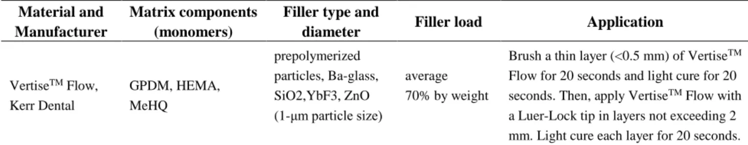
Table 1. Composition of the flowable composite and its application according to the manufacturer’s instructions Material and
Manufacturer
Matrix components (monomers)
Filler type and
diameter Filler load Application
VertiseTM Flow,
Kerr Dental
GPDM, HEMA, MeHQ
prepolymerized particles, Ba-glass, SiO2,YbF3, ZnO (1-μm particle size)
average 70% by weight
Brush a thin layer (<0.5 mm) of VertiseTM
Flow for 20 seconds and light cure for 20 seconds. Then, apply VertiseTM Flow with
a Luer-Lock tip in layers not exceeding 2 mm. Light cure each layer for 20 seconds.
GPDM=glycerol phosphate dimethacrylate, HEMA=hydroxyethyl methacrylate, MeHQ=hydroquinone monomethyl ether
Table 2. Microshear bond strength (µSBS; MPa) of the resin composite to the dentin surfaces
Study groups After 24 hours
(n=12)
After thermocycling (5,000 cycles; n=12)
Control (no plasma treatment) 19.47±7.4 19.10±5.1
Argon plasma treatment 34.20±12.12 33.64±5.6
group did not significantly decrease from the initial mean bond strength.
Discussion
In restorative dentistry, high bond strength between the restoration and dentin has the potential to decrease secondary caries, tooth sensitivity, and marginal discoloration; dental caries is known to be a major epidemiological and economic burden in the developing and developed countries [16,17]. Plasma, the fourth state of matter, can quickly change surface energy without affecting the bulk properties of materials. Non-thermal plasma has been used recently to modify enamel [18] and dentin surfaces [19] and also to improve the interfacial bonding of dental composite restorations to dentin and enamel surfaces [20,21]. Moreover, a previous study has proven that non-thermal plasma treatment would improve the bond strength between dentin and self-etch adhesives [22]. The present study assessed whether NT-APP application by itself has a potential to increase the bond strength of self-adhesive composites without using adhesives or dentin
etching prior to the application of the composite resin.
While micro-mechanical interlocking is essential for attaining a good bonding in the clinical setting, the potential advantage of supplementary chemical interaction between functional monomers and tooth-substrate components has lately reclaimed attention [23]. Each dental adhesive comprises a specific functional monomer that determines its adhesive performance to dental tissue [24]. VertiseTM Flow contains GPDM monomers which are well-known from the OptiBond FL adhesive system [25]. GPDM features a rather short spacer chain and high hydrophilicity that result in a strong etching effect and a better dentin wettability but also a relatively low chemical bonding potential to hydroxyapatite compared to other self-adhesive monomers [26].
In the present study, VertiseTM Flow resulted in thin and sparse tags compared to a different study, where tags were partially branched [27]. Not only when measured immediately after application [28], but especially after aging [29,30], self-adhesive composites present significantly lower bond
strengths than conventional composites that were applied with a bonding system in the etch-and-rinse or self-etch technique. Chemical bonding of GPDM to the calcium present in hydroxyapatite has been positively demonstrated [31]. Unlike etch-and-rinse adhesives, self-adhesive composites are applied directly on the smear layer of dentin surface; etching and subsequent penetration of monomers into the demineralized dentin are carried out as a single step, which is achieved by including polymerizable acidic monomers into the formula [32]. These polymerizable acidic monomers can react with other resin monomers in the adhesive formula to produce an effective bonding. The acidic groups in these monomers can etch mineral debris, remove smear layers, and help resin monomers to penetrate into dentinal tubules [31,32].
Because of about 70% filler content in self-adhesive composites, they have lower wettability on dentin surface and higher viscosity that lead to less resin monomer penetration and decreased bond strength of self-adhesive composites [32]. Our results showed that the µSBS for the NT-APP group (34.20 MPa) is higher than that of the control group (19.47 MPa) after 24 hours. The same patterns have been seen after 5,000 cycles of aging, with the µSBS in the NT-APP group (33.64 MPa) and in the control group (19.10 MPa). There are no studies in the literature on the evaluation of µSBS values after NT-APP surface treatment or comparing µSBS between the control (no plasma treatment) and NT-APP-treated groups with the use of VertiseTM Flow self-adhesive composite. The higher bond strength in the plasma-treated group can be justified by the high Bis-GMA content in the hybrid layer. Bis-GMA has superior
mechanical properties compared to
monomethacrylates, such as HEMA (hydroxyethyl methacrylate), but because of its hydrophobicity and large molecular size, it does not sufficiently penetrate the wet dentinal surface [33]. The plasma-treated group would transfer excess water from the interfibrillar space without collapse of the collagen network, resulting in improved Bis-GMA penetration and bond strength [34]. Treatment of dentin surfaces with NT-APP is a promising approach to improve the penetration of adhesive and resin bond strength to dentin [34].
Optical emission spectroscopy (OES) from a study by Huang et al [34] indicated that the argon plasma brush induced the production of photo-emitting species from nitrogen, oxygen, and nitrous oxide as a result of the interactions between argon plasmas and ambient air. The surface interaction of the reactive oxygen species in the plasma could be incorporated into the type I collagen molecule as carbonyl groups containing oxygen. The addition of more carbonyl groups to the collagen fibrils has been hypothesized to increase the hydrogen-bonding interactions of the collagen fibrils with the adhesive that is subsequently applied to the dentin surfaces for test specimen preparation [35]. Another potential advantage of the increased num-ber of carbonyl groups is the electrical repulsive forces that could cause the collagen fiber to be partly separated into smaller fibril collections or even single fibrils [36]; this would significantly improve the penetration of the adhesive into collagen fibrils and increase the interface area
between the adhesive and collagen and
consequently significantly enhance the interfacial
bonding strength [37]. NT-APP after
thermocycling could decrease the bond strength of the composite. Interestingly, bond strengths of the self-adhesive systems after thermal cycling were not different in the present study and in the study by Brueckner et al [20].
The effect of plasma treatment on dentin adhesion should be further investigated with different adhesive systems. In addition, future studies on plasma treatment need to evaluate the long-term durability of dentin adhesion and clinical performance.
Conclusion
Plasma treatment using NT-APP improves the adhesion of self-adhesive flowable resin composites to dentin.
References
1. Lynch CD, Frazier KB, McConnell RJ, Blum IR, Wilson NH. Minimally invasive management of dental caries: contemporary teaching of posterior resin-based composite placement in U.S. and Canadian dental schools. J Am Dent Assoc. 2011 Jun;142(6):612-20.
2. De Munck J, Van Landuyt K, Peumans M,
Poitevin A, Lambrechts P, Braem M, et al. A critical review of the durability of adhesion to tooth tissue: methods and results. J Dent Res. 2005 Feb; 84(2):118-32.
3. Armstrong SR, Vargas MA, Chung I, Pashley DH, Campbell JA, Laffoon JE, et al. Resin-dentin interfacial ultrastructure and microtensile dentin bond strength after five-year water storage. Oper Dent. 2004 Nov-Dec;29(6):705-12.
4. Fridman G, Friedman G, Gutsol A, Shekhter AB, Vasilets VN, Fridman A. Applied plasma medicine. Plasma Process Polym. 2008 Aug; 5(6): 503-33.
5. Kim JH, Lee MA, Han GJ, Cho BH. Plasma in dentistry: a review of basic concepts and applications in dentistry. Acta Odontol Scand. 2014 Jan;72(1):1-12.
6.Cheruthazhekatt S, Černák M, Slavíček P, Havel J. Gas plasmas and plasma modified materials in medicine. J Appl Biomed. 2010 Jan;8(2):55-66. 7.Shohet JL. Plasma-aided manufacturing. IEEE T Plasma Sci. 1991 Oct;19(5):725-33.
8. Yasuda H. Luminous Chemical Vapor
Deposition and Interface Engineering. Florida, Bota Racon, USA, CRC Press, Taylor & Francis Group, 2004:11-40.
9.Yu QS, Li H, Ritts AC, Yang B, Chen M, Hong L, et al. Non-thermal atmospheric plasma treatment for deactivation of oral bacteria and improvement of dental composite restoration. In Plasma for Bio-Decontamination, Medicine and Food Security. NATO Science for Peace and Security Series A: Chemistry and Biology, 2012: 215-228.
10.Ritts AC, Li H, Yu Q, Xu C, Yao X, Hong L, et al. Dentin surface treatment using a non‐thermal argon plasma brush for interfacial bonding improvement in composite restoration. Eur J Oral Sci. 2010 Oct;118(5):510-6.
11.Zhu XM, Zhou JF, Guo H, Zhang XF, Liu XQ, Li HP, et al. Effects of a modified cold atmospheric plasma jet treatment on resin-dentin bonding. Dent Mater J. 2018 Sep 30;37(5):798-804.
12. Deng D, Huang X, Huang C, Yang T, Du X, Wang Y, et al. Effects of chlorhexidine on bonding durability of different adhesive systems using a novel thermocycling method. Aust Dent J. 2013 Jun;58(2):148-55.
13. Perdigão J, Fundingsland JW, Duarte S Jr, Lopes M. Microtensile adhesion of sealants to intact enamel. Int J Paediatr Dent. 2005 Sep; 15 (5):342-8.
14. Zhang Y, Yu Q, Wang Y. Non-thermal atmospheric plasmas in dental restoration: improved resin adhesive penetration. J Dent. 2014 Aug;42(8):1033-42.
15. Bahrololoomi Z, Soleymani A, Heydari Z. In vitro comparison of microleakage of two materials used as pit and fissure sealants. J Dent Res Dent Clin Dent Prospect. 2011 Summer;5(3):83-86. 16. Lehmann A, Rueppell A, Schindler A, Zylla IM, Seifert HJ, Nothdurft F, et al. Modification of enamel and dentin surfaces by non‐thermal atmospheric plasma. Plasma Process Polym. 2013 Mar;10(3):262-70.
17. Dong X, Ritts AC, Staller C, Yu Q, Chen M, Wang Y. Evaluation of plasma treatment effects on improving adhesive-dentin bonding by using the same tooth controls and varying cross‐sectional surface areas. Eur J Oral Sci. 2013 Aug; 121(4): 355-62.
18. Teixeira HS, Coelho PG, Duarte S, Janal MN, Silva N, Thompson VP. Influence of atmospheric pressure plasma treatment on mechanical proprieties of enamel and sealant bond strength. J Biomed Mater Res B Appl Biomater. 2015 Jul; 103(5):1082-91.
19. Wang R, Shi Y, Li T, Pan Y, Cui Y, Xia W. Adhesive interfacial characteristics and the related bonding performance of four self-etching adhesives with different functional monomers applied to dentin. J Dent. 2017 Jul;62:72-80. 20. Brueckner C, Schneider H, Haak R. Shear bond strength and tooth-composite interaction with self-adhering flowable composites. Oper Dent. 2017 Jan/Feb;42(1):90-100.
21.Poitevin A, De Munck J, Van Ende A, Suyama Y, Mine A, Peumans M, et al. Bonding effectiveness of self-adhesive composites to dentin and enamel. Dent Mater. 2013 Feb;29(2):221-30. 22.Ayres AP, Freitas PH, De Munck J, Vananroye A, Clasen C, Dias CDS, et al. Benefits of Nonthermal Atmospheric Plasma Treatment on Dentin Adhesion. Oper Dent. 2018 Nov/Dec; 43 (6):E288-E299.
23. Sofan E, Sofan A, Palaia G, Tenore G, Romeo U, Migliau G. Classification review of dental
adhesive systems: from the IV generation to the universal type. Ann Stomatol (Roma). 2017 Jul 3; 8(1):1-17.
24. Marine J, Myers CP, Picquet GA, Zaidel LA, Wu D, Uhrich KE. Reduction of bacterial attachment on hydroxyapatite surfaces: Using hydrophobicity and chemical functionality to enhance surface retention and prevent attachment. Colloids Surf B Biointerfaces. 2018 Jul 1;167:531-537.
25. Peterson J, Rizk M, Hoch M, Wiegand A. Bonding performance of self-adhesive flowable composites to enamel, dentin and a nano-hybrid composite. Odontology. 2018 Apr;106(2):171-180. 26. Erli HJ, Marx R, Paar O, Niethard FU, Weber M, Wirtz DC. Surface pretreatments for medical application of adhesion. Biomed Eng Online. 2003 Sep 18;2:15.
27. Makishi P, Pacheco RR, Sadr A, Shimada Y, Sumi Y, Tagami J, et al. Assessment of self-adhesive resin composites: nondestructive imaging of resin-dentin interfacial adaptation and shear bond strength. Microsc Microanal. 2015 Dec; 21 (6):1523-1529.
28. Fu J, Kakuda S, Pan F, Hoshika S, Ting S, Fukuoka A, et al. Bonding performance of a newly developed step-less all-in-one system on dentin. Dent Mater J. 2013;32(2):203-11.
29. Vichi A, Margvelashvili M, Goracci C, Papacchini F, Ferrari M. Bonding and sealing ability of a new self-adhering flowable composite resin in class I restorations. Clin Oral Investig. 2013 Jul;17(6):1497-506.
30. Goracci C, Margvelashvili M, Giovannetti A, Vichi A, Ferrari M. Shear bond strength of orthodontic brackets bonded with a new self-
adhering flowable resin composite. Clin Oral Investig. 2013 Mar;17(2):609-17.
31. Veli I, Akin M, Kucukyilmaz E, Uysal T. Shear bond strength of a self-adhering flowable composite when used for lingual retainer bonding J Orofac Orthop. 2014 Sep;75(5):374-83.
32. Bektas OO, Eren D, Akin EG, Akin H. Evaluation of a self-adhering flowable composite in terms of micro-shear bond strength and microleakage. Acta Odontol Scand. 2013 May-Jul; 71(3-4):541-6.
33. Han GJ, Kim JH, Chung SN, Chun BH, Kim CK, Seo DG, et al. Effects of non‐thermal atmospheric pressure pulsed plasma on the adhesion and durability of resin composite to dentin. Eur J Oral Sci. 2014 Dec;122(6):417-23. 34.Huang C, Yu Q, Hsieh FH, Duan Y. Bacterial deactivation using a low temperature argon atmospheric plasma brush with oxygen addition. Plasma Process Polym. 2007 Jan;4(1):77-87. 35. Naga AA, Yousef M, Ramadan R, Fayez Bahgat S, Alshawwa L. Does the use of a novel
self-adhesive flowable composite reduce
nanoleakage? Clin Cosmet Investig Dent. 2015 Mar 27;7:55-64.
36. Pashley DH, Tay FR, Carvalho RM,
Rueggeberg FA, Agee KA, Carrilho M, et al. From dry bonding to water-wet bonding to ethanol-wet bonding. A review of the interactions between dentin matrix and solvated resins using a macromodel of the hybrid layer. Am J Dent. 2007 Feb;20(1):7-20.
37.Moszner N, Salz U, Zimmermann J. Chemical aspects of self-etching enamel-dentin adhesives: a systematic review. Dent Mater. 2005 Oct; 21(10): 895-910.