Review
Ultrasound for patients in a high HIV/tuberculosis prevalence setting:
a needs assessment and review of focused applications for
Sub-Saharan Africa
Tom Heller
a,*, Eric A. Mtemang’ombe
a, Michae¨la A.M. Huson
b, Charlotte C. Heuvelings
b,
Sabine Be´lard
b,c,d, Saskia Janssen
b, Sam Phiri
a,e,f, Martin P. Grobusch
b,ga
Lighthouse Clinic, Kamuzu Central Hospital, Area 33, Mzimba Street, PO Box 106, Lilongwe, Malawi
b
Centre of Tropical Medicine and Travel Medicine, Department of Infectious Diseases, Division of Internal Medicine, Academic Medical Centre, University of Amsterdam, Amsterdam, the Netherlands
c
Department of Paediatric Pneumology and Immunology, Charite´ – Universita¨tsmedizin Berlin, Berlin, Germany
d
Berlin Institute of Health, Berlin, Germany
e
Department of Medicine, University of North Carolina School of Medicine, North Carolina, USA
f
Department of Public Health, School of Public Health and Family Medicine, College of Medicine, University of Malawi, Lilongwe, Malawi
g[2_TD$DIFF]
Institute of Tropical Medicine, University of Tu¨bingen, Tu¨bingen, Germany
1. Introduction
Ultrasound (US) is applied as a diagnostic aid and to guide therapy in a vast number of tropical infectious diseases.1,2[1_TD$DIFF] In Malawi, it was found to be particularly useful in inpatients with HIV infection.3 During the past two decades, technological advances have improved image quality and have significantly reduced the size and price of US equipment. As a result, US is
increasingly applied at the point of care for clinical decision-making and to provide procedural guidance.4Emergency medicine physicians have pioneered and greatly advanced the point-of-care applications of US (POCUS). The ‘focused assessment with sonography for trauma’ (FAST) protocol has become well recog-nized as a standardized diagnostic test in emergency depart-ments.5 Other specialties, including infectious diseases (ID), are beginning to utilize POCUS.6
The fundamental difference between POCUS and conventional US examinations is that POCUS is performed by the treating physician who aims at answering simple, usually binary questions relevant to immediate patient management (e.g., ‘‘Is there a A R T I C L E I N F O
Article history:
Received 2 October 2016
Received in revised form 29 October 2016 Accepted 1 November 2016
Corresponding Editor:Eskild Petersen,
Aarhus, Denmark
Keywords:
Point-of-care Ultrasound TB HIV FASH
S U M M A R Y
Ultrasound is increasingly used in point-of-care applications and has great potential to support the diagnosis of infectious diseases, especially in resource-limited settings. A cross-sectional study was performed involving 100 Malawian patients with a clinical indication for ultrasound. Furthermore, the literature on point-of-care ultrasound (POCUS) in Sub-Saharan Africa was reviewed to establish its applicability, most frequent indications, findings, and implications for treatment, and therefore relevance in POCUS curricula, with a main focus on infectious diseases. In Malawi, the main indications for ultrasound were weight loss, abdominal pain, and shortness of breath. Abnormal findings were observed in 77% of patients, the most common being enlarged abdominal lymph nodes (n= 17), pericardial effusion (n= 15), splenic microabscesses (n= 15), and pleural effusion (n= 14). POCUS led to a change in treatment in 72% of patients. The literature on the various POCUS applications used in Malawi was reviewed, including focused assessment with sonography for HIV-associated TB (FASH), heart, liver, kidney, deep venous thrombosis (DVT), and gynaecology. Based on disease prevalence, impact of POCUS on treatment, and technical difficulty, it is proposed that FASH, heart, and DVT are the most relevant POCUS applications in comparable Sub-Saharan African settings and should be incorporated in POCUS curricula.
ß2016 The Authors. Published by Elsevier Ltd on behalf of International Society for Infectious Diseases. This is an open access article under the CC BY-NC-ND license ( http://creativecommons.org/licenses/by-nc-nd/4.0/).
* Corresponding author. Tel.: +265 885832022.
E-mail address:[email protected](T. Heller).
Contents lists available atScienceDirect
International Journal of Infectious Diseases
j o u r n a l h o m e p a g e :w w w . e l s e v i e r . c o m / l o c a t e / i j i d
http://dx.doi.org/10.1016/j.ijid.2016.11.001
pericardial effusion, yes or no?’’). It has been demonstrated that physicians can perform accurate US scans with a limited amount of training.5Formal training programs have been developed to teach US skills within the framework of an emergency medicine curriculum,7while the development of POCUS training programs in ID is still in its infancy. In ID, individual applications are taught in short courses,8 but a standardized curriculum has never been developed and most curricula are collated following the opinions and preferences of local leaders in the field. The selection of POCUS applications for a particular setting should apply three criteria: (1) the condition should be prevalent in the patient population; (2) its diagnosis should carry the potential to significantly change patient management; and (3) POCUS diagnosis should be fairly straight-forward for a novice to learn.9
In this study, the frequency of use of various POCUS applications, findings, and consequences for treatment were assessed in urban Malawi, a setting with high tuberculosis (TB) and human immunodeficiency virus (HIV) prevalence. The literature on common US applications was then reviewed, including focused assessment with sonography for HIV-associated TB (FASH), heart, liver, kidney, deep venous thrombosis (DVT), and gynaecology. Their applicability, most frequent indications, find-ings, and possible implications for treatment in the resource-limited setting in Sub-Saharan Africa were assessed. Finally, a ranking is provided to inform local POCUS curriculum develop-ment. The aim is to present an objective and targeted approach to determine an epidemiologically and clinically relevant curriculum in this setting.
2. Methods
2.1. Clinical setting
POCUS scans were performed in four different clinical settings in Lilongwe, Malawi: two integrated HIV and TB outpatient clinics operated by the Lighthouse Trust, the internal medicine ward of Kamuzu Central Hospital (KCH), and the TB ward at Bwaila Hospital.
Both HIV outpatient clinics of the Lighthouse Trust, a World Health Organization (WHO) recognized centre of excellence for HIV care, provide integrated HIV testing, treatment, and care. Lighthouse Clinic (LH) on the campus of KCH currently provides care for approximately 10 000 registered HIV patients on antiretroviral therapy (ART). Martin Preuss Clinic (MPC) at Bwaila Hospital (Lilongwe District Health Office) currently provides care for just over 19 000 HIV patients on ART. The two clinics combined register an average of 750 patient-visits per day. LH serves as a primary and secondary centre, as well as being a tertiary referral centre for other ART clinics within Lilongwe and the central region of Malawi. The integration of TB and HIV services is a key priority; all patients starting ART are clinically screened for TB. LH and MPC are recognized TB treatment sites and manage TB patients as outpatients in accordance with the Malawi National TB guidelines. LH, which is predominantly an ART site, has a smaller TB treatment service; in 2015, 81 TB patients were registered (63 pulmonary TB (PTB) patients (97% HIV-positive) and 18 extrapulmonary TB (EPTB) patients (94% HIV-positive)). MPC treats the largest cohort of TB patients in Malawi (2015: 1000 PTB patients (61% HIV-positive) and 322 EPTB patients (63% HIV-HIV-positive)).
KCH is the tertiary referral hospital in Lilongwe for the central region of Malawi. The internal medicine department of KCH admits approximately 250–300 patients every month. Around one third of inpatients are HIV-infected, with a third of these being newly diagnosed with HIV and two thirds being on, or having been on, ART. The LH team supports the HIV and TB diagnostics and treatment of inpatients. Diagnostic services available are generally
restricted; TB diagnostics include smear microscopy and GeneX-pert. Plain X-ray, computed tomography (CT), and US are available as imaging modalities.
The TB ward at Bwaila hospital predominantly treats TB re-treatment cases requiring injectable drugs and TB patients who are otherwise too sick to be treated on an outpatient basis. On average, 20–25 patients are admitted at any time, most of them for a 2-month period.
2.2. Patients
A cross-sectional study was performed in adult patients (age 15 years) for whom the treating clinician established an indication for US. All examinations were performed in duplicate by two clinicians (TH and EAM). A portable black-and-white scanner with a 3.5-MHz convex probe and a 7-MHz linear probe was used (DP-30; Mindray, China). A convenience sample of 100 consecutive US examinations was collected prospectively and analyzed. For scanned patients, demographic information and HIV data were recorded; indications for US examination, relevant findings, and therapy adjustment following examination were noted. The US procedures were summarized by one or more key terms, hereafter called ‘applications’.
2.3. Ethics
As examinations were part of routine care, no informed consent was sought. The extraction and analysis of the clinical data were covered under the Lighthouse blanket Ethics and Research Approval from the National Health Sciences Research Committee (NHSRC) of Malawi.
2.4. Literature review
Based on the selection of US applications relevant in this patient cohort in Malawi, the literature was reviewed for information about indications, applicability, and consequences for treatment of these applications in the settings of Sub-Saharan Africa.
3. Results
POCUS examination data of 100 patients were recorded over a 2-month period (June to August 2016). Baseline characteristics and clinical indications for POCUS are presented inTable 1.
The most frequent reason for POCUS was loss of weight (or cachexia), followed by abdominal pain and shortness of breath. Abnormal findings on US were found in 77% of patients, and there was a change in treatment following POCUS in 72% of patients
(Table 2). The initiation of TB treatment was the most common
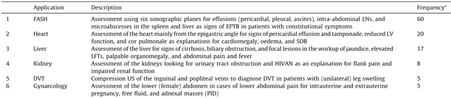
treatment change resulting from POCUS, followed by the initiation of ART, mostly in patients in whom no sign of TB was found. Other treatment changes, such as initiation of antibiotic therapy or anticoagulation, were common for specific indications such as abdominal pain and suspected DVT, respectively (Table 2). POCUS examinations were grouped into six applications; their description and frequency are shown inTable 3. FASH was the most frequently used POCUS application, followed by ‘heart’ and ‘liver’. ‘Kidney’, ‘DVT’, and ‘gynaecology’ were recorded in fewer than 10 patients.
4. Discussion and literature review
was observed, which included a high number of inpatients (n= 52) and patients who had been on ART for less than 6 months (n= 30), in whom opportunistic infections and unmasking immune reconstitution inflammatory syndrome (IRIS) are frequently seen. Furthermore, the study centre serves as a tertiary referral centre and may therefore receive a preselected population with more severe illness. Nevertheless, it is believed that the US applications used in the present cohort can be applied in various settings. The applications are discussed individually below, and conclusions are drawn on the relevance of different POCUS applications in a curriculum in Sub-Saharan African settings with a similar high prevalence of HIV and TB.
4.1. Focused assessment with sonography for HIV-associated TB (FASH)
People living with HIV are 20 to 30 times more likely to develop active TB, and one in three HIV deaths is due to TB.10TB mainly causes pulmonary disease, but can disseminate to extrapulmonary sites such as lymph nodes, pleurae, pericardium, and abdominal cavity, especially in HIV-infected patients.11,12The diagnosis of EPTB is hampered by difficulties in obtaining adequate samples and the limited availability and low sensitivity of diagnostic tests. FASH screens the patient for sonographic findings that suggest EPTB and can support the presumptive diagnosis of EPTB, especially in HIV-infected patients in endemic settings.13 Impor-tant findings include pleural effusion, pericardial effusion, free intra-abdominal fluid, enlarged abdominal lymph nodes, and microabscesses in the liver or spleen. Pleural effusions present as anechoic fluid, in the costophrenic angles, with or without fibrinous strands. In particular, unilateral effusions are highly
suggestive of TB.14 Pericardial effusions usually present as an anechoic rim around the heart (Figure 1), and in Sub-Saharan Africa, this is associated with TB in the majority of cases.3 Tuberculous peritonitis can present with ascites, with or without fibrin strands, and is most likely seen in Morrison’s (hepato-renal) pouch, the spleno-renal pouch, or pouch of Douglas.13Enlarged abdominal lymph nodes present as round, hypoechoic structures; lymph nodes larger than 1.5–2.0 cm are considered pathological
(Figure 2a). TB lymph nodes are commonly found in the
epigastrium, the para-aortic region, mesenterium, and the splenic hilum.13 Finally, microabscesses in the liver and particularly in the spleen present as small, round, hypoechoic lesions in the parenchyma (Figure 2b).13Data from various settings have shown that in a quarter of patients with FASH findings suggestive of EPTB, no signs of TB are seen on chest X-ray (CXR),15demonstrating the added value of FASH in prompting the initiation of anti-TB treatment. In South Africa, a country with a high prevalence of TB and HIV, FASH is among the most commonly used POCUS applications.9 Detecting fluid in pleura,16 pericardium,17 and abdomen5can be considered as technically ‘easy’. The detection of enlarged lymph nodes and splenic microabscesses is slightly more difficult, but can still be taught in short courses.8
4.2. Heart
Dyspnoea is a frequent reason for patients to seek medical attention and was one of the main indications for US in the present patient cohort. US constitutes an excellent tool to diagnose cardiac conditions causing dyspnoea and to differentiate between differ-ent conditions causing an image of cardiomegaly on CXR.3In the 15 patients with dyspnoea in the present study, a cardiac cause was found in seven patients. US of the heart generally uses four views: the subxyphoid view (which is also included in the FASH protocol), the left parasternal long axis and short axis views, and the apical four-chamber view.18It has been demonstrated that non-cardiologists can reliably estimate left ventricle (LV) function by ultrasound.19 Considering the different findings, the present authors would grade the difficulty of sonographic assessment of the heart as ‘moderate’.
4.2.1. Pericardial effusion
Tuberculous pericarditis is by far the commonest cause of pericardial effusion in Sub-Saharan Africa.20 Other important causes of large pericardial effusions, especially in HIV-infected patients, include pyogenic infection, lymphoma, and Kaposi’s sarcoma.21,22Pericardial effusions are usually assessed using the subxyphoid view. In addition, the parasternal long axis view can be helpful to differentiate between pleural and pericardial fluid, as pleural effusions do not cross the descending aorta while pericardial effusions do.18Tamponade is characterized by diastolic collapse of the right atrium or ventricle and requires urgent pericardial drainage, which can be performed in the resource-limited setting.23
4.2.2. Cor pulmonale
Right heart failure is an important cause of morbidity in Sub-Saharan Africa; in a South African study, it was found to be the cause of heart failure in 27% of 844 new cases.24A study among HIV-infected patients with cardiac symptoms in Tanzania ob-served pulmonary hypertension in 12.7% of 102 cases.25Infectious diseases like HIV, schistosomiasis, and TB, haemoglobinopathies, and air pollution are thought to play an important role in the pathophysiology of cor pulmonale in Sub-Saharan Africa.20,26The apical four-chamber view is best suited to assess the relative dimensions of the LV and right ventricle (RV). A RV/LV ratio>0.7 indicates a dilated RV. In addition, movement of the septum away Table 1
Baseline characteristics and indications for POCUS in Malawian patients
Total
n= 100
HIV-positive
n= 83
HIV-negative
n= 17
Male sex,n 42 35 7
Age, years, median (IQR) 34 (25–44) 33 (24–42) 38 (25–51) Location of patient,n(%)
Internal medicine ward 44 38 (86) 6 (14)
ART clinic 37 36 (97) 1 (3)
TB clinic 11 4 (36) 3 (17)
TB ward 8 5 (63) 3 (17)
On ART (% of HIV-positive) 74[3_TD$DIFF](89) ART started in last 6 months 30[4_TD$DIFF](36) ART started in last
6–12 months
4[5_TD$DIFF](5)
ART started in last 12–24 months
5[6_TD$DIFF](6)
On ART for over 24 months 35[7_TD$DIFF](42)
On TB treatment,n 8 8 0
Indications for scanning,n(%)
Weight loss 37 36 (97) 1 (3)
Abdominal paina
20 19 (95) 1 (5)
SOB 15 12 (80) 3 (20)
Cardiomegaly 11 7 (63) 4 (37)
Cough 11 10 (91) 1 (9)
Jaundice 10 9 (90) 1 (10)
Chest pain 9 6 (67) 3 (33)
Organomegaly palpable 8 7 (87) 1 (13) Bilateral leg oedema 6 4 (67) 2 (33)
Suspected ascites 5 3 (60) 2 (40)
Unilateral leg swelling 5 4 (80) 1 (20)
Abnormal CXR 4 3 (75) 1 (25)
Elevated LFT 4 4 (100) 0 (0)
Renal failure 2 1 (50) 1 (50)
Herpes zoster 1 1 (100) 0 (0)
POCUS, point-of-care applications of ultrasound; IQR, interquartile range; ART, antiretroviral therapy; TB, tuberculosis; SOB, shortness of breath; CXR, chest X-ray; LFT, liver function test. All percentages refer to the total number of patients per row.
a
from the RV during diastole indicates increased RV pressure, resulting in a ‘D’-shaped LV in the parasternal short axis.18 Treatment will be aimed at the underlying disease and at symptomatic relief of over-hydration by diuretics.
4.2.3. Dilated cardiomyopathy (DCM)
DCM is a common pathology of advanced HIV infection27and presents with shortness of breath, hypotension, and shock in advanced disease. In Sub-Saharan Africa, DCM is a common cause of heart failure, ranging from 9.8% of patients with cardiac symptoms in Tanzania,25to 28% of heart failure patients in South
Africa.28 Other important causes of cardiomyopathy in Sub-Saharan Africa include familial disease, alcoholism, and peripar-tum cardiomyopathy.20
DCM can be recognized on US using the subxyphoid view, or the apical four chamber view. Dilatation of all four chambers can be observed, and reduced inward endocardial systolic motion, which can be recognized by sonographers with limited experience.19To evaluate different causes of shock, measurement of the inferior vena cava (IVC) is incorporated as part of the RUSH protocol (rapid ultrasound in shock).29A patient in shock with a hyperdynamic LV with a small IVC (<1.5 cm) requires aggressive fluid resuscitation, Table 2
Indications for scanning, related abnormal findings, and treatment consequences
Reason for scan Total patients,n
Patients with abnormal findings,n(%)
Most common abnormal findings,n(%)
Patients with treatment change,n(%)
Most common treatment changes,n(%)
Total 100 77 (77) Enlarged abdominal LN, 17 (17)
Pericardial effusion, 15 (15) Spleen microabscesses, 15 (15) Pleural effusion, 14 (14)
72 (72) Start TB treatment, 23 (23) ART initiation/change, 10 (10) Diuretics/antihypertensives, 10 (10) Liver/LN/pleura biopsy, 5 (5) Weight loss 37 29 (78) Enlarged abdominal LN, 13 (35)
Spleen microabscesses, 12 (32) Pericardial effusion, 8 (22) Pleural effusion, 6 (16) Ascites, 4 (11)
28 (76) Start TB treatment, 18 (49) ART initiation/change, 8 (22)
Abdominal pain 20 18 (90) Enlarged abdominal LN, 7 (35) Spleen microabscesses, 3 (15) Ascites, 3 (15)
Adnexal mass/EP, 3 (15) Bowel wall thickening, 3 (15)
15 (75) Start antibiotic treatment 5 (25) Start TB treatment, 3 (15) Gynaecology/surgery referral, 3 (15)
SOB 15 11 (73) LV dysfunction, 4 (27)
Pleural effusion, 3 (20) Cor pulmonale, 3 (20) Pericardial effusion, 2 (13) Lung infiltrate, 2 (13)
11 (73) Diuretics/antihypertensives, 5 (33) Various othera
, 8 (53)
Cardiomegaly 11 9 (81) LV dysfunction, 4 (36) Pericardial effusion, 3 (27) Pleural effusion, 2 (18) Cor pulmonale, 1 (9)
7 (64) Diuretics/antihypertensives, 4 (36) Start TB treatment, 2 (18) Steroids, 1 (9)
NSAIDs, 1 (9)
Cough 11 10 (91) Pleural effusion, 4 (36)
Pericardial effusion, 4 (36) Spleen microabscesses, 3 (27) Lung infiltrate, 3 (27)
8 (72) Start TB treatment, 6 (54) Diuretics/antihypertensives, 2 (18) ART initiation/change, 2 (18)
Jaundice 10 8 (80) HCC/CCC, 3 (30)
Cirrhosis, 3 (30) Biliary dilatation, 3 (30)
7 (70) Liver/LN/pleura biopsy, 2 (20) ART initiation/change, 2 (20) Various otherb
, 3 (30)
Chest pain 9 7 (78) Pleural effusion, 4 (44)
Pericardial effusion, 4 (44) Spleen microabscesses, 1 (11)
8 (89) Start TB treatment, 5 (55) Liver/LN/pleura biopsy, 1 (11) Steroids, 1 (11)
NSAIDs, 1 (11) Organomegaly 8 8 (100) Hepato/splenomegaly, 3 (38)
HCC/CCC, 3 (38) Various otherc
, 5 (63)
6 (75) ART initiation/change, 3 (38) Liver/LN/pleura biopsy, 2 (25) Start TB treatment, 1 (13) Praziquantel, 1 (13) Bilateral oedema 6 5 (83) LV dysfunction, 2 (33)
Various otherd, 5 (83)
5 (83) Diuretics, 2 (33) Various othere, 3 (50) Suspected ascites 5 5 (100) Ascites, 2 (40)
Cirrhosis, 2 (40) Various otherf
, 2 (40)
4 (80) Variousg, 5 (100)
Unilateral leg swelling 5 3 (60) DVT, 3 (60)
Enlarged abdominal LN, 2 (40) Spleen microabscesses, 2 (40) Pericardial effusion, 1 (20)
4 (80) Anticoagulation, 3 (60) ART initiation/change, 3 (38) Liver/LN/pleura biopsy, 2 (25) Start TB treatment, 1 (13)
TB, tuberculosis; ART, antiretroviral treatment; LN, lymph nodes; EP, ectopic pregnancy; SOB, shortness of breath; LV, left ventricle; NSAIDs, non-steroidal anti-inflammatory drugs; HCC, hepatocellular carcinoma; CCC, cholangiocarcinoma; DVT, deep venous thrombosis. All percentages refer to the total number of patients per row. Some patients had more than one reason for scanning, more than one outcome, or more than one change in treatment. Indications that occurred in fewer than five patients are omitted from this table. These included elevated liver biochemistry (n= 4), an abnormal chest X-ray (n= 4), renal failure (n= 2), and herpes zoster (n= 1).
a
Including start of TB treatment, start of antibiotics, pleural biopsy, steroids, Kaposi sarcoma treatment, digoxin, and bronchodilators. b
Including stopping TB treatment, start of hyoscine butylbromide, and further diagnostics with hepatitis B serology and alpha-fetoprotein. c
Including enlarged abdominal lymph nodes, cirrhosis, ascites, pericardial effusion, biliary dilatation, and schistosomiasis. d Including pleural effusion, pericardial effusion, spleen microabscesses, cirrhosis, and cor pulmonale.
eIncluding start of TB treatment, further diagnostics with hepatitis B serology and alpha-fetoprotein, and digoxin. f
Including LV dysfunction and an adnexal mass. g
while a patient with a hypodynamic LV and a dilated IVC (>2.5 cm) requires diuretics and, if available, inotropes.18,29 Additionally angiotensin-converting enzyme (ACE) inhibitors and aldosterone antagonists may be available for the treatment of patients with cardiomyopathy.
4.3. Liver
The liver, the largest solid organ, is the organ most often scanned by US in internal medicine patients in resource-limited settings.30In the present patient cohort, abdominal pain, jaundice, and organomegaly were common indications for liver US. The complex liver anatomy, the wide variety of possible findings, and the broad differential diagnosis require a high level of expertise. Biliary dilatation, cirrhosis, and focal liver lesions are the three major findings of relevance to HIV-infected patients.
4.3.1. Focal liver lesions
Focal liver lesions can be due to infections as well as malignant and benign tumours. Focal TB abscesses are found in HIV co-infected individuals,15,31and are also well-described in patients without immunodeficiency. Tuberculomata may also be detected as part of disseminated TB disease when lymphadenopathy and spleen abscesses are present concomitantly.15 Amoebic liver abscesses occur in HIV-infected patients, but whether HIV infection is a risk factor for invasive amoebiasis remains controversial.32,33Typical US findings of liver abscesses are focal areas of different echogenicity within the liver parenchyma (which in most cases is hypoechoic) compared to the surrounding liver
tissue, as pus or necrotic material has generally lower echogeni-city.13In isolated cases, abscesses can also be iso- or hyperechoic. The detection of focal hypoechoic hepatic lesions is possible even for less experienced sonographers if the lesions are located in a sonographically accessible area of the liver, and it is therefore integrated in the FASH protocol.8Positive findings can be used in conjunction with the clinical picture to diagnose an abscess, which can be treated successfully. Nevertheless, it is difficult to rule out a focal process as it may easily be overlooked if small or located in less accessible areas like the subphrenic segments of the liver. Combined with the wide differential diagnosis of liver masses, the examination may often require input from an experienced sonographer.
4.3.2. Liver cirrhosis
Jaundice is a frequent sign in patients with HIV and TB for a variety of reasons. Common causes are liver cirrhosis, flares of hepatitis B,34and ‘drug-induced liver injury’ (DILI) due to TB drugs and ART. Liver cirrhosis is prevalent in HIV-infected patients, frequently due to co-infection with hepato-tropic viruses like hepatitis B virus35and hepatitis C virus.36In patients with HIV co-infection, the progression from fibrosis to cirrhosis due to hepatitis is accelerated compared to patients without HIV co-infection.37 Ruling in cirrhosis may influence treatment decisions, as it makes DILI less likely. Sonographic signs of cirrhosis can be subtle and in early stages undetectable; to rule out early liver cirrhosis by US is therefore not possible. Signs of advanced cirrhotic changes are (1) a nodular liver surface, best seen in the presence of ascites; (2) changes in the vasculature with amputated portal branches and a prominent central portal vein; (3) a coarse inhomogeneous echo pattern of the liver tissue; and (4) possibly an enlarged caudate lobe. Changes in the liver surface and vasculature may be discernable by a less experienced sonographer. Nevertheless, the sonographic diagnosis of cirrhosis must be considered as ‘difficult’.
4.3.3. Biliary dilatation
Dilatation of the intra- and extra-hepatic bile ducts is another explanatory finding in patients with jaundice and elevated liver enzymes. In the industrialized setting, these changes are mainly due to gallstones or tumours obstructing the bile flow,38but the differential diagnosis in HIV/TB patients is different. Obstruction by enlarged tuberculous lymph nodes may cause dilatation of the bile ducts and pancreatic duct.39HIV-associated cholangiopathy has to be considered in cases of segmental or general dilatation of the duct system.40The sonographic features of dilated bile ducts are anechoic tubular structures running parallel to the portal venous system. This is often described as ‘double-barrel’ or ‘shot-gun’ sign. Although the US picture is clear, well described, and Table 3
POCUS applications used during the examination of 100 patients in a Malawian high prevalence HIV and TB setting
Application Description Frequencya[8_TD$DIFF]
1 FASH Assessment using six sonographic planes for effusions (pericardial, pleural, ascites), intra-abdominal LNs, and microabscesses in the spleen and liver as signs of EPTB in patients with constitutional symptoms
60
2 Heart Assessment of the heart mainly from the epigastric angle for signs of pericardial effusion and tamponade, reduced LV function, and cor pulmonale as explanations for cardiomegaly, oedema, and SOB
20
3 Liver Assessment of the liver for signs of cirrhosis, biliary obstruction, and focal lesions in the workup of jaundice, elevated LFTs, palpable organomegaly, and abdominal pain and fever
17
4 Kidney Assessment of the kidneys looking for urinary tract obstruction and HIVAN as an explanation for flank pain and impaired renal function
8
5 DVT Compression US of the inguinal and popliteal veins to diagnose DVT in patients with (unilateral) leg swelling 5 6 Gynaecology Assessment of the lower (female) abdomen in cases of lower abdominal pain for intrauterine and extrauterine
pregnancy, free fluid, and adnexal masses (PID)
5
POCUS, point-of-care applications of ultrasound; TB, tuberculosis; FASH, focused assessment with sonography for HIV-associated TB; LN, lymph node; EPTB, extrapulmonary tuberculosis; LV, left ventricle; SOB, shortness of breath; LFT, liver function test; HIVAN, HIV-associated nephropathy; US, ultrasound; DVT, deep vein thrombosis; PID, pelvic inflammatory disease.
a
Sum is>100, as more than one application could be chosen per patient.
[(Figure_1)TD$FIG]
Figure 1.Pericardial effusion in a patient with TB pericarditis: an anechoic, black
unambiguous, it is often difficult to recognize for a beginner. While obstructing tuberculous nodes normally respond to treatment, treatment options for cholangiopathy are limited in the absence of advanced endoscopic services.
4.4. Kidney
Renal problems are frequent in tropical settings41and US is the imaging modality of choice to assess the kidneys. Common indications for kidney POCUS are flank pain and reduced renal function.42
Flank pain may be caused by urinary tract obstruction. Enlarged tuberculous lymph nodes are a well-known cause of compression; enlarged lymph nodes may also be due to cervical cancer or lymphoma.43In renal TB, dilatation of calyces and the renal pelvis, sometimes containing debris, can be found, as well as calcification and thickening of the ureter.44In patients receiving the protease inhibitor atazanavir, which is widely used today in second-line ART regimens in Africa, precipitation of crystals in kidney tubules with consecutive formation of stones obstructing the urinary tract are well described.45POCUS to detect urinary tract obstruction is well described;46its use is widely accepted and considered relatively easy. Therapeutic options in the resource-limited setting are usually limited to analgesia and increased fluid intake.
Renal failure is also a well-known complication in HIV patients, as a result of HIV-associated nephropathy (HIVAN) and nephro-toxic medication, such as tenofovir, which is used widely in first-line ART regimens.47,48Sonographically, HIVAN is often character-ized by slightly enlarged kidneys with an echogenic cortex and decreased cortico-medullary definition.49,50 In a setting where biopsy is not available, proteinuria and these sonographic findings may be sufficient to make a presumptive diagnosis.43In untreated HIV-patients, ART should be initiated. Additionally, the concomi-tant prescription of an ACE inhibitor is a treatment option. The sonographic assessment of causes of renal impairment is less straightforward for the beginner and is considered ‘moderate’.
4.5. Deep venous thrombosis
There is a paucity of literature on thromboembolic disease in Sub-Saharan Africa. The few studies available suggest it is an important problem, while prophylaxis is under-used.51,52Patients with HIV and/or TB are likely to have an increased risk for thromboembolic disease, as both HIV and TB activate the coagulation system, damage endothelium, and impair anticoagu-lant mechanisms.53–55A study in Kenya demonstrated that HIV
and pulmonary TB were frequent comorbidities in patients with pulmonary embolism (10.9% and 12.5%, respectively).56Therefore, the application of US for the diagnosis of DVT is relevant in Sub-Saharan Africa.
A simplified POCUS protocol to screen for DVT has been developed by emergency physicians and includes assessment of the common femoral vein and popliteal vein.57 Thrombosis is identified when the vein is not compressible while applying enough pressure to deform the adjacent arterial wall. This can be assessed reliably by emergency physicians57,58 and can be considered ‘easy’. In the present cohort, DVT was observed in three out of five patients with unilateral leg swelling, which had immediate consequences for treatment. The treatment of DVT by anticoagulation can prevent pulmonary embolism and leads to resolution of the thrombus. The implementation of anticoagulation monitoring may be challenging in resource-limited settings, particularly as drug interactions with TB drugs and ART are frequent. Nevertheless, good performance of a pharmacist-managed anticoagulation clinic was demonstrated in rural Kenya.59Furthermore, the implementation of novel anticoagulant therapies, which do not warrant monitoring, could facilitate the management of thromboembolism in Sub-Saharan Africa.
4.6. Gynaecology
Pelvic inflammatory disease (PID) and ectopic pregnancy are common gynaecological problems in HIV-infected women. In Sub-Saharan Africa, 17–40% of gynaecology admissions are due to PID.60The symptoms may vary from asymptomatic to abnormal vaginal discharge, lower abdominal pain, cervical motion tender-ness, and fulminant infection with fever and peritonitis. Ectopic pregnancy is the leading cause of maternal death in the first trimester,61,62with a very high case-fatality rate in the developing world.63
Clinical and laboratory findings often suffice to diagnose PID and initiate treatment, but trans-abdominal US is a useful diagnostic tool in cases with unexplained symptoms, chronic PID, and complicated cases, such as tubo-ovarian abscess. Trans-vaginal US has a higher sensitivity and provides a more detailed view of the uterus and adnexa, but is not readily available outside gynaecology departments, is technically more difficult, and is often not required to establish the diagnosis. In women with a suspicion of PID, one should look for enlargement of the uterus and/or adnexa, free fluid in the pouch of Douglas, fluid in the cavum uteri, and increased echogenicity of the pelvic fat. Salpingitis can present with hyperechoic structures surrounded by a hypoechoic rim of
[(Figure_2)TD$FIG]
Figure 2.(a) Enlarged para-aortal abdominal lymph nodes due to abdominal TB are seen as hypoechoic masses (arrows). (b) Multiple hypoechoic microabscesses of the spleen
in a patient with disseminated TB (arrowheads).
T. Heller et al. / International Journal of Infectious Diseases 56 (2017) 229–236
fluid, and a tubo-ovarian abscess can present as an ill-defined, multi-loculated, cystic structure, possibly with a wall.
Urinary beta human chorionic gonadotropin (
b
-HCG) plus a trans-abdominal US are simple and inexpensive diagnostic tests for ectopic pregnancy, therefore it should be used in all women of childbearing age with lower abdominal pain. The diagnostic approach is to search for an intrauterine pregnancy, as a positive urinaryb
-HCG without a visible intrauterine pregnancy is virtually diagnostic for ectopic pregnancy. Only after this should the sonographer look for the ectopic pregnancy itself. Ninety-five percent of ectopic pregnancies develop in the Fallopian tubes;64 however, they also occur in the ovaries, cervix, and peritoneal cavity and may not always be found.65Free abdominal fluid should be looked for, which can be present in ruptured and non-ruptured ectopic pregnancies, with the larger quantities usually seen after rupture.While the difficulty of sonographic diagnosis of ectopic pregnancy (or the absence of intrauterine pregnancy) may be considered as ‘moderate’, the visualization of adnexa and abscesses usually requires more advanced US skills. Treatment implications are antibiotic treatment for PID and referral to surgery for ectopic pregnancy.
4.7. Training implications
Defining POCUS curricula is important for a targeted allocation of training resources. Based on the experience with US in the present patient cohort in Malawi and the review of the literature, a ranking of the most relevant POCUS applications is proposed. To assess relevance, a method proposed by van Hoving et al. was followed.9Weights are assigned to three components (prevalence, impact, and difficulty) for each of the POCUS applications (Table 4). Next, a ‘relative weight’ of each application is calculated to rank them (Table 5). According to this ranking, FASH is the module with the most favourable balance between prevalence (as it is very frequent in HIV/TB endemic settings), impact (as it has significant effects on management), and difficulty (the detection of effusions can be considered ‘easy’, although the detection of enlarged lymph nodes and spleen abscesses was considered ‘moderate’). Although this multifactorial weighting model has not yet been validated, it is easy to use and to apply. Therefore, it has the potential to inform decisions about a relevant POCUS curriculum in various settings.
5. Conclusions
These data from Malawian patients with a high prevalence of HIV and TB demonstrate that POCUS is a very useful tool in the resource-limited setting, in particular to aid the diagnosis of EPTB, but also other conditions such as heart failure, adnexal masses and ectopic pregnancy, hepatocellular carcinoma, and DVT. POCUS informed a change in treatment in a large proportion of the patients. A further review of the literature illustrated the relevance and applicability of various POCUS applications including FASH, heart, DVT, liver, gynaecology, and kidney. Based on prevalence, impact, and difficulty, it is recommended that POCUS curricula in HIV/TB endemic settings focus on FASH, heart, and DVT, while kidney, liver, and gynaecology applications can be reserved for physicians more experienced with US and those with a specific interest in these fields. This review focused on the literature in Sub-Saharan Africa, but, depending on the epidemiology of HIV, TB, and other illnesses, POCUS has the potential to inform decisions regarding treatment management in other resource-limited settings, such as Latin America and Southeast Asia.
Funding:The authors did not receive any financial support for
this review.
Conflict of interest:The authors have no conflicts of interest to
disclose.
References
1.Richter J, Hatz C, Haussinger D. Ultrasound in tropical and parasitic diseases.
Lancet2003;362:900–2.
2.Brunetti E. Ultrasound in tropical medicine. In: Farrar J, Hotez P, Junghanss T, Kang G, Lalloo D, White NJ, editors.Manson’s tropical diseases. 23rd
ed, Elsevier; 2013. p. 60–7.
3.Brindle HE, Allain TJ, Kampondeni S, Kayange N, Faragher B, Bates I, et al. Utilization of ultrasound in medical inpatients in Malawi.Trans R Soc Trop Med Hyg2013;107:405–10.
4.Moore CL, Copel JA. Point-of-care ultrasonography.N Engl J Med2011;364: 749–57.
5.Ma OJ, Mateer JR, Ogata M, Kefer MP, Wittmann D, Aprahamian C. Prospective analysis of a rapid trauma ultrasound examination performed by emergency physicians.J Trauma1995;38:879–85.
6.Belard S, Tamarozzi F, Bustinduy AL, Wallrauch C, Grobusch MP, Kuhn W, et al. Point-of-care ultrasound assessment of tropical infectious diseases—a review of applications and perspectives.Am J Trop Med Hyg2015;94:8–21.
7.International Federation for Emergency Medicine. Point-of-care ultrasound curriculum guidelines 2014. IFEM; 2014.
8.Heller T, Wallrauch C, Lessells RJ, Goblirsch S, Brunetti E. Short course for focused assessment with sonography for human immunodeficiency virus/ tuberculosis: preliminary results in a rural setting in South Africa with high prevalence of human immunodeficiency virus and tuberculosis.Am J Trop Med Hyg2010;82:512–5.
9.van Hoving DJ, Lamprecht HH, Stander M, Vallabh K, Fredericks D, Louw P, et al. Adequacy of the emergency point-of-care ultrasound core curriculum for the local burden of disease in South Africa.Emerg Med J2012;30:312–5. 10. World Health Organization. Tuberculosis fact sheet No. 104. Geneva: WHO;
2016.
11.Gupta RK, Lucas SB, Fielding KL, Lawn SD. Prevalence of tuberculosis in post-mortem studies of HIV-infected adults and children in resource-limited set-tings: a systematic review and meta-analysis.AIDS2015;29:1987–2002. 12.Bates M, Mudenda V, Shibemba A, Kaluwaji J, Tembo J, Kabwe M, et al. Burden of
tuberculosis at post mortem in inpatients at a tertiary referral centre in Sub-Saharan Africa: a prospective descriptive autopsy study. Lancet Infect Dis
2015;15:544–51.
13.Heller T, Wallrauch C, Goblirsch S, Brunetti E. Focused assessment with sonog-raphy for HIV-associated tuberculosis (FASH): a short protocol and a pictorial review.Crit Ultrasound J2012;4:21.
14.Luzze H, Elliott AM, Joloba ML, Odida M, Oweka-Onyee J, Nakiyingi J, et al. Evaluation of suspected tuberculous pleurisy: clinical and diagnostic findings in HIV-1-positive and HIV-negative adults in Uganda. Int J Tuberc Lung Dis
2001;5:746–53.
15.Heller T, Goblirsch S, Bahlas S, Ahmed M, Giordani MT, Wallrauch C, et al. Diagnostic value of FASH ultrasound and chest X-ray in HIV-co-infected patients with abdominal tuberculosis.Int J Tuberc Lung Dis2013;17:342–4. 16.Ma OJ, Mateer JR. Trauma ultrasound examination versus chest radiography in
the detection of hemothorax.Ann Emerg Med1997;29:312–5. discussion 315-6. 17.Rozycki GS, Feliciano DV, Ochsner MG, Knudson MM, Hoyt DB, Davis F, et al. The role of ultrasound in patients with possible penetrating cardiac wounds: a prospective multicenter study.J Trauma1999;46:543–51. discussion 551-2. Table 4
Weighting of prevalence, diagnostic impact, and difficulty of POCUS applications
Weight Disease prevalence Diagnostic impact of US US difficulty
1 Rare Minor or no treatment change Advanced 2 Relatively common Treatment change Moderate 3 Very common Urgent treatment change
(possibly life-threatening)
Easy
POCUS, point-of-care applications of ultrasound; US, ultrasound.
Table 5
Weighting model applied to POCUS applications in high-prevalence HIV and TB settings
Module Prevalence (P)
Impact (I)
Difficulty (D)
PID Relative weight
Rank
FASH 3 3 2 18 0.40 1
Heart 2 3 2 12 0.27 2
DVT 1 2 3 6 0.13 3
Liver 2 2 1 4 0.09 4
Gynaecology 1 3 1 3 0.07 5
Kidney 1 1 2 2 0.04 6
Total 45 1.00
18.Noble VE. Manual of emergency and critical care ultrasound, 2nd ed, Cam-bridge: Cambridge University Press; 2011.
19.Randazzo MR, Snoey ER, Levitt MA, Binder K. Accuracy of emergency physician assessment of left ventricular ejection fraction and central venous pressure using echocardiography.Acad Emerg Med2003;10:973–7.
20.Bloomfield GS, Barasa FA, Doll JA, Velazquez EJ. Heart failure in Sub-Saharan Africa.Curr Cardiol Rev2013;9:157–73.
21.Chen Y, Brennessel D, Walters J, Johnson M, Rosner F, Raza M. Human immu-nodeficiency virus-associated pericardial effusion: report of 40 cases and review of the literature.Am Heart J1999;137:516–21.
22.Reynolds SP, Gibbs AR, Weeks R, Adams H, Davies BH. Massive pleural effusion: an unusual presentation of Castleman’s disease. Eur Respir J
1992;5:1150–3.
23.Heller T, Lessells RJ, Wallrauch C, Brunetti E. Tuberculosis pericarditis with cardiac tamponade: management in the resource-limited setting.Am J Trop Med Hyg2010;83:1311–4.
24.Stewart S, Wilkinson D, Hansen C, Vaghela V, Mvungi R, McMurray J, et al. Predominance of heart failure in the Heart of Soweto Study cohort: emerg-ing challenges for urban African communities. Circulation 2008;118: 2360–7.
25.Chillo P, Bakari M, Lwakatare J. Echocardiographic diagnoses in HIV-infected patients presenting with cardiac symptoms at Muhimbili National Hospital in Dar es Salaam, Tanzania.Cardiovasc J Afr2012;23:90–7.
26.Butrous G, Ghofrani HA, Grimminger F. Pulmonary vascular disease in the developing world.Circulation2008;118:1758–66.
27.Khunnawat C, Mukerji S, Havlichek Jr D, Touma R, Abela GS. Cardiovascular manifestations in human immunodeficiency virus-infected patients. Am J Cardiol2008;102:635–42.
28.Twagirumukiza M, Nkeramihigo E, Seminega B, Gasakure E, Boccara F, Barbaro G. Prevalence of dilated cardiomyopathy in HIV-infected African patients not receiving HAART: a multicenter, observational, prospective, cohort study in Rwanda.Curr HIV Res2007;5:129–37.
29.Perera P, Mailhot T, Riley D, Mandavia D. The RUSH exam: Rapid Ultrasound in SHock in the evaluation of the critically ill. Emerg Med Clin North Am
2010;28:29–56. vii.
30.Groen RS, Leow JJ, Sadasivam V, Kushner AL. Review: indications for ultra-sound use in low- and middle-income countries. Trop Med Int Health
2011;16:1525–35.
31.Carrara E, Brunetti E, Di Matteo A, Ravetta V, Minoli L, Youkee D. Tubercular liver abscess: an uncommon presentation of disseminated tuberculosis. Infec-tion2015;43:237–40.
32.Jessurun J, Barron-Rodriguez LP, Fernandez-Tinoco G, Hernandez-Avila M. The prevalence of invasive amebiasis is not increased in patients with AIDS.AIDS
1992;6:307–9.
33.Hung CC, Chen PJ, Hsieh SM, Wong JM, Fang CT, Chang SC, et al. Invasive amoebiasis: an emerging parasitic disease in patients infected with HIV in an area endemic for amoebic infection.AIDS1999;13:2421–8.
34.Puoti M, Torti C, Bruno R, Filice G, Carosi G. Natural history of chronic hepatitis B in co-infected patients.J Hepatol2006;44(1 Suppl):S65–70.
35.Hoffmann CJ, Thio CL. Clinical implications of HIV and hepatitis B co-infection in Asia and Africa.Lancet Infect Dis2007;7:402–9.
36.Castellares C, Barreiro P, Martin-Carbonero L, Labarga P, Vispo ME, Casado R, et al. Liver cirrhosis in HIV-infected patients: prevalence, aetiology and clinical outcome.J Viral Hepat2008;15:165–72.
37.Soto B, Sanchez-Quijano A, Rodrigo L, del Olmo JA, Garcia-Bengoechea M, Hernandez-Quero J, et al. Human immunodeficiency virus infection modifies the natural history of chronic parenterally-acquired hepatitis C with an unusu-ally rapid progression to cirrhosis.J Hepatol1997;26:1–5.
38.Pasanen PA, Partanen KP, Pikkarainen PH, Alhava EM, Janatuinen EK, Pirinen AE. A comparison of ultrasound, computed tomography and endoscopic retrograde cholangiopancreatography in the differential diagnosis of benign and malig-nant jaundice and cholestasis.Eur J Surg1993;159:23–9.
39.Sharma MP, Bhatia V. Abdominal tuberculosis.Indian J Med Res2004;120: 305–15.
40.Miller FH, Gore RM, Nemcek Jr AA, Fitzgerald SW. Pancreaticobiliary manifes-tations of AIDS.AJR Am J Roentgenol1996;166:1269–74.
41.Naicker S, Aboud O, Gharbi MB. Epidemiology of acute kidney injury in Africa.
Semin Nephrol2008;28:348–53.
42.Noble VE, Brown DF. Renal ultrasound. Emerg Med Clin North Am
2004;22:641–59.
43.Me´decins Sans Frontie`res. Renal disease in HIV—a practical manual for primary care physicians. MSF; 2012.
44.Matos MJ, Bacelar MT, Pinto P, Ramos I. Genitourinary tuberculosis.Eur J Radiol
2005;55:181–7.
45.Rockwood N, Mandalia S, Bower M, Gazzard B, Nelson M. Ritonavir-boosted atazanavir exposure is associated with an increased rate of renal stones compared with efavirenz, ritonavir-boosted lopinavir and ritonavir-boosted darunavir.AIDS2011;25:1671–3.
46.Erwin BC, Carroll BA, Sommer FG. Renal colic: the role of ultrasound in initial evaluation.Radiology1984;152:147–50.
47.Peyriere H, Reynes J, Rouanet I, Daniel N, de Boever CM, Mauboussin JM, et al. Renal tubular dysfunction associated with tenofovir therapy: report of 7 cases.
J Acquir Immune Defic Syndr2004;35:269–73.
48.Winston JA, Bruggeman LA, Ross MD, Jacobson J, Ross L, D’Agati VD, et al. Nephropathy and establishment of a renal reservoir of HIV type 1 during primary infection.N Engl J Med2001;344:1979–84.
49.Atta MG, Longenecker JC, Fine DM, Nagajothi N, Grover DS, Wu J, et al. Sonography as a predictor of human immunodeficiency virus-associated ne-phropathy.J Ultrasound Med2004;23:603–10. quiz 612-3.
50.Di Fiori JL, Rodrigue D, Kaptein EM, Ralls PW. Diagnostic sonography of HIV-associated nephropathy: new observations and clinical correlation.AJR Am J Roentgenol1998;171:713–6.
51.Ogeng’o JA, Gatonga P, Olabu BO. Cardiovascular causes of death in an East African country: an autopsy study.Cardiol J2011;18:67–72.
52.Ba SA, Badiane SB, Diop SN, Diouf FS, Fall D, Ka MM, et al. A cross-sectional evaluation of venous thromboembolism risk and use of venous thromboem-bolism prophylaxis in hospitalized patients in Senegal.Arch Cardiovasc Dis
2011;104:493–501.
53.Kager LM, Blok DC, Lede IO, Rahman W, Afroz R, Bresser P, et al. Pulmonary tuberculosis induces a systemic hypercoagulable state. J Infect2015;70: 324–34.
54.Jong E, Louw S, van Gorp EC, Meijers JC, ten Cate H, Jacobson BF. The effect of initiating combined antiretroviral therapy on endothelial cell activation and coagulation markers in South African HIV-infected individuals.Thromb Hae-most2010;104:1228–34.
55.Huson MA, Kalkman R, Hoogendijk AJ, Alabi AS, van’t Veer C, Grobusch MP, et al. Impact of HIV infection on the haemostatic response during sepsis and malaria.
Br J Haematol2016;173:918–26.
56.Ogeng’o JA, Obimbo MM, Olabu BO, Gatonga PM, Ong’era D. Pulmonary thromboembolism in an East African tertiary referral hospital.J Thromb Throm-bolysis2011;32:386–91.
57.Blaivas M, Lambert MJ, Harwood RA, Wood JP, Konicki J. Lower-extremity Doppler for deep venous thrombosis—can emergency physicians be accurate and fast?Acad Emerg Med2000;7:120–6.
58.Birdwell BG, Raskob GE, Whitsett TL, Durica SS, Comp PC, George JN, et al. The clinical validity of normal compression ultrasonography in outpatients sus-pected of having deep venous thrombosis.Ann Intern Med1998;128:1–7. 59.Manji I, Pastakia SD, Do AN, Ouma MN, Schellhase E, Karwa R, et al. Performance
outcomes of a pharmacist-managed anticoagulation clinic in the rural, resource-constrained setting of Eldoret, Kenya.J Thromb Haemost2011;9:2215–20. 60.Kano J, Adler M. Epidemiology of pelvic inflammatory disease. Raven Press;
1992.
61.Tay JI, Moore J, Walker JJ. Ectopic pregnancy.West J Med2000;173:131–4. 62.Tenore JL. Ectopic pregnancy.Am Fam Physician2000;61:1080–8.
63.Goyaux N, Leke R, Keita N, Thonneau P. Ectopic pregnancy in African developing countries.Acta Obstet Gynecol Scand2003;82:305–12.
64.Kirk E, Bourne T. Diagnosis of ectopic pregnancy with ultrasound.Best Pract Res Clin Obstet Gynaecol2009;23:501–8.