INFLUENCE OF DENTIN DESENSITIZERS ON THE MICROTENSILE BOND STRENGTHS OF SELF-ETCH AND ETCH-AND-RINSE
ADHESIVES TO DENTIN
Roopwant Kaur
A thesis submitted to the faculty of the University of North Carolina at Chapel Hill in partial fulfillment of the requirements for the degree of Master of Science in the
Department of Operative Dentistry.
Chapel Hill 2012
Approved by
Edward J. Swift, Jr., DMD, MS
Harald O. Heymann, DDS, MEd
Ceib Phillips, MPH, PhD
André V. Ritter, DDS, MS
ABSTRACT
ROOPWANT KAUR:Influence of Dentin Desensitizers on the Microtensile Bond
Strengths of Self-Etch and Etch-and-Rinse Adhesives to Dentin (Under the direction of Dr. Edward J. Swift, Jr.)
This study evaluated several dentin desensitizers’ effects on dentin
microtensile bond strengths (MTBS) of various self-etch (SE) and etch-and-rinse
(ER) adhesives. For ER, dentin from human molars was phosphoric acid-etched,
treated with a desensitizer, and coated with the adhesive. For SE, dentin was
treated with the same desensitizers, and coated with the adhesive. Composite
build-ups were placed and specimens stored in distilled water at 37°C for 24h. Specimens
were sectioned into beams and tested either immediately or after 6 months. Data
were analyzed using factorial analysis of variance. At 24h, mean MTBS’s ranged
from 20.0-46.6 MPa for ER and from 22.0-37.0 MPa for SE. At 6 months, mean
MTBS’s ranged from 22.0-45.4 MPa for ER and from 20.2-33.4 MPa for SE. The
main factors (adhesive, desensitizer) and interaction effects were not statistically
significant (p>0.25). Use of desensitizers did not affect dentin bond strengths either
Dedication
To my ‘Heroes’ my parents Smt. Amarbir Kaur and S. Devinder Singh
And
To my son -Viraaj Singh
Acknowledgement
Dr. Edward J. Swift, my mentor, Thank you for guiding me and for being a role
model. For opening my horizon to research and for accepting me as an advisee. For being patient and for always being there.
Dr. Ricardo Walter, Thank you for helping me understand the concepts of research
and for guiding me at every step in my research project and thesis.
Dr. Harald O. Heymann, Thank you for accepting me to the program and for giving
me a chance to study operative dentistry at UNC Chapel Hill.
Dr. Andre Ritter, Thank you for the opportunity to learn and explore at UNC Chapel
Hill. Your guidance and support helped me immensely.
Dr. Ceib Phillips, Thank you for being on my committee and for guiding me
regarding my project and statistics.
Dr. Terry Donovan, Thank you for the literature reviews and for educating me. I
learnt a lot about life from you.
Operative Dentistry Faculty for being tremendous role models for me.
Ms. Marie Roberts, Ms. Shannon Tate, Ms. Jamie Desoto, Ms. Ginger Couch, Ms. Barbara Walton, Ms. Dayna McNaught and Ms. Cynthia Lambert for always
making me feel part of the operative family. The last three years have been a joy because of your warmth and support.
Operative Dentistry Residents for being supportive colleagues.
My mother and father Smt. Amarbir Kaur and S. Devinder Singh for making me who I am today. You are my true inspiration. Without your love and support I wouldn’t have made it.
My son Viraaj for being the joy of my life. You make me proud each and every moment. You are a true blessing and life is beautiful because of you .I love you.
TABLE OF CONTENTS
LIST OF TABLES...vii
LIST OF FIGURES...viii
LIST OF GRAPHS……… ix
Chapter 1. INTRODUCTION...1
2. LITERATURE REVIEW...3
2.1 Adhesives 2.1.1 Dentin bonding………...3
2.1.2 History of adhesives………6
2.2. Sensitivity with current adhesives……….………8
2.3. Desensitizers……….………...11
2.4. Adhesives and desensitizers………14
3. SPECIFIC AIMS and NULL HYPOTHESES……….15
4. MATERIALS AND METHODS...16
5. RESULTS….…………...19
6. DISCUSSION…...21
7. CONCLUSIONS...29
LIST OF TABLES
TABLE 1. Experimental groups ……….30
TABLE 2. Desensitizers systems evaluated……….31
TABLE 3. Adhesive systems evaluated……….32
TABLE 4. Microtensile bond strengths for combinations of
Adhesive and Desensitizer at 24 h and 6 months in MPa………...33
TABLE 5. Microtensile bond strengths by type of adhesive in MPa………...34
TABLE 6.Microtensile bond strengths by type of desensitizer in MPa………35
TABLE 7. Results from linear mixed effects model with and
without interactions of explanatory variables and time…………...36
LIST OF FIGURES
FIGURE 1. Experimental design………...………..38
FIGURE 2. Isomet 1000 Precision saw ……….39
FIGURE 3. Ecomet 3 grinder/polisher machine ………..40
FIGURE 4. Placement of beam on the jig using adhesive………..41
LIST OF GRAPHS
GRAPH 1.Means and standard deviations (pooled data) for
adhesives at 24 h and 6months...43
GRAPH 2. Means and standard deviations (pooled data) for
1. INTRODUCTION
Post-operative sensitivity, a well-localized and momentary sensitivity often
associated with occlusal loading (e.g., chewing), is an ongoing problem with
composite resins. A prevalence of 8-19% has been reported in patients treated with
posterior composite resins with the sensitivity lasting several months in some cases.
[1,2] Possible causes of post-operative sensitivity are: (a) poor dentin sealing, (b)
toxicity of the resin, (c) marginal microleakage induced by polymerization contraction
of the resin, and (d) hydraulic stress to odontoblastic processes generated either by
the flexure of cusps due to polymerization contraction of the resin or by occlusal
loading. [3]
The most accepted mechanism for dentin sensitivity is the hydrodynamic
theory described by Brännström, which suggests that rapid movement of fluids within
the dentinal tubules produces a deformation of nerve fibers wrapped around the
odontoblast cells. [4] Thus, materials that occlude dentin tubules to any extent can
potentially reduce fluid filtration across the dentin and decrease sensitivity. [5, 6]
The use of dentin desensitizers prior to adhesive application has proved
effective resulting in adequate bond strengths with etch-and-rinse adhesive
systems.Several studies have tested the bond strengths of composite resins to dentin
after treatment with dentin desensitizers and shown no decrease in bond strengths.
the microtensile bond strength (MTBS) of four etch-and-rinse and two one-step
2. LITERATURE REVIEW
Dentin bonding has been the focus of extensive research over the past
decades. The reason being is the difficulty in achieving stable long-term bonding to
that substrate. The composition of dentin – 50% inorganic material, 30% organic
material, and 20% water [13] – complicates the bonding procedure that otherwise is
quite simple and predictable in enamel. Bonding to dentin is facilitated by dentin
hybridization that can be defined as the penetration of resin monomers into the
dentin matrix. [14] Mechanisms of adhesion are (1) mechanical adhesion where
interlocking of adhesive with irregularities in the surface of the substrate takes place;
(2) adsorption adhesion in which chemical bonding between the adhesive and the
substrate takes place; and (3) diffusion adhesion, which is the interlocking between
mobile molecules. [15] Some factors that may affect adhesion are effective
demineralization of dentin, effective smear layer removal, good wetting of the
substrate, dispersion and penetration of the adhesive. [16]
In order to form adequate bonding between dentin and restorative material,
dentin bonding agents must condition (etch), prime, and, bond the tooth structure.
[17-19] Etch-and-rinse systems utilize phosphoric acid-etching, which removes the
smear layer, opens dentinal tubules, and decalcifies dentin, as the first step.
Hydroxyapatite crystals are dissolved, leaving behind a collagen network that must
the etchant will collapse the collagen fibers preventing infiltration of the
primer/bonding resin into the collagen mesh. After the etchant is rinsed off, the
second step, which is application of primer, follows. The primer contains solvents
such as acetone, ethanol and/or water and one or more bifunctional resin monomers
such as HEMA. HEMA has two functional groups – a hydrophilic group that will
interact with dentin and a hydrophobic group that will interact with the subsequently
placed bonding resin. Aside from making the connection between dentin and bonding
resin, the primer wets the dentin and helps increasing its surface energy. Once
priming is done, the bonding resin is applied. That will make the bridge between
primed dentin and resin-based restorative material. Bonding resins contain
monomers such as Bis-GMA, HEMA, UDMA, PENTA, and 4-META. In addition, filler
particles create thicker resin layers that might be present and can potentially aid in
providing stress relief at the tooth-restoration interface. [20] The polymerized
primer/bonding resin interlocked with the collagen fibers plays a pivotal role in the
bond between composite resin and dentin. This layer is known as the ‘hybrid layer,’
‘resin reinforced zone,’ or ‘resin-infiltrated layer’ and was first described by
Nakabayashi and colleagues in 1982. [21]
Another bonding approach is the self-etch approach. Self-etch adhesive
systems have gained popularity in clinical dentistry as they reduce the clinical
application time and may be less technique-sensitive than etch-and-rinse adhesives.
[22] In two-step self-etch adhesives, the elimination of the separate acid-etching step
leads to incorporation of acid monomers and primer in a single solution. The bonding
and the underlying and partially infiltrated dentin. All components – etch, primer, and
bonding resin – also are available in one single solution (all-in-one adhesives),
although specific components may be packaged in two bottles or two reservoirs. [23,
24] Self-etch adhesives also can be subdivided according to their pH values into
2.1.2 History of adhesives
Since enamel was first phosphoric acid etched by Buonocore in the 1950’s,
bonding to the tooth structures has been focus of much research. In 1975, the first
generation of adhesives was introduced but poor clinical results were found when
used to restore cervical lesions without mechanical retention. [25] Developments led
to a second generation of adhesives that was launched in the early 1980’s and
consisted of halophosphorous esters of monomers such as Bis-GMA or HEMA. The
mechanism of action was interaction of phosphate groups in the adhesive and
calcium ions in the smear layer along with wetting which led to bond formation. [26]
However, a major shortcoming was that these adhesives bonded to smear layer,
which bonds only weakly to the underlying dentin. [27] Combined with polymerization
shrinkage, bond strengths of only 1-10 MPa [26] were likely to result in clinical failure.
[28] Better clinical performance was noted with third-generation adhesives in the mid
1980’s. [28, 29] These systems removed or modified the smear layer to cause
effective penetration of resin into dentin.
Total-etch systems, currently known as etch-and-rinse, were introduced in the
United States in the late 1980’s. Fusayama’s original concept [30] led to a technique
proposed by Kanca and Bertolotti. With this technique, phosphoric acid-etching of
dentin and enamel is done. [31, 32] At the time, potential pulpal damage was
feared.[33]. Etching of dentin was considered taboo, but new research led to new
remains in use today with products such as All-Bond 2 (Bisco, Inc), OptiBond FL
(Kerr) and Scotchbond Multi-Purpose (3M ESPE).
Not long after, self-etch adhesives were introduced to the market. While
two-step self-etch adhesives have been available since the early 1990’s, all-in-one
adhesives were launched in the late 1990’s. In all-in-one materials, all functional
steps (i.e. conditioning, priming, and bonding) are combined in a single final solution.
Through generations, various advancements in chemistry and application methods
have occurred. Steps in the technique have been shortened allowing easier
application and theoretically less technique sensitivity. However, post-operative
2.2. SENSITIVITY WITH CURRENT ADHESIVES
Etch-and-rinse adhesives require dentin to be left moist to allow proper
bonding to occur. That will avoid collapse of the exposed collagen mesh. With
two-step etch-and-rinse systems, the primer and the bonding resin are applied in a single
solution simultaneously infiltrating the exposed collagen network leading to formation
of the hybrid layer. [34-36] Even though that is a simplification of the technique, some
two-step etch-and-rinse systems are technique-sensitive because of the difficulty in
achieving adequate surface moisture, which may result in less-than-ideal bonds
when the dentin is excessively wet [19, 37] or dry [38-41]. Difficulty in bonding is
faced due to the intrinsic wetness of dentin after removal of the smear layer. [42,43]
Incomplete sealing and continuous transudation of dentinal fluid through open
dentinal tubules before polymerization of the adhesive may result in entrapment of
water-filled blisters along the adhesive interface. [44] Compression of these blisters
during mastication may cause, within the dentinal tubules [45], rapid fluid movement
that activates the intradental A-delta nerve fibers [46] resulting in post-operative
sensitivity.
In all-in-one adhesives, various acidic monomers are part of the composition
and are responsible for conditioning of the tooth structures. Resultant adhesives are
classified according to their pH and consequently their ability to demineralize enamel
and dentin. [22, 47-49] Owing to their mild acidic nature, some have low pH and
therefore can remove the smear layer and open the dentinal tubules [50]. This can
Post-operative sensitivity secondary to recently placed restorations can be a
result of number of factors like polymerization shrinkage related to the C-factor,
aggressive tooth preparation leading to overheating, microleakage, poor adhesion
protocol and contamination of the substrate during bonding etc. One of the clinical
strategies used to mitigate post-operative sensitivity is dentin desensitizers. Many
studies show that use of desensitizers under a restoration helps reduce sensitivity.
[52-54]
Three common theories found in the literature related to sensitivity are – direct
innervation theory, odontoblast receptor and fluid movement/hydrodynamic theory.
[55] According to the direct innervation theory, nerve endings penetrate dentine and
extend to the dentinoenamel junction (DEJ). [55] Action potential is initiated by direct
mechanical stimulation of the dentinal receptors. Some shortcomings of this theory
are highlighted by the fact that outer part of dentin is most sensitive yet is not
innervated which does not seem to be in agreement with this theory. Newly erupted
teeth also show sensitivity where the intratubular receptors don’t develop until tooth
has erupted itself.
The odontoblastic theory proposed by Rapp et al. states that odontoblasts act
as receptors and relay the signal to nerve terminal. [56] Studies show that
odontoblasts are scaffold/matrix-forming cells and are hence not capable of forming
synapses between odontoblasts and nerve terminals. [56] This theory is generally
lacking proof and is inconclusive.
The hydrodynamic mechanism proposed by Brännström states that pain is
tubules has been seen when SEM was done on hypersensitive dentin. [57] Presence
and movement of dentinal fluid inside dentinal tubules is the basis of this theory.
Centrifugal fluid movement activates the nerve endings at the end of the dentinal
tubules or at the pulp dentine complex. [58] Response of the pulpal nerves is
dependent on the pressure applied, i.e., intensity of stimulus applied. [58]Cooling,
drying, application of hypertonic substances, acidic fruits, tooth brushing, etc. can
also aggravate sensitivity [59].
The many treatment strategies for management of sensitivity are nerve
desensitization using potassium nitrate, covering or plugging of dentinal tubules
which includes plugging dentinal tubules using ions/salts like aluminum, aluminum
hexafluorosilicate, calcium hydroxide, calcium carbonate, calcium phosphate, calcium
silicate, dibasic sodium citrate, fluorosilicate, potassium oxalate, silicate, sodium
monofluorophosphate, sodium fluoride, sodium fluoride/stannous fluoride
combination, strontium acetate with fluoride and strontium chloride. Use of protein
precipitants like formaldehyde, glutaraldehyde, silver nitrate, and strontium chloride
hexahydrate and zinc chloride have proven beneficial. Phytocomplexes Rhubarb
rhaponicum and Spinacia oleracia have also been used. [60] Use of dentinal sealers
such as glass ionomer cements, composites, adhesives, resin-based desensitizers,
2.3. DESENSITIZERS
Dentin is a tissue containing dentinal tubules with an approximate diameter of
0.6-2.0 µm.[58] More than 2 million dentinal tubules can be exposed per cm2, which
once exposed can provide pathways to and from the pulp. How to seal the dentinal
tubules is a controversial topic.
The two main methods of treatment of sensitivity are tubular occlusion of
exposed dentin and blockage of nerve activity by means of direct ionic diffusion,
which leads to an increase of the concentration of potassium ions acting on pulpal
nerve sensorial activity. [62]
Use of oxalate particles has been studied. Liberation of calcium from dentin
occurs when acidic oxalates are applied to the dentin surface which produces
insoluble calcium oxalate crystals that block dentinal tubules. It has been shown that
if the dentin surface is treated with an oxalate solution, the hydraulic conductance is
decreased, which can efficiently prevent sensitivity. [63]
Dentinal tubule sealers are used and common examples are adhesive
restorative materials and dentinal adhesives. Dentin desensitizers have been
advocated as part of the bonding procedures for many years. Potential benefits
suggested in in vitro studies are disinfection of cavity preparations [64, 65],
desensitization, and re-wetting of cavity preparations when used in conjunction with
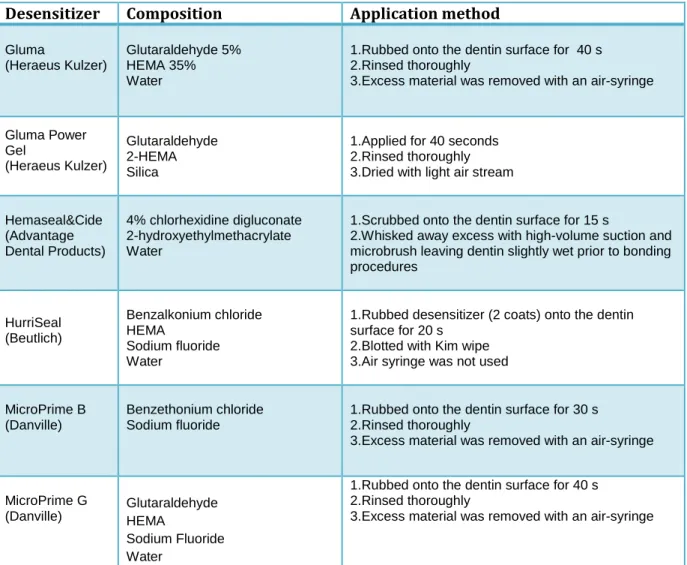
etch-and-rinse adhesives. [66, 67] Desensitizers are HEMA-containing products that
may contain glutaraldehyde and chlorhexidine, for instance. Some desensitizers have
Glutaraldehyde which is present in Gluma Desensitizer, Gluma Power Gel and
MicroPrime G, all of which are used in this study, reacts with serum albumin in the
dentinal fluid and coagulates the plasma proteins hence leading to reduction of
dentin permeability, ultimately counteracting the hydrodynamic mechanism of dentin
sensitivity. [71-74] Glutaraldehyde forms crosslinks with serum collagen and albumin
.[64]. After topical application of glutaraldehyde to the dentin surface, multiple
transverse septa occur in the lumen of the dentinal tubules down to a depth of 200
µm, effectively creating a barrier that eliminates the hydrodynamic mechanism of dentin sensitivity. [75-78] After albumin precipitation, HEMA polymerization takes
placed and may be aid in sensitivity prevention. [65]
HEMA or hydroxyethylmethacrylate, which is present in Gluma Desensitizer,
Gluma Power Gel, Hemaseal & Cide, and MicroPrime G, is an important component
as it plays the role of physically blocking the dentinal tubules. It can be absorbed by
dentin and collagen. [64] The glutaraldehyde/HEMA combination (Gluma
Desensitizer, Gluma Power Gel and MicroPrime G) is an antimicrobial, a flocculating
agent that strengthens collagen, and an agent that creates tubular occlusion, thereby
reducing post-operative sensitivity by limiting fluid movement without affecting the
strength of bonding or adhesive cements.
Other desensitizers may contain chlorhexidine or benzalkonium chloride.
Chlorhexidine is present in Hemaseal & Cide which acts by protein precipitation and
tubule occlusion. It can be chemisorbed on hydroxyapatite of tooth or can act as an
ion to form an insoluble compound with phosphate ions in plaque, saliva and
HurriSeal and MicroPrime B. It helps in crosslinking with HEMA to form a temporary
coating hence helping in dentinal bonding. HurriSeal also contains HEMA and
sodium fluoride and water. Studies have investigated the role of these materials on
2.4. ADHESIVES AND DESENSITIZERS
Adhesives and desensitizer are often used together in clinical dentistry to
reduce post-operative sensitivity. Many studies have been done in the past to
analyze whether there is any impact, positive or negative, and if any variables
associated make a difference. Recent studies show favorable results indicating either
increased bond strengths or no adverse effect.
No difference in bond strengths was found when the effect of Gluma
desensitization on dentin bond strengths was evaluated. [80] No interference on
behalf of desensitizers with resin cement to dentin was found when use of two
HEMA-containing dentin desensitizing agents was analysed with resins. [81]
Al Qahtani and colleagues studied the effect on shear bond strength of
rewetting dry dentin with two desensitizers and three etch-and-rinse dentin bonding
agents (Syntac Single-Component, OptiBond Solo Plus, and Prime & Bond NT). After
application to moist dentin and dry dentin, high bond strengths were noted in the
HurriSeal and Prime & Bond NT group. [66-68]
On the other hand, some studies have also shown decreased bond strengths
when desensitizers were used.[68,82,83] It is important to keep in mind that the type
of desensitizer used may impact the bond strength and is an important factor
affecting bond strength to dentin. [84] We also know that use of desensitizers under
3. SPECIFIC AIMS and NULL HYPOTHESES
1. To determine whether the use of dentin desensitizers has an adverse effect on
the dentin bond strengths of resin-based adhesives.
2. To determine whether time affects the dentin bond strengths of resin-based
adhesives.
The null hypotheses to be tested are that desensitizers do not adversely affect
dentin bond strengths of two-step etch-and-rinse and one-step self-etch adhesives
to dentin and that time does not affect bond strengths of these resin-based
4. MATERIALS AND METHODS
One hundred and fifty extracted human third molars were used in this study.
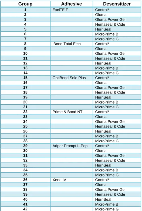
As shown in Table 1, each tooth was randomly assigned to one of six adhesive
groups (ExciTE F, iBond Total Etch, OptiBond Solo Plus, Prime & Bond NT, Adper
Prompt L-Pop, Xeno IV). Teeth in each adhesive group were further assigned to one
of six desensitizer groups (Gluma Desensitizer, Gluma Power Gel, Hemaseal & Cide,
HurriSeal, MicroPrime B, MicroPrime G) and control. Approximately four teeth were
tested per group. The control group consisted of teeth in which no desensitizers were
used. Study was designed as per Figure 1.
After disinfection with Chloramine-T solution, the crowns and roots were
separated using a low speed diamond disk (see Figure 3). Occlusal surfaces were
ground wet with 600-grit silicon carbide paper using a mechanical grinder under to
obtain flat dentin (see Figure 2).
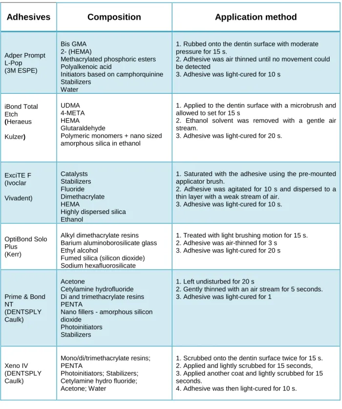
For the etch-and-rinse adhesive groups – iBond Total Etch, ExciTE F, Prime & Bond NT and OptiBond Solo Plus - Ultra-Etch (35% phosphoric acid, Ultradent
Products, Inc, Jordan,UT.) was applied for 15 s and thenthoroughly rinsed off for 10
s. Specimens for all groups were blot using KimWipes (Kimberly-Clark,Texas, USA)
prior to application of the desensitizers using micro brushes. The desensitizers were
applied after etching in the etch-and-rinse groups. Desensitizers and their application
After application of the adhesives, Venus Diamond composite resin build-ups
were placed in three 2-mm increments. Each composite increment was light-cured for
40 s with a Spectrum 800 halogen light-curing unit (Dentsply Caulk) at a minimum
intensity of 550 mW/cm2. Specimens were stored in distilled water at 37°C and
sectioned into beams using an Isomet 1000 Precision saw (Buehler,Lake
Bluff,IL)after 24 h. Beams were approximately 1 x 1 mm with half of each beam
consisting of composite resin and the other half of dentin. On average, each tooth
yielded about 15 beams. One-third of the beams were tested at 24 h and one-third at
six months aging. Remaining beams are scheduled for testing at one year; data
yielded will be presented in another report.
The beams were carefully positioned in a custom notched jig so the force
applied would be perpendicular to dentin-composite interface (see Figure 4). Scotch
Super Glue was used to fix the beams to the jig. The adhesive was sprayed with
Zap-it Accelerator Spray (Dental Ventures of America, Inc Corona,CA) to accelerate Zap-its
setting process. Each beam was loaded to failure in an EZ-Test
(Shimadzu,Tokyo,Japan) (see Figure 5). The MTBS was calculated dividing the peak
break by the bonded area (mm2) and was expressed in megapascals (MPa). The
failure mode was determined by visual inspection and classified as interfacial,
cohesive in dentin, or cohesive in composite resin.
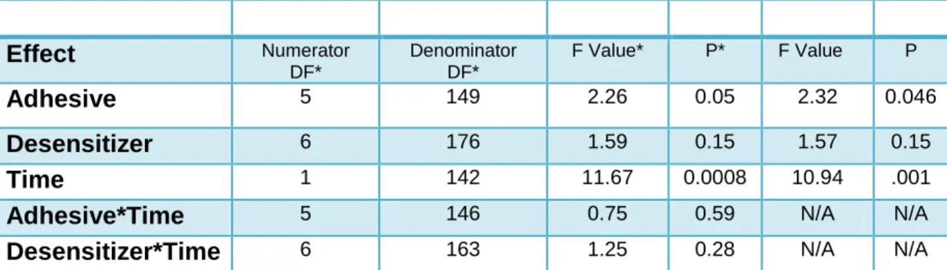
Because of the expected correlation between beams tested at 24 h and 6
months, linear mixed effects model was used with compound symmetry covariance.
Because of the small sample size, Kenward-Roger method was used to compute DF
and time were considered as main effects, and the pairwise interactions between
adhesive and time, and desensitizer and time were also considered. Pairwise
contrasts among adhesives were conducted using least square means. All analyses
were conducted using teeth as unit of analysis in SAS 9.2 at a significance level of
5. RESULTS
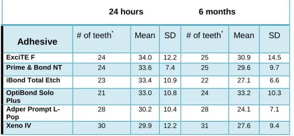
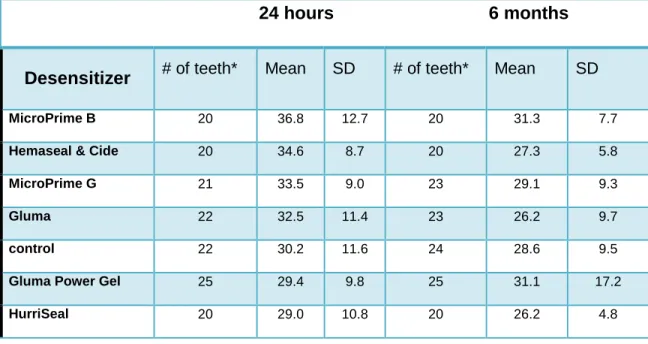
Descriptive statistics for each adhesive/desensitizer combination at each time
point are provided in Table 4. Adhesive and desensitizer MTBS at both time points
are provided in Table 5 and Table 6. 3 teeth were excluded from data as they did not
meet requirements.
At 24 h, the mean MTBS of the adhesives ranged from 29.9 MPa for Xeno IV
to 34.0 MPa for ExciTE F. At 6 months, the mean MTBS ranged from 24.1 MPa for
Adper Prompt L-Pop to 33.2 MPa for OptiBond Solo Plus (Table 5).
At 24 h, the mean MTBS for desensitizer groups ranged from 29.0 MPa for
HurriSeal to 36.8 MPa for MicroPrime B. At 6 months, the mean MTBS ranged from
26.2 MPa for Gluma Desensitizer and HurriSeal to 31.3 for MicroPrime B (Table 6).
The interactions between adhesive and time (p = 0.59) and desensitizer and
time (p = 0.29) were not statistically significant, indicating that the patterns over time
were similar for all adhesives and desensitizers. After removing the interactions,
there was a statistically significant difference among the means of the adhesive
groups (p = 0.046) but not among the desensitizers (p = 0.16) (Table 7). There was a
statistically significant difference in overall mean MTBS from 24 h to 6 months
(p=0.001), as the microtensile bond strengths decreased over time.
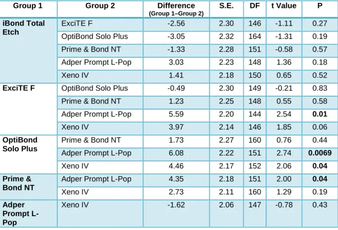
The pairwise contrasts among the adhesive groups indicated statistically
Adper Prompt L-Pop. OptiBond Solo Plus also had a higher MTBS than Xeno IV
6. DISCUSSION
Results of this study revealed that dentin desensitizers did not adversely affect
dentin bond strengths of etch-and-rinse and self-etch adhesives. However, time did
affect the findings of this study, as bond strengths were lower at six months than at
24 hours.
The problem of post-operative sensitivity commonly presents to the restorative
dentist as a transient pain that cannot be related to any other dental defect or
pathology. It can be a result of number of factors such as polymerization shrinkage
related to the C-factor, aggressive tooth preparation leading to overheating,
microleakage, poor adhesion protocol, or contamination of the substrate during
bonding. The hydrodynamic (fluid movement) theory proposed by Brännström is an
important theory related to understanding of sensitivity. Dentin is a complex structure
and has different characteristics and variations. Procedures such as root planing,
cavity preparation, or crown preparation involves stripping of cementum or the
enamel layer leading to exposure of dentinal tubules. This may lead to post-operative
sensitivity.
Many methods have been advocated to prevent post-operative sensitivity,
including use of oxalate particles, dentinal tubule sealers and dentin desensitizers. It
is known that use of certain adhesives requires conditioning which leads to
technique-sensitive, since wet bonding is required for full expansion and optimal
hybridization of the demineralized collagen matrix. [87,88 ] Intrinsic wetness of vital
deep dentin after removal of the smear layer [42] leads to difficulty in bonding, and
the increased permeability associated with the simplified version of these adhesives.
[43]Incomplete sealing and continuous transudation of dentinal fluid through open
dentinal tubules before polymerization of the adhesive may result in entrapment of
water-filled blisters along the adhesive interface. [43]Compression of these blisters
during mastication may cause, within the dentinal tubules[45], rapid fluid movement
that activates the intradental A-delta nerve fibers [46], which results in post-operative
sensitivity.
In etch-and-rinse adhesives, currently three steps or two steps are used.
Three-step etch-and-rinse adhesives have three components – etchant, primer and
adhesive resin. Two-step etch-and-rinse systems include an etchant and combined
primer and adhesive resin. Etch-and-rinse adhesives form an excellent bond to
enamel with excellent marginal integrity. For the self-etch adhesives, two-step and
one-step products are available. Two-step systems contain a self-etching primer and
an adhesive resin. One-step products combine all three functional steps. According
to Ernst, they may not be the first choice in all cases but might be a better idea in a
variety of indications due to easier application, less chance of post-operative
sensitivity and easy hybridization of dentin.
In the present study, several etch-and-rinse adhesive groups – iBond Total
Etch, ExciTE F, OptiBond Solo Plus, and Prime & Bond NT – were tested and the
Adper Prompt L-Pop (3M ESPE) is a strong water-based, self-etch adhesive that
contains acidic methacrylated phosphoric esters. Xeno IV (Dentsply Caulk) is a
milder self-etch adhesive containing mono, di, tri methacrylates and PENTA.
Results at 24 hours and 6 months for adhesives indicate that the overall mean
was highest for OptiBond Solo Plus which is an etch-and-rinse adhesive, followed by
ExciTE F which behaved similar or greater than Prime & Bond NT. The lowest mean
MTBS was observed in the self-etch adhesives where in Xeno IV and Adper Prompt
L-Pop were comparable.
Beams were evaluated at 24 hours and 6 months of storage could have
impacted the bond strengths. It is known that adhesives exhibit evidence of
mechanical and morphological degradation which leads to decrease in bond. [89]
It should be noted that number of teeth varied at 24 hours and 6 months.
Some of the samples at 24 hours broke before MTBS testing. Hence, data for 3 teeth
was excluded from data because they did not meet requirements.
The desensitizers used were Gluma Power Gel, Gluma Desensitizer,
Hemaseal & Cide, HurriSeal, MicroPrime B, or MicroPrime G. Desensitizers serve
the role of disinfection of cavity preparations, desensitization and rewetting on the
cavity. Commonly used contain glutaraldehyde, HEMA, sodium fluoride,
benzalkonium chloride and combinations. In this study, glutaraldehyde was present in
Gluma, Gluma Power Gel and MicroPrime G. It reacts with serum albumin in the
dentinal fluid, coagulates with plasma protein and then cross links with bovine serum
collagen and albumin.HEMA present in Gluma, Gluma Power Gel, Hemaseal and
collagen. Glutaraldeyhde and HEMA are present in Gluma, Gluma Power Gel and
MicroPrime G. In addition to be being an effective antimicrobial agent, it is also a
flocculating agent providing strength to collagen and creates tubular occlusion.
At 24 hours, highest mean MTBS was noted for MicroPrime B at 37 MPa to
lowest at 29 MPa for HurriSeal whereas at six months, it ranged from 31.3 MPa for
MicroPrime B to 26.2 MPa for HurriSeal. Overall, there was a decrease in MTBS over
time. Lower trend of mean MTBS was noted. Desensitizers containing
HEMA/Glutaraldehyde performed well.
We wanted to account for the main effects in this study – i.e. time, desensitizer
and adhesive – and their interactions to determine whether any combination of
factors affected bond strengths. Time was a significant factor when interactions were
accounted for and remained significant even when interactions were removed.
Adhesive also showed marginal significance at 0.0457 but was rounded off to 0.05
hence, not statistically significant. What can be interpreted from this finding is that
time impacted the findings of this study. Lower bond strengths were obtained after
specimens were stored for six months.
Many studies have been done in the past showing varied effects of use of the
desensitizer on MTBS of adhesives. Although, many have shown adverse effects,
some have shown increased bond strengths. Studies that have shown no effect of
the use of desensitizer on MTBS are relatively few. Most studies show that there is
no harm in using GLUMA which contains HEMA and glutaraldehyde.
A study investigated the influence of three dentin hypersensitivity treating
agents (Panavia Fluoro Cement and Super-Bond C & B). What they found was that
the use of the Gluma desensitizer did not affect bond strength of any of the three
adhesive systems, and the bond strength of the Panavia cement with the AD Gel
conditioning was not reduced by application of any of the three desensitizers. [90]
Another study evaluated the influence of a dentin desensitizer and ozone
application on the bond strength to dentin of a composite resin material. The dentin
desensitizing agent and ozone treatment were applied on the cervical dentin surfaces
of extracted, caries-free, erupted third molars. Statistical analysis showed no
significant influence of the different hypersensitivity treatments on shear bond
strength to dentin. The authors concluded that short-term use of dentin
hypersensitivity treatments like ozone and dentin desensitizers containing
gluteraldehyde did not affect the shear bond strength to dentin of subsequent
composite resin restorations. [91]
Investigation was done to see whether a desensitizing agent (GLUMA
Desensitizer) containing glutaraldehyde and HEMA improved the bond strength and
bonding durability of a self-etching primer adhesive to Er:YAG-irradiated dentine.
Tensile bond strengths (TBS) of 10 specimens of each treatment group were
measured after 24-h water storage, 6 months water storage and 12 months water
storage, respectively, and the failure modes were analyzed. They concluded that
application of GLUMA Desensitizer to Er:YAG-irradiated dentine increased the bond
strength and durability of the self-etching priming adhesive used. [92]
Bond strength of a self-etching dentin adhesive was evaluated for its ability to
gingival walls. The application of MicroPrime and Gluma Desensitizer to
caries-affected dentin did not show any effect on bond strength testing. According to this
study, desensitizer application on sound dentin is recommended with self-etch
bonding systems.[93]
This study evaluated the effect of rewetting dried dentin with two commercial
desensitizing agents (Protect and HurriSeal) on the dentin shear bond strength of
three total-etch dentin bonding agents (Syntac Single-Component, OptiBond Solo
Plus and Prime & Bond NT) and compared both to applying these same bonding
agents to moist dentin and dry dentin. The specimens were stored in distilled water at
37 degrees C for 24 hours prior to thermocycling 2,500 times. In most groups, no
significant difference in shear bond strength between the control and others was
noted. In the Prime & Bond NT bonding agent groups, the shear bond strength of the
HurriSeal group (20.7 MPa) was significantly higher than the mean of the other
groups: control (8.0 MPa), dry (5.7 MPa) and Protect (5. 5 MPa). [66]
Main aim of this study was to determine whether the use of two
HEMA-containing dentin desensitizing agents [Health-Dent Desensitizer with fluoride (H) or
Gluma Desensitizer (G)], when applied at simulated "cavity preparation" and
"cementation" appointments, affected the bond strength of lab processed resin
composite restorations cemented to dentin. The treatments were applied in two
sessions in order to simulate cavity preparation and cementation appointments.
Results of this in vitro study indicated that the use of desensitizers at the preparation
and/or cementation appointment does not interfere with bond strengths of resin
In our study, the microtensile bond strength method was used to assess the
bond strength. Several advantages have been advocated for use of the MTBS
method developed by Sano in 1994 [94] e.g., better stress distribution at the bonding
area, improved comparison of data from peripheral central dentin and ability for
collection of multiple micro specimens from each tooth.
Many MTBS studies have been done in the past but due to lack of
standardization, important comparisons cannot be made. Many limitations also
impacted the findings of this study since sound teeth were used which may not
reproduce most of the cases encountered clinically. Mostly when used clinically,
desensitizers are acting on more thicker and denser smear layer which can affect
how the adhesives perform.
Many factors like pulpal pressure, dentinal fluid movement and tooth dynamics
cannot be mimicked in the lab. It is well know that hydraulic conductance also has an
important role in adhesion. We were unable to determine the age of patient or time of
extraction. Young dentin might respond differently to bonding than mature dentin.
Rods within the tooth play an important role and since in our study, molars were used
owing to the fact that bond strengths may be different due to increased intertubular
dentin. An SEM analysis could be done to compare the formation of hybrid layer with
etch-and-rinse and self-etch adhesive systems.
Within the limitations of this study, we conclude that all the desensitizers and
adhesives performed equally and that time was the only factor that affected the
findings of the study. A trend of lower dentin bond strengths was noted over time.
immediately or after aging. Clinically, it is safe to use a desensitizer along with
adhesives. However, further testing needs to be done allowing more aging of
7. CONCLUSIONS
Within the limitations of our study, one can conclude that:
1. In general, the microtensile bond strengths of the various adhesive and
desensitizer combinations were not significantly different.
2. Time was the only significant factor affecting dentin bond strengths, as these
significantly decreased over time
3. Use of desensitizers did not significantly reduce or improve dentin bond
strengths either immediately or after aging.
This study failed to reject the first null hypotheses, as the desensitizers had no
adverse effect on dentin bond strengths of two-step etch-and-rinse or one-step
self-etch adhesives. The second null hypothesis was rejected, as time did affect dentin
TABLE 1. Experimental groups
*No desensitizer used
Group Adhesive Desensitizer
1 ExciTE F Control*
2 Gluma
3 Gluma Power Gel
4 Hemaseal & Cide
5 HurriSeal
6 MicroPrime B
7 MicroPrime G
8 iBond Total Etch Control*
9 Gluma
10 Gluma Power Gel
11 Hemaseal & Cide
12 HurriSeal
13 MicroPrime B
14 MicroPrime G
15 OptiBond Solo Plus Control*
16 Gluma
17 Gluma Power Gel
18 Hemaseal & Cide
19 HurriSeal
20 MicroPrime B
21 MicroPrime G
22 Prime & Bond NT Control*
23 Gluma
24 Gluma Power Gel
25 Hemaseal & Cide
26 HurriSeal
27 MicroPrime B
28 MicroPrime G
29 Adper Prompt L-Pop Control*
30 Gluma
31 Gluma Power Gel
32 Hemaseal & Cide
33 HurriSeal
34 MicroPrime B
35 MicroPrime G
36 Xeno IV Control*
37 Gluma
38 Gluma Power Gel
39 Hemaseal & Cide
40 HurriSeal
41 MicroPrime B
TABLE 2. Desensitizers evaluated
Desensitizer Composition Application method
Gluma
(Heraeus Kulzer)
Glutaraldehyde 5% HEMA 35% Water
1.Rubbed onto the dentin surface for 40 s 2.Rinsed thoroughly
3.Excess material was removed with an air-syringe
Gluma Power Gel (Heraeus Kulzer) Glutaraldehyde 2-HEMA Silica
1.Applied for 40 seconds 2.Rinsed thoroughly 3.Dried with light air stream
Hemaseal&Cide (Advantage Dental Products)
4% chlorhexidine digluconate 2-hydroxyethylmethacrylate Water
1.Scrubbed onto the dentin surface for 15 s
2.Whisked away excess with high-volume suction and microbrush leaving dentin slightly wet prior to bonding procedures HurriSeal (Beutlich) Benzalkonium chloride HEMA Sodium fluoride Water
1.Rubbed desensitizer (2 coats) onto the dentin surface for 20 s
2.Blotted with Kim wipe 3.Air syringe was not used
MicroPrime B (Danville)
Benzethonium chloride Sodium fluoride
1.Rubbed onto the dentin surface for 30 s 2.Rinsed thoroughly
3.Excess material was removed with an air-syringe
MicroPrime G (Danville) Glutaraldehyde HEMA Sodium Fluoride Water
1.Rubbed onto the dentin surface for 40 s 2.Rinsed thoroughly
3.Excess material was removed with an air-syringe
TABLE 3. Adhesive systems evaluated
Adhesives Composition Application method
Adper Prompt L-Pop (3M ESPE)
Bis GMA 2- (HEMA)
Methacrylated phosphoric esters Polyalkenoic acid
Initiators based on camphorquinine Stabilizers
Water
1. Rubbed onto the dentin surface with moderate pressure for 15 s.
2. Adhesive was air thinned until no movement could be detected
3. Adhesive was light-cured for 10 s
iBond Total Etch (Heraeus Kulzer) UDMA 4-META HEMA Glutaraldehyde
Polymeric monomers + nano sized amorphous silica in ethanol
1. Applied to the dentin surface with a microbrush and allowed to set for 15 s
2. Ethanol solvent was removed with a gentle air stream.
3. Adhesive was light-cured for 20 s.
ExciTE F (Ivoclar Vivadent) Catalysts Stabilizers Fluoride Dimethacrylate HEMA
Highly dispersed silica Ethanol
1. Saturated with the adhesive using the pre-mounted applicator brush.
2. Adhesive was agitated for 10 s and dispersed to a thin layer with a weak stream of air.
3. Adhesive was light-cured for 10 s.
OptiBond Solo Plus
(Kerr)
Alkyl dimethacrylate resins Barium aluminoborosilicate glass Ethyl alcohol
Fumed silica (silicon dioxide) Sodium hexafluorosilicate
1. Treated with light brushing motion for 15 s. 2. Adhesive was air-thinned for 3 s
3. Adhesive was light-cured for 20 s
Prime & Bond NT
(DENTSPLY Caulk)
Acetone
Cetylamine hydrofluoride Di and trimethacrylate resins PENTA
Nano fillers - amorphous silicon dioxide
Photoinitiators Stabilizers
1. Left undisturbed for 20 s
2. Gently thinned with an air stream for 5 seconds. 3. Adhesive was light-cured for 1
Xeno IV (DENTSPLY Caulk) Mono/di/trimethacrylate resins; PENTA Photoinitiators; Stabilizers; Cetylamine hydro fluoride; Acetone; Water
1. Scrubbed onto the dentin surface twice for 15 s. 2. Applied and lightly scrubbed for 15 seconds, 3. Applied another coat and lightly scrubbed for 15 seconds.
Bis-GMA - bisphenol A-glycidyl methacrylate; HEMA - hydroxyethyl methacrylate; UDMA - urethane dimethacrylate; 4-META - methacryloyloxyethy trimellitate anhydride; PENTA - dipentaerythritolpentaacrylate monophosphate
TABLE 4. Microtensile bond strengths for combinations of adhesive and desensitizer
at 24 h and 6 months in MPa*
Adhesive Desensitizer 24 hours 6 months
Mean S.D. Mean S.D.
ExciTE F Control 28.9 3.8 24.1 5.9
Gluma 33.1 9.2 28.1 8.5
Gluma Power Gel 35.8 11.7 45.4 31.2
Hemaseal & Cide 46.5 8.3 26.7 2.3
HurriSeal 22.6 9.5 26.1 5.6
MicroPrime B 43.6 18.9 28.5 11.0
MicroPrime G 34.3 7.5 36.0 5.6
iBond Total Etch
Control 36.6 5.1 34.6 7.6
Gluma 38.9 16.9 27.0 4.6
Gluma Power Gel 19.9 9.8 24.9 10.1
Hemaseal & Cide 30.0 5.3 30.0 7.5
HurriSeal 37.2 3.5 26.4 3.2
MicroPrime B 37.4 7.5 26.9 5.8
MicroPrime G 42.4 2.2 24.1 3.7
OptiBond Solo Plus
Control 42.3 18.2 38.0 9.1
Gluma 26.6 6.6 32.9 21.1
Gluma Power Gel 34.5 4.8 37.8 11.4
Hemaseal & Cide 33.1 6.8 29.4 5.12
HurriSeal 28.1 12.1 28.0 4.0
MicroPrime B 35.5 12.7 30.8 11.4
MicroPrime G 30.7 12.8 32.6 10.4
Prime & Bond NT
Control 30.3 6.6 25.1 10.8
Gluma 32.6 6.5 23.2 6.5
Gluma Power Gel 30.8 7.4 30.4 10.4
Hemaseal & Cide 39.2 5.3 29.5 6.5
HurriSeal 27.1 6.2 21.8 6.1
MicroPrime B 40.2 1.9 38.3 7.5
MicroPrime G 35.0 9.5 36.0 10.0
Adper Prompt L-Pop
Control 21.9 8.1 21.7 6.0
Gluma 36.8 16.4 23.7 9.5
Gluma Power Gel 31.0 3.8 23.1 11.0
Hemaseal & Cide 28.4 7.9 23.6 6.0
HurriSeal 27.1 11.9 30.2 3.3
MicroPrime B 31.3 9.3 28.3 1.7
MicroPrime G 37.0 3.0 20.2 6.8
Xeno IV Control 28.1 14.4 29.2 7.9
Gluma 27.4 8.4 24.8 7.5
Gluma Power Gel 28.6 11.1 29.0 18.9
Hemaseal & Cide 34.2 6.4 26.7 7.4
HurriSeal 35.9 17.6 24.7 4.2
MicroPrime B 34.4 18.9 33.4 6.2
TABLE 5. Microtensile bond strengths by type of adhesive in MPa
*Number of teeth varied at 24 hours and 6 months. Some of the specimens at 24 hours broke before MTBS testing. Data for those specimens (3 teeth) were not included.
24 hours 6 months
Adhesive # of teeth
*
Mean SD # of teeth* Mean SD
ExciTE F 24 34.0 12.2 25 30.9 14.5
Prime & Bond NT 24 33.6 7.4 25 29.6 9.7
iBond Total Etch 23 33.4 10.9 22 27.1 6.6
OptiBond Solo Plus
21 33.0 10.8 24 33.2 10.3
Adper Prompt L-Pop
28 30.2 10.4 28 24.1 7.1
TABLE 6.Microtensile bond strengths by type of desensitizer in MPa
24 hours 6 months
Desensitizer # of teeth* Mean SD # of teeth* Mean SD
MicroPrime B 20 36.8 12.7 20 31.3 7.7
Hemaseal & Cide 20 34.6 8.7 20 27.3 5.8
MicroPrime G 21 33.5 9.0 23 29.1 9.3
Gluma 22 32.5 11.4 23 26.2 9.7
control 22 30.2 11.6 24 28.6 9.5
Gluma Power Gel 25 29.4 9.8 25 31.1 17.2
HurriSeal 20 29.0 10.8 20 26.2 4.8
TABLE 7. Results from linear mixed effects model with and without interactions of
explanatory variables and time
*with variables evaluated i.e. adhesive *time and desensitizer*time
Effect Numerator
DF*
Denominator DF*
F Value* P* F Value P
Adhesive 5 149 2.26 0.05 2.32 0.046
Desensitizer 6 176 1.59 0.15 1.57 0.15
Time 1 142 11.67 0.0008 10.94 .001
Adhesive*Time 5 146 0.75 0.59 N/A N/A
TABLE 8. Least square means for the global difference between adhesive groups
Group 1 Group 2 Difference
(Group 1–Group 2)
S.E. DF t Value P
iBond Total Etch
ExciTE F -2.56 2.30 146 -1.11 0.27
OptiBond Solo Plus -3.05 2.32 164 -1.31 0.19
Prime & Bond NT -1.33 2.28 151 -0.58 0.57
Adper Prompt L-Pop 3.03 2.23 148 1.36 0.18
Xeno IV 1.41 2.18 150 0.65 0.52
ExciTE F OptiBond Solo Plus -0.49 2.30 149 -0.21 0.83
Prime & Bond NT 1.23 2.25 148 0.55 0.58
Adper Prompt L-Pop 5.59 2.20 144 2.54 0.01
Xeno IV 3.97 2.14 146 1.85 0.06
OptiBond Solo Plus
Prime & Bond NT 1.73 2.27 160 0.76 0.44
Adper Prompt L-Pop 6.08 2.22 151 2.74 0.0069
Xeno IV 4.46 2.17 152 2.06 0.04
Prime & Bond NT
Adper Prompt L-Pop 4.35 2.18 151 2.00 0.04
Xeno IV 2.73 2.11 160 1.29 0.19
Adper Prompt L-Pop
GRAPH 1. Means and standard deviatio
months
GRAPH 2. Means and standard deviations (pooled data) for desensitizers at 24 h
and 6 months
REFERENCES
1. Leinfelder KF. Wear patterns and failure mechanisms. In: Posterior Composites – Proceedings of International Symposium on Resins 1982; Taylor DF, ed., Chapel Hill, NC: The University of North Carolina Press, 329-357.
2. Johnson GH, Gordon GE, Bales DJ. Postoperative sensitivity associated with posterior composite and amalgam restorations. Oper Dent 1988; 13:66-73.
3. Suzuki M, Jordan RE, Boksman L.Posterior composite restorations - clinical considerations. In: Posterior Composite Resin Dental Restorative Materials 1985; Vanherle G and Smith DC, Ed., St. Paul, MN: 3M Co., 455-464.
4. Brännström M, Aström A. The hydrodynamics of the dentine; its possible relationship to dentinal pain. Int Dent J 1972; 22:219-227.
5. Jain P, Vargas MA, Denehy GE, Boyer DB. Dentin desensitizing agents: SEM and X-ray microanalysis assessment. Am J Dent 1997; 10:21-26.
6. Pashley DH, Livingston MJ, Greenhill JD. Regional resistances to fluid flow in human dentine in vitro. Arch Oral Biol 1978; 23:807-810.
7. Dondi dall'Orologio G, Malferrari S. Desensitizing effects of Gluma and Gluma 2000 on hypersensitive dentin. Am J Dent 1993; 6:283-286.
8. Felton DA, Bergenholtz G, Kanoy BE. Evaluation of the desensitizing effect of Gluma Dentin Bond on teeth prepared for complete-coverage restorations. Int J Prosthodont 1991; 4:292-298.
9. Soeno K, Taira Y, Matsumura H, Atsuta M. Effect of desensitizers on bond strength of adhesive luting agents to dentin. J Oral Rehabil 2001; 28:1122-1128.
10. Cobb DS, Reinhardt JW, Vargas MA. Effect of HEMA-containing dentin desensitizers on shear bond strength of a resin cement. Am J Dent 1997; 10:62-65.
12. Yu X, Liang B, Jin X, Fu B, Hannig M. Comparative in vivo study on the desensitizing efficacy of dentin desensitizers and one-bottle self-etching adhesives. Oper Dent 2010; 35:279-286.
13. Marshall GW, Inai N, Magidi IW, Balooch M, Kinney JH, Tagami J, Marshall SJ. Dentin demineralization effects of dentin depth, pH and different acids. Dent Mater 1997;13:338-343.
14. Söderholm, KJ. Correlation of in vivo and in vitro performance of adhesive restorative materials: a report of the ASC MD156 Task Group on Test Methods for the Adhesion of Restorative Materials. Dent Mater 1991; 7:74-83.
15. Allen KW: Theories of adhesion. In Packham DE, editor, Handbook of adhesion, 1992, 473-475.
16. Van Landuyt KL, Snauwaert J, De Munck J, Peumans M, Yoshida Y, Poitevin A, Coutinho E, Suzuki K, Lambrechts P, Van Meerbeek B. Systematic review of the chemical composition of contemporary dental adhesives. Biomaterials 2007; 28:3757-3785.
17. Van Meerbeek B, Inokoshi S, Braem M, Lambrechts P, Vanherle G. Aspects of the resin-dentin interdiffusion zone with different dentin adhesive systems. J Dent Res 1992; 71:1530-1540.
18. Inokoshi S, Hosoda H, Harnirattisai C, Shimada Y. Interfacial structure between dentin and seven dentin bonding systems revealed using argon ion beam etching. Oper Dent 1993; 18:8-16.
19. Swift EJ, Heymann HO, Perdigão J. Bonding to enamel and dentin: A brief history and state of the art. Quintessence Int 1995; 26:95-110.
20. Condon J.R,J.L Ferracane. Reduction of composite contraction stress through non-bonded microfiller particles. Dental Materials 1998,14:256-260.
21. Nakabayashi N, Kojima K, Masuhara E. The promotion of adhesion by the infiltration of monomers into tooth substrates. J Biomed Mater Res 1982; 16:265-273.
23. Eick JD, Gwinnett AJ, Pashley DH, Robinson SJ. Current concepts on adhesion to dentin. Crit Rev Oral Biol Med 1997; 8:306-335.
24. Albaladejo A, Osorio R, Toledano M, Ferrari M. Hybrid layers of etch-and-rinse versus self-etching adhesive systems. Med Oral Patol Oral Cir Bucal 2010; 15:112-118.
25. Jendresen MD. Clinical performance of a new composite resin for Class V erosion (abstract 1057). J Dent Res 1978; 57:339.
26. Eliades GC, Caputo AA, Vougiouklakis GJ. Composition, wetting properties and bond strength with dentin of 6 new dentin adhesives. Dent Mater 1985; 1:170-176.
27. Davidson CL, de Gee AJ, Feilzer A. The competition between the composite-dentin bond strength and the polymerization contraction stress. J Dent Res 1984; 63:1396-1399.
28. Van Meerbeek B, Peumans M, Verschueren M, Gladys S, Braem M, Lambrechts P.Clinical status of ten dentin adhesive systems. J Dent Res 1994; 73:1690-1702.
29. Van Meerbeek B, Peumans M, Gladys S, Braem M, Lambrechts P, Vanherle G. Three-year clinical effectiveness of four total-etch systems in cervical lesions. Quintessence Int 1996; 27:775-784.
30. Fusayama T, Nakamura M, Kurosaki N, Iwaku M. Non-pressure adhesion of a new adhesive restorative resin. J Dent Res 1979; 58:1364-1370.
31. Kanca J. Bonding to tooth structure: A rational rationale for a clinical protocol. J Esthet Dent 1989; 1:135-138.
32. Bertolotti RL. Total-etch—the rational dentin bonding protocol. J Esthet Dent 1991; 3:1-6.
33. Macko DJ, Rutberg M, Langeland. Pulpal response to the application of phosphoric acid to dentin. Oral Surg Oral Med Oral Pathol 1978; 45:930-946.
34. Kanca J. A method for bonding to tooth structure using phosphoric acid as a dentin-enamel conditioner. Quintessence Int 1991; 22:285-290.
36. Shimono M, Maeda T, Suda H, Takayashi K, eds. Dentin/pulp complex. Tokyo: Quintessence; 1996; 359-363.
37. Tay FR, Gwinnett AJ, Wei SH. The overwet phenomenon: an optical, micromorphological study of surface moisture in the acid-conditioned, resin-dentin interface. Am J Dent 1996; 9:43-48.
38. Nakabayashi N, Pashley DH. Hybridization of dental hard tissues. Tokyo: Quintessence; 1998.
39. Kanca J 3rd. Improving bond strength through acid etching of dentin and bonding to wet dentin surfaces. J Am Dent Assoc 1992; 123:35-43.
40. Kanca J 3rd. Wet bonding: effect of drying time and distance. Am J Dent 1996; 9:273-276.
41. Manso AP, Marquezini L Jr, Silva SM, Pashley DH, Tay FR, Carvalho RM. Stability of wet versus dry bonding with different solvent-based adhesives. Dent Mater 2008; 24:476-482.
42. Itthagarun A, Tay FR. Self-contamination of deep dentin by dentin fluid. Am J Dent 2000; 13:195-200.
43. Tay FR, Gwinnett AJ, Wei SH. The overwet phenomenon: a transmission electron microscopic study of surface moisture in the acid-conditioned, resin-dentin interface. Am J Dent 1996; 9:161-166.
44. Tay FR, Suh BI, Pashley DH, Prati C, Chuang SF, Li F. Factors contributing to the incompatibility between simplified-step self-cured or dual-cured composites. Part II. Single-bottle, total-etch adhesive. J Adhes Dent 2003; 5:91-105.
45. Brännström M, Johnson G. Movements of the dentine and pulp liquids on application of thermal stimuli. An in vitro study. Acta Odontol Scand 1970; 28:59-70.
46. Närhi M, Yamamoto H, Ngassapa D, Hirvonen T. The neurophysiological basis and the role of inflammatory reactions in dentine hypersensitivity. Arch Oral Biol 39:23S-30S.
48. Ernst, CP.Options for dentin bonding. J Esthet Restor Dent 2006; 18:61-67.
49. Peumans M, Kanumilli P, De Munck J, et al. Clinical effectiveness of contemporary adhesives: a systematic review of current clinical trials. Dent Mater 2005; 21:864-881
50. Skupien JA, Susin AH, Angst PD, Anesi R, Machado P, Bortolotto T, Krejci I. Micromorphological effects and the thickness of the hybrid layer – a comparison of current adhesive systems. J Adhes Dent 2010; 12:435-442.
51. Perdigão J, Dutra-Correa M, Anauate-Netto C, Castilhos N, Carmo AR, Lewgoy HR, Amore R, Cordeiro HJ.Two-year clinical evaluation of self-etching adhesives in posterior restorations. J Adhes Dent 2009; 11:149-159.
52. Jalalian E, Meraji N, Mirzaei M. A comparison of the efficacy of potassium nitrate and Gluma desensitizer in the reduction of hypersensitivity in teeth with full-crown preparations. J Contemp Dent Pract 2009; 10:66-73.
53. Ozen T, Orhan K, Avsever H, Tunca YM, Ulker AE, Akyol M. Dentin hypersensitivity: a randomized clinical comparison of three different agents in a short-term treatment period.Oper Dent 2009; 34:392-398.
54. Yu X, Liang B, Jin X, Fu B, Hannig M. Comparative in vivo study on the desensitizing efficacy of dentin desensitizers and one-bottle self-etching adhesives.Oper Dent 2010; 35:279-286.
55. Irvine JH. Root surface sensitivity: A review of aetiology and management. J N Z Soc Periodontol 1988; 66:15-18.
56. Rapp R, Avery JK, Strachan DS. Possible role of the acetylcholinesterase in neural conduction within the dental pulp. In: Finn SB, editor. Biology of the dental pulp organ. Birmingham: University of Alabama Press; 1968:309-331.
57. Absi EG, Addy M, Adams D. Dentine hypersensitivity: A study of the patency of dentinal tubules in sensitive and non-sensitive cervical dentine. J Clin Periodontol 1987; 14:280-284.
59. Chidchuangchai W, Vongsavan N, Matthews B. Sensory transduction mechanisms responsible for pain caused by cold stimulation of dentine in man. Arch Oral Biol 2007; 52:154-160.
60. Miglani S. Dentin hypersensitivity: recent trends in management. J Conserv Dent 2010; 13:218-224.
61. Pashley DH. Dentin permeability, dentin sensitivity and treatment through tubule occlusion. J Endod 1986; 12:465-474.
62. Brännström M. The hydrodynamic theory of dentinal pain: sensation in preparations, caries, and the dentinal crack syndrome. J Endod 1986; 12:453-457.
63. Pashley DH, Galloway SE. The effect of oxalate treatment on the smear layer of ground surfaces of human dentin. Arch Oral Biol 1985; 30:731-737.
64. Dondi dall’Orologio G, Lone A, Finger WJ. Clinical evaluation of the role of glutardialdehyde in a one-bottle adhesive. Am J Dent 2002; 15:330-334.
65. Qin C, Xu J, Zhang Y. Spectroscopic investigation of the function of aqueous 2-hydroxyethylmethacrylate/glutaraldehyde solution as a dentin desensitizer. Eur J Oral Sci 2006; 114:354-359.
66. Al Qahtani MQ, Platt JA, Moore BK, Cochran MA.The effect on shear bond strength of rewetting dry dentin with two desensitizers. Oper Dent 2003; 28:287-296.
67. Walter R, Miguez PA, Swift EJ Jr, Pereira PN. Long-term bond strength to dentin treated with different re-wetting solutions. Am J Dent 2008; 21:143-147.
68. Aranha AC, Siqueira Junior A de S, Cavalcante LM, Pimenta LA, Marchi GM. Microtensile bond strengths of composites to dentin treated with desensitizer products. J Adhes Dent 2006; 8:85-90.
69. Kobler A, Schaller HG, Gernhardt CR. Effects of the desensitizing agents Gluma and Hyposen on the tensile bond strength of dentin adhesives. Am J Dent 2008; 21:388-392.
71. Bergenholtz G, Jontell M, Tuttle A, Knutsson G. Inhibition of serum albumin flux across exposed dentine following conditioning with GLUMA primer, glutaraldehyde or potassium oxalates. J Dent 1993; 21:220-227.
72. Suh BI, Feng L, Pashley DH, Tay FR. Factors contributing to the incompatibility between simplified-step adhesives and chemically-cured or dual-cured composites. Part III. Effect of acidic resin monomers. J Adhes Dent 2003; 5:267-282.
73. Swift EJ Jr, Lloyd AH, Felton DA. The effect of resin desensitizing agents on crown retention. J Am Dent Assoc 1997; 128:195-200.
74. Swift EJ Jr, Perdigão J, Combe EC, Simpson CH 3rd, Nunes MF. Effects of restorative and adhesive curing methods on dentin bond strengths. Am J Dent 2001; 14:137-140.
75. Takahashi A, Sato Y, Uno S, Pereira PN, Sano H. Effects of mechanical properties of adhesive resins on bond strength to dentin. Dent Mater 2002; 18:263-268.
76. Tay FR, Pashley DH, Mak YF, Carvalho RM, Lai SC, Suh BI. Integrating oxalate desensitizers with total-etch two-step adhesive. J Dent Res 2003; 82:703-707.
77. Tay FR, Pashley DH, Suh BI, Carvalho RM, Itthagarun A. Single-step adhesives are permeable membranes. J Dent 2002; 30:371-382.
78. Tay FR, Pashley DH. Water treeing--a potential mechanism for degradation of dentin adhesives. Am J Dent 2003; 16:6-12.
79. Ling TY, Gillam DG. The effectiveness of desensitizing agents for the treatment of cervical dentine sensitivity (CDS)--a review. J West Soc Periodontol Periodontal Abstr 1996; 44:5-12.
80. Reinhardt JW, Stephens NH, Fortin D. Effect of Gluma desensitization on dentin bond strength. Am J Dent 1995; 8:170-172.
81. Cobb DS, Reinhardt JW, Vargas MA. Effect of HEMA-containing dentin desensitizers on shear bond strength of a resin cement. Am J Dent 1997; 10:62-65.