INTRINSIC AND EXTRINSIC REGULATION OF POTENCY IN THE INTESTINAL STEM CELL NICHE
Adam David Gracz
A thesis submitted to the faculty of the University of North Carolina at Chapel Hill in partial fulfillment of the requirements for the degree of Doctor of Philosophy in the Department of Cell Biology and Physiology in the School of Medicine.
Chapel Hill 2013
Approved by:
Scott T. Magness, Ph.D. P. Kay Lund, Ph.D.
John Rawls, Ph.D.
iii ABSTRACT
ADAM DAVID GRACZ: Intrinsic and extrinsic regulation of potency in the intestinal stem cell niche
(Under the direction of: Scott T. Magness, Ph.D)
ii
ACKNOWLEDGMENTS
The work presented in this dissertation would not have been possible without the help, guidance, and support of a number of individuals. I would like to first thank my mentors, Dr. Scott Magness and P Kay Lund for believing in me and facilitating my pursuit of an advanced education in biomedical science. I would also like to acknowledge the Gastrointestinal Stem Cell group in the Department of Medicine at UNC, including Drs. Susan Henning, Chris Dekaney, Laurianne vanLandeghem, and Victoria Newton. The cell culture microarray data presented here would have been possible without the help and guidance of Drs. Nancy Allbritton, Christopher Sims, and Yuli Wang in the UNC Department of Chemistry. Additionally, I would like to acknowledge my fellow graduate students in the Magness lab and Allbritton lab: Kyle Roche, Ian Williamson, Bailey Zwarycz, James McCann, Asad Ahmad, and Pete Attayek, for their contributions to the data presented here, as well as Danny Trotier for his significant contribution to phenotyping of Sox4 knockout mice. Finally, acknowledgement is due to the exceptional undergraduates who contribute to research in the Magness lab, including Xiao Fu Liu, Keith Murphy, Casey Collins, and Liam Gaynor.
iii
TABLE OF CONTENTS
LIST OF TABLES………...v
LIST OF FIGURES………vi
LIST OF ABBREVIATIONS……….ix
Chapter I. INTRODUCTION.………13
Intestinal epithelial structure and function………....13
Identification of intestinal stem and progenitor cells………....16
Stem cell plasticity and interconversion between CBCs and +4 ISCs…..21
Fate decisions and clonal expansion of intestinal stem cells……….25
Regulation of proliferation and differentiation in the intestinal stem cell niche……….………...29
Studying intestinal stem cells in vitro………....37
Sry-box (SOX) transcription factors and cellular potency……….39
Sox4 as a regulator of development and differentiation……….43
II. CD24 AND CD44 MARK HUMAN INTESTINAL EPITHELIAL CELL POPULATIONS WITH CHARACTERISTICS OF ACTIVE AND FACULTATIVE STEM CELLS……….…...47
Overview..………..47
Introduction………47
Results and discussion………...48
iv
Acknowledgements………....59
III. A HIGH THROUGHPUT, CLONAL PLATFORM FOR STUDYING THE INTESTINAL STEM CELL NICHE IN VITRO………..……….60
Overview..………..60
Introduction………61
Results………63
Discussion………..72
Materials and methods………...76
IV. SOX4 REGULATES PROLIFERATION, DIFFERENTIATION, AND 5-HYDROXYMETHYLCYTOSINE PATTERNS IN THE INTESTINAL EPITHELIUM………...85
Overview..………..85
Introduction………86
Results………90
Discussion………100
Materials and methods……….105
V. SCOPE OF WORK, SIGNIFICANCE, AND IMPLICATIONS FOR FUTURE STUDIES………111
Isolation strategies………...111
Intrinsic regulation of potency in ISCs………113
Outside influences: the impact of extrinsic factors on ISC behavior…..120
Key findings………124
v
LIST OF TABLES
Table
2.1. Culture conditions for human intestinal epithelial stem cell
vi
LIST OF FIGURES
Figure
1.1. Anatomy of the small intestine………...………...47 1.2. ISCs can adopt asymmetric or symmetric fate decisions….……….48 2.1. CD24 and CD44 are expressed in the stem cell zone of the
human intestinal epithelium………...62 2.2. CD45, CD326, CD44, and CD24 antibodies label positive
populations via flow cytometry……….63 2.3. CD24 and CD44 differentially label human jejunal cells………..64 2.4. CD24/CD44 sorting strategy enriches for CD24- and CD44-positive
populations……….65 2.5. CD24-/CD44+ and CD24+/CD44+ intestinal epithelial cells are
enriched for active and reserve/facultative markers, respectively……….66 2.6. CD24/CD44 populations are de-enriched for Paneth and goblet
cell markers……...……….67 2.7. CD24-/CD44+ and CD24+/CD44+ populations generate
enteroids in vitro………68 2.8. Co-culture with myofibroblasts induces long-lived enteroid
formation in CD24-/CD44+ and CD24+/CD44+ populations…………...69 2.9. Human enteroids are composed of epithelial cells………70 2.10. Enteroids derived from CD24-/CD44+ and CD24+/CD44+
populations are multipotent………71 2.11. Epithelial preparations enrich for stem cell containing crypts…………...72 3.1. Fabrication of glass-mounted microwell arrays……….99 3.2. Modified microwell arrays meet requirements for high-throughput
cell culture………....100 3.3. Microwell arrays are compatible with long-term culture of
vii
3.4. Software-assisted post-hoc analysis identifies initial well contents
of microwell array culture………....103 3.5. Sox9EGFP transgenic mice facilitate high purity FACS isolation
of Paneth cells………..105 3.6. Paneth cells do not significantly impact ISC survival at
physiologically relevant numbers in vitro………....106 4.1. Sox4 is expressed in ISC and progenitor populations………..134 4.2. SOX4high cells are not label retaining………135 4.3. Conditional deletion of Sox4 does not affect expression of other
SoxC factors in the intestinal epithelium……….136 4.4. Sox4 knockout intestines exhibit crypt hyperplasia……….137 4.5. Sox4 negatively regulates crypt proliferation and Wnt signaling………138 4.6. ISC biomarker expression is elevated in absence of Sox4………...139 4.7. Sox4 regulates secretory lineage allocation in the intestinal
epithelium………140 4.8. Expression of transcriptional regulators of differentiation is
altered following the loss of Sox4………142 4.9. RNA microarray identifies significantly regulated targets
of Sox4……….143 4.10. Tet1-3 are expressed in intestinal epithelial subpopulations…………...144 4.11. Dysregulation of Tet expression following the loss of Sox4…………....145 4.12. Sox4 knockout intestines demonstrate reduced 5hmC……….146 4.13. Proposed model for regulation of ISC potency and differentiation
by Sox4……….……147 5.1. Human Paneth cells do not express CD24………...164 5.2. WNT receptor complexes and their negative regulators are
viii
5.4. Apoptosis is impaired in Sox4 knockout intestines following
ix
LIST OF ABBREVIATIONS
5hmC 5-hydroxymethylcytosine
5mC 5-methylcytosine
APC allophycocyanin
β-NF beta naphthoflavone
BAC bacterial artifical chromosome
bHLH basic helix-loop-helix
BrdU bromodeoxyuridine
CAG cytomegalovirus/beta-actin/beta-globin
CBC crypt base columnar
CD cluster of differentiation
CDK cyclin-dependent kinase
cDNA complementary deoxyribonucleic acid
CNC computer numerical control
CRC colorectal cancer
DNA deoxyribonucleic acid
DSL Delta/Serrate/Lag2
DI deionized
DPBS Dulbecco’s phosphate-buffered saline
DMSO dimethyl sulfoxide
DTR diphtheria toxin receptor
ECM extracellular matrix
EDTA ethylenediaminetetraacetic acid
x
EE enteroendocrine
EGF epidermal growth factor
EGFP enhanced green fluorescent protein
EpCAM epithelial cell adhesion molecule
ESC embryonic stem cell
FACS fluorescent activated cell sorting
FBS fetal bovine serum
FDR false discovery rate
FSC forward scatter
GBL gamma-butyrolactone
GENSAT gene expression nervous system atlas
GI gastrointestinal
Gy gray
HBSS Hank’s balanced salt solution
HDAC histone deacetylase
HMG high mobility group
hr hour
IBD inflammatory bowel disease
IgG immunoglobulin G
iPSC induced pluripotent stem cell
IRES internal ribosomal entry site
ISC intestinal stem cell
ISEMF intestinal subepithelial myofibroblast
xi
Lgr5 leucine-rich repeat-containing G-protein coupled
receptor 5
lncRNA long non-coding ribonucleic acid
LRC label retaining cell
min minute
mRNA messenger ribonucleic acid
NDS normal donkey serum
NGS normal goat serum
NICD Notch intracellular domain
OCT optimal cutting temperature
PAA poly(acrylic acid)
PBS phosphate-buffered saline
PC Paneth cell
PCA principal component analysis
PCR polymerase chain reaction
PDMS polydimethylsiloxane
PEG polyethylene glycol
PFA paraformaldehyde
POU domain Pit-1 Oct1/2 Unc-86 domain
PRC Polycomb Repressor Complex
RNA ribonucleic acid
RPM revolutions per minute
RT-PCR reverse transcriptase polymerase chain reaction
xii
SEM standard error mean
SMA smooth muscle actin
Sox Sry Box-containing
Sry Sex determining region Y
SSC side scatter
TA transit-amplifying cell
TDF testis determining factor
TET ten-eleven translocation
TGF-β transforming growth factor β
wk week
13 CHAPTER 1 INTRODUCTION
Intestinal epithelial structure and function
14
15
“pulse label” actively dividing crypt cells and track their migration by observation at different time points following thymidine administration, and have since been confirmed using non-radioactive thymidine analogues such as BrdU and EdU 10-12.
16
characterized a fourth secretory cell lineage, tuft cells, which are identified by expression of COX1, COX2, and DCAMKL1, though their precise function remains unknown 14, 15.
Identification of intestinal stem and progenitor cells
17
ISC populations: an “active”, rapidly-cycling ISC population of CBCs, and a “quiescent” LRC or reserve ISC population located at the +4 position 18. However, proving, disproving, or reconciling these theories remained a significant hurdle for the ISC field for decades, as technological limitations prevented the functional testing of stemness.
18
expressed in a mosaic manner, with only some crypts exhibiting EGFP expression, despite the fact that Lgr5 is expressed in all intestinal crypts 19.
19
due to a recent report that Lgr5High ISCs are radioresistant and capable of initiating epithelial repair following irradiation damage 32.
The extent to which CBC and +4 ISC populations are distinct from one another remains controversial. A recent study that analyzed transcriptomic and proteomic data from intestinal epithelial cell populations expressing variable levels of Lgr5 reported that all of the previously described +4 CBC markers were most significantly upregulated in the Lgr5High population, which was initially characterized as a CBC-specific marker 19, 33. Additionally, the study demonstrated that Bmi1 is not exclusively expressed in +4 cells, but is also present in CBCs, as well as the TA zone of the crypts 33. However, these data are subject to important caveats. The principle assumption of the Munoz, et al study is that Lgr5High expression is restricted to CBCs, and it is upon this assumption that the authors conclude that previously characterized +4 markers are most strongly expressed in CBC ISCs. It is possible that, depending on their functional status, some +4 ISCs may express upregulated levels of Lgr5 that are consistent with those found in CBCs. Examination of Lgr5EGFP expression reveals that some crypts do express high levels of EGFP in the supra-Paneth cell position 19, 34. This leaves open the possibility that heterogeneity within the Lgr5High population could bias gene expression data aimed at determining the existence of two distinct ISC populations.
20
21
Stem cell plasticity and interconversion between CBCs and +4 ISCs
Due to the complexities of reconciling myriad ISC biomarker studies, each focused on individual biomarkers, there has been intense interest in generating a unifying theory for how CBC and +4 ISCs contribute to maintenance of the intestinal epithelium, and if they function independently or in a cooperative manner. There is reasonable evidence that interconversion between CBCs and +4 ISCs occurs on a semi-regular basis, as Hopx+ +4 cells are capable of giving rise to Lgr5High CBCs both in vivo and in vitro 29. To further support the concept of ISC interconversion and distinct CBC and +4 populations, Tian, et al demonstrated that Lgr5-negative, Bmi1+ cells were capable of maintaining intestinal epithelial homeostasis following complete ablation of Lgr5+ cells 37. The group developed a novel Lgr5EGFP allele that also expressed the diphtheria toxic
22
supported the existence of multiple populations with stemness potential in the intestinal crypt.
As an alternative explanation to the existence of two distinct ISC populations, emerging evidence suggests that the +4 population does not represent bona fide ISCs, but rather one or more populations of TA progenitor cells capable of dedifferentiating and adopting stem cell fate following damage. Early studies theorized that TA cells may be able to function as ISCs, and recent experiments have begun to support these hypotheses 3, 38. In a recent study, the Notch ligand Dll1 was shown to mark a population of TA cells,
23
24
25
Fate decisions and clonal expansion of intestinal stem cells
In addition to efforts focused on the identification and characterization of ISC biomarkers, there is also a strong interest in understanding the proliferative dynamics and clonal behavior of ISCs. As stemness is characterized by a cell’s ability to self-renew and differentiate in a multipotent manner, the clonal behavior of a single ISC is the functional outcome of these processes as they pertain to the production and maintanence of a given tissue area. The survival of an ISC clone is entirely dependent on the ability of the parental ISC to self-renew and produce daughter ISCs that can continue to proliferate, differentiate and maintain the epithelial monolayer. Since inadequate or excessive self-renewal could theoretically lead to a compromised epithelium or tumorigenesis, respectively, the cellular dynamics underlying ISC self-renewal and differentiation are critical in understanding intestinal epithelial homeostasis and disease.
26
to describe the production of new stem cells without an overall net change in stem cell numbers 9. Additionally, Potten supported his model of invariant asymmetry by arguing that expansion of ISC numbers by symmetric division, while a feasible possibility in the context of tissue regeneration following damage, would be impractical under physiological conditions, as it would lead to aberrant tissue growth and, perhaps, tumorigenesis 9. It was acknowledged that an alternative model could exist, in which some ISCs divide symmetrically to produce two daughter ISCs (Figure 1.2B). This expansion could exist in homeostatic conditions under which the symmetric division of other ISCs toward differentiated lineages compensated for it, though Potten argued that this stochastic process would be subject to random and aberrant tissue loss and/or expansion since random shift in ISC population dynamics could be difficult to balance (Figure 1.2C) 9. Since these theories about ISC population dynamics and self-renewal were formed long before established ISC biomarkers, they remained impossible to test for some time.
27
28
invariant asymmetry and is sufficient to maintain homeostasis in the stem cell pool. In the absence of the present experimental validation of neutral drift ISC dynamics, logic would follow Potten’s original postulation that stochasticity in ISC self-renewal seems impractical 9. However, despite seemingly random expansion and loss of ISC clones, there appear to no pathological consequences to intestinal epithelial homeostasis. Therefore, rather than behaving in a manner consistent with cell autonomous asymmetry, ISCs exhibit the property of population asymmetry, in that the net outcome of all stochastic symmetric and asymmetric divisions is such that the overall number of ISCs in the intestinal epithelium remains at equilibrium. In further support of this model, a very recent study has demonstrated that only a minority of ISCs identified by the expression of ISC markers actually exhibits functional stemness in vivo 44. These data provide experimental validation of the self-limiting nature of population asymmetry in the intestinal epithelium.
29
ISC survival and growth in vitro 8. This observation provides proof of principle that extrinsic factors can, at very least, significantly modulate ISC behavior.
Regulation of proliferation and differentiation in the intestinal stem cell niche
Though much of our current knowledge surrounding molecular signaling networks in the intestinal epithelium precedes the advent of ISC biomarkers, there is a considerable amount of data concerning the various pathways that regulate proliferation and differentiation in the intestinal epithelium. The most substantially studied pathways pertaining to intestinal epithelial proliferation and differentiation are Wnt, Notch, and Bmp 47. The molecular functions of these pathways are reviewed at great length elsewhere, and will be summarized here 48-50.
30
derepresses the transcription factor TCF, and recruits myriad co-factors to activate context-dependent targets, generally associated with cellular growth and proliferation (reviewed by 48). In addition to canonical Wnt signaling, non-canonical Wnt signaling can be activated by WNT ligands, but induces downstream effects that are unique and independent from stabilization of β-catenin 51. Because proliferation is an important steady-state process in the intestinal epithelium, the Wnt pathway is especially active throughout adult life, and has been the subject of many studies aimed at understanding ISC biology. Additionally, mutations in the Wnt pathway are common across a wide range of cancers. Inactivating mutations in Apc are one of the most common mutations found in colorectal cancer (CRC), being detected in 85% of sporadic CRCs, and resulting in the aberrant accumulation of β-catenin due to defective destruction complexes 52-55.
31
occurs normally in both wild type and Tcf712-/- mice at E14.5, demonstrating that Wnt signaling is required for the establishment of ISCs and TAs, but dispensable for the formation of a differentiated epithelium 56. Later studies examined the role of Tcf1, Tcf3, and Tcf4 in adult murine intestine using conditional, floxed alleles, and demonstrated that loss of Wnt signaling through Tcf results in the loss of Lgr5/Olfm4+ ISCs in adult intestine 57. In addition to the establishment of intestinal crypts, it was shown that Wnt signaling is required for CRC cell proliferation through the expression of dominant-negative TCF1 and TCF4 in CRC cell lines 58. Inducible expression of the dominant-negative proteins was used to show that loss of functional TCF results in a depletion of canonical Wnt signaling, a halt in proliferation, and G1 cell cycle arrest in cell lines 58. The authors showed that loss of proliferation was due to decreased TCF4 signaling causing a decrease in C-MYC and subsequent depression of p21CIP1/WAF1, a cyclin-dependent kinase (CDK) inhibitor 58. Importantly, these studies identified a number of direct genetic targets of Wnt signaling, which were in turn associated with ISC identification and function.
32
Included in this list were Lgr5, Ascl2, and the subject of a portion of this thesis, Sox4 59. Further studies by the Clevers group demonstrated that, in addition to its role in ISC establishment and proliferation, Wnt signaling exerts an influence on cell position in the crypts. Dominant-negative TCF experiments by van der Wetering, et al. demonstrated that Wnt signaling positively regulated the expression of EphB receptors, while negatively regulating the expression of their ligands, ephrinB 58, 60. In normal tissues, Eph-ephrin interactions result in repulsive forces that effectively “sort” cells into discrete compartments. While EphBs and ephrinBs were expressed as an opposing gradient in the intervillus regions of normal embryonic intestines and adult intestinal crypts, they were absent in Tcf712-/- embryonic intestines 60. Genetic or pharmacological disruption of EphB expression in the intestine resulted in the displacement of Paneth cells, as well as a decrease in crypt proliferation, and less clearly separated proliferative and differentiated zones between the crypts and villi 60, 61.
33
associated with proliferative cell populations 62, 65. Several Paneth cell genes are dependent on Tcf4 signaling, and proper Paneth cell differentiation and localization to the crypt base is dependent on expression of the WNT receptor Fzd5 65. Together, these data demonstrate that WNT provides a significant mitogenic and morphogenic signal in the intestinal epithelium that is critical to ISC survival and proliferation. Additionally, Wnt signaling works in a highly context-dependent manner, directing ISC self-renewal and proliferation, while simultaneously controlling Paneth cell differentiation. Wnt signaling appears to arise from cells that constitute the ISC niche, including ISEMFs and Paneth cells, suggesting that dynamic crosstalk between the intestinal epithelium and its underlying mesenchyme is critical for controlling ISC self-renewal and differentiation.
While much less is known about the role of Bmp signaling in the intestinal epithelium, evidence suggests that this pathway provides pro-differentiation cues that serve as a counterbalance to Wnt-induced proliferation. BMP proteins belong to the TGF-β superfamily, and act by binding to type II and type I serine/threonine kinase receptors,
34
the crypt mesenchyme 69, 70. One of the most compelling studies demonstrating the role of Bmp signaling in the intestinal epithelium employed the use of transgenic mice which over-expressed Noggin in epithelial cells 69. TGF-β inhibitory proteins, such Noggin, can negatively regulate Bmp signaling. Upon overexpression of Noggin in the epithelium, Bmp4 signals from the mesenchyme were effectively inactivated, as measured by a reduction in SMAD phosphorylation, a downstream effect of Bmp4 signaling 69. The striking physiological consequence of this inhibition was the de novo formation of crypts along the length of the intestinal villi, which the authors likened to the hyperplastic growth observed in patients with juvenile polyposis syndrome 69. Further studies examined the role of Bmpr1a in the intestinal epithelium through genetic ablation. Surprisingly, while these studies produced defects that included increased epithelial proliferation and decreased differentiation that eventually led to a polyposis phenotype, they did not phenocopy the severe de novo crypt phenotype observed by Haramis, et al 70, 71. Together, these studies suggest that the severe phenotypes associated with clinical
polyposis disorders are the result of multiple mutations in the Bmp signaling pathway, which plays a major role in regulating intestinal epithelial proliferation through the induction of differentiation 67-71. Genetic evidence indicates that the opposing regulatory elements of Wnt and Bmp signaling may act through extensive crosstalk, as a number of loci containing regulatory response elements for both Wnt (Tcf/Lef) and Bmp (Smad) have been identified 72.
35
36
37 Studying intestinal stem cells in vitro
The inability to culture primary intestinal tissue, including ISCs, was a significant hurdle in the field of intestinal biology for decades. Due to the lack of an in vitro model system for studying ISCs, much of the early data regarding molecular signaling in the intestine relied on expensive and time-consuming in vivo mouse models, or CRC cell lines, which often fail to accurately recapitulate physiologically relevant properties. However, the increased understanding of signaling networks in the intestinal epithelium led to the development of a novel, three-dimensional culture system for intestinal crypts and primary-isolated ISCs 24. These culture conditions rely on the extensive use of small molecules and growth factors targeting the Wnt, Bmp, and Notch pathways. While originally established for murine small intestinal epithelium, these conditions have now been adapted and expanded to support the growth of murine colon and single colonic stem cells, human small intestinal, colonic crypts, and colon stem cells, as well as murine stomach and adenomatous tissue 24, 82, 83. Single isolated ISCs are capable of producing complex, multicellular organoids without the need for co-culture with ISEMFs 24. These long-lived structures, termed enteroids, produce crypt-like proliferative buds and contain all of the post-mitotic lineages found in the intestinal epithelium 24, 25. Significant modifications to the original growth factor cocktail used to support ISC growth has resulted in increased survival rates that potentiate expanded in vitro studies 84.
38
39
studying ISCs in vitro 8. Despite these limitations, the development of an in vitro culture system for ISCs has provided the field with a critical tool for examining and modulating the effect of extrinsic signaling on ISC stemness and differentiation.
Sry-box (SOX) transcription factors and cellular potency
40
binding the minor groove of the DNA helix and bending chromatin 92, 93. These conformational effects on chromatin structure have been shown to be essential for sex-determination by Sry and are thought to function through joining enhancer and promoter regions that are normally separated by large distances across the genome 91, 94. Because Sox factors share a common binding motif, functional compensation has been observed between several pairs of Sox factors. Both Sox5 and Sox6 are independently dispensable for chondrogenesis, but genetic deletion of both results in defective cartilage formation 95. Similarly, Sox4 and Sox11, which both belong to the SoxC subfamily, exhibit overlapping expression in the developing nervous system and can compensate for one another in spinal cord development 96.
41
specification toward tissue-specific genes. For example, during the generation of cardiac progenitors from iPSCs, Oct4 switches from driving Sox2 expression to driving Sox17 expression, which results in the promotion of an endodermal cell fate 104. Further studies into the interaction between Sox2 and Oct4 have yielded significant insight into specificity and redundancy between Sox factors. Under normal conditions, SOX2 and SOX17 both act as OCT4 cofactors, but SOX2-OCT4 targets remain distinct from SOX17-OCT4 targets 105. Substitution of a single amino acid in the OCT4-interaction domains of SOX2 and SOX17 reverses this specificity and results in a novel SOX17 protein that is capable of inducing pluripotency, and a novel SOX2 protein that is capable of activating genes associated with endodermal specification 105. Expanded studies revealed that similar results could be achieved by modifying SOX7, but not other Sox factors, though further modification of C-terminal transactivation domains could confer some reprogramming function to SOX4 and SOX18 106. Together, these studies indicate that Sox factors have strong potential for directing genetic programs associated with stemness and potency, and despite having similar binding motifs, possess context-dependent capabilities based on association with partner proteins.
42
109. Loss of Wnt signaling results in a total loss of Sox9 expression in the crypts,
indicating that Sox9 is a Wnt target gene 108. Interestingly, Sox9 is expressed at different levels throughout the crypt, with Sox9Sublow expression marking TAs, Sox9Low expression marking ISCs, and Sox9High expression marking enteroendocrine cells 21, 35. Some Sox9High cells are present in the villi and express the enteroendocrine marker Chga, as well as the tuft cell marker Dclk1, though the heterogeneity of the Sox9High cells in the villus has not been fully examined 14, 15, 35. Interestingly, Sox9High cells in the crypts are commonly observed in the +4 position and express markers associated with reserve ISCs 35, 36. Though normally not observed to be proliferative, the Sox9High cells of the crypt
43
Sox4 as a regulator of development and differentiation
Sox4 has been studied mainly in the contexts of hematopoietic and neural development. It was originally established as a lymphocyte-associated transcription factor expressed in T and pre-B lymphocytes as well as the thymus, and was shown to have potent transactivating capabilities that led to the conclusion that Sox4 might be important for the establishment of cell fate in lymphocytes 115, 116. Later, Sox4 was established as a critical factor in cardiac development, as mice carrying constitutive deletion of Sox4 die at E14.5 due to significant defects in the cardiac outflow tract, which resemble common arterial trunk defects in humans 117. Consistent with its early association with lymphocytes, hematopoietic cells isolated from Sox4 knockout embryos and transplanted into wild-type recipients produced fewer numbers of pre- and pro-B cells, but normal number of T lymphocytes 117. Despite the normal numbers of T lymphocytes in recipient mice following transplantation, further studies on thymic explant cultures from Sox4 -/-embryos demonstrated decreased efficiency in T lymphocyte differentiation as well 118. Studies in adult mice demonstrated that induced ablation of Sox4 results in apoptosis and loss of pro-B cells, indicating that Sox4 plays an important role in fate specification in the hematopoietic system even after development 119. These phenotypic observations generated an interest in the mechanistic effects of Sox4 in B and T cell differentiation. Sox4 is known to be a target of TGF-β signaling in hematopoietic and neural tissues 120, 121. Detailed studies into the role of Sox4 in T cell development showed that Sox4 is
44
to genes required for T helper type II cells 121. Through these mechanisms, Sox4 is able to effectively block Gata3-dependent T helper type II differentiation, both through classical transcriptional repression and by direct inhibition of the GATA3 transcription factor. In pre- and pro-B cell lineages, Sox4 was shown to bind and enhance the activity of the B cell-specific λ5-VpreB1 locus, providing mechanistic evidence for how Sox4 may specify B cell fate in hematopoietic precursors 122. Interestingly, this locus was bound by Sox2 in ES cells, where it carried histone marks associated with both active and repressed gene transcription 122. These combinatorial histone marks, known as bivalent domains, are associated with inactive, but primed transcription and are thought to represent regions of the genome that are prepared for rapid initiation of transcription following specific developmental cues 123. Loss of Sox2 was associated with a loss of the active histone mark in ES cells, and SOX4 colocalized with the same active mark in pre-B cells 122. Together these data implicate a Sox2-Sox4 cascade in the epigenetic regulation of B cell differentiation, suggesting that Sox factors may affect large-scale transcriptional changes associated with development and differentiation through epigenetic mechanisms.
45
46
In the gastrointestinal tract, Sox4 is most appreciated as an important regulator of endocrine differentiation in the pancreas 129, 130. Pancreatic explant cultures grown from Sox4-/- embryos exhibit decreased expression of insulin and glucagon, as well as key bHLH transcription factors required for endocrine differentiation, including Ngn3 and NeuroD1 130. In the intestinal epithelium, Sox4 was identified as a Wnt target gene in early studies examining the role of Wnt signaling in epithelial development and crypt formation 56, 59. Like Sox9, Sox4 is expressed in the base of the intestinal crypt, though the number of Sox4 transcripts per cell appears to be slightly lower than that of Sox9, based on single molecule fluorescent in situ hybridization (ISH) 131. Studies in CRC cell lines demonstrate that Sox4 positively regulates canonical Wnt signaling by increasing the activity of a TOP-flash reporter construct 114. In vitro studies also indicate that SOX4 may bind directly to β-catenin and TCF/LEF proteins, suggesting that it may stabilize the former and act as a transcriptional cofactor with the latter 114. However, the role of Sox4 in intestinal stemness and differentiation in vivo remains unknown. Though in vitro studies in cell lines can produce meaningful data aimed at understanding the molecular biology of signaling pathways, they often fail to recapitulate physiological conditions, as immortalized CRC cell lines carry multiple mutations that may not be found in normal tissue or even tumors in vivo. Additionally, CRC cell lines do not accurately model the compartmentalized proliferation and differentiation of the intestinal epithelium. Due to its complex and conserved role in differentiation during embryonic development and across multiple different tissue types, understanding the role of Sox4 in the intestinal epithelium may provide valuable mechanistic insight into the regulation of stemness and
47
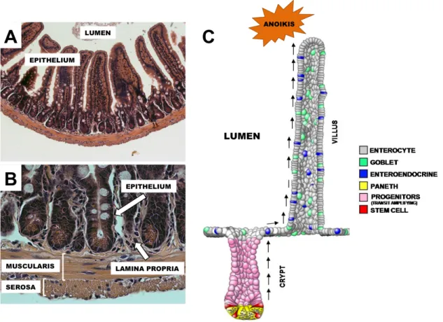
Figure 1.1: Anatomy of the small intestine. The intestinal lumen is lined by a monolayer of columnar epithelial cells, called the intestinal epithelium, which is responsible for critical nutrient processing and barrier function (A). The intestine is a complex, multilayered tissue, with the lamina propria, muscularis, and serosa acting together to support the intestinal epithelium and aid in digestive and absorptive processes (B). The intestinal epithelium forms a regular micro anatomical landscape consisting of crypts and villi (C). New epithelial cells are produced by ISCs in the base of the intestinal crypts at a rapid rate under normal conditions. These cells then undergo multiple round of differentiation and lineage specification as they migrate toward the villus tip, where they are sloughed off after ~5-7 days and undergo programmed cell death, called anoikis (C).
48
49 CHAPTER 2
CD24 AND CD44 MARK HUMAN INTESTINAL CELL POPULATIONS WITH CHARACTERISTICS OF ACTIVE AND FACULTATIVE STEM CELLS1,2
OVERVIEW
Recent seminal studies have rapidly advanced the understanding of intestinal epithelial stem cell (ISC) biology in murine models. However, the lack of techniques suitable for isolation and subsequent downstream analysis of ISCs from human tissue has hindered the application of these findings toward the development of novel diagnostics and therapies with direct clinical relevance. This study demonstrates that the cluster of differentiation genes CD24 and CD44 are differentially expressed across LGR5 positive “active” stem cells as well as HOPX positive “facultative” stem cells. Fluorescence-activated cell sorting enables differential enrichment of LGR5 cells (CD24-/CD44+) and
HOPX (CD24+/CD44+) cells for gene expression analysis and culture. These findings provide the fundamental methodology and basic cell surface signature necessary for isolating and studying intestinal stem cell populations in human physiology and disease.
1 previously published material: John Wiley & Sons, Inc., used with permission. The original citation is as follows: Gracz, et al. “CD24 and CD44 mark human intestinal cell populations with characteristics of active and facultative stem cells”, Stem Cells 31:9, 2024-2030.
2 full list of contributing authors: Gracz AD, Fuller MK, Wang F, Li L, Stelzner M, Dunn JCY, Martin MG, and Magness ST.
50 INTRODUCTION
Lgr5 was the first validated ISC biomarker shown to be expressed in actively cycling mouse crypt base columnar cells (CBCs) 19. Subsequent studies demonstrated a secondary, “reserve” population of mouse ISCs marked by Bmi1, Hopx, mTert, and Lrig
27-30. Emerging evidence indicates overlapping expression of Lgr5 with these “reserve”
ISC biomarkers; however, Lgr5-negative cell populations have also been shown to dedifferentiate in response to damage, suggesting the existence of one or more functionally competent ‘facultative’ ISC populations 33, 36, 39. Despite these advances in ISC biomarker discovery, FACS isolation and functional characterization of putative ‘”active” and “reserve/facultative” ISC populations from human intestinal tissue has been limited by the lack of validated human ISC biomarkers and in vitro assays to functionally test stemness at the single cell level.
Investigators in other stem cell fields have utilized FACS-based approaches, which rely on multiple cell surface antigens, to isolate target stem cell populations of varying purity. Notably, biomarkers comprised of cluster-of-differentiation (CD) genes have long been used to identify hematopoietic stem cells and their progenitors 132. We recently adopted a similar strategy to demonstrate that low levels of CD24 facilitate FACS of Sox9Low/Lgr5+
murine ISCs capable of forming enteroids in vitro 21. Similarly, CD44 is expressed in the stem cell zone of the murine small intestine and can be used to enrich for Lgr5+ CBCs 133. In this study we explored whether CD24 and CD44 could be used to FACS-isolate
51 RESULTS AND DISCUSSION
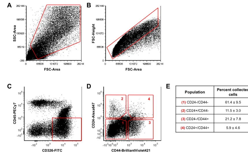
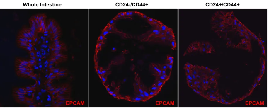
CD24 and CD44 expression was assessed on human jejunum derived from patients who had undergone roux-en-y gastric-bypass surgery. Immunostaining demonstrates that CD44 is expressed on cells from the base of the crypt to the crypt-villus junction (Figure 2.1A, C, & E). By contrast, the crypt-villus epithelium does not show appreciable CD44 staining (Figure 2.1B & D). CD24 demonstrates similar expression to CD44 in the epithelium with the notable exception that staining is primarily distributed along the apical membrane (Figure 2.1F & H, arrows). A minority population of crypt-based cells expresses high levels of cytoplasmic CD24 (Figure 2.1G & I, arrows). While CD24/44 expression is highly restricted to the stem cell zone in the epithelium, there is broad expression in non-epithelial cells in the lamina propria, sub-mucosa, and muscle (Figure 2.1A-I). EpCAM (CD326) expression is unique to all crypt and villus epithelial cells, and was deemed useful for positive FACS selection to separate epithelial from non-epithelial CD24/44-expressing cells (Figure 2.1J-L).
52
CD24/44 in the appropriate populations (Figure 2.4A & B). The data demonstrate that CD24-/CD44+ populations are most enriched for the “active” cycling ISC markers,
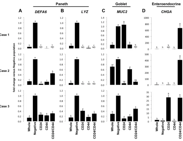
LGR5 and OLFM4 (Figure 2.5A & B); and the CD24+/CD44+ population is most enriched for the “reserve/facultative” ISC marker HOPX (Figure 2.5C). Interestingly, all CD24+ and CD44+ populations demonstrate significant de-enrichment for Paneth cell markers DefensinA6 (DEFA6) (Figure 2.6A) and Lysozyme (LYZ) (Figure 2.6B), and goblet cell marker, Mucin2 (MUC2) (Figure 2.6C), all of which associate with CD24 expression in mice 8, 39, 134. However, the CD24+/CD44+ population is highly enriched for the enteroendocrine cell marker Chromogranin A (CHGA) which is consistent with observations made in mice (Figure 2.6D) 21.
GSK-53
inhibition significantly improved 7-day enterosphere survival in the CD24-/CD44+ population, but had no appreciable effect on the CD24+/CD44+ population (Figure 2.7B & C). Initial GSK-inhibition greatly increased 14-day survival of the CD24-/CD44+ population, but was insufficient for the maintenance of CD24+/CD44+ derived enteroids, which did not survive to 14 days, regardless of GSK-inhibition (Figure 2.7C). Previous studies have shown that Lgr5-negative populations can de-differentiate and function as “facultative” stem cells when presented with the proper extrinsic cues 36, 39. Additionally, co-culture of primary human intestinal crypts with myofibroblasts enhances culture efficiency 137. In an attempt to “activate” the CD24+/CD44+ population, we co-cultured the cells with myofibroblasts isolated from human jejunal submucosa. Surprisingly, when grown in these conditions, both CD24-/CD44+ and CD24+/CD44+ cells produced long-lived enteroids, at rates of 0.07% and 0.76%, respectively (Figure 2.8).
54
occurrence of MUC2 positive cells in the enteroids. Importantly, co-culture with myofibroblasts did not induce growth in CD24-/CD44- and CD24+/CD44- populations, demonstrating that functional stemness under all tested growth conditions remains restricted to CD24-/CD44+ and CD24+/CD44+ populations.
55
In addition to FACS-enrichment of ‘active’ LGR5+ ISCs, the CD24/CD44 approach facilitates differential isolation of HOPX+ cells, which emerging evidence suggests may represent facultative ISCs 29. While several of the markers examined here exhibit a degree of variability between samples, it is clear that the population of cells most enriched for LGR5 is distinct from the population most enriched for HOPX, unlike recent observations of Hopx expression in murine Lgr5+ cells 33. Additionally, CD24-/CD44+ and CD24+CD24-/CD44+ populations exhibit differential behavior in vitro, both in response to GSK-inhibition and in basal culture conditions required for growth, further supporting the existence of phenotypically distinct ISC populations. Though overlap of
56 MATERIALS AND METHODS
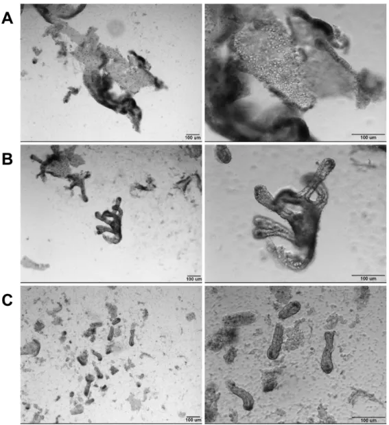
Patients/Tissue collection and preparation. De-identified tissue from female patients ranging between 33-53 yrs of age with body mass indices of 39-60 kg/m2 was used in this study. Tissue was obtained from laparoscopic roux-en-y gastric bypass surgery and represents jejunal segments of approximately 4 cm in length. Following resection, tissue was placed in a specimen cup on ice until a mucosectomy was performed, aided by injecting ice-cold saline between the mucosa and submucosa prior to careful dissection. Single cell dissociation was carried out on a small portion of the total mucosa (1 cm x 1 cm) for gene expression studies and a larger tissue area (4cm x 4cm) was dissociated for culture experiments. For an informative comparison, the mass of mucosa used for this preparation represents approximately 300- and 1200-times the mucosal mass of an average biopsy from endoscopy or colonoscopy at UNC (13 mg/biopsy; unpublished, Drs. Tope Keku/Robert Sandler), respectively. Following dissection, mucosa was placed in 3 mM EDTA in 1x PBS for 45 min at 4°C on a rocker to remove villi. The villus fraction was discarded (Figure 2.11A) and the remaining mucosa was then transferred into 5 mL of PBS and lightly shaken by hand (approximately 1 shake/sec for 2 min) to remove the remaining epithelium (Figure 2.11B). An equal volume of 2% Sorbitol made in 1x PBS (Sigma, St. Louis, MO) was added. To further deplete the solution of contaminating villi, the epithelial solution was passed through a 70µm filter. This procedure results in a ‘crypt-enriched’ epithelial preparation (Figure 2.11C). The crypts were pelleted at 150x g for 10 min at 4°C. Crypts were then digested to single cells by
57
solution was then manually shaken for 30 sec (3-4 shakes/sec). The solution was then checked for extent of dissociation to single cells. If cell clumps remained, shaking cycle was repeated every 5 minutes, then checked for extent of dissociation. Shaking cycles were stopped at the earliest time point at which 80-90% of crypts were dissociated to completion or up to 30 min maximum. An average of 1 x 10⁷ cells was obtained from a 1 cm x 1 cm mucosal segment. Single cells were filtered using a 40µm filter to remove undissociated clumps. For FACS, 1 x 10⁷ cells were placed in 500µL of ISC staining media (see Flow cytometry section, below) for antibody staining. Human tissue used in this study was deemed exempt from full Institutional Review Board review (approval #09-2159).
58
Pleasanton, CA), Mucin2 (1:100, Santa Cruz Biotechnology, Santa Cruz, CA), Sucrase Isomaltase A-17 (1:100, Santa Cruz), ChromograninA (1:500, Immunostar, Hudson, WI). Anti-Rabbit-Cy3 (1:500 Sigma, St. Louis, MO, C2306) and anti-Rat-Cy3 (1:500 Jackson Immunoresearch, Carlsbad, CA, 112-165-003) secondary detection antibodies were diluted in Dako Antibody Diluent and applied to tissue for 30 min at room temperature. Nuclei were stained for 10 minutes with bisbenzamide (1:1,000, Sigma) diluted in PBS. Background staining was negligible as determined by nonspecific IgG staining. Images were collected by capturing ~1 µm optical sections using a Zeiss LSM 710 confocal microscope.
59
DMEM/F12 (Gibco), N2 (Gibco), B27 (Gibco), Glutamax (Gibco), Penicillin/Streptomycin (Gibco), 10mM HEPES (Gibco), 10µM Y27632 (Selleck Chemicals), and 500mM N-acetyl-cysteine (Sigma)]. FACS was conducted using an iCyt Reflection (Visionary BioScience) for RNA collection or FACSAria (BD Biosciences, San Jose, CA) for cell culture experiments. Dead cells and debris were first excluded based on size via bivariate plot of forward scatter (FSC) vs. side scatter (SSC) (Supplemental Figure 3A). Doublets/multimers were excluded using a bivariate plot of FSC peak vs. FSC length (Supplemental Figure 3B). Epithelial cells were FACS-enriched by sorting EpCAM (CD326) positive, CD45 negative cells (Figure 2.3C). The remaining cell events were analyzed for CD24 and CD44 expression on a bivariate plot (Figure 2.3D). Five cell populations: CD45-EpCAM+(whole), CD44-(negative), CD45-EpCAM+CD24+CD44-(CD24+CD44-), CD45-EpCAM+CD24-CD44+(CD24-CD44+), CD45-EpCAM+CD24+CD44+(CD24+CD44+) were collected directly into 500 µl of RNA lysis buffer (Ambion RNAqueous Micro, Grand Island, NY) for gene expression analysis. For cell culture experiments, cells were collected into 500µL of ISC Staining Media.
60
and 300U/mL collagenase I (Sigma) and rotated for 25 min at room temperature. 10mL of DMEM +10%FBS was added to quench the reaction and the tissue suspension was pipetted vigorously ~50 times to further mechanically dissociate myofibroblasts. The tissue suspension was centrifuged at 300g for 5min and the resulting supernatant and tissue remnants were plated separately in DMEM +10%FBS. Media was changed every 24hrs. Cultures initiated from the supernatant of the prep produced myofibroblasts, which were passaged 3 times before use in ISC culture experiments.
Intestinal epithelial stem cell culture. Cells were pelleted and resuspended in ES-qualified Matrigel (BD Biosciences) containing ISC culture growth factors (see Table 2.1). For feeder-free cultures, 10µL Matrigel droplets were plated in 96-well plates and overlaid with 100µL ISC Sort/Culture Media, with or without 2.5µM CHIR99021 (Selleck Chemical) following 15 minute of polymerization at 37°C. For feeder co-culture, 25µL Matrigel droplets were allowed to polymerize in 12-well transwell inserts (BD Biosciences) before being placed in wells containing fibroblast feeder cells and 500µL ISC Sort/Culture Media. An additional 500µL of the same media was placed in the transwell to prevent drying of the Matrigel droplet. CHIR99021 was not used in co-culture experiments. To facilitate differentiation of enteroids, Wnt3a, SB202190, and nicotinamide were withdrawn at 14 days of culture, as previously described for whole crypt cultures83.
61
protocols. cDNA was generated using iTaq Reverse Transcription Supermix (Bio-Rad, Hercules, CA). Real-time PCR was conducted for each sample in triplicate on 1/20,000 of the total amount of cDNA generated. Taqman probes [18S, HS99999901; CD24
Hs00273561_s1; CD44, Hs01075861_m1; LGR5 Hs00173664_m1; OLFM4
HS00197437_m1; HOPX Hs04188695_m1; DEFA6 Hs00427001_m1; LYZ Hs00426232;
MUC2 Hs00159374_m1; CHGA Hs00900373_m1] for each gene were obtained from Applied Biosystems (Pleasanton, CA) and used in reactions according to the manufacturer’s protocol.
62
63
64
65
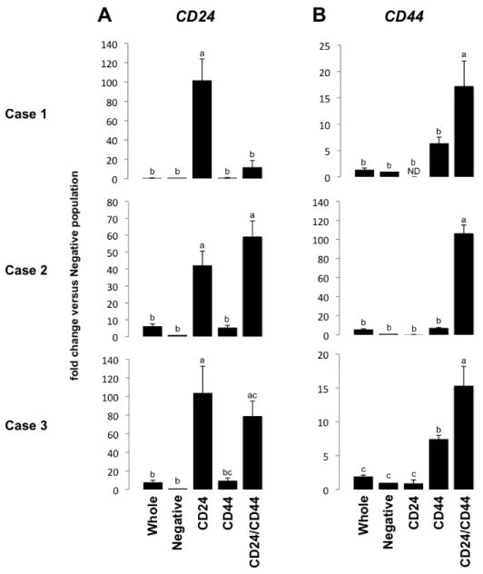
Figure 2.4 – CD24/CD44 sorting strategy enriches for CD24- and CD44-positive populations. qPCR for CD24 (A) and CD44 (B) demonstrates population-specific enrichment for each marker, validating FACS parameters. Letters a-c above each bar indicate data points that are statistically different from each other (p < 0.05).
66
Figure 2.5 - CD24-/CD44+ and CD24+/CD44+ intestinal epithelial cells are enriched for active and reserve/facultative markers, respectively. CD24-/CD44+ cells are significantly enriched for active ISC markers (A) LGR5 and (B) OLFM4, while CD24+/CD44+ cells exhibit enrichment for (C) HOPX, which is associated with reserve/facultative ISCs. Letters a-d above each bar indicate data points that are statistically different from each other (p < 0.05).
67
Figure 2.6 - CD24/CD44 populations are de-enriched for Paneth and goblet cell markers. Gene expression analysis demonstrates that CD24 and CD44 populations are significantly de-enriched for Paneth cell markers DEFA6 (A) and LYZ (B) and demonstrate enriched expression of these markers in CD24/CD44 negative populations over whole epithelium. De-enrichment is also observed for goblet cell marker MUC2 (C) in two of three cases, with no statistical difference between MUC2 in CD24-/CD44- and CD24+/CD44- populations in Case 1, which may be attributable to patient-patient heterogeneity. High levels of enteroendocrine marker CHGA are observed specifically in the CD24+/CD44+ population in two of three cases, with significant enrichment in CD24+/CD44- and CD24+/CD44+ populations in Case 3 (D), consistent with reports in mice. Letters a-g above each bar indicate data points that are statistically different from each other (p < 0.05).
68
69
Figure 2.8 - Co-culture with myofibroblasts induces long-lived enteroid formation in CD24-/CD44+ and CD24+/CD44+
populations. Both LGR5-associated CD24-/CD44+ and HOPX-associated CD24+/CD44+ cells form enteroids when co-cultured on
transwells with myofibroblasts isolated from human jejunal remnants. Enteroids are shown at 14 days post-plating.
70
71
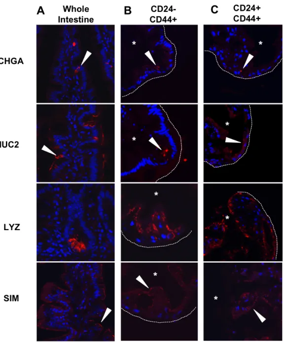
Figure 2.10 -Enteroids derived from CD24-/CD44+ and CD24+/CD44+ populations are multipotent. Enteroendocrine, goblet, and Paneth cells, as well as absorptive enterocytes, are detectable in whole human jejunum by expression of CHGA, MUC2, LYZ, and SIM, respectively (A). Similar expression patterns for each marker are observed by immunofluorescence in enteroids derived from the LGR5-associated CD24-/CD44+ population (B) and the HOPX-associated CD24+CD24-/CD44+ population (C). Arrows indicate positive staining, asterisks mark enteroid lumens, and dotted lines denote outer edge of enteroid structures. Scale bars represent 50µm.
72
Figure 2.11 - Epithelial preparations enrich for stem cell containing crypts. Initial shaking steps remove large villi (A). After villus fractions are discarded, additional shaking yields both crypts and some remaining villi (B). Filtering results in single isolated crypts, with some debris and single cells from submucosal tissues (C). Scale bars represent 100µm.
73
74
CHAPTER 3
A HIGH THROUGHPUT, CLONAL PLATFORM FOR STUDYING THE INTESTINAL STEM CELL NICHE IN VITRO3
OVERVIEW
Rapid advances in intestinal stem cell (ISC) biology driven by biomarker
identification and elegant in vivo lineage-tracing assays indicate that complex niche
dynamics govern ISC multipotency and self-renewal. While the ISC niche is emerging as
a key player in the regulation and maintenance of stemness in the intestine, current
functional assays preclude statistically meaningful studies of single ISCs and ISC-niche
interactions. Here we describe the development of an in vitro platform that facilitates
high-throughput clonogenic culture and computational identification of isolated ISCs and
niche cells in co-cultures at single cell and microscale (≤ 7 cells) resolution. This new in
vitro platform has broad applicability in studies aimed at dissecting stem cell
heterogeneity and stem cell-niche interactions at the single cell level.
3 full list of contributing authors: Gracz AD, Williamson IA, Johnston MJ, Wang F,
Wang Y. Attayek P, Balowski J, Liu XF, Laurenza RJ, Galanko JA, Sims CE, Li L, Allbritton NL, and Magness ST
75
INTRODUCTION
Throughout the life of the adult organism, somatic stem cells are responsible for
maintaining tissue homeostasis following physiological “wear and tear” and damage from
injury. Understanding how somatic stem cells self-renew and differentiate to produce the
functional cells of their resident tissue is essential for determining the mechanisms
underlying a broad range of issues related to human health and disease, including
degenerative disease, cancer, and aging. Many stem cell types reside within specialized
niches, where their behavior, including activity/quiescence, self-renewal, and
differentiation, are likely influenced by extrinsic signaling factors. While the concept of
stem cell niches was first applied to hematopoietic stem cells, the importance of niche
cells has now been demonstrated for a number of mammalian tissues, including the brain,
skeletal muscle, hair follicle, and intestine 46, 138. As signals from the niche are thought to
play a significant role in modulating stem cell fate decisions in physiology and disease,
functional assays for studying stem cell response to experimentally modulated signaling
are invaluable for understanding how stem cells behave in their native environments.
However, functional stemness remains difficult to study in a statistically meaningful way
in vitro for many adult tissue types.
The intestinal epithelium undergoes one of the most rapid rates of renewal of any
76
driven physiological renewal. Recent studies demonstrate that crypt-base columnar cells
(CBCs), marked by high levels of Lgr5 and low levels of Sox9, are capable of driving
long term regeneration in vivo and formation of “enteroid” structures in vitro, both
characteristics of functional ISCs 19-21, 24. These CBC ISCs are closely associated with
Paneth cells, which have been shown to function as niche cells and express soluble and
insoluble factors associated with stemness, such as Wnt and Notch ligands 8, 62, 70.
Additionally, Paneth cells have been shown to increase the efficiency of enteroid
formation by isolated ISCs in vitro 8.
Though the intestinal epithelium possesses many benefits as a model system for
somatic stem cell biology, technical limitations hinder efficient and detailed functional
and mechanistic studies of ISC-niche interactions. While colonic enteroids generated in
vitro have been successfully transplanted into recipient mice, the field currently lacks a
robust in vivo transplant assay to study single ISCs at a clonal level, a tool that has driven
the understanding of stem cell niches in the hematopoietic system, mammary glands, and
testis 139-141.
Recently, stem cell-niche interactions in endothelial and spermatogenic cell
populations have been probed by “recapitulating” the endogenous niche environment in
vitro through co-culturing strategies 142, 143. Similar studies have examined ISC-Paneth
cell interactions, both under normal conditions and following calorie restriction 8, 85.
However, these studies relied on the co-culture of hundreds of ISCs with hundreds of
77
crypts, where much smaller numbers of ISCs and Paneth cells (~15 per crypt) interact 8,
41, 144.
In the present study, we describe a novel platform to study large numbers of
single ISCs simultaneously, either at the clonal level or in the presence of one or more
Paneth cells. Microfabricated culture arrays modified for long-term 3-dimensional culture
are used to capture and functionally assay single ISCs and clonally derived intestinal
enteroids. Post-hoc computational identification of initial array contents coupled with
day-to-day tracking of ISCs and ISC-Paneth cell co-cultures, through the use of unique
addresses, facilitates the high-throughput analysis of ISC development into enteroid
structures. To provide proof-of-principle for interrogating ISC-niche interactions, we
co-culture ISCs with Paneth cells and assess enteroid formation from 15 different
combinations of ISCs and Paneth cell numbers. Surprisingly, we were unable to
demonstrate a statistically significant correlation between the presence of Paneth cells
and the likelihood of ISCs to form enteroids. Together, our methodology provides a novel
platform for high-throughput screening of primary ISCs and in vitro recapitulation of the
ISC niche, and suggests that ISC-Paneth cell interaction may be subtler than previously
thought.
RESULTS
Existing cell culture arrays are adaptable to long-term culture
Previous studies have utilized microfabricated devices consisting of arrayed
78
microarray format 145. We hypothesized that a similar approach could be utilized to
isolate and culture single ISCs in three-dimensional extracellular matrix required for the
growth of primary isolated ISCs. In the present study, we adapted a previously described
platform, termed microwell arrays, designed for the isolation of viable single cells
without advanced cell sorting techniques 145, 146. The arrays are fabricated from
polydimethylsiloxane (PDMS) using standard photolithography and composed of up to
12,000 microwells (Figure 3.1). As PDMS is known to be a poor substrate for some cell
types, an additional element (termed microraft) is made from Petri dish grade polystyrene
and embedded in each microwell of the array using dip-coating methods 145 (Figure
3.1D-F).
Since ISCs require several days to develop into enteroid structures, microraft
arrays had to be amenable to media and growth factor changes 21, 24. Additionally,
separate media compartments were required in order to facilitate simultaneous
examination of multiple experimental conditions on a single microraft array. To meet
these requirements, optically clear polycarbonate cassettes, with dividers to create media
reservoirs (termed culture chambers), were bonded to the microraft arrays (Figure 3.1H &
Figure 3.2A). Cassettes were fabricated with two (~5,000 microwells per culture
chamber) and four (~2,500 microwells per culture chamber) culture chambers (Figure
3.2B). Microwells were sized at 200µm2, with 30µm spacers separating microwells from
their immediate neighbors (Figure 3.2C). Microwells were designed to have a depth of
79
polystyrene rafts at 5 microwell intervals, were included in the array design to allow for
tracking of unique enteroids across many time points (Figure 3.2C).
To facilitate high-throughput analysis, we used tile-scanning microscopy to
produce high-resolution, high-magnification images of whole microraft arrays. Initial
attempts revealed significant out-of-plane sagging in the z-axis, due to the elastic
property of the PDMS array (Figure 3.2D&F). To prevent sagging of the array during
imaging, the PDMS template was bonded to a glass slide by using a thin layer of
polyacrylic acid (PAA) prior to attaching the cassette (Figure 3.1A). Microraft arrays
bound to glass with PAA, which can be dissolved in PBS to release the array and
attached cassette from the glass slide, were reproducibly imaged in a single Z-plane by
tile-scanning without any noticeable out-of-focus sagging (n = 50) (Figure 3.2F&G).
Microraft arrays are biocompatible with long-term three-dimensional ISC culture
Next, we wished to validate that the PDMS/polystyrene microraft arrays were
capable of supporting ISC growth and development into enteroids, without cytotoxic
effects. To facilitate accurate localization of isolated ISCs in the microraft arrays, we
crossed Sox9EGFP mice to CAGdsRed reporter mice, which express the dsRed
fluorescent transgene ubiquitously across all cell and tissue types, and isolated Sox9Low
ISCs from dual transgenic mice by fluorescence activated cell sorting (FACS) (Figure
3.3A) 21, 147. Additionally, we reasoned that inclusion of a constitutive transgenic reporter
in the form of dsRed fluorescence would allow us to identify any contaminating