Low Heart Rate Variability in a 2-Minute
Electrocardiogram Recording Is Associated
with an Increased Risk of Sudden Cardiac
Death in the General Population: The
Atherosclerosis Risk in Communities Study
Ankit Maheshwari1*, Faye L. Norby2, Elsayed Z. Soliman3, Selcuk Adabag4, Eric A. Whitsel5, Alvaro Alonso6, Lin Y. Chen1
1Cardiovascular Division, Department of Medicine, University of Minnesota Medical School, Minneapolis, Minnesota, United States of America,2Division of Epidemiology and Community Health, School of Public Health, University of Minnesota, Minneapolis, Minnesota, United States of America,3Epidemiological Cardiology Research Center (EPICARE), Wake Forest School of Medicine, Winston-Salem, North Carolina, United States of America,4Division of Cardiology, Veteran Affairs Medical Center, Minneapolis, Minnesota, United States of America,5Departments of Epidemiology and Medicine, University of North Carolina, Chapel Hill, North Carolina, United States of America,6Department of Epidemiology, Rollins School of Public Health, Emory University, Atlanta, Georgia, United States of America
Abstract
Low heart rate variability (HRV) has been linked to increased total mortality in the general population; however, the relationship between low HRV and sudden cardiac death (SCD) is less well-characterized. The goal of this study was to evaluate the relationship between low HRV and SCD in a community-based cohort. Our cohort consisted of 12,543 participants from the Atherosclerosis Risk in Communities (ARIC) study. HRV measures were derived from 2-minute electrocardiogram recordings obtained during the baseline exam (1987–89). Time domain measurements included the standard deviation of all normal RR intervals (SDNN) and the root mean squared successive difference (r-MSSD). Frequency domain measurements included low frequency power (LF) and high frequency (HF) power. During a median follow-up of 13 years, 215 SCDs were identified from physician adjudication of all coronary heart disease deaths through 2001. In multivariable adjusted Cox proportional hazards models, each standard deviation decrement in SDNN, LF, and HF were associated with 24%, 27% and 16% increase in SCD risk, respectively. Low HRV is independently associated with increased risk of SCD in the general population.
a11111
OPEN ACCESS
Citation:Maheshwari A, Norby FL, Soliman EZ, Adabag S, Whitsel EA, Alonso A, et al. (2016) Low Heart Rate Variability in a 2-Minute
Electrocardiogram Recording Is Associated with an Increased Risk of Sudden Cardiac Death in the General Population: The Atherosclerosis Risk in Communities Study. PLoS ONE 11(8): e0161648. doi:10.1371/journal.pone.0161648
Editor:Alena Talkachova, University of Minnesota, UNITED STATES
Received:May 3, 2016
Accepted:August 9, 2016
Published:August 23, 2016
Copyright:This is an open access article, free of all copyright, and may be freely reproduced, distributed, transmitted, modified, built upon, or otherwise used by anyone for any lawful purpose. The work is made available under theCreative Commons CC0public domain dedication.
Introduction
The cardiac autonomic nervous system is comprised of sympathetic and parasympathetic branches. Cardiac function is tightly regulated at multiple levels by the dynamic equilibrium
achieved between these systems [1]. Heart rate variability (HRV) is a non-invasive marker for
autonomic input to the heart [2]. Increased sympathetic input decreases HRV, whereas
increased parasympathetic input increases HRV. Cardiac arrhythmias are often initiated by or
occur in patients with enhanced sympathetic and diminished parasympathetic tone [2].
Distur-bances in autonomic equilibrium have been implicated in several cardiovascular conditions
including myocardial infarction (MI), heart failure, and sudden cardiac death (SCD) [1]. In
post-myocardial infarction (MI) patients and heart failure patients, low HRV has been
associ-ated with increased risk of all-cause mortality and SCD [3–8]. In the general population, low
HRV has been linked to increased risk of cardiovascular events and all-cause mortality [9–11];
however, the relationship between low HRV and SCD risk is less well-characterized. We hypothesized that attenuated HRV is an independent risk factor for SCD in the general
popula-tion. We tested our hypothesis in the Atherosclerosis Risk in Communities (ARIC) Study—a
large community-based cohort study of cardiovascular disease in the United States.
Methods
Study Population
The ARIC study is a community-based, prospective cohort study designed to identify and eval-uate risk factors, etiology, and clinical manifestations of atherosclerotic coronary heart disease
(CHD) in the general population. Between 1987 and 1989, 15,792 men and women aged 45–64
years were recruited and enrolled from four United States communities (Washington County, MD; Forsyth County, NC; Jackson, MS; and suburban Minneapolis, MN). Participants under-went an initial baseline assessment that consisted of a clinical examination, serum laboratory analysis, standard 12-lead electrocardiogram (ECG), 2-minute ECG recording, and a detailed health, behavioral, and socio-demographic history assessment. Follow-up data were obtained from study visits, annual phone calls, local hospital surveillance, and querying of the National Death Index. Approval for the study was obtained from the institutional review board on human research at each participating institution and all participants provided written informed consent. A full list of participating institutional review boards can be found in the supporting
information section (S1 Table). Further details outlining study design, outcome ascertainment
procedure, and population statistics have been previously described [12].
For our primary analysis, we considered all 15,792 participants at the baseline visit and excluded those with missing covariates (n = 15), missing HRV data (n = 812), HRV recordings of inadequate duration (n = 210), poor quality ECG recordings due to technical issues
(n = 636), HRV recordings with inadequate number of acceptable RR intervals (n = 1473), and those who were not white or black from all study sites, and nonwhite from Minneapolis and Washington County (due to small sample size; n = 103) resulting in a final cohort of 12,543 participants. For our secondary analysis, we analyzed a sample of our cohort with lower per-ceived SCD risk by further excluding those with prevalent CHD or heart failure (n = 1,012) resulting in a final cohort of 11,531 participants.
Heart Rate Variability Measurement
Measures of HRV were obtained from supine 2-minute ECG recordings obtained during the baseline exam after participants had been resting in the supine position for 20 minutes between 8:30 AM and 11:30 AM. Participants were instructed to refrain from smoking or ingesting Funding:The Atherosclerosis Risk in Communities
Study is carried out as a collaborative study supported by National Heart, Lung, and Blood Institute contracts (HHSN268201100005C, HHSN268201100006C, HHSN268201100007C, HHSN268201100008C, HHSN268201100009C, HHSN268201100010C, HHSN268201100011C, and HHSN268201100012C). The authors thank the staff and participants of the ARIC study for their important contributions.
Competing Interests:The authors have declared that no competing interests exist.
caffeine at least 12 hours prior to the procedure. Protocols for data processing, artifact identifi-cation, imputation, and quality control of HRV records have been previously described
[13,14]. Briefly, the RR interval was converted into beat-to-beat heart rate including a record of
the real time of each beat. Potential ectopy and artifact was determined by a computer algo-rithm that identified all RR intervals outside the upper and lower limits (+/- 25%) of a 5-beat moving average. These beats were removed and the intervening data was linearly smoothened and interpolated using variance preserving imputation software, PREDICT II HRVECG
(Arrhythmia Research Technology Inc, Fitchburgh, MA). Records were excluded if>20% of
RR intervals were affected to reduce the influence of data imputation. After imputation, heart rate data were converted back to RR intervals for further data processing, time domain analysis, and spectrum analysis using specialized software, PREDICT II HRVECG (Arrhythmia
Research and Technology Inc, Fitchburg, MA). In our analysis, time domain measurements included the standard deviation of all normal RR intervals (SDNN) and the root mean squared successive difference (r-MSSD). Frequency domain measurements included low frequency power (LF), defined as the energy in the heart period power spectrum between 0.04 and 0.15 Hz, and high frequency (HF) power, defined as the energy in the heart period power spectrum between 0.15 and 0.40 Hz.
Sudden Cardiac Death
The outcome of our study was SCD. In ARIC, comprehensive data on all cardiovascular events and deaths was obtained from available death certificates, coroner reports, informant inter-views, hospital records, and autopsy reports. The cause of death was adjudicated by the ARIC Morbidity and Mortality Classification Committee following a standard protocol. In order to identify SCD, all fatal CHD events through 2001 in ARIC were reviewed by an independent panel of physicians. Deaths were classified as definite sudden cardiac death, possible sudden cardiac death, not sudden cardiac death, and unclassifiable. Definite sudden cardiac death was defined as a sudden pulseless condition presumed to be of cardiac origin in a previously stable individual without evidence of non-cardiac cause of death. Possible sudden cardiac death was
defined as an un-witnessed death in a previously stable (<24 hours) individual without other
evidence indicating non-cardiac origin for instantaneous death [15,16]. All deaths classified as
SCD had to occur outside of the hospital or in the emergency room. For our analysis, SCD was defined as definite or possible sudden cardiac death.
Assessment of Other Covariates
The covariates included in our analysis were age, sex, race, study center, cigarette-smoking sta-tus, prevalent CHD, heart failure, diabetes (DM), impaired fasting glucose, borderline hyper-tension (HTN), HTN, beta-adrenergic receptor blocker use, digoxin use, use of
anti-arrhythmic drugs (AADs), left ventricular hypertrophy (LVH), and body mass index (BMI). Baseline demographic data, medication use (beta-adrenergic receptor blockers, AADs, and digoxin), and medical history were obtained by ARIC staff from participants during the study visit. AADs include type IA, type IB, type IC and type III anti-arrhythmic medications. Preva-lent CHD was defined as a self-reported history of MI, coronary artery bypass grafting,
percu-taneous coronary intervention, or ECG signs of CHD [17,18]. Heart failure was defined as
stage 3“manifest heart failure”by the Gothenburg criteria or self-reported diagnosis of heart
failure [19]. LVH was defined by the Cornell ECG criteria [20,21]. BMI was defined as weight/
height2(kg/m2) [17]. HTN was defined as systolic blood pressure140 mm Hg, diastolic
blood pressure90 mm Hg, or self-reported history of anti-hypertensive medical therapy.
pressure 80–89 mm Hg. DM was defined as a fasting (minimum of 8 hours) glucose126 gm/
dL, non-fasting glucose200 mg/dL, self-reported use of oral hypoglycemic agents or insulin,
or self-reported diagnosis of DM [22]. Impaired fasting glucose was defined as fasting
(mini-mum of 8 hours) glucose of 100–125 gm/dL. Smoking status was self-reported. Participants
were classified as current smokers and non-current smokers.
Statistical Analysis
We report means with standard deviations (SDs) for continuous variables and counts with per-centages for categorical variables. Person-years at risk were calculated from the date of baseline visit until the date of SCD, other death, loss to follow-up, or end of follow-up, whichever
occurred first. The analysis was based on data obtained from 1987–2001.
Initially, we explored the association between HRV measures (as continuous variables) and SCD incidence using restricted cubic splines (knots at the 5th, 27.5th, 50th, 72.5th and 95th percentiles). Frequency domain measures were skewed to the right. Thus, we applied a natural logarithmic transformation to normalize the distribution of HRV frequency domain indices (LF and HF) when used as continuous variables in accordance with the recommendation by
the Task Force on HRV research [2]. We used Cox proportional hazards models to estimate
the hazard ratios (HRs) and 95% confidence intervals (95% CIs) of HRV measures for SCD. HRV measures were analyzed as tertiles, with the highest tertile as the reference group, and as continuous variables (per 1-SD decrement). We constructed 2 models. Model 1 was adjusted for age, sex, race, and study center. Model 2 was additionally adjusted for BMI, LVH, CHD, heart failure, impaired fasting glucose, DM, beta-blocker use, digoxin use, smoking status (cur-rent vs. not cur(cur-rent), borderline HTN, HTN, and use of AADs. We conducted a secondary analyses applying Model 2 (without adjustment for CHD or heart failure) to our cohort of par-ticipants without prevalent CHD or heart failure. Finally, we performed sex- and race-stratified analyses.
The proportional hazards assumption was assessed with scaled Schoenfeld residuals for both graphical and numerical tests, time interaction terms, and inspection of log negative log survival curves. Modeling assumptions were not violated in any model. Statistical analysis of ARIC data was performed using SAS version 9.2 (SAS Institute Inc., Cary, NC) and STATA
13.0 (StataCorp LP, College Station, TX). AllPvalues reported were 2-sided, and statistical
sig-nificance threshold was chosen as 5%.
Results
Study Population
In our cohort of 12,543 participants, we identified 215 SCD events over a median follow-up period of 13 years. This corresponded to an incidence rate (95% confidence interval) of 1.37
(1.19–1.56) per 1000 person-years. Baseline characteristics of our study cohort are shown in
Table 1. Participants with SCD were more likely to be male, black and have a history of CHD, DM, HTN, heart failure, or LVH than those without SCD.
HRV and SCD
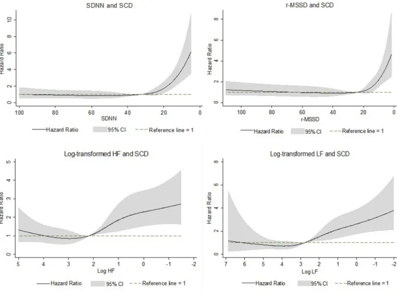
Fig 1shows the association between HRV measures and SCD risk, modeled as restricted cubic splines. For HRV values below the median, lower HRV measures were associated with higher risk of SCD after adjustment for age, sex, and race. For HRV values above the median, there
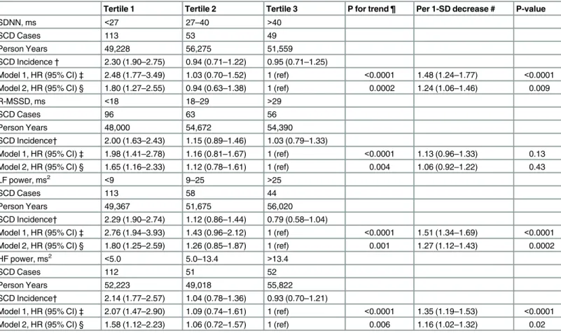
between HRV tertiles and the risk of SCD. In the demographically adjusted model (Model 1), compared with the highest tertile, the lowest tertile of SDNN and r-MSSD was significantly associated with an increased risk of SCD. This association was attenuated but remained signifi-cant after further adjustment for SCD risk factors and potential confounders (Model 2). When analyzed as a continuous variable in Model 2, each SD decrement in SDNN was associated with a significant 24% increase in SCD risk. r-MSSD was not significantly associated with SCD when analyzed in Model 2 as a continuous variable.
In the demographically adjusted model (Model 1), compared with the highest tertile, the lowest tertile of LF and HF spectral power was significantly associated with an increased risk of SCD. This association was attenuated but remained significant after further adjustment in Model 2. When analyzed as a continuous variable in Model 2, each SD decrement in LF and HF was significantly associated with 27% and 16% increase in SCD risk, respectively.
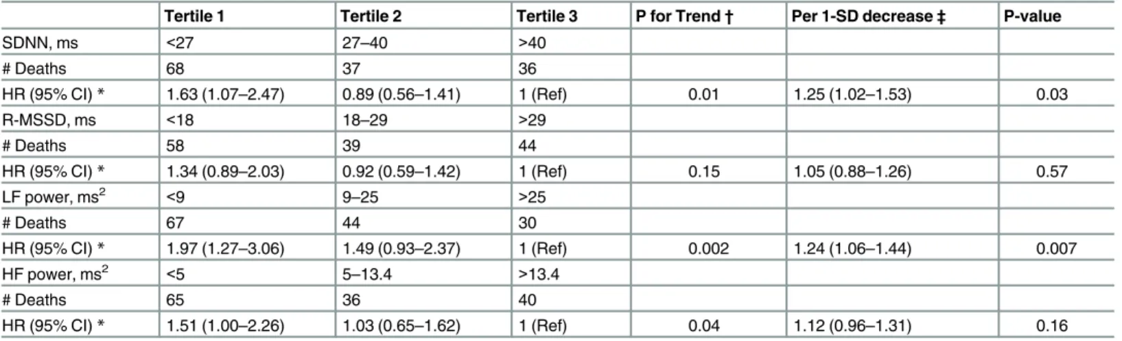
In our sample of participants without prevalent CHD or heart failure, compared with the highest tertiles, the lowest tertiles of LF spectral power, HF spectral power, and SDNN were sig-nificantly associated with an increased risk of SCD. When analyzed as a continuous variable, each SD decrement in SDNN and LF spectral power were associated with 25% and 24%
increase in SCD risk, respectively (Table 3).
We evaluated interactions with age and sex and did not find any significant sex- or
race-based interactions with respect to SCD risk (S2 Table). The association between lower HRV
and higher risk of SCD was consistently observed in women and men (S3 Table), and blacks
and whites (S4 Table).
Table 1. Baseline Participant Characteristics by Sudden Cardiac Death Status, Atherosclerosis Risk in Communities Study, 1987–2001.
Characteristic† No SCD (n = 12,328) SCD (n = 215) P-Value
Age, mean (SD), years 54.0 (5.7) 56.3 (5.7) <0.0001
Female 7,045 (57.2) 77 (35.8) <0.0001
Black race 3,173 (25.7) 91 (42.3) <0.0001
Current smoker 3,135 (25.4) 90 (41.9) <0.0001
Body Mass Index, mean (SD) kg/m2 27.6 (5.3) 28.7 (5.6) 0.003
Diabetes 1,373 (11.1) 75 (34.9) <0.0001
Hypertension 4,154 (33.7) 136 (63.3) <0.0001
Heart Failure 527 (4.3) 25 (11.6) <0.0001
Coronary Heart Disease 498 (4.0) 67 (31.2) <0.0001
Left Ventricular Hypertrophy 256 (2.1) 17 (7.9) <0.0001
Use of Digoxin 163 (1.3) 14 (6.5) <0.0001
Use of Beta-blockers 1268 (10.3) 43 (20.0) <0.0001
Use of Anti-arrhythmics 86 (0.7) 4 (1.9) 0.07
Heart Rate, mean (SD)‡ 67.7 (10.3) 70.3 (13.8) .008
SDNN, mean (SD) ms 37.2 (19.7) 31.9 (20.6) 0.002
r-MSSD, mean (SD) ms 29.2 (23.3) 27.3 (28.3) 0.25
Ln LF power, mean (SD) ms2 2.7(1.4) 2.0 (1.6) <0.0001
Ln HF power, mean (SD) ms2 2.1 (1.3) 1.6 (1.5) <0.0001
†Data are presented as no. (%) unless as otherwise stated
‡Heart rate in beats per minute obtained from 2-minute resting ECG obtained during study visit 1
Abbreviations: High Frequency (HF), Low Frequency (LF), Root Mean Squared Successive Difference (r-MSSD), Standard Deviation of Normal RR Intervals (SDNN), Sudden Cardiac Death (SCD).
Discussion
In this large biracial community-based cohort of middle-aged individuals, we found that lower HRV measured by time-domain and spectral analysis from a resting 2-minute ECG recording was independently associated with higher risk of SCD. This relationship was observed for HRV values below the median, and not above the median, suggesting a threshold effect. From race-and sex-stratified analysis, we found that this relationship was similar in whites race-and blacks race-and men and women.
The most prominent clinical risk factors for SCD are CHD and heart failure. Low HRV has been shown to be independently predictive of increased mortality in post-MI patients, heart
failure patients, and in cohorts representative of the general population [3,6,9,11,23–25]. It has
been independently associated with SCD in small studies conducted in highly selected patients
with heart failure [3,7,8,23–25] and MI [4–6,26]. The relationship between HRV and SCD has
Fig 1. Heart Rate Variability Measures and Sudden Cardiac Death Risk.Association between heart rate variability measures and sudden cardiac death incidence presented as hazard ratios (solid line) and 95% confidence intervals (shaded area). Results from Cox proportional hazards model with heart rate variability measures modeled using restricted cubic splines, adjusted for age, sex, and race. Median value of heart rate variability measures was considered the reference (HR = 1). The x-axis is presented using an inverted scale.
received limited evaluation in low risk cohorts. Algra et al demonstrated that low HRV was associated with SCD in a selected cohort of predominantly ambulatory patients (75%) with indications for 24-hour ECG monitoring including evaluation for symptoms potentially related
to cardiac arrhythmia, post-MI monitoring, and evaluation of anti-arrhythmic therapy [27].
This study, therefore, was not based on unselected subjects from the general population. Fur-ther, it was a nested case-control analysis with limited adjustment for SCD risk factors and potential confounders. Stein et al demonstrated that decreased normalized low frequency power, but not time domain HRV, derived from 24-hour ECG recordings was associated with SCD in a subset of participants from the Cardiovascular Health Study using a nested
case-con-trol analysis [28]. We extend current knowledge by demonstrating for the first time that low
HRV derived from a 2-minute ECG recording is prospectively associated with an increased
Table 2. Hazard Ratios of Sudden Cardiac Death for Heart Rate Variability Measures, Atherosclerosis Risk in Communities Study, 1987–2001.
Tertile 1 Tertile 2 Tertile 3 P for trend ¶ Per 1-SD decrease # P-value
SDNN, ms <27 27–40 >40
SCD Cases 113 53 49
Person Years 49,228 56,275 51,559
SCD Incidence† 2.30 (1.90–2.75) 0.94 (0.71–1.22) 0.95 (0.71–1.25)
Model 1, HR (95% CI)‡ 2.48 (1.77–3.49) 1.03 (0.70–1.52) 1 (ref) <0.0001 1.48 (1.24–1.77) <0.0001 Model 2, HR (95% CI) § 1.80 (1.27–2.55) 0.94 (0.63–1.38) 1 (ref) 0.0002 1.24 (1.06–1.46) 0.009
R-MSSD, ms <18 18–29 >29
SCD Cases 96 63 56
Person Years 48,000 54,672 54,390
SCD Incidence† 2.00 (1.63–2.43) 1.15 (0.89–1.46) 1.03 (0.79–1.33)
Model 1, HR (95% CI)‡ 1.98 (1.41–2.78) 1.16 (0.81–1.67) 1 (ref) <0.0001 1.13 (0.96–1.33) 0.13 Model 2, HR (95% CI) § 1.65 (1.16–2.33) 1.12 (0.78–1.61) 1 (ref) 0.004 1.06 (0.92–1.22) 0.43
LF power, ms2 <9 9–25 >25
SCD Cases 113 58 44
Person Years 49,367 51,675 56,020
SCD Incidence† 2.29 (1.90–2.74) 1.12 (0.86–1.44) 0.79 (0.58–1.04)
Model 1, HR (95% CI)‡ 2.76 (1.94–3.93) 1.43 (0.96–2.12) 1 (ref) <0.0001 1.51 (1.34–1.69) <0.0001 Model 2, HR (95% CI) § 1.80 (1.25–2.59) 1.26 (0.85–1.87) 1 (ref) 0.001 1.27 (1.12–1.43) 0.0002
HF power, ms2 <5.0 5.0–13.4 >13.4
SCD Cases 112 51 52
Person Years 52,223 49,018 55,822
SCD Incidence† 2.14 (1.77–2.57) 1.04 (0.78–1.36) 0.93 (0.70–1.21)
Model 1, HR (95% CI)‡ 2.07 (1.47–2.90) 1.09 (0.74–1.61) 1 (ref) <0.0001 1.35 (1.19–1.53) <0.0001 Model 2, HR (95% CI) § 1.58 (1.12–2.23) 1.06 (0.72–1.57) 1 (ref) 0.006 1.16 (1.02–1.32) 0.02
†Incidence is reported per 1000 person-years
‡Cox proportional hazard model adjusted for age, sex, race, and study center
§Cox proportional hazard model adjusted for age, sex, race, study center, smoking status (current vs. not current), body mass index, ECG-based left
ventricular hypertrophy, hypertension, borderline hypertension, diabetes, impaired fasting glucose, coronary heart disease, heart failure, use ofβ-blockers, use of digoxin, use of anti-arrhythmic drugs
¶P for trend calculated using the term for tertile categories
#per 1-SD decrease in log-transformed HF and LF for frequency domain
Abbreviations: Confidence Interval (CI), Hazard Ratio (HR), High Frequency (HF), Low Frequency (LF), Root Mean Squared Successive Difference (r-MSSD), Sudden Cardiac Death (SCD), Standard Deviation (SD), Standard Deviation of Normal RR Intervals (SDNN)
risk of SCD independent of established cardiovascular risk factors in a low-risk, middle-aged, community-based setting, even after excluding participants with CHD or heart failure.
We considered several mechanisms to explain this association. The majority of SCDs result from unstable ventricular tachyarrhythmias (VTAs) occurring in community-dwelling
individ-uals with undetected but significant CHD [29–35]. Such arrhythmias can be mediated by
increased sympathethic and/or decreased parasympathetic input to the heart, which may be detected by HRV analysis. In canine models, VTAs have been provoked by increased sympa-thetic activity, especially in the setting of mechanical coronary occlusion, while vagal
stimula-tion has been shown to increase the VTA threshold [36–39]. Adverse autonomic remodeling
evidenced by sympathetic nerve sprouting and hyper-innervation has been linked to the devel-opment of VTAs in canine models and observed in the myocardium of patients with a history
of VTAs [39].
Autonomic imbalance, however, is a non-specific marker linked with several cardiovascular diseases. Low HRV, in turn, has been independently associated with many SCD risk factors
including CHD [9] and obstructive CHD [40]. While we adjusted for cardiovascular risk
fac-tors in our models, we were unable to account for unreported or undetected disease. Thus, the relationship between low HRV and increased SCD risk may also be explained by underlying risk factors such as subclinical CHD.
The current paradigm for non-invasive SCD risk stratification in the general population is limited. With the majority of SCDs occurring as the first manifestation of undetected, untreated CHD, the discovery of novel SCD predictors capable of identifying at-risk individu-als in the general population earlier in the natural history of disease is paramount. Several such parameters have been investigated including proteomic, electrophysiological, genetic,
struc-tural, and autonomic variables [41,42]. While independent relationships with increased SCD
risk have been identified, large-scale investigations for purposes of risk stratification in the
Table 3. Hazard Ratios of Sudden Cardiac Death for Heart Rate Variability Measures in Low Risk Cohort (excluding participants with CHD or Heart Failure), Atherosclerosis Risk in Communities Study, 1987–2001.
Tertile 1 Tertile 2 Tertile 3 P for Trend† Per 1-SD decrease‡ P-value
SDNN, ms <27 27–40 >40
# Deaths 68 37 36
HR (95% CI)* 1.63 (1.07–2.47) 0.89 (0.56–1.41) 1 (Ref) 0.01 1.25 (1.02–1.53) 0.03
R-MSSD, ms <18 18–29 >29
# Deaths 58 39 44
HR (95% CI)* 1.34 (0.89–2.03) 0.92 (0.59–1.42) 1 (Ref) 0.15 1.05 (0.88–1.26) 0.57
LF power, ms2 <9 9–25 >25
# Deaths 67 44 30
HR (95% CI)* 1.97 (1.27–3.06) 1.49 (0.93–2.37) 1 (Ref) 0.002 1.24 (1.06–1.44) 0.007
HF power, ms2 <5 5–13.4 >13.4
# Deaths 65 36 40
HR (95% CI)* 1.51 (1.00–2.26) 1.03 (0.65–1.62) 1 (Ref) 0.04 1.12 (0.96–1.31) 0.16
*Cox Proportional Hazard Models adjusted for age, sex, race, study center, smoking status (current vs. not current), body mass index, ECG-based left ventricular hypertrophy, hypertension, borderline hypertension, diabetes, impaired fasting glucose, use ofβ-blockers, use of digoxin, use of anti-arrhythmic drugs
†P for trend calculated using the term for tertile categories
‡per 1-SD decrease in log-transformed HF and LF for frequency domain
Abbreviations: Standard deviation of normal RR intervals (SDNN), root mean squared successive difference (r-MSSD), high frequency (HF), low frequency (LF), Sudden cardiac death (SCD), hazard ration (HR), confidence interval (CI), standard deviation (SD)
general population are limited. Our findings suggest that low HRV in a 2-minute ECG is an independent risk factor for SCD. The population attributable risk, however, is modest and the clinical utility, if any, of our findings is not established and will need to be clarified.
The principal strengths of our study include the prospective design, length of follow up
(>10 years), large cohort size (>10,000 participants), and the inclusion of whites and blacks.
Some limitations should be noted. First, HRV data were only obtained from 2-minute ECG
recordings. Short (2–15 min) recordings may not encapsulate effects of the circadian rhythm
and daily activity compared with long (24 hour) recordings. However, HRV measures derived
from short and even ultra-short (10s) recordings are reliable [43–45]. As such, short recordings
have been employed in numerous epidemiological analyses as they pose a significant advantage over long recordings with respect to feasibility. Second, approximately 20% of ARIC partici-pants were excluded due to missing HRV data, technical issues with 2-minute ECG recordings, and artifact. The missing data and technical issues resulted from logistical problems that were likely random in nature; thus, it would not have biased our results. In the HRV estimation pro-tocol, potential ectopy and artifact was identified by statistical criteria imposed on RR intervals, not morphological analysis of the QRS complex. Thus, we cannot be certain that all QRS com-plexes analyzed indeed resulted from sinus node depolarization.
In summary, low HRV in a 2-minute resting ECG is independently associated with an increased risk of SCD in the general population. Further studies are needed to determine if the inclusion of HRV in a multi-marker approach would improve risk prediction of SCD in the general population.
Supporting Information
S1 Table. List of participating ARIC centers. (DOCX)
S2 Table. Sex and Race Interactions. (DOCX)
S3 Table. Sex-Stratified Hazard Ratios of Sudden Cardiac Death for Heart Rate Variability Measures, Atherosclerosis Risk in Communities Study, 1987–2001.
(DOCX)
S4 Table. Race-Stratified Hazard Ratios of Sudden Cardiac Death for Heart Rate Variabil-ity Measures, Atherosclerosis Risk in Communities Study, 1987–2001.
(DOCX)
Acknowledgments
The Atherosclerosis Risk in Communities Study is carried out as a collaborative study supported by National Heart, Lung, and Blood Institute contracts (HHSN268201100005C, HHSN2682011 00006C, HHSN268201100007C, HHSN268201100008C, HHSN268201100009C, HHSN268201 100010C, HHSN268201100011C, and HHSN268201100012C). The authors thank the staff and participants of the ARIC study for their important contributions.
Author Contributions
Conceptualization:AM LYC.
Formal analysis:FLN.
Writing–review & editing:AM FLN EZS SA EAW AA LYC.
References
1. Marmar V, Shivkumar K. The Role of the Autonomic Nervous System in Sudden Cardiac Death. Prog Cardiovasc Dis. 2008; 50: 404–419. PMID:18474284
2. Malik M, Bigger JT, Camm AJ, Kleiger RE, Malliani A, Moss AJ, et al. Heart rate variability standards of measurement, physiological interpretation, and clinical use. Eur Heart J. 1996; 17: 354–381. PMID:
8737210
3. Bilchick KC, Fetics B, Djoukeng R, Fisher SG, Fletcher RD, Singh SN, et al. Prognostic value of heart rate variability in chronic congestive heart failure (Veterans Affairs’Survival Trial of Antiarrhythmic Ther-apy in Congestive Heart Failure). Am J Cardiol. 2002; 90: 24–28. doi: 10.1016/S0002-9149(02)02380-9PMID:12088774
4. Hartikainen JEK, Malik M, Staunton A, Poloniecki J, Camm AJ. Distinction between arrhythmic and nonarrhythmic death after acute myocardial infarction based on heart rate variability, signal-averaged electrocardiogram, ventricular arrhythmias and left ventricular ejection fraction. J Am Coll Cardiol. 1996; 28: 296–304. PMID:8800101
5. Farrell TG, Bashir Y, Cripps T, Malik M, Poloniecki J, Bennett ED, et al. Risk stratification for arrhythmic events in postinfarction patients based on heart rate variability, ambulatory electrocardiographic vari-ables and the signal-averaged electrocardiogram. J Am Coll Cardiol. 1991; 18: 687–697. doi:10.1016/ 0735-1097(91)90791-7PMID:1822090
6. Bigger JT, Fleiss JL, Rolnitzky LM, Steinman RC. The ability of several short-term measures of RR vari-ability to predict mortality after myocardial infarction. Circulation. 1993; 88: 927–934. doi:10.1161/01. CIR.88.3.927PMID:8353919
7. La Rovere MT. Short-Term Heart Rate Variability Strongly Predicts Sudden Cardiac Death in Chronic Heart Failure Patients. Circulation. 2003; 107: 565–570. doi:10.1161/01.CIR.0000047275.25795.17
PMID:12566367
8. Galinier M, Pathak A, Fourcade J, Androdias C, Curnier D, Varnous S, et al. Depressed low frequency power of heart rate variability as an independent predictor of sudden death in chronic heart failure. Eur Heart J. 2000; 21: 475–482. doi:10.1053/euhj.1999.1875PMID:10681488
9. Dekker JM, Crow RS, Folsom AR, Hannan PJ, Liao D, Swenne CA, et al. Low Heart Rate Variability in a 2-Minute Rhythm Strip Predicts Risk of Coronary Heart Disease and Mortality From Several Causes : The ARIC Study. Circulation. 2000; 102: 1239–1244. doi:10.1161/01.CIR.102.11.1239
PMID:10982537
10. Tsuji H, Venditti FJ, Manders ES, Evans JC, Larson MG, Feldman CL, et al. Reduced heart rate vari-ability and mortality risk in an elderly cohort. The Framingham Heart Study. Circulation. 1994; 90: 878– 883. doi:10.1161/01.CIR.90.2.878PMID:8044959
11. Tsuji H, Larson MG, Venditti FJ, Manders ES, Evans JC, Feldman CL, et al. Impact of Reduced Heart Rate Variability on Risk for Cardiac Events: The Framingham Heart Study. Circ. 1996; 94: 2850–2855. doi:10.1161/01.CIR.94.11.2850
12. The ARIC Investigators. The Atherosclerosis Risk In Communities (ARIC) Study: Design and Objec-tives. Am J Epidemiol. 1989; 129: 687–702. Available:http://aje.oxfordjournals.org/cgi/content/ abstract/129/4/687PMID:2646917
13. Liao D, Barnes RW, Chambless LE, Simpson RJ, Sorlie P, Heiss G, et al. Age, race, and sex differ-ences in autonomic cardiac function measured by spectral analysis of heart rate variability-The ARIC study. Am J Cardiol. 1995; 76: 906–912. doi:10.1016/S0002-9149(99)80260-4PMID:7484830
14. Liao D, Barnes R, Chambless L, Heiss G. A computer algorithm to impute interrupted heart rate data for the spectral analysis of heart rate variability: the ARIC study. Comput Biomed Res. 1996; 29: 140–151. PMID:8785911
15. Chen LY, Sotoodehnia N, Bůžková P, Lopez FL, Yee LM, Heckbert SR, et al. Atrial fibrillation and the risk of sudden cardiac death: the atherosclerosis risk in communities study and cardiovascular health study. JAMA Intern Med. 2013; 173: 29–35. doi:10.1001/2013.jamainternmed.744PMID:23404043
16. Kao WHL, Arking DE, Post W, Rea TD, Sotoodehnia N, Prineas RJ, et al. Genetic variations in nitric oxide synthase 1 adaptor protein are associated with sudden cardiac death in us white community-based populations. Circulation. 2009; 119: 940–951. doi:10.1161/CIRCULATIONAHA.108.791723
PMID:19204306
17. Folsom AR, Yamagishi K, Hozawa A, Chambless LE. Absolute and attributable risks of heart failure incidence in relation to optimal risk factors. Circ Hear Fail. 2009; 2: 11–17. doi:10.1161/
18. National Heart, Lung, and Blood Institute. Atherosclerosis Risk in Communities (ARIC) study Opera-tions Manual No. 5: Electrocardiograpy. Version 1.0. Chapel Hill, NC: ARIC Coordinationg Center, School of Public Health, University of North Carolina; 1987.
19. Loehr L, Rosamond WD, Chang PP, Folsom AR, Chambless LE. Heart failure incidence and survival (from the Atherosclerosis Risk in Communities Study). Am J Cardiol. 2008; 101: 1016–1022. doi:10. 1016/j.amjcard.2007.11.061PMID:18359324
20. Crow RS, Prineas RJ, Rautaharju P, Hannan P, Liebson PR. Relation between electrocardiography and echocardiography for left ventricular mass in mild systemic hypertension: results from Treatment of Mild Hypertension Study. Am J Cardiol. 1995; 25: 2377–2383.
21. Folsom AR, Yatsuya H, Psaty BM, Shahar E, Longstreth WT. Carotid Intima-Media Thickness, Electro-cardiographic Left Ventricular Hypertrophy, and Incidence of Intracerebral Hemorrhage. Stroke. 2011; 42: 3075–3079. doi:10.1161/STROKEAHA.111.623157PMID:21940954
22. Liao D, Carnethon M, Evans GW, Cascio WE, Heiss G. Lower Heart Rate Variability Is Associated With the Development of Coronary Heart Disease in Individuals With Diabetes: The Atherosclerosis Risk in Communities (ARIC) Study. Diabetes. 2002; 51: 3524–3531. doi:10.2337/diabetes.51.12.3524PMID:
12453910
23. Nolan J, Batin PD, Andrews R, Lindsay SJ, Brooksby P, Mullen M, et al. Prospective study of heart rate variability and mortality in chronic heart failure: results of the United Kingdom heart failure evaluation and assessment of risk trial (UK-heart). Circulation. 1998; 98: 1510–1516. doi:10.1161/01.CIR.98.15. 1510PMID:9769304
24. Szabó BM, van Veldhuisen DJ, van der Veer N, Brouwer J, De Graeff PA, Crijns HJ. Prognostic value of heart rate variability in chronic congestive heart failure secondary to idiopathic or ischemic dilated cardiomyopathy. Am J Cardiol. 1997; 79: 978–980. PMID:9104918
25. Brouwer J, van Veldhuisen DJ, Man in‘t Veld AJ, Haaksma J, Dijk WA, Visser KR, et al. Prognostic value of heart rate variability during long-term follow-up in patients with mild to moderate heart failure. The Dutch Ibopamine Multicenter Trial Study Group. J Am Coll Cardiol. 1996; 28: 1183–1189. PMID:
8890814
26. Camm AJ, Pratt CM, Schwartz PJ, Al-Khalidi HR, Spyt MJ, Holroyde MJ, et al. Mortality in Patients after a Recent Myocardial Infarction: A Randomized, Placebo-Controlled Trial of Azimilide Using Heart Rate Variability for Risk Stratification. Circulation. 2004; 109: 990–996. doi:10.1161/01.CIR.0000117090. 01718.2APMID:14967728
27. Algra A, Tijssen JGP, Roelandt JRTC. Heart Rate Variabilit From 24-Hour Electrocardiography and the 2-Year Risk for Sudden Death. Circulation. 1993; 88: 180–185. doi:10.1161/01.CIR.88.1.180PMID:
8319331
28. Stein PK, Sanghavi D, Sotoodehnia N, Siscovick DS, Gottdiener J. Association of Holter-based mea-sures including T-wave alternans with risk of sudden cardiac death in the community-dwelling elderly: the Cardiovascular Health Study. J Electrocardiol. Elsevier Inc.; 2010; 43: 251–259. doi:10.1016/j. jelectrocard.2009.12.009
29. Mehta D, Curwin J, Gomes JA, Fuster V. Sudden Death in Coronary Artery Disease: Acute Ischemia Versus Myocardial Substrate. Circ. 1997; 96: 3215–3223. doi:10.1161/01.CIR.96.9.3215
30. Myerburg RJ, Junttila MJ. Sudden Cardiac Death Caused by Coronary Heart Disease. Circ. 2012; 125: 1043–1052. doi:10.1161/CIRCULATIONAHA.111.023846
31. Adabag AS, Peterson G, Apple FS, Titus J, King R, Luepker RV. Etiology of sudden death in the com-munity: results of anatomical, metabolic, and genetic evaluation. Am Hear J. 2010; 159: 33–39. doi:10. 1055/s-0029-1237430.Imprinting
32. Modi S, Krahn AD. Sudden cardiac arrest without overt heart disease. Circulation. 2011; 123: 2994– 3008. doi:10.1161/CIRCULATIONAHA.110.981381PMID:21709072
33. Deo R, Albert CM. Epidemiology and genetics of sudden cardiac death. Circulation. 2012; 125: 620– 637. doi:10.1161/CIRCULATIONAHA.111.023838PMID:22294707
34. De Vreede-Swagemakers J, Gorgels AP, Dubois-Arbouw WI, Van Ree JW, Daemen MJ, Houben LG, et al. Out-of-hospital cardiac arrest in the 1990s: a population-based study in the Maatrich area on inci-dence, characteristics and survival. J Am Coll Cardiol. 1997; 30: 1500–1505. PMID:9362408
35. Albert CM, Chae CU, Grodstein F, Rose LM, Rexrode KM, Ruskin JN, et al. Prospective study of sud-den cardiac death among women in the United States. Circulation. 2003; 107: 2096–2101. doi:10. 1161/01.CIR.0000065223.21530.11PMID:12695299
37. Myers RW, Pearlman AS, Hyman RM, Goldstein RA, Kent KM, Goldstein RE, et al. Beneficial Effects of Vagal Stimulation and Bradycardia During Experimental Acute Myocardial Ischemia. Circ. 1974; 49: 943–947. doi:10.1161/01.CIR.49.5.943
38. Vanoli E, De Ferrari GM, Stramba-Badiale M, Hull SS, Foreman RD, Schwartz PJ. Vagal stimulation and prevention of sudden death in conscious dogs with a healed myocardial infarction. Circ Res. 1991; 68: 1471–1481. doi:10.1161/01.RES.68.5.1471PMID:2019002
39. Hou Y, Zhou Q, Po SS. Neuromodulation for Cardiac Arrhythmia. Hear Rhythm. Elsevier; 2015; 13: 584–592. doi:10.1016/j.hrthm.2015.10.001
40. Kotecha D, New G, Flather M, Eccleston D, Krum H. 61 Five-min heart rate variability can predict obstructive angiographic coronary disease. Heart. 2011; 97: A38–A39. doi: 10.1136/heartjnl-2011-300198.61
41. Chugh SS. Early identification of risk factors for sudden cardiac death. Nat Rev Cardiol. 2010; 7: 318– 326. doi:10.1038/nrcardio.2010.52PMID:20421887
42. Fishman GI, Chugh SS, Dimarco JP, Albert CM, Anderson ME, Bonow RO, et al. Sudden cardiac death prediction and prevention: Report from a national heart, lung, and blood institute and heart rhythm soci-ety workshop. Circulation. 2010; 122: 2335–2348. doi:10.1161/CIRCULATIONAHA.110.976092
PMID:21147730
43. Schroeder EB, Whitsel EA, Evans GW, Prineas RJ, Chambless LE, Heiss G. Repeatability of heart rate variability measures. J Electrocardiol. 2004; 37: 163–172. doi:10.1016/j.jelectrocard.2004.04.004
PMID:15286929
44. Munoz ML, van Roon A, Riese H, Thio C, Oostenbroek E, Westrik I, et al. Validity of (Ultra-)Short Recordings for Heart Rate Variability Measurements. PLoS One. 2015; 10: e0138921. doi:10.1371/ journal.pone.0138921PMID:26414314