Development/Plasticity/Repair
Hippocampal Contribution to Context Encoding across
Development Is Disrupted following Early-Life Adversity
Hilary K. Lambert,
1Margaret A. Sheridan,
2X
Kelly A. Sambrook,
3Maya L. Rosen,
1Mary K. Askren,
1and
X
Katie A. McLaughlin
11Department of Psychology, University of Washington, Seattle, Washington 98105,2Department of Psychology, University of North Carolina, Chapel Hill,
North Carolina 27599, and3Department of Radiology, University of Washington, Seattle, Washington 98105
Context can drastically influence responses to environmental stimuli. For example, a gunshot should provoke a different response at a
public park than a shooting range. Little is known about how contextual processing and neural correlates change across human
devel-opment or about individual differences related to early environmental experiences. Children (
N
⫽
60; 8 –19 years, 24 exposed to
inter-personal violence) completed a context encoding task during fMRI scanning using a delayed match-to-sample design with neutral, happy,
and angry facial cues embedded in realistic background scenes. Outside the scanner, participants completed a memory test for
context-face pairings. Context memory and neural correlates of context encoding did not vary with age. Larger hippocampal volume was
associ-ated with better context memory. Posterior hippocampus was recruited during context encoding, and greater activation in this region
predicted better memory for contexts paired with angry faces. Children exposed to violence had poor memory of contexts paired with
angry faces, reduced hippocampal volume, and atypical neural recruitment on encoding trials with angry faces, including reduced
hippocampal activation and greater functional connectivity between hippocampus and ventrolateral prefrontal cortex (vlPFC). Greater
hippocampus-vlPFC connectivity was associated with worse memory for contexts paired with angry faces. Posterior hippocampus
appears to support context encoding, a process that does not exhibit age-related variation from middle childhood to late adolescence.
Exposure to dangerous environments in childhood is associated with poor context encoding in the presence of threat, likely due to greater
vlPFC-dependent attentional narrowing on threat cues at the expense of hippocampus-dependent processing of the broader context.
Key words:
childhood adversity; context; early-life stress; hippocampus; memory; violence
Introduction
Humans constantly interact with complex contextual stimuli,
including sights, sounds, and smells associated with particular
locations. Context encoding involves forming an integrated
rep-resentation of contextual stimuli and binding that reprep-resentation
with focal cues. Memory of contextual information from past
encounters with a cue can facilitate accurate interpretation of that
cue and appropriate responding. For example, a gunshot should
provoke a different response at a public park than a shooting
range. Remarkably little is known about how contextual
process-ing and associated neural correlates vary across development or
about individual differences related to early experience. Here we
Received Aug. 17, 2016; revised Jan. 7, 2017; accepted Jan. 12, 2017.Author contributions: H.K.L., M.A.S., and K.A.M. designed research; H.K.L., K.A.S., and M.K.A. performed re-search; H.K.L., K.A.S., M.L.R., and M.K.A. analyzed data; H.K.L., M.A.S., and K.A.M. wrote the paper.
This work was supported by a Brain and Behavior Foundation National Alliance for Research on Schizophrenia and Depression Young Investigator Grant, National Institute of Mental Health R01-MH103291, and a Jacobs Foun-dation Early Career Research Fellowship to K.A.M.
The authors declare no competing financial interests.
Correspondence should be addressed to Dr. Katie A. McLaughlin, Department of Psychology, University of Wash-ington, Box 351525, Seattle, WA 98195. E-mail:[email protected].
DOI:10.1523/JNEUROSCI.2618-16.2017
Copyright © 2017 the authors 0270-6474/17/371925-10$15.00/0
Significance Statement
examine context encoding, the earliest phase of contextual
processing.
The neural networks underlying context encoding are well
characterized in rodents and adult humans. In rodents, the dorsal
hippocampus supports context encoding (
Phillips and LeDoux,
1992
;
Young et al., 1994
;
Maren and Fanselow, 1997
) and
short-term storage of context memories (
Kim and Fanselow, 1992
;
An-agnostaras et al., 1999
). In adult humans, the parahippocampal
gyrus and anterior hippocampus are recruited during implicit
encoding of visual contexts paired with objects or faces (
Hayes et
al., 2007
,
2010
), consistent with research implicating these
re-gions in associative learning more broadly (
Davachi, 2006
).
Scant research has examined developmental variation in
neu-ral recruitment during context encoding or in recall of
context-cue pairings. We do so in the current study. We hypothesized that
the hippocampus would be involved in context encoding in
youth and that activation in this region would predict memory
for contextual information. With regard to developmental
vari-ation, two patterns are possible. First, context encoding might be
an implicit form of associative learning that remains stable across
childhood and adolescence. Implicit learning emerges early in
development and exhibits few age-related differences after early
childhood (
Meulemans et al., 1998
;
Vinter and Perruchet, 2000
;
Dixon et al., 2010
). Alternatively, context encoding may resemble
explicit forms of learning and memory, such as paired-associate
learning and declarative memory, that improve with age.
Mem-ory of explicitly encoded scenes and stimulus pairings improves
across childhood and adolescence, and associated hippocampal
activation and functional connectivity also vary developmentally
(
Ghetti et al., 2010
;
Ofen et al., 2012
;
DeMaster and Ghetti, 2013
).
Although information processing systems are tuned by
early environmental experience to maximize adaptation, little is
known about how environmental experience influences
contex-tual processing. Threatening experiences early in life (e.g.,
inter-personal violence) influence emotional processing to facilitate
rapid identification of signals of potential danger (
McLaughlin et
al., 2014
;
Sheridan and McLaughlin, 2014
). For example, children
exposed to violence exhibit heightened attention to angry faces
(
Pollak and Tolley-Schell, 2003
;
Shackman et al., 2007
). We
in-vestigated whether childhood violence exposure influences
con-text encoding.
Existing research suggests that childhood violence exposure
could result in either general context encoding deficits or
def-icits specific to contexts in which threat is present. In support
of the former hypothesis, animal models show that enhanced
corticotropin-releasing hormone binding in the hippocampus
following chronic stress reduces dendritic spines and branching
in hippocampal neurons (
Brunson et al., 2001
;
Ivy et al., 2010
)
and impairs hippocampus-dependent spatial memory (
McLaughlin
et al., 2007
) and context-modulation of fear (
Cohen et al., 2009
).
In humans, reduced hippocampal volume has been observed in
maltreated children (
Lim et al., 2014
;
Hanson et al., 2015
;
McLaughlin et al., 2016
). These findings suggest the possibility of
broad impairments in hippocampal function in children exposed
to violence. Alternatively, childhood violence exposure may
in-fluence context encoding only in the presence of threat cues, as
narrowing of attention on these cues (e.g.,
Pollak and
Tolley-Schell, 2003
;
Shackman et al., 2007
) may come at the expense of
processing the broader context. We expected that children who
experienced violence would exhibit poor retrieval of contextual
information and reduced hippocampal recruitment during
con-text encoding, and that deficits would be most pronounced in the
presence of threat cues.
Materials and Methods
Sample
A sample of 60 children 8 –19 years of age (mean⫾SD, 13.95⫾3.00 years) participated. Half of the sample was female (n⫽30, 50.0%). The sample was recruited in Seattle between February 2014 and February 2015. Youths were recruited at schools, after-school and prevention pro-grams, medical clinics, and in the general community. To ensure varia-tion in exposure to violence, recruitment targeted neighborhoods with high levels of violent crime, clinics that serve a predominantly low socioeconomic status area, and agencies that support families exposed to violence (e.g., domestic violence shelters and programs for parents mandated by Child Protective Services to receive intervention). Partici-pants were screened out during recruitment based on the presence of a pervasive developmental disorder or any contraindication to MRI scan-ning. The Institutional Review Board at the University of Washington approved all procedures. Written informed consent was obtained from legal guardians, and youths provided written assent.
For demographic characteristics of the sample according to violence exposure, seeFigure 1andTable 1. Participants with violence exposure were matched to control participants on sex and age. However, violence-exposed participants were less likely to be white, had lower IQ, were more likely to be living in poverty, and had greater exposure to neglect than participants without a history of experiencing violence. Violence-exposed participants also had higher levels of internalizing and external-izing symptoms than control participants.
Context encoding and memory task
The task involved a context encoding phase in the scanner and a context memory phase outside the scanner (Fig. 2). During the encoding phase, participants completed a working memory task using a delayed
match-Figure 1. Age distribution of violence-exposed and control participants.
Table 1. Distribution of demographics by violence exposure (Nⴝ60)
Violence-exposed (n⫽24)
Controls
(n⫽36) 2 p
Female, % (n) 54.2 (13) 47.2 (17) 0.28 0.598 Race/ethnicity, % (n) 13.03** 0.011 White, % (n) 37.5 (9) 69.4 (25)
Black, % (n) 25.0 (6) 0.0 (0) Hispanic, % (n) 25.0 (6) 13.9 (5) Asian/Pacific Islander, % (n) 8.3 (2) 13.9 (5) Biracial/other, % (n) 4.2 (1) 2.8 (1)
Poverty, % (n) 54.2 (13) 16.7 (6) 10.27** 0.001 t
Age, mean (SD) 14.44 (3.01) 13.63 (2.99) ⫺1.02 0.312 Emotional neglect, mean (SD) 17.71 (5.90) 12.86 (4.25) ⫺3.70** 0.0005 IQ, mean (SD) 96.29 (13.27) 111.86 (13.06) 4.50** ⬍0.0001 Internalizing symptoms, mean (SD) 57.04 (9.50) 46.08 (10.60) ⫺4.09** 0.0001 Externalizing symptoms, mean (SD) 55.58 (11.21) 45.33 (10.98) ⫺3.51** 0.001
to-sample design. The encoding phase occurred over two runs of 50 trials each, and each trial involved a cue (2000 ms), delay (1000 –5000 ms), and probe (2000 ms). The cue involved emotional facial stimuli presented in realistic background scenes. Evidence from rodent studies suggests that the dorsal hippocampus is involved in context encoding only when con-text is in the background of more salient foreground cues, but not in the absence of focal cues (Phillips and LeDoux, 1994). To ensure that context encoding occurred without awareness, participants were instructed to attend to the faces and their expressions, but were not instructed to focus on the background scenes. The probe involved a facial stimulus without a background scene, and participants were asked to indicate whether the probe face matched the cue face. The intertrial interval (ITI) was jittered (500 –2000 ms).
Facial stimuli were drawn from a standardized stimulus set ( Totten-ham et al., 2009) and included neutral, happy, and angry faces distributed evenly across trials and presented in a counterbalanced order across par-ticipants. Five different actors were used, each expressing a neutral, happy, and angry facial expression and each presented 6 or 7 times for each facial expression. Fifteen different contexts were used, including five outdoor street scenes, five outdoor nature scenes, and five indoor scenes. Each context type was consistently paired with an emotion type (e.g., all outdoor street scenes were paired with happy facial expressions, all out-door nature scenes were paired with neutral facial expressions, and all indoor scenes were paired with angry facial expressions), and the specific context-emotion pairings were counterbalanced across participants. Pi-lot data suggested that context memory improved when contexts and faces were paired in this way during encoding.
During the context memory phase outside of the scanner, participants were presented with trials of faces embedded within a context. Partici-pants were instructed to indicate whether the face was in the same con-text as seen during the encoding phase. There were 45 trials in total, including trials of context-face pairings that were accurate (15 trials) and trials of context-face pairings that were inaccurate (evenly distributed across three types with 15 trials each: familiar context with novel face, novel context with familiar face, and familiar context and face but inac-curate pairing). Three of each type of pairing (one for each emotion type) was presented for each of the five actors. Context memory accuracy was calculated by dividing the correct responses for all trials by the total number of trials, with a higher number indicating higher accuracy. Con-text memory accuracy was also calculated separately for trials involving facial cues with neutral, happy, and angry expressions.
Violence exposure
An interview and a self-report questionnaire were used to assess exposure to violence. Specifically, we assessed exposure to physical abuse, sexual abuse, or domestic violence, experiences that reflect a childhood envi-ronment characterized by threat. The Childhood Experiences of Care and Abuse (CECA) is an interview that assesses multiple aspects of
care-giving experiences (Bifulco et al., 1997). We used the CECA to assess physical abuse, sexual abuse, and witnessing domestic violence (i.e., directly observing violence directed at a care-giver). Inter-rater reliability for CECA mal-treatment reports is excellent, and multiple validation studies suggest high agreement be-tween siblings on reports of maltreatment ( Bi-fulco et al., 1997). We also administered the Childhood Trauma Questionnaire, a self-report questionnaire that assesses the fre-quency of child maltreatment exposure, including physical and sexual abuse, and has sound psychometric properties (Bernstein et al., 1997). Participants were classified as violence-exposed whether they reported phys-ical or sexual abuse or witnessing more than two incidents of domestic violence on the CECA or whether they received a score on the Childhood Trauma Questionnaire physical and sexual abuse subscales above a validated threshold (Walker et al., 1999). A total of 40.0% of the sample (n⫽ 24) was violence-exposed based on this definition.
Potential confounding variables
Poverty.A parent or guardian completed a socioeconomic status
mea-sure. The income-to-needs ratio was calculated by dividing total house-hold income by the 2015 U.S. Census-defined poverty line for a family of that size, with a value⬍1 indicating that a family was living below the poverty line.
Emotional neglect.The CECA includes a self-report questionnaire that assesses parental neglect. Scores on neglect items were summed for each parent figure separately. Summed scores on paternal and maternal ne-glect subscales were averaged to produce an overall nene-glect score for each participant. Higher scores indicated greater exposure to neglect.
IQ. The Wechsler Abbreviated Scale of Intelligence, Edition 2 (Wechsler, 1999) was used to estimate IQ, with higher scores indicating higher intellectual ability.
Psychopathology.Participants completed the Youth Self Report (YSR), a measure of internalizing and externalizing symptoms (Achenbach, 1991). The YSR scales are among the most widely used measures of youth emotional and behavioral problems and use extensive normative data to generate age-standardized estimates of symptom severity. Higher scores indicate worse symptom severity. Symptom scores on the Child Behavior Checklist (Achenbach, 1991), the parent-report version of the YSR, were used for two participants with missing YSR data.
Image acquisition and processing
Scanning was performed on a 3T Phillips Achieva scanner at the Univer-sity of Washington Integrated Brain Imaging Center using a 32-channel head coil. T1-weighted multiecho MPRAGE volumes were acquired (TR⫽2530 ms, TE⫽1640 –7040s, flip angle⫽7°, FOV⫽256 mm2, 176 slices, in-plane voxel size⫽1 mm3). BOLD signal during functional runs was acquired using a gradient-echo T2*-weighted EPI sequence. Thirty-two 3-mm-thick slices were acquired parallel to the AC-PC line (TR⫽2000 ms, TE⫽25 ms, flip angle⫽79°, bandwidth⫽2040 Hz, echo spacing⫽0.629 ms, FOV⫽224⫻224, matrix size⫽76⫻74). Before each scan, four images were acquired and discarded to allow lon-gitudinal magnetization to reach equilibrium.
Neural structure.T1-weighted scans were processed using FreeSurfer version 5.3 (Fischl and Dale, 2000). Automatic image segmentation was used to identify subcortical gray matter structures. The results were in-spected and manually edited to optimize accurate placement of gray/ white and gray/CSF borders. Subcortical segmentation was used to measure hippocampal volume.
Neural function.Preprocessing and statistical analysis of fMRI data was performed in FSL (Jenkinson et al., 2012) and included spatial realign-ment, simultaneous motion and slice-time correction (Roche, 2011), Figure 2. Context encoding task. Left, The context encoding phase occurred inside of the scanner. Right, The context memory
and spatial smoothing (6 mm FWHM). Data were inspected for artifacts, and volumes with motion⬎2 mm or⬎3 SD change in signal intensity were excluded from analysis. Six rigid-body motion regressors were in-cluded in person-level models. Person- and group-level models were estimated in FSL. A component-based anatomical noise correction method (Behzadi et al., 2007) was used to reduce noise associated with physiological fluctuations. Following estimation of person-level models, the resulting contrast images were normalized into standard space, and anatomical coregistration of the functional data with each participant’s T1-weighted image was performed using FSL. Normalization was imple-mented in Advanced Normalization Tools software, version 2.1.0 (Avants et al., 2011).
Statistical analysis
An outlier analysis was conducted for all behavioral and neural outcome variables with a criterion of 3 SDs above or below the group mean. Only one participant, who was a control, exhibited hippocampal activation during context encoding⬎3 SDs below the mean. Analyses involving hippocampal function were conducted both with and without this outlier.
Context memory.Linear regression was used to examine associations of age, sex, hippocampal volume, and hippocampal BOLD signal during context encoding (Cue⬎ITI) with context memory. Linear, logarith-mic, and quadratic age effects were estimated. We also examined age⫻ hippocampal volume and age⫻hippocampal BOLD signal interactions. A univariate ANOVA was used to examine context memory based on violence exposure. Variation in context memory by emotion condition was examined with a repeated-measures ANOVA with emotion of the facial cue (neutral, happy, and angry) as a within-subjects factor. Age, sex, violence exposure, and hippocampal volume were added to these models as between-subjects factors. Significant interactions with emotion were followed up with univariate ANOVAs for neutral, happy, and angry conditions, separately. Associations of hippocampal BOLD signal during context encoding with context memory were examined separately by emotion condition.
Neural function.fMRI data processing was performed using FEAT
(fMRI Expert Analysis Tool) version 6.00, part of FSL (fMRIB’s Software Library,www.fmrib.ox.ac.uk/fsl). Regressors were created by convolving a boxcar function of phase duration and amplitude one with the standard hemodynamic response function for each phase of the task (cue, delay, and probe) separately for neutral, happy, and angry facial cues. A GLM was constructed for each participant. Individual-level estimates of BOLD activity were submitted to group-level random effects models. Higher-level analyses were performed using FLAME (fMRIB’s Local Analysis of Mixed Effects) Stage 1 (Beckmann et al., 2003;Woolrich et al., 2004;
Woolrich, 2008). To examine neural recruitment during context encod-ing, we examined BOLD activity during the cue period only (Cue⬎ITI). A whole-brain analysis was conducted to identify clusters associated with context encoding in the entire sample. We used a conservative approach to cluster-level correction that is not associated with elevated risk of false-positive findings (Eklund et al., 2016). Specifically, we applied strin-gent cluster-level correction in FSL (voxel-level threshold ofz⬎2.3,p⬍
0.01; cluster-level threshold ofz⬎3.0,p⬍0.001) to our models run in FSL FLAME.
We additionally used a region of interest (ROI) analysis to examine hippocampal activation. Rodent and adult human research differs on which subregion of the hippocampus is recruited during context encod-ing (Phillips and LeDoux, 1992;Young et al., 1994;Maren and Fanselow, 1997;Hayes et al., 2007,2010). Without an a priori hypothesis of which subregion would be involved, we used an empirical approach. Specifi-cally, an ROI was created by masking functional activation during con-text encoding (Cue⬎ITI) from the whole-brain analysis in the entire sample with a structural mask of the hippocampus from the Harvard-Oxford Subcortical Atlas in FSL (20% threshold; separately for right and left hemispheres). The functional mask was created based on 58 subjects who had two runs of data (two participants completed only one run). This approach isolated the portion of the hippocampus that was signifi-cantly active during context encoding in our sample. ROI analyses in-cluded all 60 participants. Parameter estimates for each trial type (any
facial cue and neutral, happy, and angry facial cues) were extracted for each participant using FSL. Because the pattern of associations of hip-pocampal activation with task performance, age, and violence exposure was similar in direction and magnitude in the right and left hemispheres, we present all analyses with a bilateral hippocampus ROI constructed by averaging the parameter estimates from the right and left hemispheres.
We examined variation in hippocampal activation during context en-coding as a function of age using linear regression; linear, logarithmic, and quadratic age effects were estimated. Univariate ANOVA was used to examine hippocampal activation during context encoding as a function of violence exposure, and repeated-measures ANOVA with emotion as a within-subjects factor was used to examine violence⫻emotion and age⫻emotion interactions. Interactions were followed up with individ-ual univariate ANOVAs for neutral, happy, and angry conditions. Cova-riates for age and sex were not included as violence-exposed and control participants were matched on age and sex, and these factors had no influence on hippocampal activation.
Task-based functional connectivity.We conducted a whole-brain psy-chophysiological interaction (PPI) analysis to identify violence-related differences in functional connectivity of the hippocampus with other brain regions during context encoding (O’Reilly et al., 2012). PPI was only conducted for emotion conditions where significant group differ-ences emerged in behavior and neural function (i.e., the angry condi-tion). We extracted the mean BOLD signal time-series within right and left hippocampal ROI seed regions. Hippocampal vectors were multi-plied by the task condition of interest (angry facial cue⬎ITI) and then convolved with the canonical hemodynamic response function to form the PPI vector. We applied the same conservative cluster-level correction as in the whole-brain analysis (voxel-level threshold ofz⬎2.3,p⬍0.01; cluster-level threshold ofz⬎3.0,p⬍0.001).
As described below, participants exposed to violence exhibited signif-icantly greater functional connectivity between right hippocampus and right ventrolateral prefrontal cortex (vlPFC) during context encoding on angry trials than control participants. In our final analysis, we examined whether the degree of functional connectivity between these two regions was associated with context memory accuracy on trials involving angry faces. To test this, we extracted the correlation between hippocampus and vlPFC activation, specifically during angry trials, for each participant to produce subject-level estimates of functional connectivity. This was accomplished by extracting the parameter estimate for the PPI regressor (i.e., the interaction of the hippocampal BOLD signal time-series and the task regressor for angry trials) from a vlPFC ROI for each participant. Linear regression was used to examine the association of subject-level hippocampus-vlPFC seed-to-seed correlations with performance on an-gry trials of the context memory test.
We constructed the vlPFC ROI for this analysis based on MNI coor-dinates reported in a prior study on attention to threat in adolescents (Monk et al., 2006). We used this approach because significant hippocampus-vlPFC connectivity emerged only in the PPI analysis comparing children with violence exposure to controls. Using this mask to construct the vlPFC ROI for analysis with the behavioral data would have increased the likelihood of false positive findings because vio-lence exposure was associated both with hippocampus-vlPFC connec-tivity and context memory accuracy. The bias associated with this type of double-dipping used to define ROIs in fMRI studies is well established (Vul et al., 2009). As such, we used a more conservative approach that avoids this bias by using the coordinates from a related study to create the vlPFC ROI (Vul et al., 2009). We selected a study on attention to threat that included a sample of a similar age range as the present study, that used similar stimuli (i.e., angry faces), and that observed right vlPFC activation in response to angry faces (Monk et al., 2006). We drew a sphere with a 5 mm radius around the MNI coordinates of this vlPFC activation (x,y,z⫽42, 39,⫺17).
Specificity to violence exposure.Several socio-demographic and mental health characteristics differed between the violence-exposed and control groups, consistent with extensive prior research on early-life adversity. To determine whether these factors were potential confounders of the associa-tion between violence exposure and our outcomes, we first examined whether these factors were associated with context memory accuracy, hip-pocampal volume, hiphip-pocampal BOLD signal, and whole-brain functional connectivity. We controlled for any characteristics that were associated with one of our outcomes of interest in our analyses of violence exposure.
Results
Context memory
Context memory performance was above chance (68.1
⫾
11.45%).
Accuracy did not vary by emotion condition (
F
(2,116)⫽
0.16,
p
⫽
0.85). Age and sex were not associated with context memory overall
or by emotional condition (
p
values
⫽
0.11– 0.99;
Fig. 3
). Larger
bilateral hippocampal volume predicted higher context memory
ac-curacy, controlling for age, sex, and total brain volume (

⫽
0.38,
p
⫽
0.032;
Fig. 4
), and this association did not vary by age, sex, or
emotion condition (
p
values
⫽
0.41– 0.60).
Neural activation during context encoding
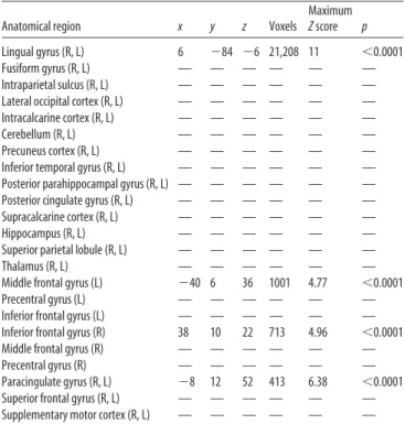
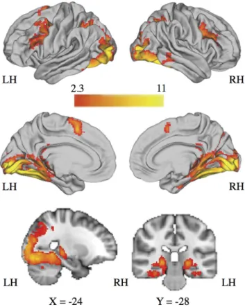
Whole-brain analysis revealed four clusters of activation during
context encoding (Cue
⬎
ITI) (
Table 2
;
Fig. 5
). The first cluster
spanned bilateral occipital, parietal, and temporal cortex,
includ-ing the posterior parahippocampal gyrus, fusiform gyrus,
precu-neus, posterior cingulate, and extending into inferior temporal
gyrus, posterior hippocampus, and thalamus. Two clusters in
PFC included right and left middle frontal gyrus. On the medial
surface, one cluster included the bilateral anterior cingulate
cor-tex/supplementary motor area.
Age was not associated with hippocampal activation during
context encoding (
p
values
⫽
0.27– 0.95), and no age
⫻
emotion
interaction was observed (
F
(2,114)⫽
0.96,
p
⫽
0.39).
Brain– behavior associations
The hippocampus ROI encompassed posterior hippocampus,
which was the only portion of the hippocampus that was active
during context encoding in the whole-brain analysis of the entire
sample.
Greater bilateral hippocampal BOLD signal during context
encoding was associated with higher context memory accuracy
specifically on trials with angry facial cues (

⫽
0.28,
p
⫽
0.030;
Fig. 6
), but not overall or for happy or neutral trials (
p
values
⫽
0.52– 0.75). There was no interaction between age and
hip-pocampal BOLD signal during encoding on context memory
overall or for angry, happy, or neutral trials (
p
values
⫽
0.45–
0.64). Results were unchanged when the single outlier was
excluded.
Violence exposure and context memory
Violence exposure was unrelated to overall context memory
(
F
(1,57)⫽
2.99,
p
⫽
0.09). However, a significant interaction
emerged between violence exposure and emotion in predicting
context memory (
F
(2,114)⫽
4.32,
p
⫽
0.016;
Fig. 7
). Violence
exposure was associated with context memory on trials involving
angry facial cues (
F
(1,57)⫽
9.88,
p
⫽
0.003), but not on trials
involving happy or neutral facial cues (
p
values
⫽
0.24 – 0.99).
Specifically, participants exposed to violence exhibited lower
context memory accuracy for contexts paired with angry facial
targets (61.11
⫾
0.12) than control participants (71.81
⫾
0.13).
Violence exposure and neural structure
Violence exposure was associated with bilateral hippocampal
volume, controlling for age, sex, and total brain volume (
F
(1,55)⫽
6.06,
p
⫽
0.017). Specifically, participants exposed to violence
Figure 3. Age and context memory accuracy on trials involving all facial targets.Figure 4. Bilateral hippocampal volume and context memory accuracy on trials involving all facial targets, adjusting for age, sex, and total brain volume.
Table 2. Whole-brain/whole-group analysisa
Anatomical region x y z Voxels
Maximum Zscore p
Lingual gyrus (R, L) 6 ⫺84 ⫺6 21,208 11 ⬍0.0001 Fusiform gyrus (R, L) — — — — — — Intraparietal sulcus (R, L) — — — — — — Lateral occipital cortex (R, L) — — — — — — Intracalcarine cortex (R, L) — — — — — — Cerebellum (R, L) — — — — — — Precuneus cortex (R, L) — — — — — — Inferior temporal gyrus (R, L) — — — — — — Posterior parahippocampal gyrus (R, L) — — — — — — Posterior cingulate gyrus (R, L) — — — — — — Supracalcarine cortex (R, L) — — — — — — Hippocampus (R, L) — — — — — — Superior parietal lobule (R, L) — — — — — — Thalamus (R, L) — — — — — — Middle frontal gyrus (L) ⫺40 6 36 1001 4.77 ⬍0.0001 Precentral gyrus (L) — — — — — — Inferior frontal gyrus (L) — — — — — — Inferior frontal gyrus (R) 38 10 22 713 4.96 ⬍0.0001 Middle frontal gyrus (R) — — — — — — Precentral gyrus (R) — — — — — — Paracingulate gyrus (R, L) ⫺8 12 52 413 6.38 ⬍0.0001 Superior frontal gyrus (R, L) — — — — — — Supplementary motor cortex (R, L) — — — — — —
had smaller average hippocampal volume (9265.8
⫾
775.1) than
control participants (10016.1
⫾
922.6).
Violence exposure and neural function
In ROI analysis, violence exposure was unrelated to hippocampal
activation during context encoding overall (
F
(1,57)⫽
1.78,
p
⫽
0.19). However, an interaction between violence exposure and
emotion was observed (
F
(2,114)⫽
3.51,
p
⫽
0.033;
Fig. 8
).
Vio-lence exposure was associated with hippocampal BOLD signal
during context encoding in the presence of angry facial cues
(
F
(1,57)⫽
4.92,
p
⫽
0.030), but not in the presence of neutral or
happy facial cues (
p
values
⫽
0.43– 0.71). Specifically,
partici-pants exposed to violence exhibited less hippocampal activation
during context encoding on angry trials (0.36
⫾
0.35) than
con-trol participants (0.56
⫾
0.34).
When the single outlier was removed, the violence by emotion
interaction was unchanged (
F
(2,112)⫽
3.46,
p
⫽
0.035). However,
an association of violence exposure with hippocampal BOLD
sig-nal during context encoding (regardless of emotion) emerged
(
F
(1,56)⫽
4.31,
p
⫽
0.043). Specifically, less hippocampal BOLD
signal during all trials of context encoding was observed in
par-ticipants exposed to violence (0.47
⫾
0.28) versus control
partic-ipants (0.62
⫾
0.28).
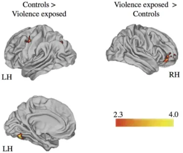
Functional connectivity
Whole-brain PPI analysis revealed that control participants
had greater functional connectivity of right hippocampus with
left middle frontal gyrus and left intraparietal sulcus during
context encoding on angry trials compared with participants
exposed to violence (
Table 3
;
Fig. 9
). Violence-exposed
par-ticipants exhibited greater functional connectivity between
right hippocampus and right vlPFC than control participants
(
Table 3
;
Fig. 9
).
Functional connectivity– behavior associations
Greater functional connectivity between right hippocampus
and right vlPFC during context encoding on trials with angry
faces predicted worse memory for contexts paired with angry
faces (

⫽ ⫺
0.49,
p
⫽
0.0001;
Fig. 10
). This association
re-mained when controlling for violence exposure (

⫽ ⫺
0.41,
p
⫽
0.001).
Figure 5. Regions of the brain with significant BOLD activation during context encoding (any facial cue⬎ITI) in the entire sample (N⫽60). Cluster-level correction applied in FSL.z⬎2.3, p⬍0.01 was the voxel-level threshold.z⬎3.0,p⬍0.001 was the cluster-level threshold.
Figure 6. Bilateral posterior hippocampal BOLD signal during context encoding (angry facial cue⬎ITI) and context memory accuracy on trials involving angry facial targets.
Figure 7. Interaction between violence exposure and emotion condition on context memory accuracy.
Mediation
We next evaluated whether the association of violence
expo-sure with context memory accuracy on angry trials was
explained by reduced hippocampal volume, reduced
hip-pocampal activation during context encoding, and increased
right hippocampus-right vlPFC functional connectivity during
context encoding. Hippocampus-vlPFC functional connectivity
mediated the association of violence exposure with context
mem-ory on angry trials (95% CI:
⫺
0.078 to
⫺
0.008), as did
hip-pocampal volume (95% CI:
⫺
0.067 to
⫺
0.003). There was no
indirect effect of violence exposure on context memory through
hippocampal BOLD signal during context encoding.
Specificity to violence exposure
We tested whether characteristics that differed between the
violence-exposed and control groups (poverty, neglect, IQ,
inter-nalizing symptoms, and exterinter-nalizing symptoms) were potential
confounders of the association of violence exposure with our four
behavioral and neural outcomes (i.e., context memory accuracy
on angry trials, hippocampal volume, hippocampal BOLD signal
during context encoding on angry trials, and
hippocampus-vlPFC functional connectivity during context encoding on angry
trials). To do so, we first examined whether these potential
con-founders were associated with each of the behavioral and neural
outcomes of interest.
Neglect, internalizing symptoms, and externalizing
symp-toms were unrelated to all outcome variables. None of the
po-tential confounders was associated with performance on the
context memory test or with hippocampus-vlPFC functional
connectivity.
Poverty was associated with one of the four outcomes. A
sig-nificant interaction emerged between poverty and emotion
con-dition in predicting hippocampal BOLD signal during context
encoding, such that poverty was associated with lower
hippocam-pal BOLD signal on angry trials (
F(1,50)
⫽
4.31,
p
⫽
0.043), but
not on neutral or happy trials. Poverty was unrelated to context
memory accuracy, hippocampal volume, and
hippocampus-vlPFC functional connectivity.
IQ was associated with two of the four outcomes. IQ was
positively associated with hippocampal volume (
F
(1,55)⫽
7.01,
p
⫽
0.011), and an IQ
⫻
emotion interaction emerged in
predict-ing hippocampal BOLD signal, such that IQ was positively
asso-ciated with hippocampal BOLD signal on angry trials (
F(1,57)
⫽
4.14,
p
⫽
0.047), but not on neutral or happy trials. IQ was
unrelated to context memory accuracy and hippocampus-vlPFC
functional connectivity.
In a final analysis, we evaluated whether the association of
violence exposure with hippocampal volume and hippocampal
BOLD signal during context encoding on angry trials persisted
after adjustment for poverty and IQ. Associations of violence
exposure, poverty, and IQ were all nonsignificant in these
mod-Table 3. Functional connectivity analysis by groupaPosterior hippocampal seed region Anatomical region x y z Voxels MaximumZscore p
Controls⬎violence-exposed
R Fusiform gyrus (L) ⫺28 ⫺72 ⫺16 140 3.98 0.010 Temporal occipital fusiform Cortex (L)
Cerebellum (L)
Precentral gyrus (L) ⫺46 ⫺8 52 133 3.48 0.014 Middle frontal gyrus (L)
Lateral occipital cortex (L) ⫺32 ⫺72 24 117 3.36 0.031 Intraparietal sulcus (L)
L —
Violence-exposed⬎controls
R Ventrolateral prefrontal cortex/frontal pole (R) 24 56 ⫺4 149 3.41 0.006
L — — — — — — —
aSignificantly different clusters exhibiting functional connectivity with the right and left posterior hippocampus during context encoding (angry facial cues⬎ITI) for participants exposed to violence versus control participants. Cluster-level correction applied in FSL.z⬎2.3, p⬍0.01 was the voxel-level threshold, andz⬎3.0,p⬍0.001 was the cluster-level threshold. Laterality of regions is specified: R, Right; L, left.
Figure 9. Regions of the brain with significantly different functional connectivity with the right posterior hippocampus during context encoding (angry facial cue⬎ITI) for participants exposed to violence and control participants. Cluster-level correction applied in FSL.z⬎2.3, p⬍0.01 was the voxel-level threshold.z⬎3.0,p⬍0.001 was the cluster-level threshold.
els, likely due to high collinearity among these factors in our
sample. After adjusting for IQ, the indirect effect of trauma
ex-posure on task performance through hippocampal volume was
no longer significant (95% CI:
⫺
0.049 to 0.003).
Hippocampus-vlPFC functional connectivity was unrelated
to all confounders and was the sole mediator of trauma-related
differences in task performance that remained significant in our
final model.
Discussion
Remarkably little is known about how contextual processing and
associated neural correlates vary across development and
whether early-life experiences influence contextual processing.
Our study demonstrates that the posterior hippocampus is
in-volved in context encoding in youth and that context encoding
does not change with age from middle childhood to late
adoles-cence. Specifically, we found that memory for contextual
infor-mation, hippocampal activation during context encoding, and
associations of hippocampal activation with context memory
did not change after age 8 years. In contrast, environmental
experiences during childhood were associated with contextual
encoding and retrieval. Specifically, children raised in
threaten-ing environments exhibited worse contextual memory and
atyp-ical neural recruitment during context encoding in the presence
of threat cues.
We observed activation in the bilateral posterior
hippocam-pus in a whole-brain analysis of context encoding. Greater
pos-terior hippocampal activation during encoding was associated
with better memory for contexts paired with angry facial cues,
and larger hippocampal volume was associated with better
con-text memory for all trial types. These findings are consistent with
evidence for dorsal hippocampal involvement in context
encod-ing in rodents (
Phillips and LeDoux, 1992
;
Young et al., 1994
;
Maren and Fanselow, 1997
), but not with evidence for anterior
hippocampal involvement in context encoding in adult humans
(
Hayes et al., 2007
,
2010
). It is possible that anterior and posterior
portions of the hippocampus contribute differently to context
encoding across development. Indeed, posterior hippocampus
increases in volume with age, whereas anterior hippocampus
de-creases (
Gogtay et al., 2006
). Posterior hippocampal activation
supports retrieval of explicitly encoded stimulus pairings in
childhood, whereas anterior hippocampal activation supports
re-trieval in adulthood (
DeMaster and Ghetti, 2013
). Future
re-search is needed to determine whether a similar developmental
posterior-anterior shift occurs for context encoding.
The fact that hippocampal activation during context encoding
was associated with context memory only in the presence of angry
faces is consistent with rodent work indicating that hippocampus
involvement in context encoding increases in the presence of
threat cues (
Phillips and LeDoux, 1994
). In humans, context
memory is enhanced when encoding occurs in the presence of
negative facial cues (
Barrett and Kensinger, 2010
). These results
suggest that threat cues might enhance processing of the
environ-ment, potentially to facilitate avoidance of future threats. Given
that context memory did not vary across emotion conditions in
the current study and that the hippocampus was activated during
context encoding in the presence of all facial cues, future research
is needed to clarify whether the valence of foreground cues
mod-ulates contextual processing.
Performance on the context memory test and hippocampal
activation during context encoding did not vary with age.
Asso-ciations of hippocampal structure and function with context
memory also did not vary with age. The lack of developmental
variation suggests that context encoding emerges early in
de-velopment. A change in context impairs explicit memory in
6-month-olds (
Hayne et al., 2000
;
Robinson and Pascalis, 2004
),
suggesting that context encoding may be intact as early as infancy.
The absence of explicit instruction to attend to background
con-text in our task and the lack of developmental variation in concon-text
encoding and retrieval suggest that context encoding may occur
implicitly. Implicit learning does not change after early
child-hood (
Meulemans et al., 1998
;
Vinter and Perruchet, 2000
;
Dixon
et al., 2010
), whereas retrieval of explicitly encoded scenes and
stimulus pairings and associated hippocampal correlates vary
de-velopmentally (
Ghetti et al., 2010
;
Ofen et al., 2012
;
DeMaster
and Ghetti, 2013
). Future research examining context encoding
and retrieval in a younger sample is needed to evaluate
age-related variation earlier in development.
Exposure to violence influenced context retrieval and neural
correlates of context encoding, suggesting influences of early
ex-perience on contextual processing. Children exposed to violence
had worse memory for contexts paired with angry faces, but
not happy or neutral faces, than children who had never
expe-rienced violence. Children exposed to violence had smaller
hippocampal volume and less hippocampal activation during
encoding on trials involving angry faces than children without
violence exposure. These findings replicate prior findings of
reduced hippocampal volume in maltreated children (
Lim et
al., 2014
;
Hanson et al., 2015
;
McLaughlin et al., 2016
) and
extend this work by documenting atypical hippocampal
func-tion during context encoding following childhood violence
exposure.
It is also possible that alterations in hippocampal structure
and function due to toxic effects of glucocorticoids (
Brunson et al.,
2001
;
Ivy et al., 2010
) following childhood violence exposure
con-tributed to general context encoding deficits. These general
deficits may have only emerged on angry trials whether
hippocampal-dependent contextual processing was greatest in
the presence of threat, as previously discussed. Future research is
needed to clarify the underlying mechanisms contributing to
poor context encoding and memory following childhood
vio-lence exposure.
We evaluated a range of potential confounders that might
have contributed to individual differences in context memory
and neural correlates of context encoding related to violence
exposure. Most of these potential confounders were unrelated
to behavioral and neural outcomes, although poverty was
as-sociated with hippocampal activation and IQ was asas-sociated
with hippocampal volume and activation. Strong correlations
among violence exposure, poverty, and IQ in our sample make
it difficult to determine conclusively that the observed
associ-ations are truly the result of violence exposure and not poverty
or IQ. However, violence exposure was the only factor
associ-ated with context memory as well as hippocampal structure,
function, and connectivity. Extensive evidence documents
at-tention bias toward angry faces in children exposed to violence
(
Pollak and Tolley-Schell, 2003
;
Shackman et al., 2007
),
con-sistent with our interpretation that attention bias toward
threat interfered with context encoding in children with
vio-lence exposure. Nonetheless, future research with larger
sam-ples and less overlap among violence exposure, other forms of
adversity, and IQ is needed to disentangle their unique
influ-ences on context encoding.
Several limitations are worth noting. First, we used realistic
pictures of indoor and outdoor scenes appearing in the
back-ground of salient facial cues, similar to methods used in adults
(
Hayes et al., 2007
,
2010
). However, pictures lack the
multisen-sory experience of real-world contexts. Future research should
use ecologically valid contextual stimuli appropriate for an MRI
scanner, such as immersive 3D virtual reality (
Åhs et al., 2015
).
Second, we examined neural activation during context encoding,
but not context retrieval. Different neural regions underlie
con-text encoding and retrieval in adults (
Hayes et al., 2010
),
high-lighting the importance of examining neural correlates of context
retrieval developmentally. Third, the study was not designed to
examine post-traumatic stress disorder or whether difficulties
with context encoding influence later stages of contextual
pro-cessing relevant to post-traumatic stress disorder, such as
context-modulation of fear. Future research should examine
whether heightened attention to threat and poor context
encod-ing in the presence of threat contribute to impaired
context-modulation of fear.
In conclusion, context encoding recruits the posterior
hip-pocampus in youth, and hippocampal structure and function are
associated with context memory. Context encoding appears to be
an implicit process that emerges early in human development. A
history of exposure to threatening environments is associated
with worse memory for contexts encoded in the presence of
threat, which may be explained by smaller hippocampal volume
and atypical hippocampal function during context encoding.
Fu-ture research should examine whether context encoding deficits
contribute to persistent fear in safe contexts among children who
have been raised in dangerous environments.
References
Achenbach TM (1991) Integrative guide for the 1991 CBCL/4 –18, YSR and TRF profiles. Burlington, VT: Department of Psychiatry, University of Vermont.
Åhs F, Kragel PA, Zielinski DJ, Brady R, LaBar KS (2015) Medial prefrontal pathways for the contextual regulation of extinguished fear in humans. Neuroimage 122:262–271.CrossRef Medline
Anagnostaras SG, Maren S, Fanselow MS (1999) Temporally graded retro-grade amnesia of contextual fear after hippocampal damage in rats: within-subjects examination. J Neurosci 19:1106 –1114.Medline
Avants BB, Tustison NJ, Song G, Cook PA, Klein A, Gee JC (2011) A repro-ducible evaluation of ANTs similarity metric performance in brain image registration. Neuroimage 54:2033–2044.CrossRef Medline
Barredo J, O¨ ztekin I, Badre D (2015) Ventral fronto-temporal pathway sup-porting cognitive control of episodic memory retrieval. Cereb Cortex 25:1004 –1019.CrossRef Medline
Barrett LF, Kensinger EA (2010) Context is routinely encoded during emo-tion percepemo-tion. Psychol Sci 21:595–599.CrossRef Medline
Beckmann CF, Jenkinson M, Smith SM (2003) General multi-level linear modelling for group analysis in fMRI. Neuroimage 20:1052–1063.
CrossRef Medline
Behzadi Y, Restom K, Liau J, Liu TT (2007) A component based noise cor-rection method (CompCor) for BOLD and perfusion based fMRI. Neu-roimage 37:90 –101.CrossRef Medline
Bernstein DP, Ahluvalia T, Pogge D, Handelsman L (1997) Validity of the Childhood Trauma Questionnaire in an adolescent psychiatric popula-tion. J Am Acad Child Adolesc Psychiatry 36:340 –348.CrossRef Medline
Bifulco A, Brown GW, Lillie A, Jarvis J (1997) Memories of childhood ne-glect and abuse: corroboration in a series of sisters. J Child Psychol Psy-chiatry 38:365–374.CrossRef Medline
Bishop SJ (2008) Neural mechanisms underlying selective attention to threat. Ann N Y Acad Sci 1129:141–152.CrossRef Medline
Brunson KL, Eghbal-Ahmadi M, Bender R, Chen Y, Baram TZ (2001) Long-term, progressive hippocampal cell loss and dysfunction induced by early-life administration of corticotropin-releasing hormone reproduce the effects of early-life stress. Proc Natl Acad Sci U S A 98:8856 – 8861.
CrossRef Medline
Cohen H, Liberzon I, Richter-Levin G (2009) Exposure to extreme stress impairs contextual odour discrimination in an animal model of PTSD. Int J Neuropsychopharmacol 12:291–303.CrossRef Medline
Davachi L (2006) Item, context and relational episodic encoding in humans. Curr Opin Neurobiol 16:693–700.CrossRef Medline
DeMaster DM, Ghetti S (2013) Developmental differences in hippocampal and cortical contributions to episodic retrieval. Cortex 49:1482–1493.
CrossRef Medline
Dixon ML, Zelazo PD, De Rosa E (2010) Evidence for intact memory-guided attention in school-aged children. Dev Sci 13:161–169.CrossRef Medline
Eklund A, Nichols TE, Knutsson H (2016) Cluster failure: why fMRI infer-ences for spatial extent have inflated false-positive rates. Proc Natl Acad Sci U S A 113:7900 –7905.CrossRef Medline
Fischl B, Dale AM (2000) Measuring the thickness of the human cerebral cortex from magnetic resonance images. Proc Natl Acad Sci U S A 97: 11050 –11055.CrossRef Medline
Ghetti S, Demaster DM, Yonelinas AP, Bunge SA (2010) Developmental differences in medial temporal lobe function during memory encoding. J Neurosci 30:9548 –9556.CrossRef Medline
Gogtay N, Nugent TF 3rd, Herman DH, Ordonez A, Greenstein D, Hayashi KM, Clasen L, Toga AW, Giedd JN, Rapoport JL, Thompson PM (2006) Dynamic mapping of normal human hippocampal development. Hip-pocampus 16:664 – 672.CrossRef Medline
Hanson JL, Nacewicz BM, Sutterer MJ, Cayo AA, Schaefer SM, Rudolph KD, Shirtcliff EA, Pollak SD, Davidson RJ (2015) Behavioral problems after early life stress: contributions of the hippocampus and amygdala. Biol Psychiatry 77:314 –323.CrossRef Medline
Hayes A (2013) Introduction to mediation, moderation, and conditional process analysis. New York: Guilford.
Hayes SM, Nadel L, Ryan L (2007) The effect of scene context on episodic object recognition: parahippocampal cortex mediates memory encoding and retrieval success. Hippocampus 17:873– 889.CrossRef Medline
context effects on face recognition: automatic binding and context shift decrements. J Cogn Neurosci 22:2541–2554.CrossRef Medline
Hayne H, Boniface J, Barr R (2000) The development of declarative memory in human infants: age-related changes in deffered imitation. Behav Neu-rosci 114:77– 83.CrossRef Medline
Ivy AS, Rex CS, Chen Y, Dube´ C, Maras PM, Grigoriadis DE, Gall CM, Lynch G, Baram TZ (2010) Hippocampal dysfunction and cognitive impair-ments provoked by chronic early-life stress involve excessive activation of CRH receptors. J Neurosci 30:13005–13015.CrossRef Medline
Jenkinson M, Beckmann CF, Behrens TE, Woolrich MW, Smith SM (2012) Fsl. Neuroimage 62:782–790.CrossRef Medline
Kim JJ, Fanselow MS (1992) Modality-specific retrograde amnesia of fear. Science 256:675– 677.CrossRef Medline
Lim L, Radua J, Rubia K (2014) Gray matter abnormalities in childhood maltreatment: a voxelwise metaanalysis. Am J Psychiatry 171:854 – 863.
CrossRef Medline
Maren S, Fanselow MS (1997) Electrolytic lesions of the fimbria/fornix, dorsal hippocampus, or entorhinal cortex produce anterograde deficits in contextual fear conditioning in rats. Neurobiol Learn Mem 67:142–149.
CrossRef Medline
McLaughlin KA, Sheridan MA, Lambert HK (2014) Childhood adversity and neural development: deprivation and threat as distinct dimensions of early experience. Neurosci Biobehav Rev 47:578 –591.CrossRef Medline
McLaughlin KA, Sheridan MA, Gold AL, Duys A, Lambert HK, Peverill M, Heleniak C, Shechner T, Wojcieszak Z, Pine DS (2016) Maltreatment exposure, brain structure, and fear conditioning in children and adoles-cents. Neuropsychopharmacology 41:1956 –1964.CrossRef Medline
McLaughlin KJ, Gomez JL, Baran SE, Conrad CD (2007) The effects of chronic stress on hippocampal morphology and function: an evaluation of chronic restraint paradigms. Brain Res 1161:56 – 64.CrossRef Medline
Meulemans T, Van der Linden M, Perruchet P (1998) Implicit sequence learning in children. J Exp Child Psychol 69:199 –221.CrossRef Medline
Monk CS, Nelson EE, McClure EB, Mogg K, Bradley BP, Leibenluft E, Blair RJ, Chen G, Charney DS, Ernst M, Pine DS (2006) Ventrolateral pre-frontal cortex activation and attentional bias in response to angry faces in adolescents with generalized anxiety disorder. Am J Psychiatry 163:1091– 1097.CrossRef Medline
Monk CS, Telzer EH, Mogg K, Bradley BP, Mai X, Louro HM, Chen G, McClure-Tone EB, Ernst M, Pine DS (2008) Amygdala and ventrolat-eral prefrontal cortex activation to masked angry faces in children and adolescents with generalized anxiety disorder. Arch Gen Psychiatry 65: 568 –576.CrossRef Medline
Ofen N, Chai XJ, Schuil KD, Whitfield-Gabrieli S, Gabrieli JD (2012) The development of brain systems associated with successful memory re-trieval of scenes. J Neurosci 32:10012–10020.CrossRef Medline
O’Reilly JX, Woolrich MW, Behrens TE, Smith SM, Johansen-Berg H (2012) Tools of the trade: psychophysiological interactions and functional con-nectivity. Soc Cogn Affect Neurosci 7:604 – 609.CrossRef Medline
Petrides M, Pandya DN (2002) Comparative cytoarchitectonic analysis of the human and the macaque ventrolateral prefrontal cortex and cortico-cortical connection patterns in the monkey. Eur J Neurosci 16:291–310.
CrossRef Medline
Phillips RG, LeDoux JE (1992) Differential contribution of amygdala and
hippocampus to cued and contextual fear conditioning. Behav Neurosci 106:274 –285.CrossRef Medline
Phillips RG, LeDoux JE (1994) Lesions of the dorsal hippocampal forma-tion interfere with background but not foreground contextual fear con-ditioning. Learn Mem 1:34 – 44.CrossRef Medline
Pollak SD, Tolley-Schell SA (2003) Selective attention to facial emotion in physically abused children. J Abnorm Psychol 112:323–338.CrossRef Medline
Robinson AJ, Pascalis O (2004) Development of flexible visual recognition memory in human infants. Dev Sci 7:527–533.CrossRef Medline
Roche A (2011) A four-dimensional registration algorithm with application to joint correction of motion and slice timing in fMRI. IEEE Trans Med Imaging 30:1546 –1554.CrossRef Medline
Shackman JE, Shackman AJ, Pollak SD (2007) Physical abuse amplifies at-tention to threat and increases anxiety in children. Emotion 7:838 – 852.
CrossRef Medline
Sheridan MA, McLaughlin KA (2014) Dimensions of early experience and neural development: deprivation and threat. Trends Cogn Sci 18:580 – 585.CrossRef Medline
Shiba Y, Santangelo AM, Roberts AC (2016) Beyond the medial regions of the prefrontal cortex in the regulation of fear and anxiety. Front Syst Neurosci 10:12.CrossRef Medline
Telzer EH, Mogg K, Bradley BP, Mai X, Ernst M, Pine DS, Monk CS (2008) Relationship between trait anxiety, prefrontal cortex, and attention bias to angry faces in children and adolescents. Biol Psychol 79:216 –222.
CrossRef Medline
Tottenham N, Tanaka JW, Leon AC, McCarry T, Nurse M, Hare TA, Marcus DJ, Westerlund A, Casey BJ, Nelson C (2009) The NimStim set of facial expressions: judgments from untrained research participants. Psychiatry Res 168:242–249.CrossRef Medline
van Marle HJ, Hermans EJ, Qin S, Ferna´ndez G (2009) From specificity to sensitivity: how acute stress affects amygdala processing of biologically salient stimuli. Biol Psychiatry 66:649 – 655.CrossRef Medline
Vinter A, Perruchet P (2000) Implicit learning in children is not related to age: evidence from drawing behavior. Child Dev 71:1223–1240.CrossRef Medline
Vul E, Harris C, Winkielman P, Pashler H (2009) Puzzlingly high correla-tions in fMRI studies of emotion, personality, and social cognition. Per-spect Psychol Sci 4:274 –290.CrossRef Medline
Walker EA, Unutzer J, Rutter C, Gelfand A, Saunders K, VonKorff M, Koss MP, Katon W (1999) Costs of health care use by women HMO members with a history of childhood abuse and neglect. Arch Gen Psychiatry 56: 609 – 613.CrossRef Medline
Wechsler D (1999) Wechsler Abbreviated Scale of Intelligence. San Anto-nio, TX: Psychological Corporation.
Woolrich M (2008) Robust group analysis using outlier inference. Neuro-image 41:286 –301.CrossRef Medline
Woolrich MW, Behrens TE, Beckmann CF, Jenkinson M, Smith SM (2004) Multi-level linear modelling for fMRI group analysis using Bayesian in-ference. Neuroimage 21:1732–1747.CrossRef Medline