Emergency Triage
Assessment
and
Treatment
(ETAT)
Manual for
participants
World Health
Organization
For further information, please contact:
Department of Child and Adolescent Health and Development (CAH) World Health Organization
20, Avenue Appia 1211 Geneva 27 Switzerland Tel: +41 22 791 2632 Fax: +41 22 791 4853 E-mail: [email protected]
Web site: http://www.who.int/child-adolescent-health
Emergency Triage
Assessment
and
Treatment
(ETAT)
Manual for
participants
World Health
Organization
The WHO Department of Child and Adolescent Health and Development wishes to acknowledge the help and support of Professor Elizabeth Molyneux, who developed the training course for the Emergency Triage Assessment and Treatment on which these course materials are based. We would like to extend our thanks to Patricia Whitesell, ACT, USA, who wrote a first draft of the materials, and to Dr. Jan Eshuis, Royal Tropical Institute, Amsterdam, who developed the training materials further. In addition, we are grateful to the participants and facilitators of several field tests of these materials for their comments. We wish to thank Dr Carolyn Maclennan, Melbourne, Australia, Dr. Diana Silimperi and Lauri Winter, Quality Assurance Project, Bethesda, USA for providing substantial comments and revisions during these field tests, and the WHO Regional Offices for Africa, Southeast Asia and the Western Pacific for their support.
WHO Library Cataloguing-in-Publication Data
Emergency triage assessment and treatment (ETAT). Contents: Manual for participants — Facilitator guide.
1. Triage. 2. Emergency treatment. 3. Child health services. 4. Teaching materials. I. World Health Organization.
ISBN 92 4 154687 5 (participants) (NLM classification: WS 205) ISBN 92 4 154688 3 (facilitator)
© World Health Organization 2005
All rights reserved. Publications of the World Health Organization can be obtained from WHO Press, World Health Organization, 20 Avenue Appia, 1211 Geneva 27, Switzerland (tel: +41 22 791 2476; fax: +41 22 791 4857; email: [email protected]). Requests for permission to reproduce or translate WHO publications – whether for sale or for noncommercial distribution – should be addressed to WHO Press, at the above address (fax: +41 22 791 4806; email: [email protected]).
The designations employed and the presentation of the material in this publication do not imply the expression of any opinion whatsoever on the part of the World Health Organization concerning the legal status of any country, territory, city or area or of its authorities, or concerning the delimitation of its frontiers or boundaries. Dotted lines on maps represent approximate border lines for which there may not yet be full agreement.
The mention of specific companies or of certain manufacturers’ products does not imply that they are endorsed or recommended by the World Health Organization in preference to others of a similar nature that are not mentioned. Errors and omissions excepted, the names of proprietary products are distinguished by initial capital letters.
All reasonable precautions have been taken by WHO to verify the information contained in this publication. However, the published material is being distributed without warranty of any kind, either express or implied. The responsibility for the interpretation and use of the material lies with the reader. In no event shall the World Health Organization be liable for damages arising from its use.
Table of contents
Introduction 1
Learning objectives for the training course 2
Module One: Triage and the “ABCD” concept 3
The “ABCD” concept 4
Priority signs 4
The triaging process 4
When and where should triaging takes place? 5
Who should triage? 5
How to triage? 5
Assessing priority signs 6
General treatment for priority signs 8
The need for frequent reassessment 9
Assessment questions: Triage 10
Module Two: Airway and breathing 13
Assessment of the airway 13
Is the child breathing? Is the airway obstructed? 13
Management of the airway 14
Management of the choking child 14
Positioning to improve the airway 15
Is trauma of the neck a possibility? 16
Assessment breathing 17
Is the child breathing? 17
Does the child show central cyanosis? 17
Does the child have severe respiratory distress? 18
Management of breathing problems 18
Ventilate with bag and mask 18
Insertion of an oropharyngeal (Guedel) airway 19
Give oxygen 20
Assessment questions: Airway and breathing 23
Module Three: Circulation 25
Assess the circulation 26
Are the child’s hands warm? 26
Is the capillary refill longer than 3 seconds? 26
Is the pulse weak and fast? 27
Shock 27
Treatment of shock 27
Is the child in coma? 35
Is the child convulsing now? 36
Treatment of coma and convulsion 36
Manage the airway 37
Position the child 37
Check blood sugar 37
Give IV glucose 38
Give an anticonvulsant 39
Assessment questions: Coma and convulsion 41
Module Five: Dehydration 43
Assess for severe dehydration 43
Is the child lethargic 44
Does the child have sunken eyes? 44
Does a skin pinch go back very slowly (longer than 2 seconds)? 44 Treatment of severe dehydration in an emergency setting 45 Severe dehydration (without shock or severe malnutrition) 45 Give fluids by nasogastric tube if you cannot set up an intravenous infusion 45
Severe dehydration with severe malnutrition 47
Assessment questions: Dehydration 49
Module Six: Case management scenarios 53
Module Seven: Implementing ETAT 55
Objectives of the chapter/session 55
Implementing ETAT in your hospital 56
Advocacy 56
Patient flow and tasks 57
Material resources 57
Developing individual plans of actions 57
Annexes
Annex 1. Practical procedures 59
Giving parenteral fluids 59
Insertion of a nasogastric tube 63
Annex 2. Resources required to implement emergegncy care of children in hospitals 65
Annex 3. ETAT charts 67
Chart 2: Triage of all sick children 67
Chart 3: How to manage a choking child 69
Chart 4: How to manage the airway in a child with obstructed breathing
(or who has just stopped breathing) 70
Chart 5: How to give oxygen 71
Chart 6: How to position the unconscious child 72
Chart 7: How to give IV fluids rapidly for shock in a child without severe
malnutrition 73
Chart 8: How to give IV fluids for shock in a child with severe malnutrition 74 Chart 9: How to give diazepam (or paraldehyde) rectally 75
Chart 10: How to give IV glucose 76
Introduction
A nine-month old baby boy is carried into the children’s section of the outpatient department in his mother’s arms. He appears to be asleep. At the triage desk he is seen by a nurse and found to have lips and tongue that are grey/blue in colour, and he is taken straight into the resuscitation room as an emergency.
In the resuscitation room he is given oxygen from an oxygen concentrator. He is noted to be grunting and breathing very fast. His hands are cold to touch and the capillary refill time is prolonged to four seconds. An intravenous cannula is placed. A blood sample is taken at the same time for blood glucose, haematocrit and other investigations. An intravenous infusion of normal saline is commenced at 20ml/kg to run as fast as it can go.
Other treatments are given, depending on the result of the investigations and the response to the treatment he receives. It is now 18 minutes since the baby came through the outpatient department’s door, and his situation is stable. It is now time to take a full history and carry out a full examination to make a definitive diagnosis. He is diagnosed as having very severe pneumonia, and receives specific treatment for this. However, before coming to this diagnosis, no time was wasted, his status was stabilized, based on a few leading signs and symptoms, even when the medical staff did not know exactly what was wrong with him.
This was good triage and emergency management. Would it have happened like this in your hospital? In this training course, you are going to acquire the necessary knowledge and skills for the triage and emergency management of sick children, and you will consider what is needed to introduce this to your hospital. Deaths in hospital often occur within 24 hours of admission. Many of these deaths could be prevented if very sick children are identified soon after their arrival in the health facility, and treatment is started immediately. Therefore, a process of rapid triage for all children presenting to hospital needs to be put in place, to determine whether any emergency or priority signs are present. Triage may be done in 15-20 seconds by medical staff or by non-medical staff (after appropriate training) as soon as the child arrives, and no special equipment is needed for this. Once emergency signs are identified, prompt emergency treatment needs to be given to stabilize the condition of the child.
WHO has developed Emergency Triage Assessment and Treatment (ETAT) guidelines. These are adapted from the Advanced Paediatric Life Support guidelines used in western countries, and they identify children with immediately life-threatening conditions which are most frequently seen in developing countries, such as obstruction of the airway and other breathing problems caused by infections, shock, severely altered central nervous system function (coma or convulsions), and severe dehydration.
These guidelines were developed in Malawi, and were field-tested in several other countries including Angola, Brazil, Cambodia, Indonesia, Kenya and Niger. The guidelines are contained in the manual “Management of the child with a serious infection or severe malnutrition” and in the “Pocketbook of hospital care for children”, on which this training course is based. This course manual is primarily meant for the participants of a 3 1/2 days training course in Emergency Triage, Assessment and Treatment. It provides participants with the reading materials to prepare themselves for the modules taught in the course. Some of the reading might be done during the course. In addition, it gives questions for self-assessment which participants can respond to after having gone through the training. Apart from use in a full-time training course, the reading will be useful for trainers and participants who take part in training as a series of seminars. Guidance on how to conduct such training is contained in a parallel facilitator’s guide.
This training course does not stand on its own. It can be included in a quality improvement process which targets the whole hospital or it can start such a process. At the end of the course, participants plan for introducing an ETAT process at their institution, by comparing the existing situation with international standards, and suggesting actions to solve identified problems and to document and evaluate such a process. Lessons learned in this process can be applied to other areas of child health in hospital and to care of other patient groups. Emergency management is by team, rather than by individual players, so team work is emphasized and practised throughout the course.
Learning objectives for the training course
At the end of the course you will be able to: Triage all sick children when they arrive at a health facility, into the following categories:
- those with emergency signs - those with priority signs
- those who are non-urgent cases.
Assess a child’s airway and breathing and give emergency treatments. Assess the child’s status of circulation and level of consciousness.
Manage shock, coma, and convulsions in a child.
Assess and manage severe dehydration in a child with diarrhoea.
Triage and the "ABCD"
concept
Many deaths in hospital occur within 24 hours of admission. Some of these deaths can be prevented if very sick children are quickly identified on their arrival and treatment is started without delay. In many hospitals around the world, children are not checked before a senior health worker examines them; as a result, some seriously ill patients have to wait a very long time before they are seen and treated. Children are known to have died of a treatable condition when waiting in the queue for their turn. The idea of triage is to prevent this from happening. The word “triage” means sorting. The use of triage to prioritize the critically ill dates back to the early 19th century, when this was developed by
military surgeons in the Napoleonic war between France and Russia.
Triage is the process of rapidly examining all sick children when they first arrive in hospital in order to place them in one of the following categories:
Those with EMERGENCY SIGNS who require immediate emergency
treatment.
If you find any emergency signs, do the following immediately: - Start to give appropriate emergency treatment.
- Call a senior health worker and other health workers to help. - Carry out emergency laboratory investigations.
Those with PRIORITY SIGNS, indicating that they should be given
priority in the queue, so that they can rapidly be assessed and treated without delay.
Those who have no emergency or priority signs and therefore are NON-URGENT cases. These children can wait their turn in the queue for assessment and treatment.1 The majority of sick children will be
non-urgent and will not require emergency treatment.
After these steps are completed, proceed with general assessment and further treatment according to the child’s priority.
In an ideal situation, all children should be checked on their arrival in hospital by a person who is trained to assess how ill they are. This person decides whether the patient will be seen immediately and will receive life-saving treatment, or will be seen soon, or can safely wait his/her turn to be examined.
1 Sometimes it is discovered that a child in the queue is waiting for immunization. These children do not need
assessment and can be referred to the right department without delay.
Module One
TRIAGE
is the sorting of patientsinto priority groups according to their need
and the resources available
E Emergency
P Priority
The ABCD concept
Triage of patients involves looking for signs of serious illness or injury. These emergency signs relate to the Airway-Breathing-Circulation/Consciousness-Dehydration and are easily remembered as “ABCD”.
Each letter refers to an emergency sign which, when positive, should alert you to a patient who is seriously ill and needs immediate assessment and treatment.
Priority signs
Besides the group of emergency signs described above, there are priority signs, which should alert you to a child who needs prompt, but not emergency assessment. These signs can be remembered with the symbols 3 TPR - MOB:
Tiny baby: any sick child aged under two months Temperature: child is very hot
Trauma or other urgent surgical condition
Pallor (severe)
Poisoning Pain (severe)
Respiratory distress
Restless, continuously irritable, or lethargic
Referral (urgent)
Malnutrition: Visible severe wasting
Oedema of both feet
Burns
The frequency with which children showing some of these priority signs appear in the outpatient department depends on the local epidemiology. The signs might need to be adapted accordingly, for example by including signs for common severe conditions which cannot wait in your setting.
The triaging process
Triaging should not take much time. For a child who does not have emergency signs, it takes on average 20 seconds. The health worker should learn to assess several signs at the same time. A child who is smiling or crying does not have severe respiratory distress, shock or coma. The health worker looks at the child, observes the chest for breathing and priority signs such as severe malnutrition and listens to abnormal sounds such as stridor or grunting.
Categories after triage Action required
EMERGENCY CASES Need immediate emergency treatment PRIORITY CASES Need assessment and rapid attention NON-URGENT CASES Can wait their turn in the queue
A Airway B Breathing C Circulation Coma Convulsion D Dehydration (severe)
Several methods are available to facilitate the triaging process. One example is a stamp being used in Malawi consisting of the “ABCD” signs in which the health worker circles the correct step and initiates emergency treatment “E” or puts them in priority groups “P” or “Q” for children who can wait in the queue. Colours can also be used for differentiating the three groups, giving a red sticker to emergency cases, a yellow for priority and green for the queue.
WHEN AND WHERE SHOULD TRIAGING TAKE PLACE?
Triage should be carried out as soon as a sick child arrives in the hospital, well before any administrative procedure such as registration. This may require reorganizing the flow of patients in some locations.
Triage can be carried out in different locations – e.g. in the outpatient queue, in the emergency room, or in a ward if the child has been brought directly to the ward at night. In some settings, triage is done in all these places. Emergency treatment can be given wherever there is room for a bed or trolley for the sick child and enough space for the staff to work on the patient, and where appropriate drugs and supplies are easily accessible. If a child with emergency signs is identified in the outpatient queue, he/she must quickly be taken to a place where treatment can be provided immediately, e.g. the emergency room or ward.
WHO SHOULD TRIAGE?
All clinical staff involved in the care of sick children should be prepared to carry out rapid assessment in order to identify the few who are severely ill and require emergency treatment. If possible, all such staff should be able to give initial emergency treatment, as described in the flowchart and treatment charts. In addition, people such as gatemen, record clerks, cleaners, janitors who have early patient contact should be trained in triage for emergency signs and should know where to send people for immediate management.
HOW TO TRIAGE?
Keep in mind the ABCD steps: Airway, Breathing, Circulation, Coma, Convulsion, and Dehydration.
To assess if the child has airway or breathing problems you need to know:
Is the child breathing?
Is the airway obstructed?
Is the child blue (centrally cyanosed)?
Look, listen and feel for air movement. Obstructed breathing can be due to blockage by the tongue, a foreign body, a swelling around the upper airway (retropharyngeal abscess) or severe croup which may present with abnormal sounds such as stridor.
Does the child have severe respiratory distress?
Is the child having trouble getting breath so that it is difficult to talk, eat or breastfeed? Is he breathing very fast and getting tired, does he have severe chest indrawing or is he using auxiliary respiratory muscles?
A Airway B Breathing C Circulation Cm Coma Cn Convulsion D Dehydration (severe) E Emergency P Priority Q Queue
To assess if the child has circulation problems you need to know:
Does the child have warm hands?
If not, is the capillary refill time longer than 3 seconds? And is the pulse weak and fast?
In the older child the radial pulse may be used; however, in the infant, the brachial or femoral pulses may need to be felt.
To assess for coma you need to know:
A rapid assessment of conscious level can be made by assigning the patient to one of the AVPU categories:
A Alert
V responds to Voice P responds to Pain U Unresponsive
A child who is not alert but responds to voice is lethargic. If the assessment shows that the child does not respond to voice and only responds to pain (with targeted or untargeted movements), or does not respond at all, the level is at “P” or “U”. We then refer to that child as having coma and the child needs to be treated accordingly.
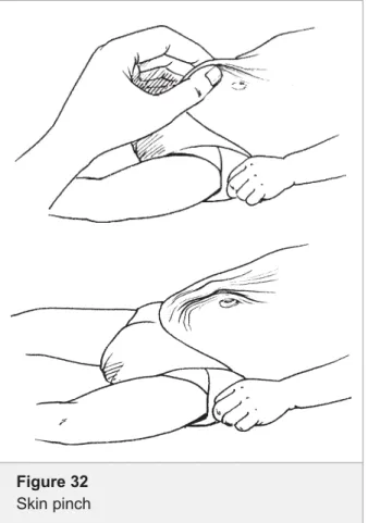
To assess for dehydration you need to know:
If the child is lethargic or unconscious
If the child has sunken eyes
If the skin pinch goes back very slowly
When ABCD has been completed and there are no emergency signs, continue to assess the priority signs.
Assessing priority signs
If the child does not have any of the E signs, the health worker proceeds to assess the child on the priority signs. This should not take more than few seconds. Some of these signs will have been noticed during the ABCD triage discussed so far, and others need to be rechecked. Follow the 3 TPR-MOB to quickly complete this section.
Tiny infant (less than two months of age)
If the child appears very young, ask the mother his age. If the child is obviously not a young infant, you do not need to ask this question.
Small infants are more difficult to assess properly, more prone to getting infections (from other patients), and more likely to deteriorate quickly if unwell. All tiny babies of under two months should therefore be seen as a priority.
Temperature: Hot (fever - high Temperature)
A child that feels very hot may have high fever. Children with high fever on touch need prompt treatment. Take the waiting child to the front of the queue and take locally adopted action, like having the temperature checked by thermometer, giving an antipyretic, or doing investigations like a blood film for malaria.
If the child has any sign of the ABCD, it means the child has an emergency “E” sign and
emergency treatment
should start
SevereTrauma (or other urgent surgical condition)
Usually this is an obvious case, but one needs to think of acute abdomen, fractures and head injuries in this category.
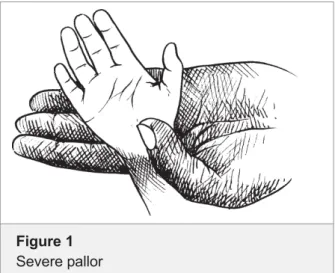
SeverePallor
Pallor is unusual paleness of the skin, and severe pallor is a sign of severe anaemia which might need urgent transfusion. It can be detected by comparing the child’s palms with your own. If the palms are very pale (almost paper-white), the child is severely anaemic.
Poisoning
A child with a history of swallowing drugs or other dangerous substances needs to be assessed immediately, as he can deteriorate rapidly and might need specific treatments depending on the substance taken. The mother will tell you if she has brought the child because of possible intoxication.
SeverePain
If a child has severe pain and is in agony, she/he should be prioritized to receive early full assessment and pain relief. Severe pain may be due to severe conditions such as acute abdomen, meningitis, etc.
Lethargy or Irritable and Restless
Recall from your assessment of coma with the AVPU scale whether the child was lethargic. A lethargic child responds to voice but is drowsy and uninterested (V in the AVPU scale).
The continuously irritable or restless child is conscious but cries constantly and will not settle.
Respiratory distress
When you assessed the airway and breathing, did you observe any respiratory distress? If the child has severe respiratory distress, it is an emergency. There may be signs present that you do not think are severe, e.g. lower chest wall indrawing (not severe), or difficulty in breathing. In this case, the child does not require emergency treatment but will need urgent assessment. Decisions on the severity of respiratory distress come with practice. If you have any doubts, have the child seen and treated immediately.
UrgentReferral
The child may have been sent from another clinic. Ask the mother if she was referred from another facility and for any note that may have been given to her. Read the note carefully and determine if the child has an urgent problem. Severe wasting (Severe Malnutrition)
A child with visible severe wasting has a form of malnutrition called marasmus. To assess for this sign, look rapidly at the arms and legs as well as the child’s chest. A detailed description is given in the section on dehydration (see pages 28/29).
Figure 1
Oedema of both feet
Oedema of both feet is an important diagnostic feature of kwashiorkor, another form of severe malnutrition. Other signs are changes in the skin and hair. MajorBurn
Burns are extremely painful and children who seem quite well can deteriorate rapidly. If the burn occurred recently, it is still worthwhile to cool the burnt area with water, for example, by sitting the child in a bathtub with cool water. Any child with a major burn, trauma or other surgical condition needs to be seen quickly. Get surgical help or follow surgical guidelines.
Triage all sick children. When a child with emergency signs is identified, take to the emergency room or treatment area and start the appropriate emergency treatments immediately. Do not proceed to the next step before treatment is begun for a positive sign.
If the child has no emergency signs, check for priority signs. After the examination for priority signs has been completed, the child will be assigned to one of:
Priority (P): the child should be put at the front of the queue Queue (Q): if the child has no emergency or priority signs Once appropriate emergency treatments have been initiated:
call for a senior health worker;
draw blood for emergency laboratory investigations such as blood glucose, haemoglobin and malaria smear;
ask about head or neck trauma before providing treatment;
take careful note if the child is severely malnourished, because this will affect the treatment of shock and dehydration caused by diarrhoea. It is essential to act as quickly as possible and to start the emergency treatments. The team needs to stay calm and work together efficiently. The person in charge of the emergency management of the child assigns tasks so that the assessment can continue and treatments can be initiated as quickly as possible. Other health workers help to give the treatment needed, especially since a very sick child may need several treatments at once. The senior health worker will direct the treatment and immediately continue with a core assessment and follow-up of the child, identifying all the child’s problems and developing the treatment plan.
GENERAL TREATMENT FOR PRIORITY SIGNS
Priority signs lead to quicker assessment of the child by moving the child to the front of the queue. While waiting, some supportive treatments may already be given. For example, a child found to have a hot body may receive an antipyretic
Triage Steps Treat when any sign is positive Assess A If positive, treat. If negative, proceed to B Assess B If positive, treat. If negative, proceed to C Assess C If positive, treat. If negative, proceed to D
Assess D If positive, treat. If negative, proceed to priority signs
Treatment begins
when
any emergency sign is identified
More than one treatment may have to
be administered as quickly as possible, and
several people may have to work together
such as paracetamol. Similarly, a child with a burn may have severe pain and the pain could be controlled while waiting for definitive treatment.
If a child has no emergency signs or priority signs, she/he may return to the queue.
THE NEED FOR FREQUENT REASSESSMENT
During and following emergency treatment, the child should be re-assessed using the complete ABCD process. The disease course is dynamic and there could be new developments within a short time. Reassessment should begin with assessment of the airway and through the ABCD.
Triage is the sorting of patients into priority groups according to their need. All children should undergo triage. The main steps in triage are:
Look for emergency signs.
Treat any emergency signs you find.
Call a senior health worker to see any emergency.
Look for any priority signs.
Place priority patients at the front of the queue.
Move on to the next patient.
Triage should be carried out quickly. You will soon learn to observe several things at once. For example, when assessing the airway and breathing you may note that the baby is very small or is restless. With practice, a complete triage (if no emergency treatment is needed) takes less than a minute.
Assessment questions: Triage
Answer all the questions on this page, writing in the given spaces. If you have a problem, ask for help from one of the facilitators.
ASSESSMENT QUESTIONS: TRIAGE
1. Define “triage”.
2. When and where should triage take place?
3. Who should do triaging?
4. What do the letters A, B, C and D in "ABCD" stand for?
6. Put the actions in the right chronological order: what will you do first, what next, what after that, and so on, and what last?
Ask about head or neck trauma
Call a senior health worker to see any emergency Have blood specimens taken for laboratory analysis Look for any priority signs
Look for emergency signs Move on to the next patient
Place priority patients at the front of the queue Start treatment of any emergency signs you find
ASSESSMENT QUESTIONS: TRIAGE
7. A three-year old girl is carried in her mother's arms wrapped in a blanket, in the queue. Her airway and breathing are OK. She has cold hands. Her capillary refill is 1.5 seconds. She is alert. Asked if the child has had diarrhoea, the mother answered "YES. Four loose stools per day". The skin pinch takes 3 seconds. How do you triage this child?
8. A four-year old male child was rushed in. He convulsed one hour ago. He is
breathing fast but there is no cyanosis and no respiratory distress. He feels very hot, but responds quickly to questions. He has no diarrhoea or vomiting. How do you triage this child?
ASSESSMENT QUESTIONS: TRIAGE
14. What should you do if the child has a priority sign? 13. Below what age is a child always a priority? 12. Where do you look for signs of severe wasting? 11. What signs of malnutrition do you check during triage?
10. A two-year old male is rushed to your clinic acutely convulsing. How do you triage this child?
9. A one-year old had a seizure at home; then again outside the clinic. He became unconscious. His breathing sounds very wet and noisy and there is drooling coming from his mouth. He is looking blue. How do you triage this child?
Airway and breathing
Module Two
The letters A and B in “ABCD” represent “airway and breathing”. It is evident that an open (patent) airway is needed for breathing. An airway or breathing problem is life-threatening and must receive your attention before you move on to other systems. It is therefore convenient that the first two letters of the alphabet represent the two most important areas to look for emergency or priority signs. If there is no problem with the airway or breathing, you should look for signs in the areas represented by C.
This section examines the assessment of the signs concerning the child’s airway and breathing that suggest a need for emergency treatments, and what treatments to give.
To assess if the child has an airway or breathing problem you need to know:
Is the child breathing?
Is the airway obstructed?
Is the child blue (centrally cyanosed)?
Does the child have severe respiratory distress?
If the child is not breathing or if the airway appears obstructed, you must first open the airway.
Assessment of the airway
IS THE CHILD BREATHING? IS THE AIRWAY OBSTRUCTED?
If the child is not breathing, or if the child has severe respiratory distress, is there an obstruction to the flow of air? Obstruction can occur at several levels.
Table 1
Assessment and treatment of airway and breathing
A B
AIRWAY AND BREATHING
Not breathing
Central cyanosis, or
Severe respiratory distress
If yes, is the breathing obstructed?
¾ Manage airway
¾ Give oxygen
¾ Make sure the child is warm
Check for head/neck trauma before treating the child; do not move neck if cervical spine injury is possible. Any sign positive
A Airway B Breathing C Circulation Coma Convulsion D Dehydration (severe)
The tongue can fall back and obstruct the pharynx, or a foreign body (such as a piece of fruit) can lodge in the upper airway. Croup can also cause upper airway obstruction. Coins and peanuts are notorious causes of aspiration and subsequent choking. Ask the child’s caretaker explicitly for a history of choking. Techniques to remove foreign bodies are based on support of forced expiration rather than a blind finger sweep of the mouth or other mechanical. A blind finger sweep in infants and children should not be done, as it might cause serious bleeding. Attempts to force the foreign body out of the airway should be done immediately, because airflow may be halted completely and sudden death could be imminent.
Management of the airway
MANAGEMENT OF THE CHOKING CHILDA child with a history of aspiration of a foreign body who shows increasing respiratory distress is in immediate danger of choking. Attempts to remove the foreign body should be made instantly. Do not hesitate. Apply back slaps or Heimlich manoeuvre. The treatment differs depending on whether there is a foreign body causing respiratory obstruction or some other cause for the obstruction or respiratory distress.
If a foreign body is causing the obstruction, the treatment depends on the age of the child.
Management of young infant (see Figure 2)
Lay the infant on your arm or thigh in a head down position
Give 5 blows to the infant’s back with heel of hand
If obstruction persists, turn infant over and give 5 chest thrusts with 2 fingers, one finger breadth below nipple level in midline
If obstruction persists, check infant’s mouth for any obstruction which can be removed
If necessary, repeat sequence with back slaps again If the child is choking,
do not hesitate, call for help and immediately
manage the airway
according to the instructions
Chest thrusts
Figure 2
Management of young infant
Management of child (see Figure 3)
Give 5 blows to the child’s back with heel of hand with child sitting, kneeling or lying
If the obstruction persists, go behind the child and pass your arms around the child’s body; form a fist with one hand immediately below the child’s sternum; place the other hand over the fist and pull upwards into the abdomen; repeat this Heimlich manoeuvre 5 times
If the obstruction persists, check the child’s mouth for any obstruction which can be removed
If necessary, repeat this sequence with back slaps again
After you have performed this procedure you should check inside the mouth for any foreign body. Obvious foreign bodies should be removed. Secretions should be cleared from the throat of all children. The breathing should be checked again.
POSITIONING TO IMPROVE THE AIRWAY
An airway or breathing problem is life-threatening. This child needs immediate treatment to improve or restore the breathing, even before you continue with the assessment of emergency signs. To treat an airway or breathing problem you should first open the airway and then begin giving the child oxygen. The drawings below show the chin lift. This is a way of opening the airway in children who have not been subjected to trauma. The drawings illustrate two different positions. Figure 4 shows the position for infants, the nose pointing upwards. Figure 5 shows the position for children, the chin pointing up. In both cases, place your hand on the child’s forehead and apply a little pressure
Figure 4
Neutral position to open airway in an infant (“nose up”) Heimlich manoeuvre in a choking older child Figure 3 Management of child
Slapping the back to clear airway obstruction in a choking child
to achieve the tilt. The fingers of the other hand are used to gently lift the chin.
To do this safely you must know if the child has been subjected to any trauma. In such a case, it is important not to tilt the head or move the neck. It is also important to know the child’s age because you will position an infant (under 12 months of age) differently from a child.
IS TRAUMA OF THE NECK A POSSIBILITY?
Any child with an emergency sign needs emergency treatment. However, always ask and check for head or neck trauma before treating, as this will determine how much a child can be moved. If a child has trauma you must avoid further injury during assessment or treatment. To check for head or neck trauma:
Ask if the child has had trauma to the head or
neck, or a fall which could have damaged the spine
Look for bruises or other signs of head or neck trauma
Stabilize the neck if trauma is suspected
If you suspect trauma which might have affected the neck or spine, do not move the head or neck as you treat the child and continue the assessment. This child may have a spinal injury, which could be made worse by moving him. To open and manage the airway when trauma is suspected ajaw thrust is used, as is illustrated in Figure 6. This is a way of opening the airway without moving the head. It is safe to use in cases of trauma for children of all ages. The jaw thrust is achieved by placing two or three fingers under the angle of the jaw on both sides, and lifting the jaw upwards.
If neck trauma is suspected, stabilize the neck (Figure 7):
Stabilize the child’s neck and keep the child lying on the back
Tape the child’s forehead to the sides of a firm
board to secure this position
Prevent the neck from moving by supporting the child’s head (e.g. using litre bags of IV fluid on each side)
Place a strap over the chin
If vomiting, turn on the side, keeping the head in line with the body (see Figure 8 {log roll}). If the child is restless, ask an attendant to stabilize the neck without upsetting the child more.
Log roll
Move a patient with a suspected cervical spine injury carefully. Avoid rotation and extremes of flexion and extension. One person, usually the most senior
Figure 6
Jaw thrust without head tilt
Figure 5
Sniffing position to open up airway in an older child (“chin up”)
Figure 7
attendant, should assume responsibility for the neck. He should stand at the top end of the patient, hold the patient’s head, and place the fingers under the angle of the mandible with the palm over the ears and parietal region and maintain gentle traction to keep the neck straight and in line with the body. When the patient is not being moved, a sandbag1 placed on each side or a cervical collar
can splint the neck.
Assessment of breathing
IS THE CHILD BREATHING?To assess whether or not the child is breathing there are three things you must do (see Figure 9):
LOOK
If active, talking, or crying, the child is obviously breathing. If none of these, look again to see whether the chest is moving.
LISTEN
Listen for any breath sounds. Are they normal?
FEEL
Can you feel the breath at the nose or mouth of the child?
If the child is not breathing, you need to support the breathing artificially by ventilating the child with a bag and mask.
DOES THE CHILD SHOW CENTRAL CYANOSIS?
Cyanosis occurs when there is an abnormally low level of oxygen in the blood. This produces a bluish or purplish discoloration of the tongue, the inside of the mouth and the skin. This sign may be absent in a child who has severe anaemia. To assess for central cyanosis, look at the mouth and tongue. A bluish or purplish discoloration of the tongue and the inside of the mouth indicates central cyanosis.
Before moving to the next section you will be given the opportunity to
practise the skills needed for moving a patient with an injury of
the neck
1 Use bottles or rolled towels in case sandbags are not available.
Figure 9
Look, listen and feel for breathing
Child
Figure 8
Log roll: stabilizing the neck of the patient while moving the body
DOES THE CHILD HAVE SEVERE RESPIRATORY DISTRESS?
If the child has severe respiratory distress there is increased work during breathing. The child may appear tired and distressed from the effort of trying to get enough oxygen into the lungs, and the breathing appears fast.
If the child is talking, drinking or feeding comfortably, or appears to be happy, there is no severe respiratory distress (or obstructed breathing).
Observe whether the child has significant discomfort from not getting enough air into the lungs. Is there difficulty in breathing while talking, eating or breastfeeding? Is the child breathing very fast, have severe lower chest wall indrawing, or using the auxiliary muscles for breathing which cause the head to nod or bob with every inspiration? The latter is particularly seen in young infants. If you see this, the child has severe respiratory distress.
Is the child’s breathing very laboured – i.e. needing much more effort to breathe than normal? Is the child exhausted (tired)? Are any of the signs of severe respiratory distress present?
Abnormal respiratory noises
Are there any noises heard when breathing in? A harsh noise on breathing in is called stridor, a short noise when breathing out in young infants is called grunting. Both noises are signs of severe respiratory problems.
If the child is breathing adequately, go to the next section to quickly continue the assessment for emergency signs. If the child has an airway or breathing problem, you should initiate appropriate treatment and then quickly resume the assessment.
Management of breathing problems
VENTILATE WITH BAG AND MASKIf the child is not breathing after management of the airway, ventilate with a self-inflating bag and mask. Such a bag fills itself with room air when released, and when squeezed again, pushes air through an outlet, to which a mask is attached for inflating the lungs. The bag is used together with a facemask. It is important to use the right size of facemask to prevent leakage. Before use, check the bag and valve by closing the patient’s connection with your thumb and attempt to expel air from the bag. If the bag and valve are in order, this will not be possible until you release your thumb. If either the bag or valve is faulty, the bag will empty easily. If you have oxygen, this should be connected to the mask (see Figure 10).
It is important for the mask to be the correct size for the child and that it is placed correctly over the face. The correct size and position are shown in the illustration. There are several sizes of mask, and a selection of these should be available (see Figure 11).
Figure 10
Ventilating a baby with a self-inflating mask
Signs of severe respiratory distress
- Very fast breathing - Severe lower chest wall
indrawing
- Use of auxiliary muscles - Head nodding
- Inability to feed because of respiratory problems
INSERTION OF AN OROPHARYNGEAL (GUEDEL) AIRWAY
The oropharyngeal or Guedel airway can be used in an unconscious patient to improve airway opening. It may not be tolerated in a patient who is awake and may induce choking or vomiting. Guedel airways come in different sizes. An appropriate sized airway goes from the centre of the teeth (incisors) to the angle of the jaw when laid on the face with the raised curved side (concave) up (“the right side up”). Infant
Select an appropriate sized airway
Position the child to open the airway (p.15), taking care not to move the neck if trauma suspected
Using a tongue depressor, insert the oropharyngeal airway the convex side up
Re-check airway opening
Use a different sized airway or reposition if necessary Give oxygen
Child
Select an appropriate sized oropharyngeal airway
Open the child’s airway, taking care not to move the neck if trauma suspected
Using a tongue depressor, insert the airway “upside down”
(concave side up) until the tip reaches the soft palate
Rotate through 180° and slide back over the tongue
Re-check airway opening
Use a different sized airway or reposition if necessary Give oxygen
Figure 11
Choosing the right mask size
1 2 3 4
1. Correct size and position 2. Mask too large, overlaps the
chin
3. Mask too small, nostrils not covered
4. Mask too large, overlap with eyes
Figure 12
Guedel tubes of different sizes and types
Figure 13
Selecting the right size of an oropharyngeal airway
Figure 14
Inserting an oropharyngeal airway in an infant: convex side up
GIVE OXYGEN
For all children who have any problem with their airway or breathing, always give oxygen first, while you continue to assess for other problems. Central cyanosis is a sign of deficient oxygenation (desaturation) and these children need oxygen urgently; however, children who are anaemic and desaturated may not show cyanosis, but also need oxygen. Many children with severe respiratory distress and with shock are also desaturated or not delivering enough oxygen to the brain and other vital organs and will benefit from oxygen treatment.
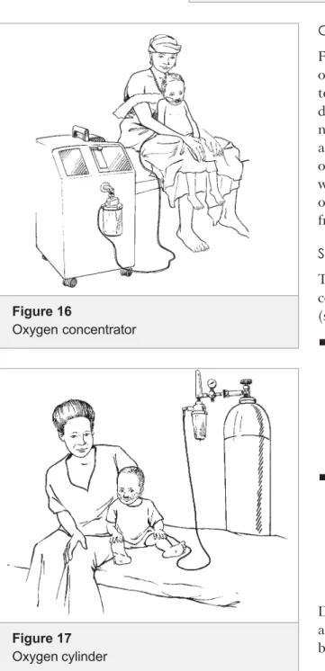
Sources of oxygen to treat hypoxaemia
There are two possible sources of oxygen: oxygen concentrators (see Figure 16) and oxygen-filled cylinders (see Figure 17):
Oxygen concentrators work by pumping room air through a zeolite canister to remove nitrogen, thus concentrating the oxygen. The device is of moderate cost, requires little maintenance, and, once purchased, produces oxygen continuously at low cost. A continuous electrical supply is
required, however, to operate the pump.
Oxygen cylinders are easy to use, requiring only
a flow meter and appropriate tubing, and can operate even when there is no electrical supply. The oxygen in cylinders is, however, relatively expensive and maintaining a constant supply is often difficult, especially at peripheral hospitals and health centres.
Depending on the availability or possibility of introduction at your health facility, you will be demonstrated one or both during the course.
Figure 16
Oxygen concentrator
Figure 17
Oxygen cylinder
Figure 15
Inserting an oropharyngeal airway in an older child
Oxygen delivery
Two methods are recommended for the delivery of oxygen in an emergency setting: nasal prongs and nasal catheter. Nasal prongs are best for delivering oxygen to young infants and children with severe croup or pertussis; do not use a nasal catheter as they provoke paroxysms of coughing.
An alternative method in emergency settings is the use of a face mask, which requires higher flow rates. It is therefore not suitable for permanent oxygen delivery on the ward, as it wastes a precious resource. It is important to have the proper equipment to control oxygen flow rates (0.5-2 litres/ minute).
Nasal prongs (see Figure 18) are short tubes
inserted into the nostrils. Place them just inside the nostrils and secure with a piece of tape on the cheeks near the nose (see Figure 19). Care should be taken to keep the nostrils clear of mucus, which could block the flow of oxygen. Set a flow rate of 0.5-1 litres/min in infants and 1-2 litres/min if older in order to deliver 30-35% oxygen concentration in the inspired air.
Prongs come in different sizes for adults and children. If you have only adult-size prongs, and the outlet tubes are too far apart to fit into the child's nostrils, cut the outlet tubes off and direct the jet of the oxygen into the nostrils.
Anasal catheter is made from tubing of 6 or 8
FG size such as a nasogastric tube or suction catheter. The tubing is inserted into either nostril a distance equivalent to that from the child’s nostril to the inner eyebrow (see Figure 20). It must then be firmly secured using tape, and connected to the oxygen. The tip of the catheter
should NOT be visible below the uvula. Set a flow rate of 0.5-1 litres for infants and 1-2 litres/min for older children, which delivers an oxygen concentration of 45-60% in the inspired air.
Figure 19
Nasal prongs correctly positioned and secured
Figure 20
Correct position of nasal catheter (cross-sectional view)
Figure 18
Nasal prongs with tubing
A B C
A Infant size prongs B Adult size prongs
Note: the distance between the outlet tubes is larger and the tubes are thicker
C Joined prongs for adults with a connector between 2 separate pieces of tubing
Practice (using equipment)
At this point it is important for you to be familiar with the equipment in your own department. You should be able to put together and take apart a self-inflating bag. You should be able to work the oxygen cylinder or oxygen concentrator. You can find out which size masks you have and try them on real patients to get an idea which size suits which age group. You can also practise the chin lift.
To assess the airway and breathing you need to know:
Is the airway obstructed?
Is the child breathing?
Is the child cyanosed?
Are there signs of severe respiratory distress?
If the patient is not breathing you need to:
Open the airway
Remove any foreign body
Ventilate with a bag and mask.
In all cases of airway or breathing problems:
Give oxygen: 0.5 1 litre/min (<1-year-olds) and 1 2 litres/minute (older children).
SUMMARY
Figure 21
Yankhauer (A) catheter and Suction (B) catheter for clearing secretions from the airway
A
ASSESSMENT QUESTIONS: AIRWAY AND BREATHING
Assessment questions: Airway and Breathing
Answer all the questions on this page, writing in the given spaces. If you have a problem, ask for help from one of the facilitators.
5. What size of tubing should you use for a nasal catheter?
4. When opening the airway of an infant (<12 months) who has not been subjected to trauma, name the part of the body that should point upwards.
3. Does stridor occur in inspiration or expiration? 2. List three signs of severe respiratory distress:
ASSESSMENT QUESTIONS: AIRWAY AND BREATHING
10. A three-year old boy is carried into the outpatient department in his father's arms. He is pale, floppy and having difficulty breathing. His father says he has been unwell and coughing for 3 days. Weight 14kg. He breathes fast with heavy severe chest indrawing. The airway is patent. He is alert. How do you triage this child? What do you do?
8. A 4-year old boy hit by a bicycle was carried in on a blanket. The child was unconscious responding only to pain. His breathing was noisy. What do you do?
7. You have successfully removed a coin from the trachea (windpipe) of a three-year old boy by applying Heimlich's manoeuvre. You checked his respiration and found that he was breathing normally. What do you do next?
6. At what flow (volume/time) should oxygen be started?
9. A nine-month girl and her older brother have been playing in the emergency department with an old bead necklace, suddenly the child is brought to you by one of the nurses, the child is choking. What do you do?
Circulation
Module Three
The letter C in “ABCD” stands for
circulation (assessment and management of shock);
assessment and management of coma; and convulsions.
With experience you can assess these emergency signs very quickly, almost simultaneously. You can recognize some immediately, such as coma (unconsciousness) and convulsions.
These assessments are done if the assessment of airway and breathing was normal, or after emergency treatments have been given for any respiratory problems encountered.
In the table below, the signs are listed on the left and the corresponding treatments on the right. Complete the assessment of all the signs on the left before deciding on and initiating treatment. However, because the assessment is done quickly (in less than a minute) there is hardly any delay in beginning the necessary treatment. You always need to check whether the child may have head or neck trauma, because this will affect how you treat the child.
Table 2
Assessment and treatment of circulation
C
1CIRCULATION
Cold hand with:
Capillary refill longer than 3
seconds, and
Weak and fast pulse
¾ Stop any bleeding
¾ Give oxygen
¾ Make sure the child is warm
IF NO SEVERE MALNUTRITION:
Insert IV and begin giving fluids
rapidly.
If not able to insert peripheral IV, insert an external jugular or intra-osseous line.
IF SEVERE MALNUTRITION:
Give IV glucose
Proceed immediately to full
assessment and treatment Check for head/neck trauma before treating the child; do not move neck if cervical spine injury is possible.
Any sign positive
Check for severe malnutrition
A Airway B Breathing C Circulation Coma Convulsion D Dehydration (severe)
Assess the circulation
First in this section we will look at the assessment of circulation and signs of shock.
To assess if a child has a circulation problem you need to know:
Does the child have warm hands?
If not, is the capillary refill time longer than 3 seconds?
And is the pulse weak and fast?
ARE THE CHILD’S HANDS WARM?
If the child’s hands are warm, there is no problem with the circulation and you can move to the next assessment. If they are cold, you need to assess the circulation further. If the circulation is poor, as during shock, the blood flow moves to the most important parts of the body. So the hands, feet and skin get less blood and often feel cold.
To assess the circulation, take the child’s hand in your own. If it feels warm, the child has no circulation problem and you do not need to assess capillary refill or pulse. If the child’s hands feel cold, you need to assess the capillary refill.
IS THE CAPILLARY REFILL TIME LONGER THAN 3 SECONDS?
Capillary refill (see Figure 22) is a simple test that assesses how quickly blood returns to the skin after pressure is applied. It is carried out by applying pressure to the pink part of the nail bed of the thumb or big toe. The capillary refill time is the time from release of pressure to complete return of the pink colour. It should be less than 3 seconds. If it is more than 3 seconds the child is shocked. Capillary refill is prolonged in shock because the body tries to maintain blood flow to vital organs and reduces the blood supply to less important parts of the body like the skin (peripheral vasoconstriction). The vessels open slowly because of low pulse pressure. This sign is reliable except when the room temperature is low; a cold environment may also cause vasoconstriction and thus cause a delayed capillary refill. So you will need to check the pulse only if the room is cold.1
To assess capillary refill, grasp the child’s thumb or big toe between finger and thumb. Look at the pink of the nail bed. Apply minimal pressure necessary for 3 seconds to produce blanching of the nail bed. The pressure should be sufficient to cause blanching (a change in colour from pink to white). The pressure is applied for 3 seconds and then released. Time the capillary refill from the moment of release until total return of the pink colour. If the refill time is longer than 3 seconds, the child may have a circulation problem with shock. To confirm, it is necessary to check the pulses.
Figure 22
Checking capillary refill
A. Applying pressure to the nail bed for 3
seconds
B. Check the time to the return of the pink
colour after releasing the pressure
A
IS THE PULSE WEAK AND FAST?
The radial pulse (the pulse at the wrist) should be felt. If this is strong and not obviously fast, the pulse is adequate; no further assessment is needed.
If the radial pulse is difficult to find, you need to look for a more central pulse (a pulse nearer to the heart). In an infant (less than one year of age) the best place to look is at the middle of the upper arm, the brachial
pulse. If the child is lying down you could look for the femoral pulse in the groin. In an older child you should feel for the carotid pulse in the neck. The pulse should be strong. If the more central pulse feels weak, decide if it also seems fast. This is a subjective judgement and an exact count is not taken. If the central pulse is weak and fast, the child needs treatment for shock.
All these procedures can and should be practised on yourself, your friends, your children and family, and finally on real patients. This is the best way to improve in testing capillary refill and finding pulses.
Note that we do not recommend blood pressure to assess for shock because of two reasons: 1) Low blood pressure is
a late sign in children and may not help identify treatable cases and 2) the BP cuff necessary in children of different age groups is mostly unavailable in many district hospitals.
Shock
The most common cause of shock in children is due to loss of fluid from circulation, either through loss from the body as in severe diarrhoea or when the child is bleeding, or through capillary leak in a disease such as severe Dengue fever. In all cases, it is important to replace this fluid quickly. An intravenous line must be inserted and fluids given rapidly in shocked children without severe malnutrition. The recommended volumes of fluids to treat shock depending on the age/weight of child are shown in Chart 7 (see Annex 3).
If the child has severe malnutrition, you must use a different fluid and a different rate of administration and monitor the child very closely. Therefore a different regime is used for these children. Treatment of shock in the malnourished child is shown in Chart 8 (see Annex 3).
TREATMENT OF SHOCK
If the child has cold hands, a capillary refill time more than 3 seconds, and afast weak pulse, then he or she is in shock.
Treatment of shock requires teamwork. The following actions need to be started simultaneously:
If the child has any bleeding, apply pressure to stop the bleeding Give oxygen
Make sure the child is warm
Figure 23
Feeling the brachial pulse in an infant
To assess the circulation, you need to
know: 1. Does the child have warm hands?
2. If not, is the capillary refill more than 3 seconds? 3. And is the pulse weak and fast?
In other words, is the child shocked?
Select an appropriate site for administration of fluids Establish IV or intraosseous access
Take blood samples for emergency laboratory tests
Begin giving fluids for shock.
Stop any bleeding
The best and safest way to stop bleeding is to apply firm and direct pressure to the point that is bleeding. Do not use a tourniquet. Give oxygen
All children who are in shock require oxygen. It can be given in any of the ways discussed in the previous section.
Make sure the child is warm
This should be done by ensuring that the child is dry and covered with blankets or warm clothing.
Select an appropriate site for administration of fluids
The most appropriate route for administration is intravenous and a peripheral vein is preferable but not always accessible. Alternatives are intraosseous infusion or a central vein catheter. Read Annex 1: Practical procedures for establishing IV access.
Give intravenous fluid
Firstly consider if the child also has severe malnutrition before selecting treatment as shown on Table 3.
Children with severe malnutrition are difficult to assess and manage. The malnourished child may appear lethargic and have sunken eyes and a very slow skin pinch as he/she has no subcutaneous fat. Malnutrition not only affects the muscles but also the organs we cannot see. The heart can become very weak and may fail if it has to pump large volumes of fluid. When this happens fluid accumulates in the lungs (lung oedema) and makes breathing difficult with the child geting worse or even critical.
Therefore, a child who is severely malnourished should not be treated by rapid IV infusion of fluids. The signs of dehydration overestimate the degree of dehydration in the severely malnourished child. It is important to involve a senior heath worker early in the management of these children.
To check for severe malnutrition:
Look for visible severe wasting
A child with visible severe wasting has a form of malnutrition called marasmus. To assess for this sign, look rapidly at the arms and legs and pull up the shirt to look at the chest (see Figure 25). The marasmic child does not look just thin, but appears to be all skin and bone. The skin looks too large for the body, there is no fat on the child and you will see the outlines of the ribs. There is also severe muscle wasting of the arms, legs and buttocks. The head may appear relatively large because of wasting of the body.
Before giving the IV fluid check for severe
malnutrition Figure 24
Check for oedema of both feet
Oedema is a major sign of kwashiorkor, a severe form of long-standing malnutrition. To assess for oedema you first need to look at the feet after removing the booties or shoes. Press the top of the foot gently with your thumb for a few seconds. Oedema is present if a definite dent is left in the tissues. Look and feel to determine if the child has oedema of both feet. Use your thumb to press gently for a few seconds on the topside of each foot (see Figure 26). The child has oedema if there is an impression when you lift your thumb. Check if the other foot also has oedema. Localized oedema can be due to injury or infection.
The recommended fluids and rates of administration are shown in Charts 7 and 8 (see Annex 3) and summarized in the tables on the next two pages. However, if the child has severe malnutrition, you must use a different fluid and a different rate of administration and monitor the child very closely. Children with severe malnutrition are very delicate and can easily go into congestive heart failure from intravenous fluids. Sometimes children with severe malnutrition have circulatory signs suggesting shock, but have sepsis rather than hypovolemia. It is important to involve a clinician who understands the guidelines for caring for a child with severe malnutrition; the clinician should immediately carry out a full assessment to understand the clinical situation of the child. If at all possible, avoid IV – use a nasogastric (NG) tube or oral fluids. If the child cannot swallow or tolerate an NG tube (e.g. vomiting), use ½-strength normal saline with 5% glucose at 15 ml/kg in 1 hr, but monitor carefully and remove as soon as it is safe to do so. Stay with the child and check the pulse and breathing rate every 5 minutes. Discontinue the intravenous infusion if either of these increase (pulse by 15, respiratory rate by 5/min).
Before giving the IV fluids, check for severe malnutrition. Note the differences in fluid type and volume between the well-nourished and the severely malnourished.
Charts 7 and 8 give approximate volumes by age groups. This is a useful guideline in the emergency situation, when you may not have a chance to weigh the child. It may be helpful to put this chart on the wall in your department.
If you reassess the circulation and find a definite improvement at any stage, the pulse has slowed or the capillary refill has improved, you can prescribe maintenance fluids and move onto the next stage of triage.
Figure 26
Pitting oedema on dorsum foot After applying pressure for a few seconds, a pit remains after the finger is removed
Figure 25
Table 3
Treatment of shock
If the child has NO severe malnutrition
See Chart 7
Insert an intravenous line (and draw blood for
emergency laboratory investigations).
Fix the cannula and immobilize the extremity
with a splint.
Attach Ringer’s lactate or normal saline
-make sure the infusion is running well.
Infuse 20 ml/kg as rapidly as possible. The circulation should be reassessed as
described before.
Improvement: warmer hands, pulse slows and
capillary refill faster.
If there is NO improvement:
Give another 20 ml/kg of Ringer’s lactate or
normal saline as quickly as possible.
Reassess the circulation again, and if there is still no improvement.
Give another 20 ml/kg of Ringer’s lactate or
normal saline, as quickly as possible. The circulation should be assessed again.
If there is still NO improvement:
Give 20 ml/kg of blood over 30 minutes
unless there is profuse watery diarrhoea. In this case, repeat Ringer's lactate. The circulation should be assessed again.
If there is still NO improvement:
See inpatient treatment guidelines for
underlying condition.
If the child HAS severe malnutrition
See Chart 8
Avoid IV, find out if the child can drink or use a
nasogastric tube (NGT).
Weigh the child.
Insert an intravenous line (and draw blood for
emergency laboratory investigations).
Fix the cannula and immobilize the extremity with
a splint.
Give ReSoMal rehydration fluid orally or by NGT:
- 5 ml/kg every 30 min for 2 hours, then - 5-10 ml/kg/hour for 4-10 hours, or
- give half-strength normal saline (or half strength Darrows with 5% glucose) with 5% glucose at 15 ml/kg give over 1 hour
Stay with the child and check the pulse and
breathing rate every 5-10 minutes.
Discontinue the intravenous infusion if either of
these increase (pulse by 15, respiratory rate by 5/ min).
If there IS improvement:
pulse and breathing rate fall.
Repeat 15ml/kg over 1 hour.
Switch to oral or NGT rehydration with ReSoMal
10ml/kg/hour.
If there is NO improvement:
Call senior health worker.
Give maintenance IV fluid 4ml/kg/hour while
waiting for blood.
Transfuse fresh whole blood at 10ml/kg/hour
slowly over 3 hours (use packed cells if in cardiac failure).
Table 4
Maintenance fluids
If the child has NO severe malnutrition
See Chart 11
Give 70 ml/kg of Ringer's lactate solution (or, if not available, normal saline)
over 5 hours in infants (aged <12
months);
over 2½ hours in children (aged 12
months to 5 years).
Reassess the child every 1-2 hours. If the condition is not improving, give the IV fluids more rapidly.
Also give ORS solution (about 5 ml/kg/ hour) as soon as the child can drink; this is usually:
after 3-4 hours (in infants); after 1-2 hours (in children).
Encourage breastfeeding:
Reassess after 6 hours (infants) and after 3 hours (children)
If the child HAS severe malnutrition
Continue ReSoMal 5-10ml/kg/hour for the next 4-10 hours.
See guidelines in the "Pocket book of hospital care for children" or the manual "Management of the child with a serious infection or severe malnutrition."
IF SHOCKED
If the child has NO severe malnutrition If the child HAS severe malnutrition
Does the child have warm hands?
Is the capillary refill time more than 3 seconds?
Is the pulse fast and weak?
In other words, is the child shocked?
Stop any bleeding
Give oxygen
Keep child warm
Give IV fluids rapidly
Stop any bleeding
Give oxygen
Keep child warm
Assess if child can drink oral or NGT fluids
Give IV fluids if child unable to tolerate oral or fluids by nasogastric tube
SUMMARY
ASSESSMENT QUESTIONS: CIRCULATION
Assessment questions: Circulation
Answer all the questions on this page, writing in the given spaces. If you have a problem, ask for help from one of the facilitators.
1. Define a normal capillary refill time.
2. If you cannot feel the radial pulse in an older child, which pulse should you look for next?
3. Name the two types of fluid you can give to treat shock initially.
4. Which fluid would you give to a child in shock with signs of severe malnutrition?
ASSESSMENT QUESTIONS: CIRCULATION
6. How many times can you give this bolus of fluid in shock before calling a senior health worker?
7. In triage of a two-year old girl you find her hands are warm, what do you do next?
9. In triage of a 10-year old boy who was rushed to emergency after falling from a coconut palm half an hour earlier, you find his hands are cold and the capillary refill time is longer than three seconds. What do you do next?
8. In triage of an 18-month old, well-fed boy, you find his hands are cold. What do you do next?
12. A four-months old baby is brought to hospital with fever, rapid breathing and refusing to breastfeed. She has had 2 episodes of vomiting and watery diarrhoea. Weight 5 kg. Her hands are cold. The capillary refill is 6 seconds. The femoral pulse is palpable but fast and weak. There is no chest indrawing and there are no abormal respiratory noises. How do you triage the baby? How do you manage the baby? 11. Can you give blood through an intraosseous infusion? And antibiotics, in case these are needed?