Available online on 15.06.2019 at http://jddtonline.info
Journal of Drug Delivery and Therapeutics
Open Access to Pharmaceutical and Medical Research© 2011-18, publisher and licensee JDDT, This is an Open Access article which permits unrestricted non-commercial use, provided the original work is properly cited
Open Access
Research Article
Formulation and Evalution of Levamisole Niosomes by using Sonication
method
Sahore Ruchika
*1, Dua Jagdeep Singh
1, Prasad D.N.
2, Sharma Diksha
1, Hans Mansi
11 Department of Pharmaceutics, Shivalik College of Pharmacy Nangal, Punjab, India
2 Department of Pharmaceutical Chemistry, Shivalik College of Pharmacy, Nangal, Punjab, India
ABSTRACT
Niosomes or non- ionic surfactants vesicles are one of the many different carriers for transporting a drug molecule to its site of action. Niosomes are vesicular system similar to liposomes that can be used for amphiphilic and lipophilic drugs. Niosomes are biocompatible, biodegradable, non- immunogenic and exhibit flexibility. Niosomes has been widely used for controlled release drug delivery system. Niosomes can entrap both hydrophobic and hydrophilic drugs. Niosomes are chemically stable drug delivery systems. Niosomes are biocompatible, biodegradable, non- immunogenic and exhibit flexibility in structure. Niosomes have been widely used for controlled drug delivery system. They have been prepared with different ratios of surfactants and cholesterol and their properties have been determined by scanning electron microscopy. There are five batches of Levamisole niosomal preparations were prepared by changing the surfactant concentration but keeping the cholesterol concentration constant. The surfactant used Span40 and the five batches of niosomal preparations in the ratios of 1:1:1, 1:2:1, 1:3:1, 1:4:1 and 1:5:1 (Surfactant: cholesterol: drug). Furthermore, the release profiles, entrapment efficiency, size distribution and stability of these niosomes under various temperatures were studied. Niosomes were prepared using Span40 by using sonication method. The test changes in the characteristics of the liposomes.
Keywords- Niosomes, Compositions, Preparation methods, Factors affecting, characterizations, in- vitro methods, Applications.
Article Info:Received 07 May 2019; Review Completed 06 June 2019; Accepted 09 June 2019; Available online 15 June 2019 Cite this article as:
Sahore E, Dua JS, Prasad DN, Sharma D, Hans M, Formulation and Evalution of Levamisole Niosomes by using Sonication method , Journal of Drug Delivery and Therapeutics. 2019; 9(3-s):553-559 http://dx.doi.org/10.22270/jddt.v9i3-s.2916
*Address for Correspondence:
Ruchika Sahore, Department of Pharmaceutics, Shivalik College of Pharmacy Nangal, Punjab, India
INTRODUCTION
Novel Drug Delivery System: Novel drug delivery system is
based on physical mechanisms and biological mechanisms. Physical mechanisms referred as controlled drug delivery system includes osmosis, dissolution, erosion and electro- transport. Biochemical mechanisms include monoclonal antibodies, drug addicts and liposomes. New drug delivery system consists of duration of action of drug, increasing dose frequency. Drug carrier’s soluble polymers are made up of biodegradable natural and synthetic polymers. These carriers can slowly stimuli (example- PH Sensitive) and targeted (conjugate specific antibodies). These two mechanisms can be represented as- a) passive b) Drug targeting. Novel drug delivery system may be divided into 2
classes-a) Sustained release drug delivery system b) Controlled release drug delivery system
Drug Delivery Carriers
Drug carrier systems such as crystal dispersions as well as 10-400nm diameter. They are developed by formulations used to system with optimized properties.
Drug Targeting
Drug targeting may be defined as the ability to directly a therapeutic agent specifically to desired a site of action with non- target tissue. The vesicles forming amphiphile is a non- ionic surfactants such as Span40 which is usually stabilized by addition of cholesterol. The first report of non- ionic surfactant vesicles come from the cosmetics applications described by Loreal. Various types of drug deliveries can be possible using niosomes like targeting, ophthalmic, topical, parentral etc.
powder, and in pastes gels, tablets, feed premixes and topical and injectable solutions.
A dose of 2.5mg/kg has been recommended for antihelminthic therapy in humans and a dose of 50mg. 3 times a day, for 3 day every other week for colon cancer therapy in Levamisole approved countries.
2) MATERIALS AND METHODS
2.1) Materials-Cholesterol, sorbitan monopalmitate (Span40), chloroform, Methanol, n- propanol were procured from central drug house Lab Reagents, Delhi, India and drug Levamisole was obtained as a gift sample for Life Sciences Pvt. Ltd., Gurugram, India.
3) Pre- formulation
studies-Preformulation study is the process of optimizing the drug delivery through determination of various physicochemical properties of active compound. It is an investigation to determine the physicochemical properties of active compound which could affect the drug performance and development. It is an investigation to determine the physical and chemical properties of drug substance alone and with excipients to ensure its good quality. Preformulation studies consist of melting point determination, solubility and partition coefficient. The drug- excipient compatibility was carried using FT-IR Spectroscopy3, 4.
Determination of λ MAX
The absorption maxima of Levamisole hydrochloride were scanned between 200- 400 nm. λ max of the drug was determined by UV- Visible spectrophotometric method to obtain the structural information regarding of Levamisole Hydrochloride.
Fourier Transform Infra- Red (FTIR) Spectroscopy-
IR study was performed for identification and structural analysis of procured drug and polymer by using Perkin Fourier Transform Infrared Spectroscopy. The absorption maxima spectrum was compared with the reference spectrum.
Scanning Electron Microscopy (SEM)-
The sizes of the vesicles were measured by scanning electron microscopy (HITACHI-150). A small amount of sample of niosomes was taken in a cover slip on the specimen stub. It was coated with carbon and then gold vapour using HITACHI vaccum evaporator, model HITACHI S GB.
Preparation of Standard Curve of Levamisole-
100 mg of Levamisole was accurately weighed and dissolved in a small quantity of methanol and made up to 100ml with methanol. From this primary solution, 10ml was pipetted out and made up to 100ml with water. From this secondary solution aliquots were taken to produce concentration of 2,4,6,8 and 10ug/ml. The absorbance of the resulting solution was measured at 215nm in the UV- Visible Spectrophotometer (Shimadzu).
Preparation of Niosomes by Sonication Method11.
The niosomes were prepared by sonication method. Accurately weighed quantities of drug (200mg), non- ionic surfacatnts Span40 (200mg) and cholesterol were dissolved in quantity (10ml) of solvent mixture, chloroform (50ml): Methanol (25ml) 2:1 to give a clear solution. The resulting solution is poured into a sonicator. The sonicator was removed from the bath and allowed to return at room temperature. The film formed was hydrated with 20ml of distilled water while sonicator at gentle agitation at a temperature. The resulting niosomes suspension was stored in tightly closed container in a refrigerator.
Formulation Code Non-Ionic Surfacatnts Drug: Surfactant: Cholesterol (m moles)
LVS40-1 SPAN 40 1:1:1
LVS40-2 SPAN 40 1:2:1
LVS40-3 SPAN 40 1:3:1
LVS40-4 SPAN 40 1:4:1
LVS40-5 SPAN 40 1:5:1
Characterization of Drug Loaded Span40 Niosomes-
Determination of drug content-The amount of drug (100mg) in the formulation was determined by lysing the niosomes using 5% of N- Propanol 1ml of the niosomes preparation was pipetted out, sufficient quantity of 50% N- Propanol was added and shaken well for the complete lysis of the vesicles. After suitable dilution with water containing 10% methanol, the absorbance of the solution was measured at 215nm in the UV- Visible Spectrophotometer. The excess mixture without the drug treated in the similar manner as the niosome suspension was used as blank. The drug content was calculated.
Determination of Entrapment Efficiency- Niosomes containing Levamisole was separated from unentrapped drug by centrifugation. The Entrapment Efficiency of the formulation was determined by centrifuging 1ml of the suspension diluted to 10ml with distilled water at 15,000 rpm for 60 minutes at 40 C using a high speed cooling in order to separate niosomes from unentrapped drug. The free drug concentration at 215nm using UV- Visible
spectrophotometer after suitable dilution. The percentage of drug entrapment in niosomes was calculated by using the following formula-
% Drug Entrapment = (Total drug- Drug in supernatant liquid)/ Total drug X 100
Drug Dissolution
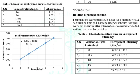
Studies-Kinetics modelling of in- vitro drug release 1) Zero- order kinetics model-
Drug release study can be expressed by the equation- Qo – Qt = Kot………. (1)
Rearrangement of equation I Qt = Qo + Kot……… (2)
Data obtained from in- vitro drug release were plotted as cumulative amount of drug release versus time.
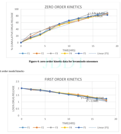
2) First – order kinetics model
This model has been described by the absorption of some drugs. The release of the drug which followed first order kinetics can be expressed as:
Dc/ DT = - Kt ………. (3)
Where, X is the first order rate constant, and t is the time. The data obtained as plotted as log cumulative %age of drug remaining versus time which would yields a straight line with a slope of –k/2.303.
3) Higuchi Model Kinetics Model-
This model is based on hypothesis that-
1) Initial drug concentration in matrix is much higher than drug solubility.
2) Drug diffusion takes place.
3) Drug diffusion are much smaller than system. 4) Perfect sink conditions are always attained in the
release environment.
F1= Q √ ( ) --- (5) Where, Qis the amount of drug released in time t per unit area A, C is the drug solubility in the matrix media and D is the diffusivity of the drug molecules (diffusion coefficient) in the matrix substance.
The relation is valid during all the time, except when the total depletion of the drug in the therapeutic a planar heterogenous matrix system, where the drug concentration in the matrix is lower than its solubility and the release occurs through pores in the matrix, the expression is given equation-
Ƒ1=Q=√
( ) --- (6)
Where D is the diffusion coefficient of the drug molecule in the solvent, S is the porosity of the matrix, T is the tortuosity of the matrix and Q, A, C and t have the meaning assigned above. Tortuisity is defined as the dimensions of radius and branching of the pores and canals in the matrix. In a general way it is possible to simplify the Higuchi model as generally known as the simplified Higuchi model:
Ƒ1 = Q = KH X t1/2--- (7) Where, KH is the Higuchi dissolution constant.
The data obtained were plotted as cumulative percentage drug release versus square root of time.
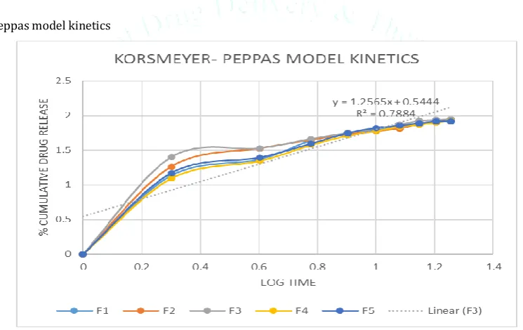
D) KORSMEYER- PEPPAS MODEL KINETICS-
To find out the mechanism of drug release, first 60% drug release data were fitted in Korsmeyer- peppas model. Mt/ M∞ = Ktn--- (8) Where, Mt/M∞ is a fraction of drug released at a time t, k is the release rate constant and n is the release exponent. The n value is used to characterize different release for cylindrical shaped matrices.
In this model, the value of n characterize the release mechanism of drug. For the case of cylindrical, 0.45<n< 0.89 to non- Fickian transport, and n= o.89 to case 2 transport. To find out the exponent of n the portion of the release curve, where Mt/ M∞ < 0.6 should only be used. To study the release kinetics, data obtained from in- vitro drug release studies were plotted as log cumulative %age drug release versus log time.
Stability
The niosomal dispersions were kept in the air tight containers and stored at 2-8.C and at room temperature (30±2.C) for 30 days and 2ml samples were withdrawn every 15 days and at the end of 45 days. The samples were analysed spectrophotometrically at Ϡ max 210nm after disrupting the vesicles with 50% n- propanol.
Optimization of Process- Related Variables-
a) Effect of hydration time102, 103
The niosomal formulations containing Span40 at different ratios and a fixed amount of cholesterol (1:1:1, 1:2:1, 1:3:1) were hydrated with 10ml of distilled water for 30minutes. The vesicle formations and entrapment efficiency of the formulations were calculated by centrifugation method.
b) Effect of sonication method-
The niosomal formulations containing Span40 at different ratios and a fixed amount of cholesterol (1:1:1, 1:2:1, 1:3:1, 1:4:1, 1:5:1) were subjected to ultrasonic vibration using ultra sonicator. To study the effect of sonication time, the formulations were subjected to sonicator for various time intervals (1min, 2min, 3min, 4min, and 5min). The entrapment efficiency of the formulations were measured.
RESULTS AND DISCUSSION
FT-IR Spectrophotometry-. The FT-IR spectra of the drug and the formulations LVS40- 5 are shown in following figures, tables.
Scanning Electron Microscopy (SEM)
Figure 2: SEM Photograph Preparation of Standard Curve of Levamisole-
The UV- Spectrophotometer was used to analyser
Levamisole. The absorbance of the drug in water with small amount of methanol was measured at a wavelength of 215nm
Table 1: Data for calibration curve of Levamisole
S.N. Concentration(µg/Ml) Absorbance
1 1ml 0.011
2 2ml 0.021
3 3ml 0.031
4 4ml 0.041
5 5ml 0.051
Optimization of Process Related Variables-
A) Effect of Hydration time-
The niosomes formulations were hydrated with 20ml distilled water for 40, 45, 70 and 90 minutes. The parameters like vesicle formulation and entrapment efficiency was studied. The results indicated that vesicles were not formed properly but resulted in aggregates with hydration time of 30 minutes. On increase in hydration time, the spherical vesicles were formed and the entrapment efficiency also increased with increase in hydration time from 30 minutes to 60 minutes.
Table 2: Effect of Hydration time on entrapment efficiency
S.N. Hydration Time Entrapment Efficiency
(%w/w)
1) 30 32.06± 1.52
2) 45 40.5 ± 1.16
3) 60 76.2 ± 0.13
4) 75 72.52 ± 1.7
5) 90 70.27 ± 1.15
*Mean SD (n=3)
B) Effect of sonication time –
Formulations were sonicated 3 times for 5 minutes with 2 sec running time and 1 second interval spherical vesicles were not observed after 10 minutes of sonication resulted uniform uni-lamellar vesicles.
Table 3: Effect of sonication time on Entrapment efficiency
S.N, Sonication Time
( minutes) Entrapment Efficiency (%w/w)
1) 0 42.06 ± 0.122
2) 0.5 52.12 ± 0.010
3) 10 61.16 ± 0.042
4) 15 52.21 ± 0.089
5) 20 55.23 ± 1.13
Characterization of Drug Loaded Span40 Niosomes
a) Determination of drug content and Entrapment Efficiency-
Table 4: Drug content and Entrapment Efficiency of Levamisole niosomes containing span-40
FORMULATION CODE DRUG CONTENT (%W/W) %ENTRAPMENT EFFICIENCY (%W/W)
LVS40-1 55.63 63.2
LVS40-2 56.98 64.7
LVS40-3 57.32 65.22
LVS40- 4 58.22 66.54
LVS40- 5 61.34 68.32
The drug content was found to be 55.63%, 56.98%, 57.32%, 58.22%, and 61.34% for the formulations LVS40-1. The entrapment efficiency was found to be 63.2%, 64.7%, 65.22%, 66.54%, and 68.32% for the formulations LVS40-1. The highest entrapment efficiency was observed with Span 40 formulations niosomes in the similar range. This may be due to the higher HLB value of span 40 as compared to
Span60. The entrapment efficiency as the HLB Value decreased from 6.7 of Span40. This may be due to the difference in phase transition temperature. The order of non- ionic surfactants that resulted in better entrapment efficiency is as follows:-
SPAN40 > SPAN60
y = 0.01x + 0.001 R² = 1
0 0.01 0.02 0.03 0.04 0.05 0.06
0 1 2 3 4 5 6
Ab
so
rb
an
ce
a
t
2
1
5
n
m
Conc(µg/ml)
Figure 3: Drug content of LVS 40 Drug Dissolution Studies
Kinetic modelling of drug
1) Zero- order kinetic model-
Figure 4: zero order kinetic data for levamisole niosomes
2) First order model kinetic-
Figure 5: First order kinetics data for levamisole Niosomes
0 10 20 30 40 50 60 70 80
1 2 3 4
Drug(%w/w) %EE
y = 4.8x + 8.256 R² = 0.9426
0 20 40 60 80 100
0 5 10 15 20
%
C
UM
UL
A
TI
V
E
D
R
UG
R
EL
EA
SE
TIME(HRS)
ZERO ORDER KINETICS
F1 F2 F3 F4 F5 Linear (F5)
y = -0.0464x + 2.0248 R² = 0.9881
0 0.5 1 1.5 2 2.5
0 5 10 15 20
LO
G
%
D
R
UG
R
EL
EA
SE
TIME(HRS)
FIRST ORDER KINETICS
3) Higuchi Model kinetic-
Figure 6: Higuchi model kinetic data for Levamisole niosomes
4) Korsmeyer- Peppas model kinetics
Figure 7: Korsmeyer peppas model kinetic data for levamisole niosomes
Kinetic Modelling of Levamisole Niosomes-
Table 5: Kinetic modelling of niosomes formulations Formulations Zero order
Kinetic data (r2)
First order Kinetic data (r2)
Higuchi model Kinetic data (r2)
Peppas model Kinetic data (r2)
Peppas model Kinetic Data (n value)
Best fit model
F1 0.9608 0.9889 0.9558 0.8815 1.2565 Zero order
F2 0.9603 0.9778 0.9559 0.5886 1.3345 Zero order
F3 0.9514 0.9848 0.9514 0.5622 1.2687 Zero order
F4 0.9542 0.9942 0.9655 0.6703 1.3552 Zero order
F5 0.9426 0.9881 0.9433 0.6384 1.2262 Zero order
y = 22.356x - 5.1256 R² = 0.986
-20 0 20 40 60 80 100
0 0.5 1 1.5 2 2.5 3 3.5 4 4.5
%
D
R
UG
R
EL
EA
SE
SQUARE ROOT OF TIME
HIGUCHI MODEL KINETICS
CONCLUSION-
This research affirmed the sustained release of Levamisole hydrochloride loaded span40 niosomes can be attained by Sonication method using Span40 as polymer. DSC and FTIR studies exposed that there is no interaction between the drug and polymer which implies that the drug and polymer. The five batches were evaluated for drug content, drug loading and entrapment efficiency. The batch 3 (CLP-3) with the drug and polymer ratio 1:3:1 was considered to be
superior which showed maximum drug content and entrapment efficiency and prolonged release of drug. The drug release from niosomes was affected by various Span40 concentrations. Scanning electron microscopy (SEM) studies revealed that the niosomes were spherical. The drug release was found to be in sustained manner from batch 3 (LVS-3). Therefore, it was concluded the Levamisole hydrochloride loaded Span40 niosomes were found to be best in treatment of worm skin infections with reduced the side effect on skin and better diagnosis a sustained release manner.
REFERENCES
1) Manconi, M., Sinico, C., Donatella, V., Loy, G., Fadda, A.M., Niosomes as carriers for tritenion. I. Preparation and properties. Int. J. Pharm. 234, 2002, 237-248.
2) Khandare J.N, Madhavi G, Tamhankar B.M., Niosomes Novel Drug Delivery system. The eastern Pharmacists, 1994 37: 61-64.
3) Cherukuri S, Batchu R.U., Ganapuram R.K. Formulation and evaluation of transdermal drug delivery of Levamisole. International Journal of pharmaceutical investigation. 2017: 5-23.
4) Sachan R, Bajpai M, Transdermal drug delivery system: A Review, International journal of research and development in Pharmacy and Life sciences. 2013; 3: 748-765.
5) Gomathi T, Sudha P.N., Florence A.K., Venkatesan J, Sukumaran A. Fabrication of Letrozole formulation using Span40 polymer through sonication method.
6) Yoshika TB, Stemberg and A. Florence 1994. Preparation and properties of vesicles (niosomes) of sorbitan monopalmitate (Span40). International journal of 105(1): 1-6.
7) Madhav NVS, and Saini A, Niosomes, A Novel drug delivery system, Int. J. rpc. 2011 1(3): 498-511.
8) Chien YW, Novel drug delivery system, 2nd edition New York:
Marcel Dekker, Inc. 2007, p.631-637.
9) Indian Pharmacopoeia 2007. Vol-I on behalf of ministry of Health and Family welfare Government of India.
10) Tripathi KD, Essential of medicinal pharmacology 6th edition
Jay pee publications, 2008 254- 274.
11) K. Sabari Kumar, P. Vartharajan, P. Vartharajan, P. Flavarasan and SM Shaik, “ Bioavailability enhancement of Aceclofenac containing surfactants and cholesterol,” International Journal of Biological and Pharmaceutical Research, Vol3 No3, pp. 354-359, 2012.
ACKNOWLEDGEMENTS-The author wishes to acknowledge the Principal, Shivalik College of Pharmacy, Nangal for
providing the chemicals and infrastructure and also Register, IKG Punjab Technical