Frontiers in Dentistry
This work is published as an open access article distributed under the terms of the Creative Commons Attribution 4.0 License (http://creativecommons.org/licenses/by-nc/4). Non-commercial uses of the work are permitted, provided the original work is properly cited.
Shear Bond Strength of Self-Adhesive Flowable
Composite, Conventional Flowable
Composite and
Resin-Modified Glass Ionomer Cement to Primary
Dentin
Kiana Poorzandpoush
1, Mehdi Shahrabi
1,2*, Alireza Heidari
1, Zohre
Sadat Hosseinipour
11. Department of Pediatric Dentistry, School of Dentistry, Tehran University of Medical Sciences, Tehran, Iran 2. Dental Research Center, Dentistry Research Institute, Tehran University of Medical Sciences, Tehran, Iran
Article Info A B S T R A C T
Article type:
Original Article Objectives: adhesive and conventional flowable composites and resin-modified glass-ionomer This study aimed to compare the shear bond strength (SBS) of self-cement (RMGIC) to primary dentin.
Materials and Methods: In this in vitro, experimental study, the buccal surface of 48 primary canine and first molar teeth was longitudinally sectioned to expose dentin. The teeth were randomly divided into three groups (n=16) of 37.5% phosphoric acid+ OptiBond+ Premise Flow composite (group 1), Vertise Flow composite (group 2) and RMGIC (group 3). A plastic cylindrical mold was placed on the exposed dentin and filled with restorative materials. The samples were then immersed in distilled water at 37°C for 24 hours, subjected to 1000 thermal cycles between 5-55°C and underwent SBS test. The mode of failure was determined under a stereomicroscope. Data were analyzed using one-way ANOVA and Tukey’s test.
Results: A significant difference was noted in SBS of the groups (P<0.05). The SBS of conventional flowable composite was significantly higher that of RMGIC and adhesive flowable composite (P<0.05). The difference in SBS of RMGIC and self-adhesive flowable composite was not significant (P>0.05). Failure at the dentin-restoration interface (adhesive failure) had the highest frequency in groups 1 and 2. The frequency of adhesive failure was 100% in group 3.
Conclusion: Within the limitations of this study, the conventional flowable composite yielded the highest SBS to primary dentin. Self-adhesive flowable composite and RMGIC showed the lowest SBS with no significant difference with each other.
Keywords: Composite Resins; Dentin; Glass Ionomer Cements; Shear Strength Article History:
Received: 8 July 2018 Accepted: 4 August 2018 Published: 20 January 2019
* Corresponding author:
Dental Research Center, Dentistry Research Institute, Tehran University of Medical Sciences; Department of Pediatric Dentistry, School of Dentistry, Tehran University of Medical Sciences, Tehran, Iran
Email: [email protected]
Cite this article as: Poorzandpoush K, Shahrabi M, Heidari A, Hosseinipour ZS. Shear Bond Strength of Self-Adhesive Flowable Composite, Conventional Flowable Composite and Resin-Modified Glass Ionomer Cement to
INTRODUCTION
Tooth-colored restorative materials are increasingly used for tooth restoration due to excellent esthetics. Demand for tooth-colored restorations has greatly increased in pediatric dentistry. Conventional flowable composites and resin modified glass ionomer cements (RMGICs) are among the commonly used
Poorzandpoush K, et al.
63 Front Dent, Vol. 16, No. 1, Jan-Feb 2019
hydrophobicity of composite resins, their bonding to tooth structure has always been a challenge. Attempts have been made to change the morphology and chemical composition of tooth structure aiming to improve bond strength [2]. Conventional composite resins are bonded to dentin using the bonding systems, which aim to minimize marginal gap and increase the fracture strength and durability of restorations [3].
Researchers are trying to simplify the bonding procedure, and all-in-one bonding agents have been introduced that contain etchant, primer and adhesive all in one bottle to simplify the procedure of composite restoration [4]. Recently, self-adhesive flowable composites were introduced to the market that possess the advantages of all-in-one bonding systems and flowable composites altogether [4-6]. The clinical advantages of this type of composite include easy use (not requiring separate etching, priming or adhesive application), prevention of procedural errors related to clinical application of conventional bonding agents (such as over-drying and over-wetting) and reduction of chair time [4-7]. However, durability and clinical service of these composites remain a concern for many dental clinicians [8]. Studies on physical and mechanical properties of self-adhesive composites are limited [5, 6, 9, 10] and studies on the bonding properties and other characteristics of self-adhesive flowable composites in primary teeth are scarce [11]. Considering all the above, this study aimed to assess the shear bond strength (SBS) of self-adhesive flowable composite, conventional flowable composite and RMGIC to primary dentin. The mode of failure was also determined under a stereomicroscope.
MATERIALS AND METHODS
This in vitro, experimental study eval-uated 48 extracted primary canine and first molar teeth. The study was approved in the ethics committee of our university (code: IR.TUMS.DENTISTRY.REC.1396.3345). Sample size was calculated to be 12 in each of the three groups according to a study by Sachdeva et al, [11] using one-way ANOVA power analysis feature of PASS 11 software assuming alpha=0.05, beta=0.2, standard deviation of 2.11 and effect size of 0.56. To increase the
accur-acy of the results, 48 teeth (n=16 in each group) were evaluated in this study.
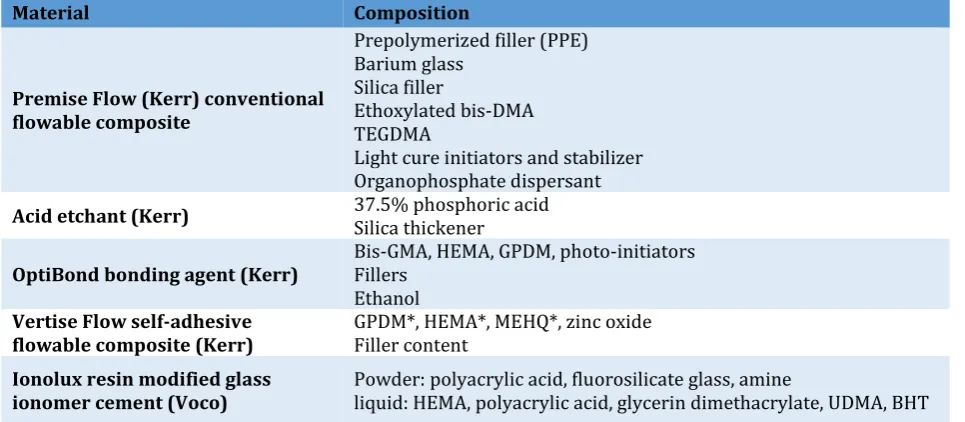
All teeth were collected within 3 months and stored in distilled water. The inclusion criteria were primary canine and first molars close to their physiologic exfolia-tion time, over-retained teeth and teeth that were candidates for serial extraction. Teeth with developmental anomalies, extensive caries, and with broken crowns were excluded. The teeth were immersed in 0.5% chloramine T solution for 7 days. After disinfection, the teeth were rinsed with distilled water, dried and mounted in polyester blocks so that their long-itudinal axis was perpendicular to the surface. After mounting, a longitudinal section was made at the buccal surface to expose dentin using a cutting machine (T201A Mecatome; Presi, France). The teeth were then cleaned, rinsed and dried. The samples were randomly divided into three groups of 16 for the use of three restorative materials. Table 1 shows the characteristics of the three restorative materials used in this study.
RMGIC (Ionolux, Voco, Germany) was used in group 1. No surface treatment was performed on dentin. The glass ionomer powder and liquid were mixed in 3.2/1 g ratio or one scoop of powder and two drops of liquid according to the manufacturer’s instructions. The powder was divided into two portions. The first portion was mixed with liquid using a plastic spatula. The second portion of powder was then added and mixed. Mixing time was 30 seconds. After mixing, the homogenous paste was packed into a cylindrical transparent plastic mold with an internal diameter of 3 mm and 2 mm height to reach 2 mm thickness. Care was taken to avoid void formation. Light curing was then performed using a LED light curing unit (Blue Phase; Ivoclar Vivadent, Schaan, Lichtenstein) with a light intensity of 600 mW/cm2 for 20
seconds. After polymerization, plastic molds were removed using a scalpel.
Conventional flowable composite (Premise Flow; Kerr, Bolzano, Italy) was used in group 2. Dentin was etched with 37.5% phosphoric acid (Kerr, Bolzano, Italy) for 15 seconds. It was then rinsed according to the manufacturer’s instructions.
64
Front Dent, Vol. 16, No. 1, Jan-Feb 2019
according to the manufacturer’s instructions. It was rubbed on the surface and gently air-sprayed for 3 seconds. Light curing was performed for 20 seconds.
Flowable composite was applied into the transparent cylindrical plastic molds with 2 mm thickness and polymerized for 20 seconds. The mold was then removed using a scalpel.
Table 1. Characteristics of the three restorative materials used in this study
Material Composition
Premise Flow (Kerr) conventional flowable composite
Prepolymerized filler (PPE) Barium glass
Silica filler
Ethoxylated bis-DMA TEGDMA
Light cure initiators and stabilizer Organophosphate dispersant
Acid etchant (Kerr) 37.5% phosphoric acid Silica thickener
OptiBond bonding agent (Kerr) Bis-GMA, HEMA, GPDM, photo-initiators Fillers
Ethanol
Vertise Flow self-adhesive
flowable composite (Kerr) GPDM*, HEMA*, MEHQ*, zinc oxide Filler content
Ionolux resin modified glass
ionomer cement (Voco) Powder: polyacrylic acid, fluorosilicate glass, amine liquid: HEMA, polyacrylic acid, glycerin dimethacrylate, UDMA, BHT
GPDM: Glycerol phosphate dimethacrylate; HEMA: Hydroxyethyl methacrylate; MEHQ: 4- methoxyphenol
Self-adhesive flowable composite (Vertise, Kerr, Italy) was used in group 3. In this group, first a 0.5 mm increment was applied on the dentin surface and rubbed with moderate pressure using a microbrush for 15 to 20 seconds. This layer was cured for 20 seconds. The next increment was applied in 1.5 mm thickness and curing was performed for 20 seconds. The mold was then removed. Care was taken to prevent void or crack formation in samples. The teeth were immersed in distilled water at 37°C for 24 hours and thermocycled for 1000 cycles between 5-55°C in a thermocycler (TC-300; Vafaei Industrial, Tehran, Iran) with a dwell time of 30 seconds and transfer time of 5 seconds [12]. The SBS was measured using a universal testing machine (Z250; Zwick/Roell, Germany). Load was applied with a blade perpendicular to the tooth-restoration interface at a crosshead speed of 1 mm/minute and the load cell applied load until bond failure. The mode of failure was evaluated under a stereomicroscope (SMZ800; Nikon, Tokyo, Japan) at x20 magnification. The mode of failure was categorized as adhesive (at the dentin-restorative material interface), cohesive (within the restorative material or dentin substrate) or mixed (a combination of both adhesive and cohesive failures).
Data were analyzed using SPSS version 22 (SPSS
Inc., IL, USA). One-way ANOVA was applied to compare the SBS among the three groups. Tukey’s post hoc test was applied for pairwise comparisons. P<0.05 was considered statistically significant.
RESULTS
Table 2 shows the mean and standard deviation of SBS in the three groups. The highest SBS was noted in the conventional flowable composite (14.87±3.4 MPa) while the lowest SBS was noted in RMGIC group (5.39±2.6 MPa). One-way ANOVA showed a significant difference in SBS among the three groups (P<0.001).
Table 2. Mean and standard deviation (SD) of shear
bond strength (MPa) in the three groups (n=16)
Study group* Min Max Mean SD
Conventional flowable
composite 7.12 18.99 14.87 3.42
Self-adhesive flowable
composite 3.69 9.92 6.60 1.97
Resin-modified glass ionomer
cement 0.53 9.35 5.39 2.63
* The mean difference is significant at the 0.05 level.
Poorzandpoush K, et al.
65 Front Dent, Vol. 16, No. 1, Jan-Feb 2019
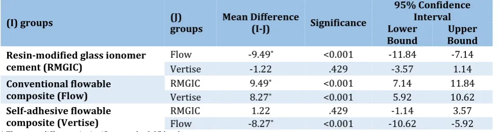
Table 3. Pairwise comparisons of the groups in terms of shear bond strength
(I) groups (J) groups Mean Difference (I-J) Significance
95% Confidence Interval Lower
Bound Bound Upper
Resin-modified glass ionomer cement (RMGIC)
Flow -9.49* <0.001 -11.84 -7.14
Vertise -1.22 .429 -3.57 1.14
Conventional flowable
composite (Flow) RMGIC 9.49
* <0.001 7.14 11.84
Vertise 8.27* <0.001 5.92 10.62
Self-adhesive flowable
composite (Vertise) RMGIC Flow -8.271.22 * <0.001 .429 -10.62 -1.14 -5.92 3.57
* The mean difference is significant at the 0.05 level
Table 3 shows pairwise comparisons of the groups in terms of SBS. Tukey’s HSD post-hoc test showed that conventional flowable composite had a significantly higher SBS to primary dentin compared to the other two groups (P<0.001). Self-adhesive flowable composite and RMGIC groups showed the lowest SBS, with no significant difference with each other (P=0.429). Table 4 shows the mode of failure of samples in the three groups.
Table 4. Mode of failure in the three groups (n=16)
Group Adhesive Mixed Cohesive
Conventional flowable
composite 11 5 0
Self-adhesive flowable
composite 13 3 0
Resin-modified glass ionomer
cement 16 - 0
The adhesive type was the most frequent mode of failure in the conventional composite and self-adhesive groups. Adhesive failure was noted in 100% of samples in RMGIC group (Fig. 1).
Fig 1. Mixed (A) and adhesive (B) modes of failure
DISCUSSION
This study assessed and compared the SBS of self-adhesive flowable composite, conventional flowable composite and RMGIC to primary dentin. A significant difference was noted in SBS of the groups. Our findings showed that the SBS of conventional flowable composite was significantly higher than that of self-adhesive flowable composite, which is in agreement with the results of previous studies [4,7,9,11,13-15]. Our findings showed that the SBS of conventional flowable composite was signify-cantly higher than that of self-adhesive flowable composite, which is in agreement with the results of previous studies [4,7,9,11,13-15]. Etch and rinse bonding system was used for the conventional composite in our study. Separate etching eliminates the smear layer and mineral contents of dentin to 5-8 µ depth [16]. It increases the permeability of dentin and enhances subsequent penetration of adhesive monomers [17]. Formation of the hybrid layer at the interface of intertubular demineralized dentin and bonding agent enhances the physical properties [18]. Also, etching with phosphoric acid in etch and rinse systems results in formation of longer and thicker resin tags compared to those in self-etch systems (as in self-adhesive flowable composites) [11].
Front Dent, Vol. 16, No. 1, Jan-Feb 2019 66 Studies regarding the reaction of GPDMA
monomer with dentin hydroxyapatite are limited [19]. However, it seems that GPDMA monomer can etch the enamel and dentin [8]. However, the acidity of Vertise Flow is not high enough to modify the smear layer and allow the penetration of resin into the substrate. Resultantly, the micromechanical retention would be lower and subsequently a lower bond strength is achieved by self-adhesive composite resins [20].
The manufacturer of OptiBond claims that it contains 15% barium glass filler (0.04 µ) that not only reinforces the hybrid layer but also penetrates well into dentinal tubules and forms a structural bonding, which is not seen in unfilled or nanofilled composite resins. This filler increases the bond strength to tooth surfaces and prevents microleakage. This can also explain the higher bond strength of conventional composite/OptiBond compared to self-adhesive composite with no separate bonding agent. Premise Flow conventional composite has low-viscosity TEGDMA monomer in its composition. Since this monomer is a cross-linker, it can effectively increase the bond strength and enhance the penetration of resin into the dentin structure [21].
Uekusa et al. [22] stated that the smear layer is rapidly removed by etching of primary dentin. Thus, shorter etching time by using a weaker etchant is recommended for primary teeth. However, etch and rinse bonding systems have phosphoric acid as etchant, which is a strong acid and can be more invasive for primary dentin. Also, due to lower thickness of dentin in primary teeth, it can cause exposure of dense and wide dentinal tubules close to dental pulp and consequently limit the efficacy of bonding process. Therefore, self-adhesive composite systems are expected to be suitable for application on primary dentin due to their lower acidity than etch and rinse systems. However, our results showed higher bond strength of conventional composite and etch and rinse bonding system to primary dentin, which may be due to high viscosity and absence of solvent in self-adhesive flowable composite and its subsequent limited penetration into dentin structure [7].
Makishi et al. [9] evaluated confocal laser scanning microscopy images and did not detect formation of hybrid layer following the use of Vertise Flow composite.
Thus, it may be concluded that high viscosity, less wettability and limited penetration of self-adhesive composites can result in lower bond strength compared to the use of conventional composites and total etch bonding systems [23]. Our findings also revealed lower SBS of RMGIC than that of conventional flowable composite, which was in line with the findings of a previous study [13]. Studies comparing the bond strength of RMGIC and self-adhesive composite are scarce. In our study, the SBS values of RMGIC and self-adhesive composite were comparable; this finding was in agreement with that of Pacifici et al, [13] and Scaminaci et al [14]. RMGIC and self-adhesive composite have easier application than the conventional composite. Application of self-adhesive composite is even easier and faster than RMGIC. Although self-adhesive composite does not release fluoride, it has high filler content and is believed to have a higher wear resistance than RMGIC. Moreover, Vertise Flow composite has a less porous surface than RMGIC, which can result in higher esthetics and less plaque accumulation. Decreased postoperative tooth hypersensitivity is another advantage of Vertise self-adhesive composite [7,13]. Scaminaci et al. [15] reported that the SBS of Vertise Flow was significantly higher than that of glass ionomer cement, which was in contrast to our finding. This difference in the results may be due to the use of conventional cement (Ketac Fil), which has a lower SBS than RMGIC used in our study because the HEMA molecule in the composition of RMGIC increases the bond strength. However, Scaminaci et al, [14] in another study compared the SBS of restorative materials to permanent dentin and found results in line with our findings. Although the SBS of Vertise Flow was slightly higher than that of GIC, this difference was not significant. Since the type of tooth (primary versus permanent) was different in the two studies by Scaminaci et al, [14,15] it may be stated that in addition to difference in sample size, difference in structure of permanent and primary dentin is another reason explaining the difference in the results of the two studies.
Poorzandpoush K, et al.
67 Front Dent, Vol. 16, No. 1, Jan-Feb 2019
dentin with bonding agent, no formation of hybrid layer or formation of a thin, non-homogenous hybrid layer [25]. Considering the presence of chemical bonding mechanism between glass ionomer cement and dentin, the adhesive bond at the interface is expected to be stronger than the cohesive bond within the cement. However, considering the dominant mode of failure being the adhesive type in RMGIC group, it seems that high viscosity of this material causes less mechanical interlocking and consequently less contact of material with the porosities and irregularities of the dentin surface. This study had an in vitro design. Oral conditions cannot be well simulated in vitro in terms of thermal changes, masticatory forces, water sorption, and pH alterations. Thus, generalization of results to the clinical setting must be done with caution. Future in vivo studies are required to compare the clinical success of these restorative materials in a larger sample size.
CONCLUSION
Within the limitations of this study, the conventional flowable composite yielded the highest SBS to primary dentin. Self-adhesive flowable composite and RMGIC showed the lowest SBS with no significant difference with each other.
ACKNOWLEDGMENTS
This study has been funded and supported by Tehran university of medical sciences (TUMS); Grant No: 9411276001.
REFERENCES
1. Bücher K, Metz I, Pitchika V, Hickel R, Kühnisch J. Survival characteristics of composite restorations in primary teeth. Clin Oral Investig. 2015 Sep;19(7):1653-62.
2. Korkmaz Y, Gurgan S, Firat E, Nathanson D. Shear bond strength of three different nano-restorative materials to dentin. Oper Dent. 2010 Jan-Feb;35(1):50-7.
3. Türkmen C, Sazak-Oveçoğlu H, Günday M, Güngör G, Durkan M, Oksüz M. Shear bond strength of composite bonded with three adhesives to Er,Cr:YSGG laser-prepared enamel. Quintessence Int. 2010 Jun;41(6):e119-24. 4. Memarpour M, Shafiei F, Razmjoei F, Kianimanesh N. Effect of laser preparation on adhesion of a self-adhesive flowable composite
resin to primary teeth. Microsc Res Tech. 2016 Apr;79(4):334-41.
5. Poitevin A, De Munck J, Van Ende A, Suyama Y, Mine A, Peumans M, et al. Bonding effectiveness of self-adhesive composites to dentin and enamel. Dent Mater. 2013 Feb;29(2):221-30.
6. Wajdowicz MN, Vandewalle KS, Means MT. Shear bond strength of new self-adhesive flowable composite resins. Gen Dent. 2012 Mar-Apr;60(2):e104-8.
7. Vichi A, Margvelashvili M, Goracci C, Papacchini F, Ferrari M. Bonding and sealing ability of a new self-adhering flowable composite resin in class I restorations. Clin Oral Investig. 2013 Jul;17(6):1497-506.
8. Naga AA, Yousef M, Ramadan R, Fayez Bahgat S, Alshawwa L. Does the use of a novel self-adhesive flowable composite reduce nanoleakage? Clin Cosmet Investig Dent. 2015 Mar 27;7:55-64.
9. Makishi P, Pacheco RR, Sadr A, Shimada Y, Sumi Y, Tagami J, et al. Assessment of Self-Adhesive Resin Composites: Nondestructive Imaging of Resin-Dentin Interfacial Adaptation and Shear Bond Strength. Microsc Microanal. 2015 Dec;21(6):1523-9.
10. Salerno M, Derchi G, Thorat S, Ceseracciu L, Ruffilli R, Barone AC. Surface morphology and mechanical properties of new-generation flowable resin composites for dental restoration. Dent Mater. 2011 Dec;27(12):1221-8.
11. Sachdeva P, Goswami M, Singh D. Comparative evaluation of shear bond strength and nanoleakage of conventional and self-adhering flowable composites to primary teeth dentin. Contemp Clin Dent. 2016 Jul-Sep;7(3):326-31.
12. Goracci C, Margvelashvili M, Giovannetti A, Vichi A, Ferrari M. Shear bond strength of orthodontic brackets bonded with a new self-adhering flowable resin composite. Clin Oral Investig. 2013 Mar;17(2):609-17.
13. Pacifici E, Chazine M, Vichi A, Grandini S, Goracci C, Ferrari M. Shear-bond strength of a new self-adhering flowable restorative material to dentin of primary molars. J Clin Pediatr Dent. 2013 Winter;38(2):149-54.
Front Dent, Vol. 16, No. 1, Jan-Feb 2019 68 15. Scaminaci Russo D, Iuliano V, Franchi L,
Ferrari M, Giachetti L. Adhesion to primary dentin: microshear bond strength and scanning electron microscopic observation. Am J Dent. 2013 Dec;26(6):341-6.
16. Pashley DH, Tay FR, Breschi L, Tjäderhane L, Carvalho RM, Carrilho M, et al. State of the art etch-and-rinse adhesives. Dent Mater. 2011 Jan;27(1):1-16.
17. Nakabayashi N, Kojima K, Masuhara E. The promotion of adhesion by the infiltration of monomers into tooth substrates. J Biomed Mater Res. 1982 May;16(3):265-73.
18. Van Meerbeek B, Willems G, Celis JP, Roos JR, Braem M, Lambrechts P, et al. Assessment by nano-indentation of the hardness and elasticity of the resin-dentin bonding area. J Dent Res. 1993 Oct;72(10):1434-42.
19. Van Meerbeek B, De Munck J, Yoshida Y, Inoue S, Vargas M, Vijay P, et al. Adhesion to enamel and dentin: current status and future challenges. Oper Dent. 2003 Oct;28(3):215-35. 20. Yazici AR, Agarwal I, Campillo-Funollet M, Munoz-Viveros C, Antonson SA, Antonson
DE, et al. Effect of laser preparation on bond strength of a self-adhesive flowable resin. Lasers Med Sci. 2013 Jan;28(1):343-7.
21. Van Landuyt KL, Snauwaert J, De Munck J, Peumans M, Yoshida Y, Poitevin A, et al. Systematic review of the chemical composition of contemporary dental adhesives. Biomaterials. 2007 Sep;28(26):3757-85. 22. Uekusa S, Yamaguchi K, Miyazaki M, Tsubota K, Kurokawa H, Hosoya Y. Bonding efficacy of single-step self-etch systems to sound primary and permanent tooth dentin. Oper Dent. 2006 Sep-Oct;31(5):569-76.