University of Pennsylvania
ScholarlyCommons
Publicly Accessible Penn Dissertations
1-1-2016
Molecular and Cellular Approaches Toward
Understanding Dynein-Driven Motility
Swathi Ayloo
University of Pennsylvania, [email protected]
Follow this and additional works at:
http://repository.upenn.edu/edissertations
Part of the
Biophysics Commons,
Cell Biology Commons, and the
Molecular Biology Commons
This paper is posted at ScholarlyCommons.http://repository.upenn.edu/edissertations/1595
For more information, please [email protected].
Recommended Citation
Ayloo, Swathi, "Molecular and Cellular Approaches Toward Understanding Dynein-Driven Motility" (2016).Publicly Accessible Penn Dissertations. 1595.
Molecular and Cellular Approaches Toward Understanding
Dynein-Driven Motility
Abstract
Active transport is integral to organelle localization and their distribution within the cell. Kinesins, myosins and dynein are the molecular motors that drive this long range transport on the actin and microtubule cytoskeleton. Although several families of kinesins and myosins have evolved, there is only one form of cytoplasmic dynein driving active retrograde transport in cells. While dynactin is an essential co-factor for most cellular functions of dynein, the mechanistic basis for this evolutionarily well conserved interaction remains unclear. Here, I use single molecule approaches with purified dynein to reconstitute processes in vitro, and implement an optogenetic tool in neurons to further dissect regulatory mechanisms of dynein-driven transport in cells. I demonstrate for the first time, at the single molecule level, that dynactin functions as a tether to enhance the initial recruitment of dynein onto microtubules but also acts as a brake to slow the motor. I then extend this work in neurons to understand regulation of the dynein motor at the cellular level. Neurons are particulary dependent on long-range transport as organelles and macromolecules must be efficiently moved over the extended length of the axon and further, have mechanisms in place for the
compartment-specific regulation of trafficking in axons and dendrites. I use a light-inducible dimerization tool to recruit motor proteins or motor adaptors to organelles in real time to examine downstream effects of organelle motility and compartment-specific regulation of motors. I find that while dynein works efficiently in both axons and dendrites, kinesins are differentially regulated in a compartment-specific manner. I further demonstrate that dynein-driven motility in neurons is largely governed by microtubule orientation and requires microtubule dynamics for efficient navigation in axons and dendrites. Together, this work sheds light on the molecular and cellular mechanisms of dynein function both in vitro and in vivo using a combination of approaches. My findings converge to a model wherein dynactin enhances the recruitment of dynein onto microtubule plus ends, leading to efficient minus-end directed motility of dynein. This becomes especially critical in neuronal growth cones and dendrites owing to the large number of highly dynamic microtubules in these compartments.
Degree Type Dissertation
Degree Name
Doctor of Philosophy (PhD)
Graduate Group Biology
First Advisor Erika L. Holzbaur
Subject Categories
Biophysics | Cell Biology | Molecular Biology
MOLECULAR AND CELLULAR APPROACHES TOWARD
UNDERSTANDING DYNEIN-DRIVEN MOTILITY
Swathi Ayloo
A DISSERTATION in
Biology
Presented to the Faculties of the University of Pennsylvania In Partial Fulfillment of the Requirements for the
Degree of Doctor of Philosophy 2016
Supervisor of Dissertation
_______________________________
Erika L.F. Holzbaur, PhD, Professor of Physiology
Graduate Group Chairperson
_______________________________
Michael A. Lampson, PhD, Associate Professor of Biology
Dissertation Committee
Erfei Bi, PhD, Professor of Cell and Developmental Biology E. Michael Ostap, PhD, Professor of Physiology
Tatyana Svitkina, PhD, Professor of Biology
MOLECULAR AND CELLULAR APPROACHES TOWARD UNDERSTANDING
DYNEIN-DRIVEN MOTILITY
iii
ACKNOWLEDGMENT
This work would not have been possible without the support and help from several people. I would first like to acknowledge my advisor, Erika Holzbaur for her wonderful mentorship. She has always held me to high standards and constantly challenged me to think deeply about my science. She has always encouraged me and I greatly appreciate all the opportunities she provided me with, ensuring my success. Thank you, Erika, for your faith in me and teaching me several things throughout my graduate training. I would also like to thank my thesis committee members: Erfei Bi, Mike Ostap, Tanya Svitkina and Mike Lampson for their insights and advice. Special thanks to Mike Ostap and Mike Lampson for terrific collaborations on both my projects. Both of them were instrumental in providing key reagents and scientific advice which made this thesis work possible.
I would especially like to thank all the past and current members of the Holzbaur lab: Mariko Tokito for making DNA constructs and for all the endless conversations she had with me about protein purification and several other biochemical assays; Karen Wallace for her assistance with mouse work; Adam Hendricks who first introduced me to single molecule assays and was my go-to person for all things biophysics; Meredith Wilson, Betsy McIntosh, Alison Twelvetrees, Amy Ghiretti, Mara Olenick and Eva Klinman for their friendship and for putting up with several of my random bouts of craziness.
iv
I would also like to thank Aditya Dodda and Ed Ballister for their fantastic collaborations. Aditya spent several hours and days looking at my data to develop new ways to analyze it. What started out with Aditya fixing errors in the basic code I wrote, turned into him developing new algorithms and simulations for my data. Ed Ballister developed the light-inducible dimerization system and was the first one to think about applying it to motors. Working with him was a lot of fun and without him, my second project would never exist.
I would also like to acknowledge the Pennsylvania Muscle Institute (PMI) at Penn, especially Mike Ostap, Yale Goldman, Katya Grishchuck and their lab members. Being part of the PMI gave me access to several key resources and opportunities. I always enjoyed our journal clubs and learned how to think about science through these meetings. The PMI community made me feel at home and inculcated a sense of belonging in me.
Big thanks to all my friends in Boston who was my family away from home. Needless to say, I am quite excited to be surrounded by them the next few years. Their friendship through the years, since my undergrad days has been invaluable. I had a lot of fun with you guys!
I am grateful to all my family members – parents, uncle and aunt, my siblings and my cousins. They were always there for me and made everything possible for me. I would not be where I am today without them. Thank you for believing in me and giving me the freedom to pursue what I wanted.
v ABSTRACT
MOLECULAR AND CELLULAR APPROACHES TOWARD UNDERSTANDING DYNEIN-DRIVEN MOTILITY
Swathi Ayloo Erika L.F. Holzbaur
vi
kinesins are differentially regulated in a compartment-specific manner. I further demonstrate that dynein-driven motility in neurons is largely governed by microtubule orientation and requires microtubule dynamics for efficient navigation in axons and dendrites. Together, this work sheds light on the molecular and cellular mechanisms of dynein function both in vitro
vii
TABLE OF CONTENTS
ACKNOWLEDGMENT ...III
ABSTRACT... V
LIST OF FIGURES ... VIII
CHAPTER 1: INTRODUCTION ... 1
I. Intracellular Transport – An Introduction ... 1
II. The Microtubule Cytoskeleton ... 3
III. Kinesin Motors ... 7
IV. Cytoplasmic dynein ... 10
V. Microtubule Modifications and Microtubule Associated Proteins ... 21
VI. Neuronal Transport ... 31
CHAPTER 2: DYNACTIN FUNCTIONS AS BOTH A DYNAMIC TETHER AND BRAKE DURING DYNEIN-DRIVEN MOTILITY ...40
I. Summary ... 41
II. Introduction ... 42
III. Results ... 45
IV. Discussion ... 66
V. Material and Methods ... 70
CHAPTER 3: OPTOGENETIC CONTROL OF ORGANELLE TRANSPORT USING A PHOTOCAGED CHEMICAL INDUCER OF DIMERIZATION ...78
I. Summary ... 79
II. Introduction ... 80
III. Results ... 81
IV. Discussion ... 88
V. Material and Methods ... 89
CHAPTER 4: OPTOGENETIC RECRUITMENT OF MOTORS REVEALS DIFFERENTIAL AXO-DENDRITIC REGULATION OF TRAFFICKING IN NEURONS .94 I. Summary ... 95
II. Introduction ... 96
III. Results ... 98
IV. Discussion ... 112
V. Material and Methods ... 119
CHAPTER 5: CONCLUSIONS AND FUTURE DIRECTIONS ... 122
viii
LIST OF FIGURES
FIGURE 1.1SCHEMATIC ILLUSTRATING DYNAMIC INSTABILITY OF MICROTUBULES ... 5
FIGURE 1.2SCHEMATIC ILLUSTRATING THE DYNEIN AND DYNACTIN COMPLEXES ...14
FIGURE 1.3SCHEMATIC ILLUSTRATING TUBULIN POST-TRANSLATIONAL MODIFICATIONS
(ADAPTED FROM JANKE,2014) ...23
FIGURE 1.4ILLUSTRATION OF MECHANISMS REGULATING POLARIZED NEURONAL TRAFFICKING
...38
FIGURE 2.1 DIC1-GFP IS EFFICIENTLY INCORPORATED INTO THE DYNEIN COMPLEX. ...46
FIGURE 2.2 DYNEIN-GFP SWITCHES STOCHASTICALLY FROM PROCESSIVE TO DIFFUSIVE STATES OF MOTION...48
FIGURE 2.3 SIMULATIONS TO VALIDATE OUR ANALYSIS. ...49
FIGURE 2.4 GENERATION OF PURIFIED RECOMBINANT P150GLUED
CONSTRUCTS. ...51
FIGURE 2.5 P150GLUED
CO-MIGRATES WITH DYNEIN AND ENHANCES ITS RECRUITMENT ONTO MICROTUBULES ...53
FIGURE 2.6 P150GLUED
REGULATES THE ASSOCIATION AND DISSOCIATION OF DYNEIN FROM MICROTUBULES. ...55
FIGURE 2.7THE MOTILITY OF P150GLUED
IN CELL EXTRACTS IS DYNEIN-DRIVEN AND ATP
DEPENDENT. ...58
FIGURE 2.8THE MOTILITY OF DYNACTIN IN CELL EXTRACTS IS SIMILAR TO IN VITRO MOTILITY OF PURIFIED DYNEIN-GFP...59
FIGURE 2.9 P150GLUED1-CC1
SHOWS MOTILITY CHARACTERISTICS SIMILAR TO FULL LENGTH P150GLUED. ...61
FIGURE 2.10THE CAP-GLY DOMAIN OF DYNACTIN ENHANCES THE ENGAGEMENT OF DYNEIN ONTO MICROTUBULES. ...63
FIGURE 2.11 MODEL DEPICTING THE ROLES OF DYNACTIN IN DYNEIN-DRIVEN TRANSPORT...69
ix
FIGURE 3.2PHOTOACTIVATION OF PEROXISOMES RECRUITS MOTORS AND INDUCES MOTILITY OF PEROXISOMES. ...83
FIGURE 3.3RECRUITMENT OF MOTORS TO PEROXISOMES IN AXONS INDUCES ROBUST
MOTILITY. ...85
FIGURE 3.4RECRUITMENT OF MOTORS TO MITOCHONDRIA IN AXONS OF NEURONS. ...86
FIGURE 4.1RECRUITMENT OF DYNEIN OR KINESIN TO PEROXISOMES IN DENDRITES INDUCES ROBUST BIDIRECTIONAL MOTILITY. ... 100
FIGURE 4.2KINESIN IS NOT AS EFFICIENT AS DYNEIN IN INDUCING MOTILITY IN DENDRITES. 101
FIGURE 4.3DYNEIN-DRIVEN MOTILITY IN DENDRITES HAS A RETROGRADE BIAS. ... 103
FIGURE 4.4DYNEIN REUIRES DYNAMIC MICROTUBULES FOR EFFICIENT INITIATION OF
TRANSPORT. ... 106
FIGURE 4.5BOTH KIF13A AND KIF13B, KINESIN-3 FAMILY MOTORS ARE EQUALLY EFFICIENT IN AXONS ... 108
FIGURE 4.6DIFFERENTIAL REGULATION OF KIF13A AND KIF13B IN DENDRITES. ... 110
1
CHAPTER 1: Introduction
I. Intracellular Transport – An Introduction
Cellular organization of macromolecules and organelles in space and time is critical for cellular function. Accurate localization of organelles is often achieved by the process of intracellular transport. Initial experiments by George Palade and colleagues in pancreatic exocrine cells, professional secretory cells, laid the foundation for intracellular transport. This work included pulse-chase experiments in vivo (Caro and Palade, 1964) followed by pulse-chase in in vitro tissue slices (Jamieson and Palade, 1967a, 1967b) which demonstrated that proteins shuttled from one compartment to another. These experiments also showed that intracellular transport requires energy, as absence of ATP synthesis caused proteins to remain in the endoplasmic reticulum (Jamieson and Palade, 1968), an intriguing finding at the time. Evidence from electron microscopy and other studies in the next few years suggested that the protein transport occurred via vesicular trafficking (Palade, 1975). The discovery of clathrin (Pearse, 1976) followed by elegant work with cell-free systems (Freis and Rothman, 1980) and yeast genetics quickly established the players involved in the secretory trafficking pathway (reviewed in Rothman, 1994).
2
We now know that molecular motors kinesins and cytoplasmic dynein drive long range transport of vesicles along microtubules. Most kinesins move toward the microtubule plus ends and dynein moves towards the minus ends of microtubules. Further, while several families of kinesins have evolved for specialized functions, there is only form of cytoplasmic dynein driving retrograde transport.
3 II. The Microtubule Cytoskeleton
The cytoskeleton is a dynamic network of cytoplasmic filaments that maintains cell shape, enables cellular motion and plays important roles in intracellular transport and cell division. The cytoskeleton provides a mechanical framework and structural integrity to the cell. The diverse activities of the cytoskeleton depend on three types of protein filaments - actin filaments, microtubules and intermediate filaments. Each type of filament is formed from a different protein subunit: actin for actin filaments, tubulin for microtubules and a family of related fibrous proteins for intermediate filaments. Actin and tubulin have been especially highly conserved throughout the evolution of eukaryotes
.
Each of these cytoskeletal proteins assemble into distinct linear filaments which serve as tracks for intracellular transport. As the focus of my project is microtubule-based transport, the next set of sections will discuss this in greater detail.Early work on microtubule structures came from transmission EM studies and aldehyde fixation methods (Fawcett and Porter, 1954; Sabatini et al., 1963). The first physical evidence of these structures was shown by Inoue and co-workers in sea urchin eggs using polarization microscopy (Inoue and Dan, 1951). They also established through a series of experimental perturbations (Inoue and Sato, 1967) that the microtubule structure seen during mitosis was not an artifact of fixation. It was these set of experiments that provided convincing evidence that microtubule polymerization dynamics can indeed pull chromosomes apart, a model which still holds.
4
and disrupted the formation of myotubes in chick muscle (Okazaki and Holtzer, 1965). A concern at the time was colchicine had off-target effects. However, elegant biochemistry work established tubulin as the target of colchicine and it was found abundantly in dividing cells, cilia and brain tissue (Borisy and Taylor, 1967a, 1967b). The abundance of this protein made it easy to purify and subsequent biochemical work from various labs identified several properties of these cytoskeletal structures.
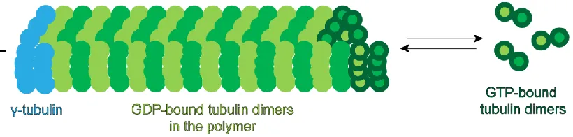
Microtubules are composed of heterodimers of - and -tubulin. Tubulin heterodimers form linear protofilaments that associate laterally and form 25 nm diameter hollow tubes composed of 10-15 protofilaments (usually 13) arranged in a lattice (Tilney et al., 1973). These individual protofilaments have longitudinal as well as lateral contacts making them rigid structures necessary for their cellular function. Microtubules are polarized filaments with the -tubulin being the stable minus-end and the -tubulin end is the dynamic plus-end (Mitchison, 1993). Although ‘treadmilling’ was proposed as a model to explain microtubule polymerization (Margolis and Wilson, 1978), in vitro work described a novel phenomenon of dynamic instability - where microtubules frequently switch between periods of slow growth, called rescue and phases of rapid depolymerization, termed catastrophe (Mitchison and Kirschner, 1984). This was subsequently confirmed in real time by other groups (Horio and Hotani, 1986; Walker et al., 1988).
5
the nucleotide state of the tubulin subunits. Tubulin heterodimers co-purify with guanine nucleotide (Weisenberg et al., 1968). Both - and -tubulin monomers bind guanosine triphosphate (GTP) in solution. Upon incorporation into the polymer, the GTP in the -tubulin is hydrolyzed to guanosine diphosphate (GDP) while the GTP in the -tubulin is not (Spiegelman et al., 1977). Soluble tubulin has a very slow rate of GTP hydrolysis. However, upon incorporation into the polymer, this rate is very high which likely leads to disassembly of the polymer (David-Pfeuty et al., 1977). Thus, the hydrolysis of GTP to GDP at the plus-end of microtubule is thought to promote depolymerization of the filament while the subunits when in GTP state resist depolymerization. Pioneering work from Walker et al., 1988 established that polymerization was dependent on tubulin concentration while depolymerization was independent of tubulin concentration. Although this work laid the foundation for several other experiments and models to explain each step in dynamic instability, we lack a unified understanding of this process (reviewed in Gardner et al., 2008).
Figure 1.1 Schematic illustrating dynamic instability of microtubules
6
periphery. The MTOC is composed of several proteins including -tubulin, pericentrin and ninein (reviewed in Kollman et al., 2011).
7 III. Kinesin Motors
Studies in early 1980s using extruded axoplasm of giant squid axons established that microtubules provided the tracks for bidirectional vesicle movement along the axon between the neuronal cell body and synaptic termini (Allen et al., 1982; Brady et al., 1982). Shortly thereafter, kinesin-1 was identified as an ATPase that moves along the microtubule plus-ends (Vale et al., 1985a, 1985b, 1985c). Since this initial discovery, the kinesin family now has expanded to include 45 different kinesins (Hirokawa et al., 2009) classified into 14 different classes with commonly agreed nomenclature (Lawrence et al., 2004).
Structurally, kinesins have a motor domain that binds to microtubules and also hydrolyzes ATP (adenosine triphosphate), neck-linker which co-ordinates the two motor heads and a tail which binds to cargos and adaptors (Hirokawa, et al., 1989). Based on their structure, kinesins are generally classified into three types: N-terminal kinesins which have the motor domain at the N-terminus and are generally plus-end directed; C-terminal kinesins which move toward the microtubule minus-end and function in mitosis and M-kinesins which have the motor domain in the middle as in kinesin-13 family which are known to depolymerize microtubules. Functionally, kinesins can be categorized into three broad classes: kinesin that power organelle transport, mitotic kinesins and kinesins that alter microtubule dynamics.
8
form heterotrimers composed of KIF3A, KIF3B and a kinesin-associated protein, Kap3 (Cole et al., 1993). Kinesin-3 family motors do not dimerize efficiently (Okada et al., 1995) but when dimerized artificially in vitro, are processive motors (Okada and Hirokawa, 1999; Tomishige et al., 2003) and it is now thought that they undergo cargo-mediated dimerization
in vivo (Soppina et al., 2014). Kinesin-5 family motors are homotetrameric (Kashina et al., 1996) that can cross-link and slide anti-parallel microtubules (Kapitein et al., 2005). MCAK and other kinesin-13 familiy proteins do not move processivley on microtubules. Rather, they diffuse along the length of the microtubule and remove tubulin subunits from the ends acting as depolymerases (Hunter et al., 2003). In contrast, Ncd which is a kinesin-14 family member, diffuses along microtubules in a tail-dependent manner and slides anti-parallel microtubules (Fink et al., 2009).
Much of our understanding of the mechanochemical properties of kinesins comes from the conventional kinesin, kinesin-1. Kinesin-1 has been shown to walk in a hand-over-hand motion along the length of the microtubule protofilament in 8 nm steps (which is the length of the tubulin heterodimer), going through one hydrolysis cycle per step (Svoboda et al., 1993; Schnitzer and Block; 1997; Yildiz et al., 2004). This stepping mechanism indicates regulated co-ordination of the biochemical cycles of both motor heads which allows the front head to remain bound to the microtubule while the rear head detaches. Single molecule studies have demonstrated that kinesin-1 has high velocities ranging from 0.5-1.0 µm/s and high stall forces of about 5-7 pN (Visscher et al., 1999), properties which make it an efficient motor for long-range transport. Consistent with this, recent studies have shown that cellular cargo are driven by relatively small teams of kinesin motors (Hendricks et al., 2012; Rai et al., 2013).
9
truncated versions of the KHC with deleted tail exhibited robust motility (Freidman and Vale, 1999). This hinted at an autoinhibitory mechanism which in fact was consistent with the initial micrographs of kinesin structure where in some cases, the tail folded back onto the motor domain (Hirokawa et al., 1989). A short basic motif, IAK in the KHC tail is sufficient to bind to the motor domain (Stock et al., 1999) and subsequently it was shown that full length KHC had several fold lower ATPase activity than the truncated KHC lacking the IAK motif (Hackney and Stock, 2009). This led to the suggestion that the tail of KHC could be folding back onto the ATPase site of the motor, preventing its binding to microtubules (Dietrich et al., 2008). Crystal structures of the kinesin-1 motor domain with and without the tail domain indicated that the tail cross-links the motor domain which potentially inhibits ADP release, thus preventing movement of the motor heads (Kaan et al., 2011). K560, constituting the first 560 amino acids of the kinesin heavy chain is now routinely used as a constitutively active form of the kinesin-1 motor. It is now thought that several kinesins are autoinhibited which serves as a general regulatory mechanism for kinesins (reviewed in Verhey and Hammond, 2009).
10 IV. Cytoplasmic dynein
Long before the discovery of kinesins, dynein was first discovered in the cilia of
Tetrahymena (Gibbons, 1963). It was quickly shown that dynein is an ATPase that drives microtubule sliding in the cilia (Gibbons and Rowe, 1965). Very quickly, sixteen genes were identified encoding the dynein heavy chain with fourteen of them functioning in the axoneme driving cilia beating, one of them is involved in the intraflagellar transport and another one that encodes for the cytoplasmic dynein that drives retrograde transport in cells (Wickstead and Gull, 2007).
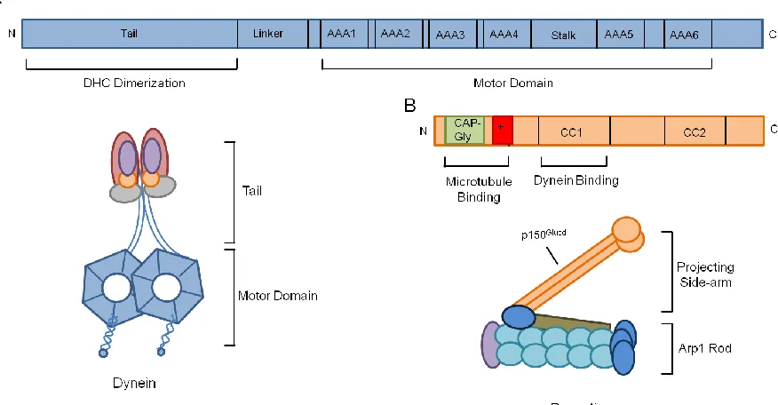
Cytoplasmic dynein (referred to as dynein from now on) was initially identified as MAP1C, a microtubule-ATPase which translocated microtubules in the direction opposite to that of kinesin (Paschal et al., 1987; Paschal and Vallee, 1987). Following these initial observations, it was quickly established that MAP1C was infact, the cytoplasmic analogue of axonemal dynein (Vallee et al., 1988). Although kinesins and myosins are structurally similar, dynein has a much more complicated structure (Vale and Milligan, 2000). Dynein is a ~1.6 MDa protein complex with several subunits. The dynein heavy chain constitutes the motor domain, linker, tail and microtubule binding stalk. The tail is required for dimerization and acts as a scaffold for the other subunits of dynein – intermediate chains, light intermediate chains.
11
AAA4 (Gee et al., 1997). Recently, it has been shown that nucleotide binding to AAA1 and microtubule binding by the stalk are allosterically coupled explaining how hydrolysis at AAA1 affects the binding and unbinding of the stalk to microtubules (Carter et al., 2008; Kon et al., 2009).
Single molecule studies in the last decade have begun to elucidate the biophysical properties of dynein. Studies with recombinant yeast dynein showed that dimerization is necessary for processive movement (Reck-Peterson et al., 2006). Further, unlike kinesin, dynein can take variable step sizes of 8-32 nm with sideways stepping on adjacent protofilaments as well as steps toward the microtubule plus-end (Mallik et al., 2004; Ross et al., 2006; DeWitt et al., 2012; Qiu et al., 2012). While the heads of kinesins and myosins are usually tightly co-ordinated, these studies indicate that dynein stepping is not as co-ordinated between its heads. It is likely that this un-coordinated stepping mechanism makes dynein efficient in navigating obstacles such as microtubule associated proteins (MAPs) on the microtubule tracks (Dixit et al., 2008).
12
In contrast to single molecule motion of dynein, studies have shown that beads coated with dynein are processive indicating dynein works efficiently in teams (King and Schroer, 2000; Mallik et al., 2005; Ross et al., 2006). More recently, optical trapping of endogenous cargos has demonstrated that dynein molecules work in teams generating large forces that increase linearly with motor number (Hendricks et al., 2012; Rai et al., 2013). This capacity of dynein to work effectively in teams likely enables it to perform the diverse cellular functions it has been implicated in (reviewed in Mallik et al., 2013).
As the major minus-end directed motor in cells, dynein has been shown to transport several organelles including endosomes, lysosomes, mitochondria, lipid droplets. (reviewed in Allan, 2011). Other functions include neuronal migration (reviewed in Vallee et al., 2009), mRNA localization (reviewed in Holt and Bullock, 2009), Golgi positioning (reviewed in Yadav and Lindstedt, 2011) and also in orientation of the mitotic spindle and cell division (reviewed in Siller and Doe, 2009). To perform these various tasks and to achieve specificity, dynein relies on several adaptors that regulate its motility and function. One such ubiquitous adaptor is the dynactin complex highlighted in the next section.
Dynactin
13
Dynactin is a 1.2 MDa complex with 11 subunits and based on its structure can be divided into two parts, the projecting side-arm and the Arp1 rod. The Arp1 rod is a 40 nm polymer made of Arp1 subunits and is flanked by CapZ on one end and p62 on the other (Schafer et al., 1994). The Arp1 filament was subsequently shown to be the domain of dynactin that interacted with most cargoes (Holleran et al., 1996; Holleran et al., 2001). The rest of the components making up the Arp1 rod were identified in the next few years (reviewed in Schroer, 2004). The flexible, projecting side-arm constitutes the largest subunit of the dynactin complex, p150Glued which is also the site of dynein interaction (Karki and Holzbaur, 1995; Vaughan and Vallee, 1995). Although p150Glued is the protein that binds dynein, the entire dynactin complex is required for dynein function as over-expression of p50, dynamitin which disrupts the two structural parts of dynactin results in impaired dynein function in cells (Echeverri et al., 1996; Burkhardt et al., 1997).
14
Figure 1.2 Schematic illustrating the dynein and dynactin complexes
15
p150Glued has 32 exons (Tokito and Holzbaur, 1998). Several alternatively spliced isoforms of p150Glued with differential exons exist in many tissues (Dixit et al., 2008). An isoform that is unique to the brain tissue is p135 which lacks the N-terminal CAP-Gly and basic domains (Tokito et al., 1996). Although this isoform cannot bind microtubules, it can still bind dynein and distinct dynactin complexes with either p150Glued or p135 exist in the brain tissue. (Tokito et al., 1996). We know very little about this isoform and an attractive hypothesis is that p135 has a specialized function that p150Glued cannot accomplish. The intriguing question of why neurons need this unique isoform still remains an open question.
16
The second coiled-coil (CC2) domain of the C-terminus of p150Glued is the domain that incorporates p150Glued into the rest of the dynactin complex (Waterman-Storer et al., 1995). The rest of the C-terminus acts as a binding site for several cargo adaptors and scaffolding proteins. Few examples include RILP which mediates endosome motility (Jordens et al., 2001), Huntingtin and HAP1 (Engelender, 1997; Li et al., 1998); JIP3 (Cavalli et al., 2005) and JIP1 (Fu and Holzbaur; 2013). All of these proteins have been implicated in regulating motility of various organelles (reviewed in Fu and Holzbaur, 2014). Thus, the C-terminus of p150Glued acts as a hub for several adaptor/scaffolding protein interactions and this is likely a key determinant of cargo specificity for dynein-mediated organelle transport.
Adaptors Regulating Motility of Dynein-Dynactin Complexes
Recent work has shown that addition of certain adaptors to dynein-dynactin complexes converts the weakly processive motor to a highly processive one (McKenney et al., 2014; Schlager et al., 2014). One such adaptor is the Bicaudal D protein (called BICD from here on). BICD was initially identified in Drosophila, where mutations in this gene caused defective embryos (Mohler and Wieschaus, 1986). It was quickly established that BICD was essential for fly oogenesis and played an important role in mRNA localization (Claussen and Suter, 2005). Two orthologs of BICD, BICD1 and BICD2 exist in mammalian cells. Biochemical characterization of BICD2 revealed that it interacted with dynein-dynactin complexes and localized to the Golgi (Hoogenraad et al., 2001). Subsequent work demonstrated that the two N-terminal coiled-coil domains of BICD2 was sufficient to bind to both dynein and dynactin. Recruitment of this N-terminal fragment to organelles in cells induced dynein-mediated transport toward the centrosome (Hoogenraad et al., 2003) and further promoted stable interaction between dynein and dynactin complexes both in vitro and
17
The C-terminal portion of BICD2 has been shown to bind to various proteins that dictate cargo selectivity. Some examples include with Rab6 (Matanis et al., 2002) and Egalatarian which promotes mRNA localization (Mach and Lehmann, 1997). In addition, the C-terminus has also been shown to bind to the N-terminus (Hoogenraad et al., 2003; Splinter et al., 2012) which in turn competes with binding to dynein-dynactin complexes. This suggests a potential autoinhibitory mechanism in cells and indicates that BICD binds to dynein-dynactin only when the C-terminus is bound to cargo adaptors relieving the autoinhibition.
18
Structural Understanding of the Dynein-Dynactin Complexes
The large size and complexity of both dynein and dynactin made structural investigations of these complexes challenging. Although initial structure work provided a framework for our understanding of the dynein motor and dynactin (reviewed earlier in this chapter), it is only now that we are beginning to explore the structural aspects of these protein complexes in part due to technical advancements in electron microscopy (EM).
Two recent studies have now solved structures of dynactin using cryo-EM and both these studies also obtained structures of dynein bound to dynactin and the adaptor protein BICD2 (Chowdhury et al., 2015; Urnavicius et al., 2015). These studies provide some insights into how an adaptor protein can induce such robust motility of the dynein motor when bound to dynactin. Both these reports showed that dynactin and BICD2 bind to the tail of the dynein motor and have overlapping binding sites. Further, this interaction of dynein tail with dynactin seems to be mediated by the N-terminus of BICD2. This explains the need for an adapotor protein facilitating the interaction between dynein and dynactin, thus forming a stable complex.
19
These two studies are the first to demonstrate structural aspects of dynein-dynactin-adaptor complexes both on microtubules (Chowdhury et al., 2015) and off microtubules (Urnavicius et al., 2015). Future studies with higher resolutions structures of dynein-dynactin complexes with other adaptors in conjunction with single molecule motility assays will inform us about the differential modulation of the dynein-dynactin complexes via cargo adaptors.
Dynein-Dynactin in Disease
Mouse mutants generated by chemical mutagenesis experiments were the first dynein mutants that gave us insight into the role of dynein in neuronal development. The initial mouse mutants were Legs at odd angles (Loa), Cramping and Sprawling mice (Duchen, 1974; Hafezparast et al., 2003; Chen et al., 2007), all of them being mutations (point mutations in Loa and Cramping, deletion in Sprawling) in the tail of the dynein heavy chain (Dync1h1).
The first dynein mutation to be discovered in humans was in 2010 and the phenotype was developmental delay and intellectual disability (Vissers et al., 2010). A subsequent study identified several mutations in the stalk and microtubule binding domain of dynein in patients with malformations of cortical development (MCD) (Poirier et al., 2013). In the last few years, several mutations have been identified in patients associated with sensory and motor neuropathy (reviewed in Schiavo et al., 2013). While some mutations are located in the motor domain and the stalk, several others are located in the tail highlighting the importance of the several interactions of the dynein tail with adaptors and co-factors.
20
21
V. Microtubule Modifications and Microtubule Associated Proteins
Microtubules are involved in several cellular processes including intracellular transport, cellular morphogenesis, organelle localization and cell division. Although the fundamental components of microtubules, the heterodimers - and -tubulin are the same across all eukaryotic species, microtubules in cells are extensively modified and modulated by various biochemical modifications and interacting proteins respectively. The modifications are post-translation modifications (PTMs) and the interacting proteins are called microtubule associated proteins (MAPs). In the previous sections I reviewed two such MAPs – kinesins and dynein. In this section, I will highlight the various biochemical modifications that microtubules undergo followed by a review of MAPs highlighting the roles PTMs and non-motor MAPs play in establishing and marking the identity of microtubules in cells.
Tubulin Tyrosination
22
1977). Detyrosinating enzymes act predominantly on polymerized microtubules and elegant experiments in cells examining microtubules right after depolymerization indicated that newly made microtubules are generally tyrosinated (Gundersen et al., 1987). Detyrosinated tubulin can be further modified to Δ2-tubulin by the removal of the glutamic acid residue which cannot go back to the tyrosinated form (Paturle-Lafanechere et al., 1991).
Some initial studies that examined the correlation between various microtubule modifications and microtubule stability, suggested that detyrosination of microtubules promoted their stability (Gundersen et al., 1987; Schulze et al., 1987). However, this was not true in all cell types (Schulze et al., 1987) and more recently, it was shown that depolymerizing kinesins MCAK and KIF2, belonging to kinesin-13 family have a preference for tyrosinated microtubules (Peris et al., 2009; Sirajuddin et al., 2014). Thus, detyrosination of microtubules protects them from depolymerizing kinesins which could explain the stability of these microtubules.
23
Figure 1.3 Schematic illustrating tubulin post-translational modifications (Adapted from Janke, 2014)
24 Tubulin Acetylation
Acetylation was the second PTM on tubulin to be discovered, first identified in the -tubulin of Chlamydomonas on lysine40 residue (L'Hernault and Rosenbaum, 1985). Lys40 is located in the lumen of the microtubule polymer (Nogales et al., 1998) and hence it is intriguing that acteylation of microtubules can affect the interaction of any motor protein or MAP as these interact with microtubules on the cytoplasmic face of the polymer. Initial functional studies identified HDAC6 (Hubbert et al., 2002) and Sirt2 (North et al., 2003) as microtubule deacetylase enzymes. However, both these enzymes have several substrates besides microtubules alone and hence it is possible that effects downstream of altering these enzymes is not a direct consequence of altered microtubule modification. More recently, tubulin acetyltransferase (TAT) has been discovered as an enzyme that specifically acts on
-tubulin (Akella et al., 2010). Recent work using X-ray crystallography and single molecule assays demonstrated that TAT does indeed enter the microtubule lumen and acetylates from the inside. Further, acetylation marking stable microtubules can be explained by the slow catalytic rate of the enzyme compared to its diffusion rate (Szyk et al., 2014).
25 Tubulin Polyglutamylation and Polyglycylation
Another modification of tubulin discovered in the early 90s was the covalent addition of chains of glutamate and glycine residues added to the C-terminal tails of tubulin termed as polyglutamylation (Edde et al., 1990) and polyglycylation (Redeker et al., 1994) respectively. In contrast to the other modifications, these modifications are the addition of several amino acids and can occur on both - and -tubulin. While glutamylation is found everywhere, glycylation seems restricted to cilia and flagella (Fouquet et al., 1994).
The high levels of polyglutamylation in neurons made possible the discovery of the first polyglutamylase enzyme (Janke et al., 2005). This is a multi-subunit protein complex with its catalytic subunit similar to the tubulin tyrosine ligase enzyme (Janke et al., 2005). Subsequently several other glutamylating and glycylating enzymes have been identified and more recently a deglutamylase enzyme has been discovered which removes the Glu side chains from tubulin (Rogowski et al., 2010). Although several enzymes responsible for glutamylation and glycylation have been identified in recent years, the spatial and temporal localization and function of these enzymes is still unclear.
26
preferential binding of Tau to polyglutamylated tubulin (Boucher et al., 1994) could be one such mechanism as Tau has been shown to protect microtubules from severing (Qiang et al., 2006). It is clear that polyglutamylation and polyglycylation can generate microtubules with varying complexity levels as these are the only modifications that can occur on any residue of either - or -tubulin and also the length of the glutamine and glycine chains can be modulated. However, we do not fully understand the cellular cues that dictate these changes and if there are specific tubulin residues that are targeted more often than others.
Other Tubulin PTMs
Several other modifications of tubulin have been reported like arginylation, methylation (reviewed in Janke 2014) and more recently polyamination (Song et al., 2013). It is clear that we are only beginning to understand the several modifications that tubulin can undergo and further studies are needed to gain insights into their functional importance.
27 Structural MAPs
MAPs were initially identified as proteins that were enriched in microtubule fractions through repeated cycles of tubulin assembly and disassembly (Shelanski et al., 1973; Borisy et al., 1975). Some of the initial MAPs identified were MAP1, MAP2 (Sloboda et al., 1975) and Tau (Weingarten et al., 1975; Cleveland et al., 1977). While MAP1 consists of polypeptides MAP1A, 1B, 1C; MAP2 consists of 2A and 2B and Tau is the most heterogenous among them ranging from 3-6 polypeptides (reviewed in Olmsted, 1986)
A common feature of all these proteins was the ability to stabilize microtubules and promote tubulin polymerization even at concentrations lower than needed for tubulin alone. These initial studies on MAPs suggested that MAPs could alter microtubule properties in cells. To test this, in vitro assays to examine dynamic instability of microtubules were carried out in the presence of purified MAPs. MAPs increase polymerization rates and frequency of rescue, suppress catastrophe rate and frequency of catastrophe (Pryer et al., 1992; Drechsel et al., 1992).
28
An attractive hypothesis is that MAPs bound to the microtubule polymer add another layer of regulation to motor proteins. Although this has been explored for Tau with kinesin-1 and dynein (Dixit et al., 2008), we know very little about this regulation with other MAPs and the various ways in which they modulate motor proteins. Besides the role of Tau in microtubule stability and motor transport, Tau is a major protein that is aggregated and modified in several ways in patients with Alzeheimer’s disease. This has generated considerable interest in Tau, and Tau dysfunction has now been proposed to be an early marker for neurodegenerative diseases (reviewed in Frost et al., 2015). Future studies focusing on structure-function correlation of these MAPs and the biological implications in neuronal development are needed to further our understanding of their cellular function.
Microtubule Plus-end Binding Proteins
Microtubule plus-end binding proteins or +TIPS are a distinct class of proteins that specifically localize to the plus-ends of growing microtubules. Since the discovery of CLIP-170 (Perez et al., 1999), the first protein shown to localize to microtubule plus-ends, different families of +TIPS have been identified.
29
the C-terminus of EB proteins, CLIP-170 and tubulin itself (Weisbrich et al., 2007). The first class includes proteins like APC, STIM1 and MCAK and the second class includes proteins like CLIP-170 and p150Glued of dynactin. Thus, through these interactions EBs form the core of the +TIP network of proteins.
In vitro studies with EB1 has demonstrated that EB1 does not bind tubulin dimers and its tip tracking behavior on growing microtubules is independent of tubulin concentrations suggesting that EBs do not co-polymerize with tubulin (Bieling et al., 2007). This indicated that EB proteins were recognizing structural features at the microtubule plus-end that was different from the rest of the microtubule lattice. Consistent with this, a recent cryo-EM study showed that the calponin homology domain of EB1 binds to four tubulin subunits in way that it bridges protofilaments except at the seam (Maurer et al., 2012). This suggests that EB1 binds to microtubules in a strategic manner that allows it to sense GTP-hydrolysis induced conformational changes in the microtubule plus-end (Maurer et al., 2012).
Several +TIP proteins undergo auto-inhibition via intramolecular interactions. CLIP-170 which binds to the EEY/F motif of EB1 also has an EEY/F motif at its C-terminus. This allows the CAP-Gly domain of CLIP-170 to interact with its C-terminus (Lansbergen et al., 2004). The autoinhibition can be relieved by proteins containing EEY/F motif like EBs and tubulin which allows the C-terminus of CLIP-170 to bind to p150Glued or Lis1 (Lansbergen et al., 2004; Weisbrich et al., 2007). Further, EB1 itself can be autoinhibited which can be relieved by its binding to tubulin or other +TIPs that bind to its C-terminus (Hayashi et al., 2007).
30
al., 2009). This finding is consistent with structural findings implicating the binding of +TIPs to EEY/F motifs (Weisbrich et al., 2007).
31 VI. Neuronal Transport
Ever since Cajal drew out the anatomy of cells in the central nervous system, scientists have always been fascinated with neurons. The polarized and extended morphology of each neuron and the contacts between neurons is integral for propagation of neuronal signals. A key aspect in proper neuronal function is the efficient supply of material synthesized in the cell body to the individual neurites and clearance of misfolded and aggregated proteins from neurites to the soma for their degradation. Thus, neurons employ mechanisms of active transport to constantly supply and clear material to and from neurites, which sometimes can be over a meter away from the cell body.
32
section I will only focus on the fast axonal transport driven by microtubule motors. I will first introduce features of the neuronal cytoskeleton followed by polarized sorting and trafficking highlighting compartment specific differences between axons and dendrites.
Microtubule Organization in Neurons
Initial studies on microtubule organization in axons were done in the early 80s by independent groups showing that microtubules in axons were uniformly polarized (Heidemann et al., 1981; Burton and Paige, 1981). A few years later, examination of mammalian neuronal cultures in vitro revealed that dendrites have mixed polarity microtubules while axons have uniformly oriented microtubules (Baas et al., 1988; Burton, 1988). Around the same time, the developmental timing of neuronal growth in vitro was documented (Dotti et al., 1988). Neuronal development followed a stereotypical sequence of events – the cells first establish small, immature processes and once one of them becomes the axon, the other processes begin to elongate to become dendrites (Dotti et al., 1988). Initially, microtubules start out uniform in all the processes with their plus-ends out and once processes mature into dendrites, minus-end out microtubules get added, establishing mixed polarity microtubules in dendrites (Baas et al., 1989). These initial findings were subsequently confirmed using live-cell imaging of EB3-GFP which tracks microtubule plus-tips (Stepanova et al., 2003).
33
a mixed polarity pattern. Dynein and certain mitotic kinesins have been implicated in setting up this organization in dendrites (reviewed in Baas and Lin, 2011).
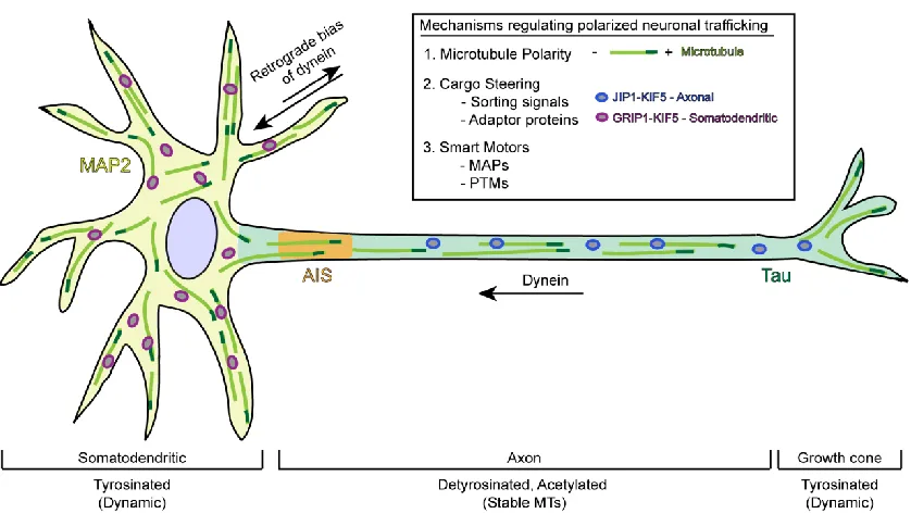
Besides the orientation, axons and dendrites also differ in their microtubule stability, post-translational modifications and the complement of MAPs associated with them in these compartments. Microtubules in axons are known to be enriched in the more stable acetylated and detyrosinated tubulin whereas dendrites and growth cones are enriched in tyrosinated microtubules (Konishi and Setou, 2009; Hammond et al., 2010). Further, structural MAPs localize differentially to axons and dendrites with Tau being axonal only (Binder et al., 1985) and MAP2 marking dendrites (Caceres et al., 1984; see section V for review on tubulin modifications and MAPs). In the section, I will introduce polarized trafficking in neurons and highlight how the underlying microtubule cytoskeleton contributes to the navigation of motor proteins.
Polarized Trafficking in Neurons
34 Sorting Signals
Early work dissecting selective transport of membrane proteins in neurons focused on identifying sorting signals that conferred compartment-specificity. The sorting mechanisms well established in epithelial cell biology (reviewed in Matter and Mellman, 1994; Mellman and Nelson, 2008) provided a foundation for these initial studies. Initial work observed the localization of basolateral marker membrane proteins and apical proteins and demonstrated that basolateral proteins are targeted to dendrites; mutations in the sequences disrupted the dendritic localization (Jareb and Banker, 1998). Interestingly, the apical proteins were targeted uniformly indicating that the sorting signal in apical proteins was not enough to make them axon-specific in neurons (Jareb and Banker, 1998). Although an exciting result at the time, it became apparent very quickly that neurons employ more complicated mechanisms for polarized sorting than just basolateral being equivalent to dendritic targeting. Key examples include identification of somatodendritic targeting signal in the cytoplasmic domain of transferrin receptor which was distinct from the basolateral targeting sequence (West et al., 1997), previously unknown targeting sequences identified in metabotropic glutamate receptors (mGluRs) (Stowell and Craig, 1999).
35
excluded from axons but this was not the case for axonal proteins (Jareb and Banker, 1998; Stowell and Craig, 1999; Burack et al., 2000). This suggested that dendritic transport was selective, achieved by motor proteins that selectively entered dendrites while axonal proteins were targeted non-selectively and a downstream step following this initial transport, likely exocytosis made them axon-specific. In the years following these initial studies, selective trafficking of motor proteins in neurons was extensively studied. Several models for how motors drive selective trafficking have been proposed and there seems to be evidence for all of these models. All these models were based on investigations of various kinesins and very little is known about selective transport of dynein-driven cargos. In this section, I will review the models for selective transport with specific examples in each case and conclude with findings from my own work on dynein-driven trafficking in neurons.
(i) Motor-cargo interactions
36
receptor 2B subunit (NR2B) to postsynaptic sites; KIF17 accomplishes this by binding to a PDZ domain of one of the proteins in complex with NR2B (Setou et al., 2000).
More recently, the C-terminus coiled-coil domains and the C-terminal PX domain of KIF16B, kinesin-3 motor have been shown to be important in the accurate localization of KIF16B to somatodendrites (Farkhondeh et al., 2015), implicating the cargo-binding region as domains containing targeting information. Adaptors bound to cargo have also been shown to play a role in targeting the motor-cargo complex to the final destination (van Spronsen et al., 2013). In this study, the authors showed that TRAK/Milton adaptor proteins establish uniform distribution of the mitochondria in neurons with TRAK1 binding to kinesin-1 and dynein which trafficks mitochondria to axons, while TRAK2 binds to dynein-dynactin alone steering these mitochondria to dendrites (van Spronsen et al., 2013).
In all the above examples, the cargo or adaptor protein bound to cargo determine the targeting of the cargo complex. However, there is also evidence that motor-microtubule interactions determine the targeting of the motor, indicating that targeting of motor-cargo complex is intrinsic to the motor-microtubule interactions as reviewed in the examples below.
(ii) Motor-microtubule interactions
Another model explaining differential trafficking of proteins in neuronal transport is the ‘smart-motor’ hypothesis which suggests that motors are capable of navigating to the axon or dendrites solely based on their interactions with axonal and dendritic microtubules. These interactions are modulated by microtubule polarity, post-translational modifications and MAPs associated with microtubules.
37
K560, the constitutively active motor domain of kinesin-1. K560 in neurons preferentially accumulated at axon tips (Nakata and Hirokawa, 2003). Further, this preferential accumulation of K560 in axons acts as an early marker for axon specification (Jacobson et al., 2006). Consistent with these initial observations, subsequent work indicated the increased affinity of kinesin with detyrosinated (Konishi and Setou, 2009) and acetylated microtubules (Reed et al., 2006), both of which are enriched in axons. However, it is important to note that correlating motor affinity to tubulin modifications has not been very straightforward due to other conflicting data: study reporting that K560 has no preference for acetylated microtubules in vitro (Walter et al., 2012); specifically increasing acetylation levels in neurons did not change the axon-selectivity of kinesin (Hammond et al., 2010). Hence, it is likely that a combination of biochemical cues between motors and microtubules determine the targeting of the motor.
Localization studies with constitutively active motor domains of a subset of kinesin-3 and kinesin-4 motors also lend support to this model – KIF13A and KIF13B, kinesin-3 motors and KIF21A and KIF21B, kinesin-4 motors localize differentially with KIF13A, KIF21A localizing to only axons while KIF13B and KIF21B localize to both axons and dendrites (Huang and Banker, 2012). This study also identified sequences in the loop12 of the motor domain that confer axon selectivity, which when mutated, abolish the selectivity (Huang and Banker, 2012). It is likely that the non-selective kinesins, KIF13B and KIF21B are likely dendritic kinesins as unlike the other kinesins tested these were the only ones that could navigate the dendritic microtubule cytoskeleton and since the motors are constitutively active, axonal accumulation could be a default for all plus-end directed kinesins. Consistent with this idea, full length KIF21B has shown to be enriched in dendrites only (Jenkins et al., 2012) implicating it as a ‘smart-motor’ that can preferentially enter dendrites.
38
membrane region in the axon, enriched in Ankyrin G, is the filter that determines which motors are axon-specific (Song et al., 2009). This study used chimeras of KIF5 and KIF17 with the corresponding cargos in different combinations and concluded that the efficacy of the cargo-motor complex determines their ability to navigate through the AIS, thus being targeted to the axon. However, a caveat with this model is that the AIS is set up later in neuronal development and a recent study demonstrated that polarized transport is established early on in neurons, even before the establishment of the AIS (Petersen et al., 2014). The AIS model also does not explain how the same KIF5 motor is axonal when bound to JIP1 for instance (Verhey et al., 2001) and is dendritic when bound to GRIP1 (Setou et al., 2002).
Figure 1.4 Illustration of mechanisms regulating polarized neuronal trafficking
39
dendrites (Black and Baas, 1989; Kapitein et al., 2010), this model does not explain the localization of cargoes that are predominantly dendritic but have kinesins on them (Setou et al., 2000; Setou et al., 2002; Jenkins et al., 2012). In conclusion, it is becoming increasingly clear that polarized trafficking in neurons is regulated at many levels and it is likely that this is controlled via co-ordinated regulation of motor-cargo and motor-microtubule interactions.
40
CHAPTER 2: DYNACTIN FUNCTIONS AS BOTH A DYNAMIC TETHER AND BRAKE DURING DYNEIN-DRIVEN MOTILITY
This chapter is adapted from:
Ayloo, S., Lazarus, J.E., Dodda, A., Tokito, M., Ostap, E.M., Holzbaur, E.L.F (2014). Dynactin functions as both a dynamic tether and brake during dynein-driven motility.
Nature Communications 5:4807
41 I. Summary
Dynactin is an essential cofactor for most cellular functions of the microtubule motor cytoplasmic dynein, but the mechanism by which dynactin activates dynein remains unclear. Here we use single molecule approaches to investigate dynein regulation by the dynactin subunit p150Glued. We investigate the formation and motility motility of a dynein-p150Glued co-complex using dual-color total internal reflection fluorescence microscopy. p150Glued recruits and tethers dynein to the microtubule in a concentration-dependent manner. Single molecule imaging of motility in cell extracts demonstrates that the CAP-Gly domain of
42 II. Introduction
Cytoplasmic dynein is the major microtubule minus-end directed motor in higher eukaryotes. Dynactin, a large multi-subunit protein complex, interacts with dynein and is essential for a broad range of cellular functions including organelle transport and mitotic spindle assembly (reviewed in Schroer, 2004). Dynactin was first identified as an activator of dynein that increased the ability of dynein to transport organelles (Gill et al., 1991) but the mechanisms involved in dynein activation are not yet understood.
Structurally, dynactin can be divided into two parts—an actin-like Arp1 rod that along with associated subunits forms the base of the complex and a projecting side arm formed from the dimerization of the largest subunit in the complex, p150Glued (Holzbaur et al., 1991; Schafer et al., 1994). p150Glued interacts directly with the dynein intermediate chain (DIC) of the dynein motor (Karki et al., 1995; Vaughan et al., 1995). p150Glued also interacts with microtubules via its Cytoskeletal Associated Protein, Glycine-rich (CAP-Gly) domain at the N terminus (Waterman et al., 1995) which is followed by a highly basic region that has a lower affinity interaction with microtubules (Culver-Hanlon et al., 2004; Lazarus et al., 2013). In vitro motility studies using beads coated with purified proteins demonstrated that the microtubule-binding capacity of dynactin increases the processivity of mammalian dynein (King and Schroer, 2000; Culver-Hanlon et al., 2004).
43
(Kardon et al., 2009), although at the cellular level this highly conserved domain contributes to the initiation and persistence of dynein-dependent nuclear movement (Moore et al., 2009). Studies in higher eukaryotes also suggest that the CAP-Gly domain may be dispensable for some cellular functions, including the trafficking and localization of organelles in S2 and HeLa cells (Kim et al., 2007; Dixit et al., 2008).
In contrast, both cellular and in vivo studies have demonstrated that the CAP-Gly domain of dynactin is essential for dynein function in neurons. In Drosophila and mammalian neurons, the CAP-Gly domain enhances the retrograde flux of cargoes from the distal axon (Lloyd et al., 2012; Moughamian and Holzbaur, 2012). An ordered recruitment pathway has been proposed, in which binding of dynactin to dynamic microtubules enriched in the distal axon leads to the enhanced recruitment of dynein, permitting the efficient initiation of retrograde transport (Moughamian et al., 2013). Importantly, mutations in the CAP-Gly domain of p150Glued cause human disease, including the motor neuron degenerative disease HMN7B and a lethal and rapidly progressive variant of parkinsonism known as Perry syndrome (Puls et al., 2003; Farrer et al., 2009). While the HMN7B-associated G59S mutation induces misfolding and aggregation (Levy et al., 2006), the Perry syndrome mutations cause a loss of CAP-Gly function in cellular assays (Moughamian and Holzbaur, 2012). Together, these genetic findings indicate a key role for the CAP-Gly domain of dynactin in neurons in vivo.
44
45 III. Results
Dynein-GFP switches between processive and diffusive motion
46
Figure 2.1 DIC1-GFP is efficiently incorporated into the dynein complex.
(A) Schematic of the DIC1–eGFP–FLAG gene knocked into the DIC1 locus. High speed supernatant (HSS) of homogenized mouse brain tissue was probed for DIC, GFP and FLAG. (B) Immunoblot analysis of sucrose gradient purified dynein. DIC1–GFP–FLAG is efficiently incorporated into the dynein complex as indicated by co-sedimentation with dynein heavy chain (DHC) at 20 S. (C) A representative time course of photobleaching showing two steps of decay. Grey lines indicate the steps. (D) Frequency distribution of the photobleaching steps for n=40 particles fit with a binomial distribution.
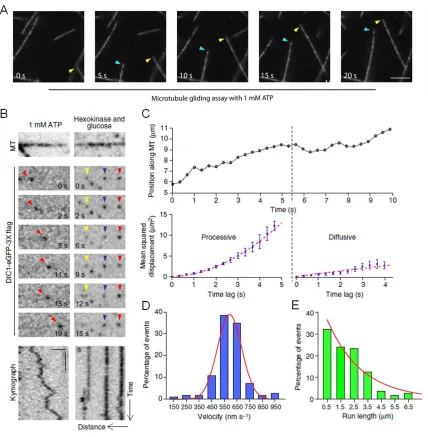
We performed gliding assays (Fig. 2.2A) and single molecule motility assays (Fig. 2.2B) to confirm that isolated dynein–dynactin complexes are functional. We observed processive, bi-directional ATP-dependent runs along the length of microtubules (Fig. 2.2C). These runs are punctuated by frequent directional switches as well as periods of apparent diffusion. While single dynein motors often diffuse along microtubules, two to three dyneins working together exhibit robust minus-end directed motility (Mallik et al., 2005; Ross et al., 2006) as supported by our gliding assays with isolated dynein–dynactin complexes (Fig. 2.2A). This behaviour may be a functional consequence of the flexible structure of the dynein motor, and may permit multiple dynein motors bound to a single cargo to function effectively in cells (Hendricks et al., 2012; Rai et al., 2013).
47
then switched to diffusion for the last few seconds of the track (Fig. 2.2C) We performed this analysis on all the particles tracked and obtained velocities and run lengths from all processive segments. The resulting data are well fit by a Gaussian distribution and a single exponential decay curve (Fig. 2.2D, E).
48
Figure 2.2 Dynein-GFP switches stochastically from processive to diffusive states of motion.
49
Vertical bar, 5 s. All the images are contrast inverted to represent the signal as black on a white background. (C) A representative trajectory of a dynein–GFP molecule. Following gradient analysis for node detection (see Methods), mean-squared displacement (MSD) analysis shows that the motion switches from processive (MSD=v2t2) to a diffusive state (MSD=2Dt). Error bars indicate SEM. (D, E) Distributions of velocities and run lengths of particles tracked (n=112, three independent experiments). Curves represent a Gaussian distributionand an exponential decay curve respectively.
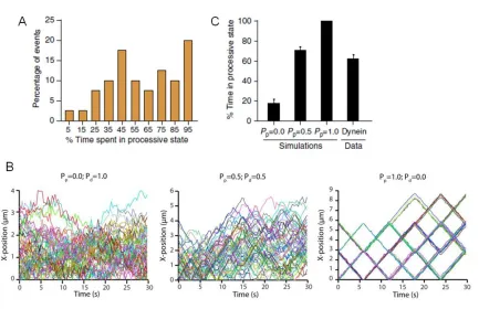
Figure 2.3 Simulations to validate our analysis.
50
motion (Pp=1, Pd=0). (C) Quantitation of the per cent time spent in processive state for
simulated tracks alongside the average for experimental data obtained from Fig. 2.3A. Mean ± SEM. n=50 for the simulations and n=40 for the experimental data.
p150Glued enhances dynein recruitment onto microtubules
51
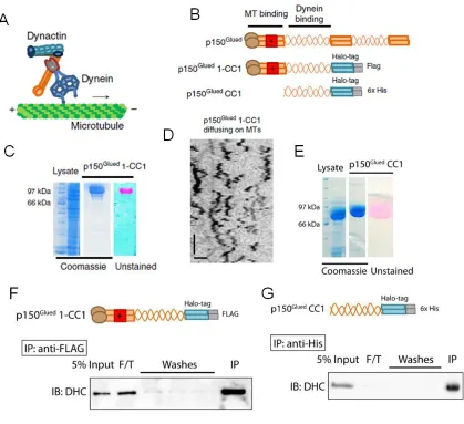
Figure 2.4 Generation of purified recombinant p150Glued constructs.
52
gel shows the labeling of the protein with TMR. (G) Recombinant p150Glued CC1 binds to purified bovine dynein as shown by the immunoblot.
We next performed dual-colour imaging of dynein–GFP and p150Glued
53
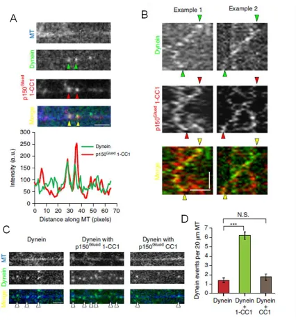
Figure 2.5 p150Glued co-migrates with dynein and enhances its recruitment onto microtubules
54
of dynein. Horizontal bar, 2 µm. (D) Quantitation of the landing events of dynein in each condition shows that p150Glued 1-CC1 increases the landing events greater than fourfold. Mean ± SEM, n=25, two independent experiments, ***p<0.001, one-way analysis of variance with Tukey’s post-hoc test. Concentration of the recombinant protein used in each case is 4.5 nM. N.S., not significant.
p150Glued regulates the dynein-microtubule interaction
55
Figure 2.6 p150Glued regulates the association and dissociation of dynein from microtubules.
56
Motility of p150Glued in cell extracts is dynein-dependent
To further dissect the domains of p150Glued that are critical for the regulation of dynein motility, we sought to develop a more versatile single molecule assay. We performed motility assays using Halo-tagged constructs expressed in cell extracts (Fig. 2.7A). Similar assays have been used extensively to study kinesin motors and their regulation (Blasius et al., 2007; Sun et al., 2011; Fu and Holzbaur; 2013) but have not yet been applied to the dynein motor to test the function of its adaptors and cofactors like dynactin.
58
Figure 2.7 The motility of p150Glued in cell extracts is dynein-driven and ATP dependent.
(A) Schematic of the experimental design with an illustration of p150Glued constructs. (B) COS7 cell extracts were precipitated with a HaloLink resin that binds specifically to the Halo tag. Protein complexes bound to the resin were analyzed by immunoblotting. The dynein complex co-precipitates with p150Glued-Halo and the addition of the Halo tag at the C-terminus does not interfere with the incorporation of p150Glued into the dynactin complex (as shown by p50). (C) A representative time course of photobleaching of TMR-labelled p150Glued shows two decay steps. Grey lines indicate the steps. (D) Frequency distribution of the photobleaching steps for n=42 particles fit with a binomial distribution. (E) Representative time series and corresponding kymographs of the motility of p150Glued-Halo in 10mM Mg-ATP and in the presence of hexokinase and glucose (an ATP depletion system). Horizontal bar, 2 µm. Vertical bar, 5 s. All images are contrast inverted. (F) Representative immunoblot of cell extracts from COS7 cells treated with Mock or DHC siRNA followed by transient transfection of p150Glued-Halo. (G) Quantitation of the number of active, motile events of p150Glued-Halo in the mock and dynein knock-down conditions. The number of events was normalized to the length of the microtubules and the expression levels of p150Glued-Halo in extracts (densitometry analysis using ImageJ). DHC knockdown resulted in a reduction in the number of motile events. Mean ± SEM, n=3 experiments, **p<0.01, Student’s t-test.
59
frequency distribution for the fraction of time spent in a processive state (Fig. 2.8D). In close agreement with the motility characteristics observed for isolated dynein–dynactin complexes (Fig. 2.3A), about ~20% of the p150Glued-Halo-TMR particles tracked were processive throughout their motion along the microtubule while the remainder exhibited stochastic switching between processive and diffusive states. The average diffusion coefficient obtained from the diffusive phases was 0.055 µm2/s, consistent with previous reports (Mallik et al., 2005; Culver-Hanlon et al., 2006; Ross et al., 2006) Together, these results indicate that we have established a robust assay to study the properties of the dynein motor in complex with its adaptor dynactin; this assay will be useful in exploring the effects of other dynein adaptors in future.
Figure 2.8 The motility of dynactin in cell extracts is similar to in vitro motility of purified dynein-GFP.