O R I G I N A L R E S E A R C H
Establishment and Evaluation of a Novel Method
Based on Loop-Mediated Isothermal Ampli
fi
cation
for the Rapid Diagnosis of Thalassemia Genes
This article was published in the following Dove Press journal:
Risk Management and Healthcare Policy
Wei-hua Wang,1,* Min Lin,2,* Hai-liang Li,3Jun-yun Huang,1 Jiang-tao Chen,4,5Xian-song Fang,6 Dong-mei Huang,1Xu-xiang Xi,1 Qing-fei Zhao,1Fang-li Song,7 Shao Huang,7Tian-yu Zhong 1
1Department of Laboratory Medicine, First
Affiliated Hospital of Gannan Medical University, Ganzhou, Jiangxi Province, People’s Republic of China;2School of Food
Engineering and Biotechnology, Hanshan Normal University, Chaozhou, Guangdong Province, People’s Republic of China;
3Department of Hematology, First Affiliated
Hospital of Gannan Medical University, Ganzhou, Jiangxi Province, People’s Republic of China;4The Chinese Medical Aid Team to
the Republic of Equatorial Guinea, Guangzhou, Guangdong Province, People’s Republic of China;5Department of Medical Laboratory,
Huizhou Central Hospital, Huizhou, Guangdong Province, People’s Republic of China;6Department of Blood Transfusion,
First Affiliated Hospital of Gannan Medical University, Ganzhou, Jiangxi Province, People’s Republic of China;7Jiangxi Shiningmed Medical
Technology Ltd, Ganzhou, Jiangxi Province, People’s Republic of China
*These authors contributed equally to this work
Purpose:Currently, thalassemia is commonly detected using gap-polymerase chain reaction (PCR) and deoxyribonucleic acid (DNA) reverse dot blot, which have high requirements of space, instruments, and personnel. Therefore, it is necessary to develop a new method for thalassemia detection with high sensitivity, low cost, and simple and fast operation. In this study, we aimed to design and evaluate a new method for detecting threeα-thalassemia genes including–Southeast Asian (SEA), -α3.7, and -α4.2 andfiveβ-thalassemia genes including 654M, 41/42M,−28M, 17M, and 27/28M based on loop-mediated isothermal amplification (LAMP).
Methods: Primer sequences were designed using Primer Explorer V4 software. Blood samples (5 mL) were collected from all participants in EDTA. DNA was extracted using Chelex 100 and was subjected to LAMP. LAMP products were detected by fluorescence development in ultraviolet light.
Results:We found that LAMP assays for positive samples of thalassemia reached a plateau before 60 minutes, whereas the negative control samples entered the plateau after 70 minutes or showed no amplification. The concentration range of positive reactions was between 20–60 pg/μL and 20–60 ng/μL. Additionally, there were no cross-reactivities among 8 thalassemia subtypes. For clinical samples, the positive sample tube showed strong green
fluorescence, whereas the negative tube showed light greenfluorescence. According to these results, the LAMP method has high sensitivity for detecting thalassemia (252/254). However, 43 false-positive results were obtained in the LAMP test. The LAMP assay was also of low cost and with simple and fast operation.
Conclusion:The novel LAMP assay can be completed within 60 min using a heating block or a water bath, and the result can be read visually based on color change to detect thalassemia. The LAMP assay fulfills the requirements of field application and resource-limited areas, especially those with primary hospitals and rural areas.
Keywords:loop-mediated isothermal amplification, diagnosis, thalassemia genes, fast, low cost
Introduction
Thalassemia is one of the most common hereditary blood diseases that pose a serious threat to human health withα- andβ-thalassemia being the most common.1 The most effective way to control thalassemia is to prevent the birth of severely ill children; thus, rapid and accurate screening for thalassemia is the key to its prevention and control. At present, the deletions of α-thalassemia are detected by gap-polymerase chain reaction (PCR) and α- and β-thalassemia mutations are
Correspondence: Shao Huang Jiangxi Shiningmed Medical Technology Ltd, Ganzhou, Jiangxi Province, People’s Republic of China
Tel +86-18602004914
Email [email protected]
Tian-yu Zhong
Department of Laboratory Medicine, First Affiliated Hospital of Gannan Medical University, Ganzhou 341000, Jiangxi, People’s Republic of China Tel +86-797-8680632 Email [email protected]
Risk Management and Healthcare Policy
Dove
press
open access to scientific and medical research
Open Access Full Text Article
Risk Management and Healthcare Policy downloaded from https://www.dovepress.com/ by 118.70.13.36 on 27-Aug-2020
detected by DNA reverse dot blot (RDB).2These methods have high requirements for space, instruments, and per-sonnel, and it is difficult to promote their use in primary hospitals and rural areas.3 Therefore, it is necessary to develop a new method for thalassemia detection with high sensitivity, low cost, and simple and rapid operation. Loop-mediated isothermal amplification (LAMP) is a novel method for nucleic acid amplification. It is performed by designing 4–6 specific primers for 6 regions of the target gene, and usingBacillus stearothermophilus(Bst) deoxyr-ibonucleic acid (DNA) polymerase to catalyze the nucleic acid amplification reaction for 30–60 min under isothermal conditions (60–65°C).4 Compared to traditional PCR, LAMP does not require a special PCR instrument, and does not need detection by electrophoresis. The complete experiment only needs a constant temperature water bath or metal bath. The results can be directly observed under a UV lamp with the naked eye to judge the green fluorescence emitted by Calcein.5 Therefore, compared with other nucleic acid amplification methods, LAMP has the advan-tages of specificity and rapidity. In recent years, the LAMP assay has been widely used in food safety, pathogen and microbiological testing, clinical diagnosis, and so on.6–9 LAMP assays are also an effective tool for screening genetic mutations.10 A previous study detected the -SEA (Southeast Asian) deletion, and IVSII-I G>A mutation indi-cating thalassemia using LAMP assays.11,12 Thus, new LAMP methods capable of detecting thalassemia could be alternative methods to other techniques, and could be used in primary hospitals and rural areas.
Previous studies have found that the mutations found in thalassemia differ between ethnic groups and regions in China because it has a vast territory with a complicated geography leading to significant variation in hereditary traits. In Wuhan China, –SEA/deletion, -α3.7/deletion, and -α4.2/deletion were the most common α-thalassemia mutations and IVS-2-654 mutation, CD41-42, CD17, CD26, and CD27-28 were the most commonβ-thalassemia mutations.13 In Quanzhou region of Southeast China, -SEA/deletion, -α3.7/deletion, and -α4.2/deletion are the most common β-thalassemia mutations whereasβ IVS-II-654/βN,βCD41-42/βN,βCD17/βN,βCD26/βN, andβ-28/
βN are the most common α-thalassemia mutations.2 In Ganzhou, Jiangxi, -SEA/deletion, -α3.7/deletion, and -α4.2/deletion were the most commonα-thalassemia muta-tions whereas 654M, 41/42M, −28M, 17M, and 27/28M were the most common β-thalassemia mutations. In this study, we aimed to design and evaluate a new method for
detecting three α- thalassemia mutations including –SEA, -α3.7, and -α4.2, andfiveβ-thalassemia mutations includ-ing 654M, 41/42M, −28M, 17M, and 27/28M using LAMP.
Materials and Methods
This study was approved by the Ethics Committee of First Affiliated Hospital of Gannan Medical University (No.201317) and conducted in accordance with the Declaration of Helsinki.
Design of LAMP Primers
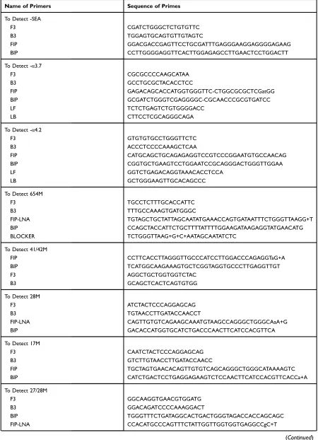
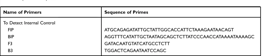
The LAMP primer sequences, including forward inner mer (FIP), backward inner primer (BIP), forward outer pri-mer (F3), and backward outer pripri-mer (B3), were designed using Primer Explorer V4 software to detect the 3 deletions ofαthalassemia including–SEA, -α3.7, and -α4.2 andfive mutants ofβ-thalassemia including 654M, 41/42M,−28M, 17M, and 27/28M. The design principle of LAMP primers was shown athttp://loopamp.eiken.co.jp/e/lamp/primer.html. Besides LAMP primers, blocker was designed to reduce the probability of non-specific amplification for detecting 654M. Blocker can bind to wild-type genomic DNA before LAMP primers, thus preventing LAMP primers non-specifically binding to wild-type genomic DNA. In addition, loop primer B (LB) and loop primer F (LF) were designed to improve the amplification efficiency for detecting -α4.2. The design prin-ciple of LB and LF was shown athttp://loopamp.eiken.co.jp/ e/lamp/loop.html. The primer sequences used for LAMP were synthesized by Sangon (Shanghai, China), and are shown inTable 1.
Clinical Specimens and Genomic DNA
Extraction
For this study, 400 individuals were recruited between January 2016 and December 2018 from First Affiliated Hospital of Gannan Medical University. All participants provided written informed consent. All individuals were diagnosed by α-thalassemia Deletion Test Kit (China Medical Device Registration: 20,173,403,211) andβ -thalas-semia Test Kit (China Medical Device Registration: 20,173,401,093) (Zeesan Biotech, Xiamen, China), then all individuals were diagnosed by LAMP for detecting the three
α-thalassemia deletions including–SEA, -α3.7, and -α4.2, andfiveβ-thalassemia mutations including 654M, 41/42M,
−28M, 17M, and 27/28M. The procedures for sample collec-tion and processing were as follows: Briefly, 5 mL blood
Risk Management and Healthcare Policy downloaded from https://www.dovepress.com/ by 118.70.13.36 on 27-Aug-2020
Table 1The Sequences of Primers Used for LAMP Method
Name of Primers Sequence of Primes
To Detect -SEA
F3 CGATCTGGGCTCTGTGTTC
B3 TGGAGTGCAGTGTTGTAGTC
FIP GGACGACCGAGTTCCTGCGATTTGAGGGAAGGAGGGGAGAAG
BIP CCTTGGGGAGGTTCACTTGGAGAGCCTTGAACTCCTGGACTT
To Detect -α3.7
F3 CGCGCCCCAAGCATAA
B3 GCCTGCGCTACACCTCC
FIP GAGACAGCACCATGGTGGGTTC-CTGGCGCGCTCGatGG
BIP GCGATCTGGGTCGAGGGGC-CGCAACCCGCGTGATCC
LF TCTCTGAGTCTGTGGGGACC
LB CTTCCTCGCAGGGCAGA
To Detect -α4.2
F3 GTGTGTGCCTGGGTTCTC
B3 ACCCTCCCCAAAGCTCAA
FIP CATGCAGCTGCAGAGAGGTCCGTCCCGGAATGTGCCAACAG
BIP CGGTGCTGAAGTCCTGGAATCCGCAGGGACTGGGTTGGAA
LF GGTCTGAGACAGGTAAACACCTCCA
LB GCTGGGAAGTTGCACAGCCC
To Detect 654M
F3 TGCCTCTTTGCACCATTC
B3 TTTGCCAAAGTGATGGGC
FIP-LNA TGTAGCTGCTATTAGCAATATGAAACCAGTGATAATTTCTGGGTTAAGG+T
BIP CCAGCTACCATTCTGCTTTTATTTTGGAAGATAAGAGGTATGAACATG
BLOCKER TCTGGGTTAAG+G+C+AATAGCAATATCTC
To Detect 41/42M
FIP CCTTCACCTTAGGGTTGCCCATCCTTGGACCCAGAGGTaG+A
BIP TCATGGCAAGAAAGTGCTCGGTAGGTGCCCTTGAGGTTGT
F3 AGGCTGCTGGTGGTCTAC
B3 GCAGCTCACTCAGTGTGG
To Detect 28M
F3 ATCTACTCCCAGGAGCAG
B3 TGTAACCTTGATACCAACCT
FIP-LNA CAGTTGTGTCAGAAGCAAATGTAAGCCAGGGCTGGGCAaA+G
BIP GACACCATGGTGCATCTGACCCAACTTCATCCACGTTCA
To Detect 17M
F3 CAATCTACTCCCAGGAGCAG
B3 GTCTTGTAACCTTGATACCAACC
FIP TGCTAGTGAACACAGTTGTGTCAGCAGGGCTGGGCATAAAAGTC
BIP CATCTGACTCCTGAGGAGAAGTCTCCAACTTCATCCACGTTCACCa+A
To Detect 27/28M
F3 GGCAAGGTGAACGTGGATG
B3 GGACAGATCCCCAAAGGACT
BIP TGGGTTTCTGATAGGCACTGACTGGGTAGACCACCAGCAGC
FIP-LNA CCACATGCCCAGTTTCTATTGGTTGGTGGTGAGGCCgC+T
(Continued)
Risk Management and Healthcare Policy downloaded from https://www.dovepress.com/ by 118.70.13.36 on 27-Aug-2020
samples in EDTA were collected from all participants and subjected to DNA extraction using Chelex 100. Briefly, 300 μL of whole blood was added to 200μL NaOH/SDS and then centrifuged at 13,000 ×gfor 3 min. The precipitate was collected, 5% Chelex-100 was added, and incubated at 56°C for 30 min. The samples were vortexed at high speed for 5–10 s and then centrifuged at 13,000 ×gfor 2 min; the supernatant was collected and stored at−20°C until use.
LAMP Reaction and Analysis of LAMP
Products
To carry out LAMP reaction, 25 μL LAMP reaction system was prepared as shown in Table 2. The tube to
detect internal control HBB gene was named as control tube, and the tubes to respectively detect 3 deletions of
α thalassemia (-SEA, -α3.7, and -α4.2) or five mutants of β-thalassemia (654M, 41/42M, −28M, 17M, and 27/28M) were named as detection tube. The DNA tem-plate added in control tube and detection tube is same DNA from same patient. The LAMP reaction system mixture was incubated at 56°C for 45 min in a water bath (Kuansons, Shanghai, China). LAMP products in the control tube and the detection tube were detected by
fluorescence color development under ultraviolet light. The definition of the positive and negative samples is shown below: The definition of the positive and
Table 1(Continued).
Name of Primers Sequence of Primes
To Detect Internal Control
FIP ATGCAGAGATATTGCTATTGGCACCATTCTAAAGAATAACAGT
BIP AGGTTTCATATTGCTAATAGCAGCTCTTATCCCAACCATAAAATAAAAGC
F3 GATACAATGTATCATGCCTCTT
B3 TGGACTCAGAATAATCCAGC
Note:Base which preceded by a“+”sign indicating locked nucleic acid (LNA) modification.
Abbreviations:LAMP, Loop-mediated isothermal amplification; SEA, Southeast Asian; BLOCKER, BLOCKER is an oligonucleotide that hybridizes perfectly to the wildtype template, and thereby suppressing unintended hybridization of the LAMP primer to the wildtype; FIP, forward inner primer; BIP, backward inner primer; F3, forward outer primer; B3, backward outer primer; LF, forward loop primers; LB, backward loop primers.
Table 2LAMP Reaction System
Component To Detect–SEA, -α3.7, 41/42M,
28M, 17M, 27/28M, and Internal
Control
To Detect -α4.2,
To Detect 654M
Volume (μL) Volume (μL) Volume (μL)
10×Bst DNA polymerase reaction buffer (NEB, Ipswich, MA, USA) 2.5 2.5 2.5
dNTP Mixture (10mM) (Sangon, Shanghai, China) 3.5 3.5 3.5
MgSO4 Solution (100mM) (NEB) 1.5 1.5 1.5
Betaine (5M) (Solarbio, Beijing, China) 4.0 4.0 4.0
Bst DNA polymerase (NEB) 1 1 1
FIP (40μM) 1 1 1
BIP (40μM) 1 1 1
F3 (10uM)) 0.5 0.5 0.5
B3(10μM) 0.5 0.5 0.5
Blocker (10μM) – – 1
LF (10μM) – 1 –
LB (10μM) – 1 –
Calcein (100μM) (Aladdin, Shanghai, China) 0.5 0.5 0.5
DNA template 2 2 2
ddH2O Up to 25 Up to 25 Up to 25
Abbreviations:LAMP, loop-mediated isothermal amplification; SEA, Southeast Asian; dNTP, deoxy-ribonucleoside triphosphate; Bst,Bacillus stearothermophilus; FIP, forward inner primer; BIP, backward inner primer; F3, forward outer primer; B3, backward outer primer; LF, forward loop primers; LB, backward loop primers; ddH2O, double distilled H2O.
Risk Management and Healthcare Policy downloaded from https://www.dovepress.com/ by 118.70.13.36 on 27-Aug-2020
negative samples is shown below: when only LAMP products of control sample show green fluorescence, this tested sample was determined to be Thalassemia negative sample; when LAMP products of both tested and control sample showed green fluorescence, the tested sample was determined to be Thalassemia posi-tive sample, and the type of thalassemia can be esti-mated according which detection tube to detect 3 deletions of α thalassemia (-SEA, -α3.7, and -α4.2) orfive mutants ofβ-thalassemia (654M, 41/42M,−28M, 17M, and 27/28M) showed green fluorescence. When LAMP products of control sample did not show green
fluorescence, the reaction was considered to be failed and the sample needed to be retested.
PCR and Analysis of PCR Products
The PCR system was the same as the LAMP reaction system. The mixture was incubated for 90 cycles at 65° C for 1 min/cycle (ABI 7500). The Ct values of wild-type and mutant wild-types were then analyzed to calculate the ΔCT values (Ct values of mutant type - Ct value of wild type).
Statistical Analyses
Using the Sanger sequencing results as the control group, the Stata 12.0 software was used to calculate the sensitivity and specificity of the assay method, and the 95% confidence interval. The consistency of the method’s test results with those of Sanger sequencing was evaluated using the Kappa test method with the SPSS software.
Results
Speci
fi
city of LAMP Primers
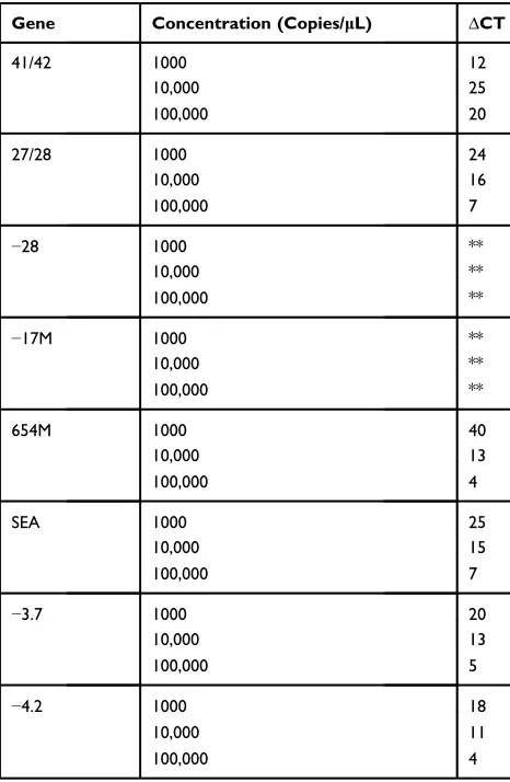
PCR was performed using the wild-type and mutant/defi -ciency plasmids at concentrations of 1000/10,000/100,000 copies/μL as templates. Under these same real-time con-ditions as the LAMP reaction, successful amplification of the mutant/deficiency sequence was observed, indicating that the primers designed for the LAMP assay could be used for PCR amplification. Additionally, genes −28 and
−17M of wild type had not been amplified. The ΔCT values are shown in Table 3. The results showed that the higher template concentrations (≥100,000 copies/μL) had lower ΔCT values whereas the lower template concentra-tions (≤100,000 copies/μL) had higherΔCT values. These
results showed that LAMP primers had high specificity to distinguish between wild-type and mutant/deficiency gene.
Reaction Time of LAMP Assays
DNA was extracted from clinical specimens and used for LAMP assays to determine the reaction time of LAMP assays. The results showed that LAMP assays for positive samples of thalassemia reached a plateau before 60 min-utes, whereas the negative control samples entered the plateau phase after 70 minutes or showed no amplification (Figure 1). The LAMP reaction tube with a reaction time of 60 minutes was exposed to ultraviolet light for obser-ving the development of color. The positive sample tube showed strong green fluorescence, whereas the negative tube showed light green fluorescence (Figure 2). The negative and positive samples could be distinguished visually byfluorescence intensity.
Table 3TheΔCT Values
Gene Concentration (Copies/μL) ΔCT
41/42 1000 12
10,000 25
100,000 20
27/28 1000 24
10,000 16
100,000 7
−28 1000 **
10,000 **
100,000 **
−17M 1000 **
10,000 **
100,000 **
654M 1000 40
10,000 13
100,000 4
SEA 1000 25
10,000 15
100,000 7
−3.7 1000 20
10,000 13
100,000 5
−4.2 1000 18
10,000 11
100,000 4
Note:ΔCT values=Ct values of mutant types−Ct value of wild type; the larger the
ΔCT value, the better the discrimination between the wild type and the mutant plasmid. **Indicates that the wild type template has not been amplified. Abbreviations:SEA, Southeast Asian; CT, cycle threshold.
Risk Management and Healthcare Policy downloaded from https://www.dovepress.com/ by 118.70.13.36 on 27-Aug-2020
-SEA
WILD TYPE
WILD TYPE
WILD TYPE
WILD TYPE WILD TYPE
WILD TYPE WILD TYPE WILD TYPE -3.7
-4.2
41/42M
17M
654M
-28M
27/27M
A B
C D
E F
G H
Figure 1LAMP assays for the positive and negative samples of thalassemia. (A–H) PCR reactions were performed using positive samples and negative samples of thalassemia as templates under these same real-time LAMP reaction conditions.
Abbreviations:SEA, Southeast Asian; LAMP, loop-mediated isothermal amplification; PCR, polymerase chain reaction.
A B C
Positive Negative Positive Negative Positive NegativeFigure 2The LAMP reaction tube with a reaction time of 60 minutes was exposed to ultraviolet light for observing color. The positive sample tube showed strong green fluorescence while the negative tube showed light greenfluorescence. (A)–SEA positive sample and negative sample; (B) 654M positive sample and negative sample; (C) 27/ 28 positive sample and negative sample.
Abbreviations:SEA, Southeast Asian; LAMP, loop-mediated isothermal amplification.
Risk Management and Healthcare Policy downloaded from https://www.dovepress.com/ by 118.70.13.36 on 27-Aug-2020
Sensitivity of LAMP Assays
DNA extracted from positive samples at concentrations of 20–60 ng/μL, was serially diluted 10-fold as 100% 10%, 1%, 0.1%, 0.01%, and wild-type plasmid was used as the negative control. The results showed that positive clinical samples at concentrations of 100% 10%, 1%, and 0.1% emitted bright green fluorescence, whereas positive sam-ples at concentrations of 0.01% and the wild-type plasmid group emitted light yellowfluorescence. The concentration range of positive reactions was between 20–60 pg/μL and 20–60 ng/μL. According to these results, the detection limit of the PCR was 20–60 pg/µL.
Speci
fi
city of LAMP Assays
Eight positive and negative clinical samples were added to 8 groups of the LAMP reaction system, respectively. For example: 8 positive and negative clinical samples were added to the HBB–SEA LAMP reaction system, respec-tively. The results showed that only HBB–SEA positive clinical samples emitted bright greenfluorescence whereas other positive and negative clinical samples emitted light yellow fluorescence. The results showed that the LAMP method which detected the HBB–SEA subtype had high specificity. Similar to these results, the LAMP method could also detect the other 7 subtypes with high specificity.
Sensitivity and Speci
fi
city of LAMP Assays
in Clinical Assay
In total, 400 clinical samples were collected and tested by LAMP assay (Table 4). Among these, there were 254 positive mutation specimens (including heterozygous) and LAMP detected 252 positive mutant specimens; this indicates that the LAMP method has high sensitivity for detecting thalassemia (252/254). However, 43
false-positive results were obtained with the LAMP test; the
−3.7 and −4.2 subtypes showed especially more false-positive results, indicating that the LAMP detection method was less specific. These results showed that the sensitivity and specificity were 99.2% (252/254) and 29.5% (43/146), respectively.
Discussion
In this study, we report a novel LAMP method to detect the three α- thalassemia deletions include –SEA, -α3.7, and -α4.2, and five β-thalassemia mutations including 654M, 41/42M, −28M, 17M, and 27/28M. Four primers were designed specifically to bind to six distinct regions of the target gene. In this study, for primers with low
speci-ficity, locked nucleic acid (LNA) technology was used for primer modification. LNA is a class of nucleic acid ana-logs that contain a methylene bridge connecting the 2′ oxy-gen and 4′carbon in the ribose moiety, which increases the thermal stability of hybrid strands.14 The use of LNA technology in molecular diagnostic methods improves the specificity and sensitivity of assays.15 Using the primers designed for the present study, the LAMP method showed high sensitivity for detecting thalassemia (252/254). However, there were 43 false-positive results in the LAMP test; in particular, the −3.7 and −4.2 subtypes showed more false-positive results, indicating that the LAMP detection method was less specific for detecting the−3.7 and −4.2 subtypes.
LAMP assay has been widely used for screening genetic mutations and deletions. In previous studies, LAMP assays were used to detect the F167Y mutation in
Fusarium graminearumand KRAS mutation patients with
cancer.16,17 Additionally, LAMP assays were used to detect the haptoglobin gene deletion in alkaline-denatured blood.18 In this study, we found that the target sequence was successfully amplified and that the wild type and target could be distinguished. LAMP assays for positive samples of thalassemia reached a plateau before 60 min-utes, whereas the negative control samples entered the plateau after 70 minutes or showed no amplification. The concentration range of positive reactions was between 20–60 pg/μL and 20–60 ng/μL. These results suggest that the designed LAMP assays had high sensitivity and low reaction time. Additionally, there were no cross-reac-tivities between 8 thalassemia subtypes, indicating that LAMP assays had high specificity. For clinical samples, the positive sample tube showed strong green fl uores-cence, whereas the negative tube showed light green
Table 4Clinical Performance of LAMP Assays
Gene Clinical +/LAMP+
Clinical +/LAMP−
Clinical −/LAMP+
Clinical −/LAMP−
41/42 34 1 2 363
27/28 3 0 0 397
−28 3 0 0 397
−17M 12 0 1 387
654M 46 1 1 352
SEA 117 0 2 281
−3.7 25 0 20 355
−4.2 12 0 17 371
Abbreviations: LAMP, loop-mediated isothermal amplification; SEA, Southeast Asian.
Risk Management and Healthcare Policy downloaded from https://www.dovepress.com/ by 118.70.13.36 on 27-Aug-2020
fluorescence; the negative and positive samples could thus be distinguished visually, based onfluorescence intensity. At present, deletions ofα-thalassemia are detected by Gap-PCR and the α- and β-thalassemia mutations are detected by DNA reverse dot blot (RDB).2,19,20 Compared to these methods, the LAMP assay has many advantages such as high sensitivity, and simple and fast operation, and it does not need a special instrument. Furthermore, the cost of detecting thalassemia (include
–SEA, -α3.7, and -α4.2 α- thalassemia, and 654M, 41/42M, −28M, 17M, 27/28Mβ-thalassemia) using com-monly clinical test kits is about 300 CNY in our hospital, while the cost of detecting thalassemia using the LAPM method in the manuscript is about 30 CNY. Compared to commonly clinical test kits, the LAMP assay has the advantage of low cost. Although the present study suc-cessfully used LAMP to detect three α- thalassemia deletions include –SEA, -α3.7, and -α4.2, and five
β-thalassemia mutations including 654M, 41/42M,
−28M, 17M, 27/28M, the−3.7 and−4.2 subtypes showed more false-positive results, possibly because the−3.7 and
−4.2 deletion genes were closely related to the wild type gene sequence before mutation, which is prone to false amplification. Another important reason is that the posi-tions and lengths of the missing fragments of −3.7 and
−4.2 are not fixed, so there may occur erroneous
ampli-fication. Hence, further studies with redesigned primer sets are needed to detect these mutations with high spe-cificity. In addition, we recommend that patients with 3.7 and 4.2 deletion mutations need to further screen in the superior hospital.
In conclusion, the novel LAMP assay can be com-pleted within 60 min using a heating block or a water bath and the result can be read visually based on color changes to detect thalassemia. The LAMP assay had high sensitivity, low cost, and simple and fast operation,
ful-filling the requirements of field application in resource-limited areas, especially those with primary hospitals and in rural areas.
Acknowledgments
The study was financially supported by the National Natural Science Foundation of China (No.81360265 and No.81702580) and the Open Project of Key Laboratory of Prevention and Treatment of Cardiovascular and Cerebrovascular Diseases, Ministry of Education, China (No. XN201812).
Disclosure
Fang-li Song and Shao Huang were employed by company Jiangxi Shiningmed Medical Technology Ltd. All other authors declare no competing interests in this work.
References
1. Taher AT, Weatherall DJ, Cappellini MD. Thalassaemia. Lancet.
2018;391(10116):155–167. doi:10.1016/S0140-6736(17)31822-6 2. Zhuang J, Jiang Y, Wang Y, et al. Molecular analysis of
alpha-thalassemia and beta-alpha-thalassemia in Quanzhou region Southeast China.J Clin Pathol.2019. doi:10.1136/jclinpath-2019-206179 3. Lin M, Zhu JJ, Wang Q, et al. Development and evaluation of a
reverse dot blot assay for the simultaneous detection of common alpha and beta thalassemia in Chinese. Blood Cells Mol Dis.
2012;48(2):86–90. doi:10.1016/j.bcmd.2011.12.001
4. Notomi T, Okayama H, Masubuchi H, et al. Loop-mediated isother-mal amplification of DNA. Nucleic Acids Res. 2000;28(12):E63. doi:10.1093/nar/28.12.e63
5. Ma L, Chen Z, Guan W, Chen Q, Liu D. Rapid and specific detection of all known Nipah virus strains’sequences with reverse transcrip-tion-loop-mediated isothermal amplification. Front Microbiol.
2019;10:418. doi:10.3389/fmicb.2019.00418
6. Nzelu CO, Kato H, Peters NC. Loop-mediated isothermal amplifi ca-tion (LAMP): an advanced molecular point-of-care technique for the detection of Leishmania infection.PLoS Negl Trop Dis.2019;13(11): e0007698.
7. Mori Y, Notomi T. Loop-mediated isothermal amplification (LAMP): expansion of its practical application as a tool to achieve universal health coverage.J Infect Chemother.2019.
8. Waterfield T, Fairley D, Lynn F, Blackwood B, Shields MD. A protocol for a systematic review of the diagnostic accuracy of Loop-mediated-isothermal AMPlification (LAMP) in diagnosis of invasive meningococcal disease in children.Syst Rev.2018;7(1):86. doi:10.1186/s13643-018-0747-0
9. Yang Q, Domesle KJ, Ge B. Loop-mediated isothermal amplification for salmonella detection in food and feed: current applications and future directions. Foodborne Pathog Dis. 2018;15(6):309–331. doi:10.1089/fpd.2018.2445
10. Srividya A, Maiti B, Chakraborty A, Chakraborty G. Loop mediated isothermal amplification: a promising tool for screening genetic mutations.Mol Diagn Ther.2019;23:723–733. doi:10.1007/s40291-019-00422-0
11. Chomean S, Pholyiam K, Thamwarokun A, Kaset C. Development of visual detection of alpha-thalassemia-1 (the - -(SEA) deletion) using ph-sensitive loop-mediated isothermal amplification. Hemoglobin.
2018;42(3):171–177. doi:10.1080/03630269.2018.1488723 12. Gill P, Amree AH. Allele-specific loop-mediated isothermal amplifi
-cation for the detection of IVSII-I G>A mutation on beta-globin gene. Open Access Maced J Med Sci. 2019;7(10):1582–1587. doi:10.3889/oamjms.2019.285
13. Cai W, Xiong Q, Tong J, et al. Prevalence and genetic analysis of thalassemia in neonates in Wuhan area: a national megacity in central China.J Matern Fetal Neonatal Med.2019;1–8.
14. Hagedorn PH, Persson R, Funder ED, et al. Locked nucleic acid: modality, diversity, and drug discovery.Drug Discov Today.2018;23 (1):101–114. doi:10.1016/j.drudis.2017.09.018
15. Ishige T, Itoga S, Matsushita K. Locked nucleic acid technology for highly sensitive detection of somatic mutations in cancer.Adv Clin Chem.2018;83:53–72.
16. Duan Y, Zhang X, Ge C, et al. Development and application of loop-mediated isothermal amplification for detection of the F167Y muta-tion of carbendazim-resistant isolates in Fusarium graminearum.Sci Rep.2014;4:7094. doi:10.1038/srep07094
Risk Management and Healthcare Policy downloaded from https://www.dovepress.com/ by 118.70.13.36 on 27-Aug-2020
17. Itonaga M, Matsuzaki I, Warigaya K, et al. Novel methodology for rapid detection of KRAS mutation using PNA-LNA mediated loop-mediated isothermal amplification.PLoS One.2016;11(3):e0151654. doi:10.1371/journal.pone.0151654
18. Soejima M, Egashira K, Kawano H, Kawaguchi A, Sagawa K, Koda Y. Rapid detection of haptoglobin gene deletion in alkaline-denatured blood by loop-mediated isothermal amplification reaction. J Mol Diagn.2011;13(3):334–339. doi:10.1016/j.jmoldx.2011.01.005
19. Laghmich A, Alaoui Ismaili FZ, Barakat A, Ghailani Nourouti N, Khattab M, Bennani Mechita M. Alpha-Thalassemia in North Morocco: prevalence and molecular spectrum. Biomed Res Int.
2019;2019:2080352. doi:10.1155/2019/2080352
20. Faraon R, Daraghmah M, Samarah F, Srour MA. Molecular charac-terization of beta-thalassemia intermedia in the West Bank, Palestine. BMC Hematol.2019;19:4. doi:10.1186/s12878-019-0135-6
Risk Management and Healthcare Policy
Dove
press
Publish your work in this journal
Risk Management and Healthcare Policy is an international, peer-reviewed, open access journal focusing on all aspects of public health, policy, and preventative measures to promote good health and improve morbidity and mortality in the population. The journal welcomes submitted papers covering original research, basic science, clinical & epidemiological studies, reviews and evaluations,
guidelines, expert opinion and commentary, case reports and extended reports. The manuscript management system is completely online and includes a very quick and fair peer-review system, which is all easy to use. Visit http://www.dovepress.com/testimonials.php to read real quotes from published authors.
Submit your manuscript here:https://www.dovepress.com/risk-management-and-healthcare-policy-journal
Risk Management and Healthcare Policy downloaded from https://www.dovepress.com/ by 118.70.13.36 on 27-Aug-2020