R E S E A R C H
Open Access
Inter/intra-frame constrained vascular
segmentation in X-ray angiographic image
sequence
Shuang Song
1, Chenbing Du
1, Ying Chen
1, Danni Ai
1, Hong Song
2, Yong Huang
1, Yongtian Wang
1,3and
Jian Yang
1*FromIEEE International Conference on Bioinformatics and Biomedicine 2018 Madrid, Spain. 3-6 December 2018
Abstract
Background:Automatic vascular segmentation in X-ray angiographic image sequence is of crucial interest, for instance, for better quantifying coronary arteries in diagnostic and interventional procedures.
Methods:A novel inter/intra-frame constrained vascular segmentation method is proposed to automatically segment vessels in coronary X-ray angiographic image sequence. First, a morphological filter operator is applied to remove structures undergoing the respiratory motion from the original image sequence. Second, an inter-frame constrained robust principal component analysis (RPCA) is utilized to remove the quasi-static structures from the image sequence. Third, an intra-frame constrained RPCA is employed to smooth the final extracted vascular sequence. Fourth, a multi-feature fusion is designed to improve the vascular contrast and the final vascular segmentation is realized by thresholding-based method.
Results: Experiments are conducted on 22 clinical X-ray angiographic image sequences. The global and local contrast-to-noise ratio of the proposed method are 6.6344 and 4.2882, respectively. And the precision, sensitivity and F1 value are 0.7378, 0.7960 and 0.7658, respectively. It demonstrates that our method is effective and robust for vascular segmentation from image sequence.
Conclusions: The proposed method is effective to remove non-vascular structures, reduce motion artefacts and other non-uniform illumination caused noises. Also, the proposed method is online which can just process one image per time without re-optimizing the model.
Keywords: X-ray angiographic image sequence, Vascular enhancement, Multi-feature, Vascular segmentation
Background
Nowadays, coronary artery disease (CAD) is greatly threatening human health [1]. Since X-ray angiography (XRA) has better imaging quality and faster imaging speed, it is regarded as the gold standard for the diagno-sis and treatment of CAD. However, due to the perspec-tive projection of 3D anatomic structures, much 3D information has been lost and different anatomical
structures are overlapped in the XRA images. Moreover, the injection of contrast agent and the blood flow vari-ation bring in-homogeneous intensity of coronary artery. And the cardiorespiratory motion and patient movement also introduce motion artefacts to the XRA images. To improve the image-guided diagnosis and interventional procedures of CAD, the automatic and robust vessel seg-mentation is of great significance and meanwhile a chal-lenging problem.
Vascular segmentation technique can be divided into two classes, including the model-based and learning-based methods. Based on the spatial continuity of
© The Author(s). 2019Open AccessThis article is distributed under the terms of the Creative Commons Attribution 4.0 International License (http://creativecommons.org/licenses/by/4.0/), which permits unrestricted use, distribution, and reproduction in any medium, provided you give appropriate credit to the original author(s) and the source, provide a link to the Creative Commons license, and indicate if changes were made. The Creative Commons Public Domain Dedication waiver (http://creativecommons.org/publicdomain/zero/1.0/) applies to the data made available in this article, unless otherwise stated. * Correspondence:[email protected]
1Beijing Engineering Research Center of Mixed Reality and Advanced Display, School of Optics and Photonics, Beijing Institute of Technology, Beijing 100081, China
vessels, level-set and active contour-based methods are commonly utilized. Wang et al. [2] utilized the level-set algorithm to segment the coronary artery by constructing the speed function with the curvature, intensity and model term. Sun et al. [3] proposed the local region based active contour method by shape fitting the energy function. It improved the segmenta-tion accuracy and is much more robust to the non-uniform intensity distribution and fitting initialization. Based on the shape of vascular section, Cheng et al. [4] employed a B-snake model to accurately segment the small-scale vessels in the low-contrast images. Lee et al. [5] utilized the Kalman filter to initialize the contour and segmented the vessels by the active con-tour model. The initialization improved the time effi-ciency. The learning-based methods usually compute the classification model based on the image hidden information. Hassouna et al. [6] modeled the background with two Gaussian and a rayleigh distri-butions and the vessels with a Gaussian distribution, respectively. Then they utilized the Expectation-Maximization algorithm to estimate the distribution parameters and employed the Markov Random Field to be the spatial constraint to realize the final vascu-lar segmentation. Goceri et al. [7] clustered the ves-sels based on the K-means approach and improved the segmentation accuracy by the morphology based iterative optimization. Lupascu et al. [8] delivered the high order features to the AdaBoost classifier to speed the segmentation. Orlando et al. [9, 10] computed the fused feature map by the Fully-Connected Conditional Random Field to ensure the continuity of different vascular segments. In recent years, Convolutional Neural Network (CNN) based vascular segmentation has attracted much researcher’s attention. Wang et al. extracted the vascular features based on CNN to seg-ment the vessels with a stochastic decision forest. Fu et al. [11] combined CNN with Conditional Random Field (CRF) and developed a DeepVessel network to improve the segmentation accuracy. Luo et al. [12] improved the DeepVessel network by considering the non-uniform intensity and noise coexistence.
To the authors’knowledge, the model-based segmentation methods are sensitive to the initial contour and learning-based approaches require large amounts of labeled datasets. Moreover, the methods mentioned above have a significant limitation in angiographic images with low contrast and noisy background. Vascular enhancement can greatly ease the vascular segmentation by enhancing the vascular struc-tures and compress the background noise. The single-image based enhancement easily introduces the non-vascular noise and motion artefacts when dealing with X-ray angiogram im-ages. While subtraction-based enhancement utilizes the an-giograms with and without vessels. It can effectively remove
the motion artefacts in the final enhanced vascular angio-grams and improve the subsequent segmentation accuracy.
Current vessel subtraction methods can be classified into two categories, including image registration based methods and layer separation based methods. In the im-aging of coronary artery, mask images are taken prior to the perfusion of the contrast agent and coronary arteries are not visible in them. While live images are taken dur-ing the contrast agent passdur-ing through the coronary ar-tery. Image registration based methods [13, 14] only need a live image and a mask image whose motion is the most similar to the live image. Such methods are usually realized by template matching, similarity measure maximization, image warping and subtraction technol-ogy sequentially. Though the technique largely reduces the motion artifacts and non-vascular noise, it is likely to be interrupted by the patient motion or contrast agent leakage when computing the correspondences be-tween images. Motion layer separation based method supposes an image in the sequence can be decomposed into motion layers. The key part of the first type methods [15–17] is motion estimation of each layer. Zhu et al. [15] divided the sequence into the vascular and non-vascular layers and applied optical flow to the non-vascular layer to compute the deformation filed. Zhang et al. [16] separated the sequence into three layers, including static, lung (slow motion) and vessel (rapid motion) layer and constructed a motion trans-formation model for each layer. Nevertheless, the struc-tures in the XRA sequence participate in different motion patterns. Specific motion model in each layers cannot cover all the motions of a structure, especially vascular motion in the XRA sequences includes the car-diac, respiratory, patient and camera motions. Another kind of methods [18–20] supposes that the image is under specific prior constraint and directly separates the sequence into background and vessel layer. Many math-ematical expressions, such asL1norm,L2norm, nuclear norm and so on, have been applied to model the specific prior. Robust principal component analysis (RPCA) model, composed of sparse and low-rank prior, has be-come a common tool in medical image analysis of vari-ous imaging modalities.
Methods
In this section, the vascular images is distinguished from the XRA sequence by removing the structures that are static or undergoes the respiratory motion. Then, a multi-feature fused descriptor is employed to further compress the background noise in the vascular images and the vascular structures are finally segmented by a thresholding-based approach.
Inter/intra-frame constrained RPCA
To extract the vascular images from the XRA sequence
I, structures that are static or undergo the respiratory motion should be removed. To reduce the disturbance (lung, diaphragm) caused by the respiratory motion, a circular structural element based morphological close operation [18] is applied to the sequenceIto obtain the respiratory sequence R. By subtracting the sequence R
from sequence I, the respiratory disturbance can be re-moved and the obtained sequence is denoted asDI. The sequenceDI is composed of two components, including the moving vascular component and the quasi-static non-vascular component. In addition, the vessels in se-quence DI only occupy a small portion. Considering RPCA aims to decompose the matrix into a low-rank component and an overall sparse component by search-ing for a low-dimensional subspace, it is suitable to sep-arate sequence DI into the moving vascular component and the quasi-static non-vascular component [21]. Hence, we have:
B;E
f g ¼ arg min1
2kDI−B−Ek
2
Fþβ1k kB
þβ2k kE 1 ð1Þ
where E refers to the vascular component, and Bis the
quasi-static non-vascular component. β1 and β2 are
regularization coefficients. ‖∙‖F is the Frobenius norm, ‖∙‖∗is the nuclear norm and‖∙‖1is L1 norm.
Considering dealing with the steaming X-ray images for coronary interventions, the online processing of XRA sequence is essential on the basis of the motion informa-tion in inter-frames. Hence, we utilize the explicit low-rank factorization [21] to describe B by the subspace basis Lr and the corresponding coefficients Ce. The factorization can be denoted as B=Lr×CeT. After this, solving Eq. (1) equals to minimize the empirical cost functiong(Ce,E,Lr), and we have:
g Ceð ;E;LrÞ ¼ λ1
2Nk kLr 2 F þ1 N XN i¼1
DIi−LriCeTi−Ei
2
2þ λ1
2 kCeik
2
2þλ2k kE 1
ð2Þ
where DIi is the ith image of sequence DI, N is the
number of images in sequenceDI.Lri∈RDr, Cei∈Rr,D
is the dimension of an image in sequence DI, r is the
upper bounded rank ofB. In the optimization of Eq. (2),
coefficients Ce, vascular component E and basis Lr are
disposed in an alternative manner. In the alternative manner, {Cei,Ei} of the ithimage in DI is computed by the ithimage and Lri−1of the (i-1) th image. Then,Lri of the ithimage is re-computed on the basis of {Cei,Ei}. By repeating the procedure, vascular component in each
image of sequence DI can be computed by combining
the motion information of vascular structures as an inter-frame constraint. After the inter-frame constrained RPCA, the vascular component is separated from the
quasi-static non-vascular component in the XRA
sequence.
However, due to the non-rigid motion between the frames in the XRA sequence, large amount of motion ar-tefacts and noises may still exist in the vascular compo-nent. Hence, we utilize the same morphological close operation to remove the motion artefacts around the catheter and obtain another difference sequenceDI′. To remove more motion artefacts and noises, we introduce the intra-frame constrained RPCA and denote it accord-ing to Eq. (2) as follows:
g Ce0;V;Lr0
¼ 1
N
XN
i¼1 DI
0
i−Lr
0
iCe
0T
i −Vi
2
2
þλ1
2 Ce
0
i
2
2þλ2k kVi 1
þ λ1
2N Lr
0 2
F
ð3Þ
whereLr0i∈RDr0,Ce0i∈Rr0. In addition, since most static structures have been removed in the inter-frame strained RPCA, the optimization of intra-frame
con-strained RPCA will not depend on the motion
information across the image sequence DI′. Hence, we
utilize a 1 matrix as Lr0i and the optimization of intra-frame constrained RPCA only need to update fCe0i;E0ig, as follows:
Ce0i;Vi
n o
¼ arg min1
2 DI 0 i−Lr 0 iCe 0
i−Vi
2 2 þλ 0 1 2 Ce 0 i 2 2þλ
0
2k kVi 1 ð4Þ
Through the intra-frame constrained RPCA, the final
enhanced vascular sequence V is obtained. In sequence
V, the contrast of vessels in the images is improved and the background is smooth and clean.
Multi-feature fused vascular segmentation
For each imageViinV, we utilizeVIi(x) to represent the
refers to the pixel coordinate. The Hessian matrix at scaleσcan be computed as follows:
H xð ;σÞ ¼σ2VIið Þx ∂
2
∂x1∂x2
expð−xTx=2σ2Þ
2πσ2 ð5Þ
We use λ1 and λ2to be the eigenvalues of matrix H, and v1 and v2 to be the corresponding eigenvectors of matrix H. For the pixels belong to the vascular struc-tures, the eigenvalues should satisfy the principle |λ1|≈ 0, |λ1|≪|λ2|. The directions of eigenvectors v1 and v2 are along with the vascular centerline and perpendicular to the vascular tangential direction, respectively.
Since the vascular structures contain the elongate and round-sectional segments (bending, bifurcations and diseased vascular segments), a good vascular feature descriptor should distinguish the vascular segments with other structures. Hence, we design a new vascular feature descriptor. To avoid the compression of feature descriptor, the first feature de-scriptor [22]F1(x,σ) at each pixel is computed as follows:
F1ðx;σÞ ¼ ln λ 2
2ðx;σÞ þ1
λ2ðx;σÞ<−
ffiffiffiffiffiffi 2π p
σ
0 else
ð6Þ
However, F1(x,σ) has a non-uniform response when the bending and bifurcation of vascular segments appear and is easily changed by the non-uniform illumination introduced by the contrast agent infusion. To avoid these conditions, another feature descriptor [23] F2(x,σ) at each pixel is calculated as follows:
F2ðx;σÞ ¼
0 λ2ðx;σÞ≤0;λrðx;σÞ≤0
1 λ2ðx;σÞ≥λr
x;σ
ð Þ
2 >0
λ2
2ðx;σÞðλrðx;σÞ−λ2ðx;σÞÞ
3
λrðx;σÞ þλ2ðx;σÞ
3
else
8 > > > > < > > > > :
ð7Þ
And,
λrðx;σÞ ¼
λ2ðx;σÞ λ2ðx;σÞ> max
x λ2ðx;σÞ
max
x λ2ðx;σÞ 0<λ2ðx;σÞ≤ maxx λ2ðx;σÞ
0 else
8 > < > :
ð8Þ
ButF2(x,σ) appears serious blurring when the vascular segments are overlapped or very close to each other. Hence, to improve the vascular contrast and compress the non-vascular structures, we fuse the two feature de-scriptor with a weighted pattern to produce the uniform response of vascular segments and improve the bound-ary accuracy of vascular segments:
F xð Þ ¼ max
σmin≤σ≤σmax
α1F1ðx;σÞ þα2F2ðx;σÞ
ð Þ ð9Þ
Until now, we obtain a feature value for each pixel in image VIi. Since the new feature descriptor can
ef-fectively distinguish the vascular and non-vascular pixels, the segmented vascular structure image SIi can
then be obtained from image VIi by only utilizing a
threshold value.
Experimental results
All the experiments were implemented in MATLAB (The MathWorks, Inc.) under the Windows 10 envir-onment, and all the experiments were conducted on a relatively low-cost PC with 16 GB RAM and 3.2 GHz Intel CPU.
Dataset and evaluation criteria
The proposed method was evaluated on 22 XRA sequences collected from the Peking Union Medical College Hospital. The size of all the images in the sequences is 512 × 512 and the resolution of each image is 0.3 × 0.3 mm2. In all 22 XRA sequences, the inflow and wash out of contrast agent
in the whole coronary artery are all recorded during the im-aging procedure.
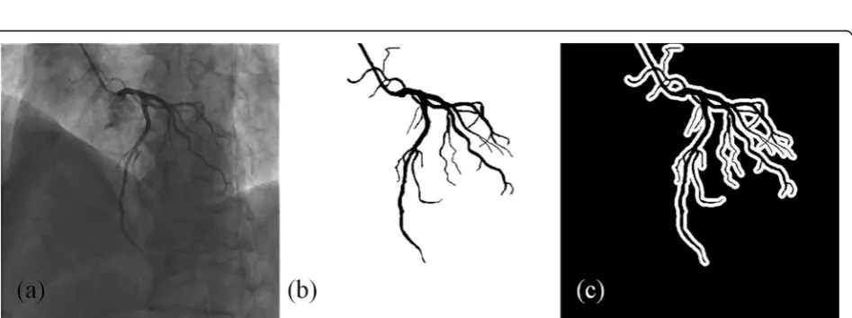
To quantitatively evaluate the proposed IFC-RPCA method, two kinds of masks are generated. One is used to evaluate the global vascular contrast and pixels within and outside the annotated vascular region consist of the vessels and background, as shown in Fig.1b. The other one is used to evaluate the local vascular contrast. As shown in Fig. 1c, vascular region is the same with the global mask, and the background region is comprised of the pixels in the white region, which is the 7-pixel-wide neighborhood of the vascular region boundary.
To evaluate the performance of the proposed method, it will be compared with the multiresolution elastic registration (MER) method [14], the online robust prin-cipal component analysis (ORPCA) method [19], the graduated RPCA with motion coherency constraint (MCR-RPCA) method [20]. To evaluate the proposed segmentation method, it will be compared with Fully-Connected Conditional Random Field (FC-CRF) method [10] and level-set-based method (LevelSet) [24].
Contrast-to-noise ratio (CNR) [18] is utilized to evalu-ate the vascular contrast of the vessels and can be de-fined as follows:
CNR¼jμF−μBj σB
ð10Þ
whereμFandμBare the mean gray values in the vascular and background regions in the extracted vascular im-ages. σB is the standard deviation of the gray values in the background region. We compute the global CNR and local CNR based on global mask and local mask, respectively.
To evaluate the proposed segmentation method, we utilize five metrics including the precision (pre), sensitiv-ity (sen) and F1 value. In addition, the metrics are com-puted as follows:
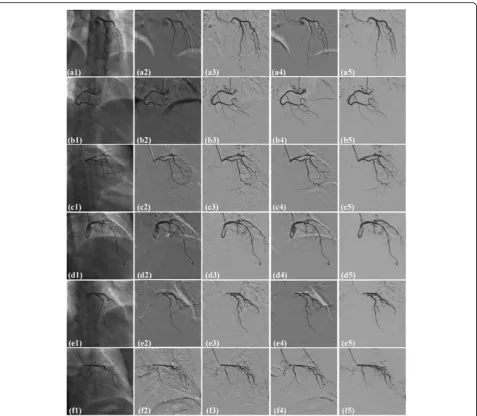
Fig. 3Comparison of the extracted vascular images on six randomly selected images from six different sequences by four methods. (a1-f1): original XRA images; (a2-f2), (a3-f3), (a4-f4) and (a5-f5): extracted vascular images by MER, ORPCA, MCR-RPCA and
IFC-RPCA methods
Table 1Comparison of local and global CNR by four different methods, including MER, ORPCA, MCR-RPCA and IFC-RPCA over 22 XRA images
Methods Local CNR Global CNR
Original Image 1.2175 ± 0.3838 0.8259 ± 0.2685
MER 0.1259 ± 0.0597 0.1709 ± 0.1143
ORPCA 3.8914 ± 0.5323 5.6527 ± 1.0719
MCR-RPCA 2.9081 ± 0.7021 4.8105 ± 1.3528
pre¼ TP TPþFP sen¼ TP
TPþFN F1¼ 2∙pre∙sen
preþsen
8 > > > > > < > > > > > :
ð11Þ
where TP, FP and FN indicate the true positive
(cor-rectly identified vessel pixels), false positive (incor(cor-rectly identified vessel pixels) and false negative (incorrectly identified background pixels), respectively.
Results
In our experiments, all the parameter settings are empir-ical. In detail, diameter of the circular structural element
d¼d′¼8:5=ð2pÞ;λ1¼λ2¼λ′1¼λ′2¼2:1=maxðM1;
M2Þ, r=r′= 5, p= 0.3, α1=α2= 0.5, M1 and M2 where are the size in each dimension of an image.
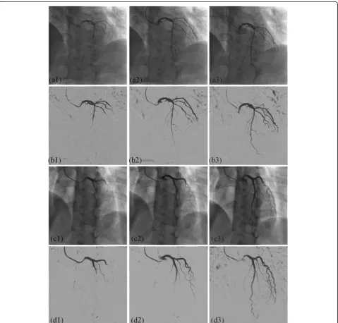
Figure 2 shows the extracted vascular images by the proposed IFC-RPCA method. The order numbers of the three random selected images in the first two rows are
17th, 23th, 31th. The order numbers of images in the last two rows are 19th, 24th and 41th, respectively. Ac-cording to the order, the inflow of contrast agent is gradually infused within the coronary artery. In Figs. 2(a1) and (c1), the contrast agent is not fully in-fused within the coronary artery. In Figs.2(a2) and (c2),
vessels are in the diastole stage, while in Figs.2(a3) and (c3), vessels are in the systole stage. As can be seen from the extracted vascular images, the vascular structures are preserved throughout the XRA sequences and present a very high contrast. In addition, motion artefacts and other non-vascular noise are also removed and vascular segments with small scales are also preserved.
Figure 3 shows the comparison results by four methods, including MER, ORPCA, MCR-PCA and IFC-RPCA, respectively. Six XRA images, as shown in Figs. 3(a1) to (f1), are randomly selected from six differ-ent sequences. In Figs.3(a2)-(f2), MER introduces much motion artefacts near diaphragm, catheter and other large intensity variation regions. In Figs. 3(a3)-(f3), ORPCA removes much non-vascular noise, but intro-duces serious motion artefacts around the catheter and diaphragm. In Figs. 3(a4)-(f4), MCR-RPCA produces strong artefacts near the vessels, diaphragm, and lung boundaries. Images in Figs. 3(a5)-(f5) are computed by the proposed IFC-RPCA method. Artefacts caused by the catheter, lung tissues, diaphragm and vessels are almostly removed.
Table 1compares the global CNR and local CNR over all the annotated XRA images by four different methods, including MER, ORPCA, MCR-RPCA and IFC-RPCA, respectively. All the methods obtain larger global CNRs than the original images and greatly improve the Fig. 4Simulated image with low dose contrast agent. (a) original image; (b) enhanced image; (c) simulated image
contrast of vessels. The global CNR of IFC-RPCA is much larger than MER and yields 17.37 and 37.91% im-provement by comparing with ORPCA and MCR-RPCA, respectively. The performance of IFC-RPCA is achieved by removing the artefacts near the vessels, catheters and non-vascular noise. For the local CNR, the values by MER is smaller than the local CNR of original images which demonstrates that MER cannot improve the con-trast within perivascular regions. IFC-RPCA obtains 10.20 and 47.46% improvement by comparing with ORPCA and MCR-RPCA, respectively. The proposed IFC-RPCA can also make the boundaries of the vascular structures much clearer.
We also simulate the angiograms with low dose con-trast agent which has significant clinical value for the cli-nicians and patients. The simulated images are generated by linearly subtracting the enhanced vascular image from the original image. Figure 4 shows an ex-ample of the simulated image with low dose contrast agent. As can be seen from Fig.4(c), the contrast of the vessels is greatly reduced.
Figure 5 shows the subtraction results based on the simulated images with low dose contrast agent. In Fig.5a, it is very difficult to distinguish the vascular structures from the background. While in Fig. 5c, the intensity of
the vessels is very close to the bones. As can be seen from Figs. 5b and d, the proposed IFC-RPCA method can improve the contrast of the vessels and meanwhile, remove the diaphragm and bones.
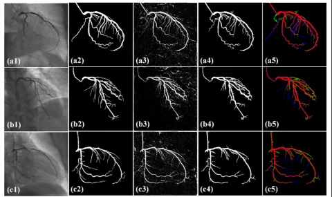
Fig.6 shows the segmented vascular structures by the proposed method. Angiograms in the first column are randomly selected from image sequences. Images in the third column are computed by the proposed feature de-scriptor. In the images, there are uniform responses when the vascular segments appear bifurcation, overlap-ping or are very close to each other. In addition, the re-sponses in vascular regions are much larger than the background which brings the vessels high contrast. Im-ages in the fourth column refer to the segmentation re-sults by a threshold value from images in the third column. In the images, the vascular edges are preserved even when different vascular segments are very close. In addition, vascular segments with small scales are also ac-curately segmented. In the fifth column, vascular seg-ments in green color refer to over-segmentation, while vascular segments in blue color refer to under-segmentation. As can be seen from the figures, the ves-sels will be fractured when the intensity of vascular seg-ments is close to the background. For the vessels with large scales, they have precise boundaries and are
consistent with the ground truth. As also can be seen from the segmented results, the non-vascular noise is al-most removed.
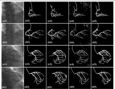
Figure7shows the comparison results by methods, in-cluding LevelSet, FC-CRF and the proposed model. In the first column, two right and two left coronary artery angiograms are randomly selected from the image se-quences. In the third column, vascular segments with large scales are precisely segmented and vascular seg-ments with small scales present serious noise. In addition, LevelSet method is semi-automatic and re-quires the manual labeled seed points. In the fourth col-umn, large amounts of noise appears in the non-vascular regions and there are many holes in the vascular seg-ments. After comparing with the ground truth, the di-ameters of the obtained vascular segments are smaller than the actual vessels which make a great influence to the subsequent parameter measurement of vascular seg-ments. In the fifth column, there are fractures when the
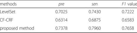
vascular segments appear small-scales. But the vascular segments with large scales are precisely segmented. In addition, when the vascular segment is overlapped with the diaphragm, the segmentation results doesn’t intro-duce motion artefacts. For each angiogram in the fig-ures, the segmentation results by the proposed method present a clean background without non-vascular noise. Table 2provides the quantitative comparison inpre, sen
and F1 value by LevelSet, FC-CRF and the proposed method. Pre, sen and F1 value of the proposed method are all the largest, the vessels and noise are both dis-posed best. All the metrics of LevelSet are all superior to those of FC-CRF. The vascular structure preservation and background noise removing are all better than these of FC-CRF.
Conclusion and discussion
In the paper, we propose an inter/intra-frame con-strained vascular segmentation method and demonstrate
its application in the XRA sequences. Experimental re-sults demonstrate the effectiveness of the proposed method in accurate vessel segmentation in the XRA se-quence. As can be seen from the experimental results, the proposed IFC-RPCA effectively reduces the lung tis-sues, diaphragm and vertebral bodies and removes the motion artefacts near the catheter and non-vascular noises from the XRA sequence. The proposed IFC-RPCA yields 17.37 and 37.79% improvement in global CNR and 10.20 and 47.46% improvement by comparing with ORPCA and MCR-RPCA methods. The proposed multi-feature fused feature descriptor produces uniform response in different vascular segments and makes the vascular structures high contrast with the background. Based on this, the vascular structures can be simply seg-mented with only a threshold value. We obtain 0.7378, 0.7960 and 0.7658 with respect to the precision, sensitiv-ity and F1 value, respectively. It demonstrates the pro-posed method can both effectively dispose the vessels and background. The proposed vessel segmentation method is online without re-optimizing the whole model and automatic, it is very suitable to be applied in the intra-operative image guided surgical navigation.
Abbreviations
CAD:Coronary artery disease; CNN: Convolutional neural network; CNR: Contrast-to-noise ratio; CRF: Conditional random field; FC-CRF: Fully-connected conditional random field; FN: False negative; FP: False positive; IFC-RPCA: Inter/infra-frame constrained RPCA; LevelSet: Level-set-based method; MCR-RPCA: The graduated RPCA with motion coherency constraint; MER: Multiresolution elastic registration; ORPCA: Online robust principal component analysis; pre: Precision; RPCA: Robust principal component analysis; sen: Sensitivity; TP: True positive; XRA: X-ray angiography
Acknowledgements
The authors would like to thank the staff of Peking Union Medical College Hospital for welcoming and assisting the research team and the interview participants for their time.
About this supplement
This article has been published as part ofBMC Medical informatics and Decision Making Volume 19 Supplement 6, 2019: Selected articles from the IEEE BIBM International Conference on Bioinformatics & Biomedicine (BIBM) 2018: medical informatics and decision making. The full contents of the supplement are available online athttps://bmcmedinformdecismak.biomedcentral.com/ articles/supplements/volume-19-supplement-6.
Authors’contributions
SS and YJ conceived and conducted the experiments; SS, DC, CY and AD analyzed the results and wrote the paper; YJ, AD, SH, HY and WY reviewed the manuscript and provided many thoughtful suggestions to improve the manuscript. All authors read and approved the final manuscript.
Funding
Publication costs are funded by the National Key R&D Program of China (2017YFC0112000) and the National Science Foundation Program of China (81627803, 61501030, 61672099).
Availability of data and materials
The data is not shared with outside institutions.
Ethics approval and consent to participate
The study was conducted in accordance with the Declaration of Helsinki, and was approved by the local (Peking Union Medical College Hospital) ethics committee. Informed consent to participate in the study was obtained from all participants. Data access was granted in the context of project National Key R&D Program of China (2017YFC0112000) and the National Science Foundation Program of China (81627803, 61501030, 61672099), where the authors’institutions participate.
Consent for publication
Not applicable.
Competing interests
The authors declare that they have no competing interests.
Author details
1Beijing Engineering Research Center of Mixed Reality and Advanced Display, School of Optics and Photonics, Beijing Institute of Technology, Beijing 100081, China.2AICFVE of Beijing Film Academy, 4 Xitucheng Rd, Haidian, Beijing 100088, China.3School of Computer Science & Technology, Beijing Institute of Technology, Beijing 100081, China.
Published: 19 December 2019
References
1. Cavaye D, White R. Imaging technologies in cardiovascular interventions. J Cardiovasc Surg. 1993;34:13–22.
2. Wang C, Moreno R, Smedby Ö. Vessel segmentation using implicit model-guided level sets. In: MICCAI Workshop" 3D Cardiovascular Imaging: a MICCAI segmentation Challenge", Nice France, 1st of October 2012; 2012. 3. Sun K, Chen Z, Jiang S. Local morphology fitting active contour for
automatic vascular segmentation. IEEE Trans Biomed Eng. 2012;59:464–73. 4. Cheng Y, Hu X, Wang J, Wang Y, Tamura S. Accurate vessel segmentation
with constrained B-snake. IEEE Trans Image Process. 2015;24:2440–55. 5. Lee S-H, Lee S. Adaptive Kalman snake for semi-autonomous 3D vessel
tracking. Comput Methods Prog Biomed. 2015;122:56–75. 6. Hassouna MS, Farag AA, Hushek S, Moriarty T. Cerebrovascular
segmentation from TOF using stochastic models. Med Image Anal. 2006;10:2–18.
7. Goceri E, Shah ZK, Gurcan MN. Vessel segmentation from abdominal magnetic resonance images: adaptive and reconstructive approach. Int J Numer Method Biomed Eng. 2017;33:e2811.
8. Lupascu CA, Tegolo D, Trucco E. FABC: retinal vessel segmentation using AdaBoost. IEEE Trans Inf Technol Biomed. 2010;14:1267–74.
9. Orlando JI, Blaschko M. Learning fully-connected CRFs for blood vessel segmentation in retinal images. In: international conference on medical image computing and computer-assisted intervention; 2014. p. 634–41. 10. Orlando JI, Prokofyeva E, Blaschko MB. A discriminatively trained fully
connected conditional random field model for blood vessel segmentation in fundus images. IEEE Trans Biomed Eng. 2017;64:16–27.
11. Fu H, Xu Y, Lin S, Wong DWK, Liu J. Deepvessel: Retinal vessel segmentation via deep learning and conditional random field. In: International Conference on Medical Image Computing and Computer-Assisted Intervention; 2016. p. 132–9.
12. Luo Y, Yang L, Wang L, Cheng H. Efficient CNN-CRF network for retinal image segmentation. In: International Conference on Cognitive Systems and Signal Processing; 2016. p. 157–65.
13. Bentoutou Y, Taleb N. Automatic extraction of control points for digital subtraction angiography image enhancement. IEEE Trans Nucl Sci. 2005;52: 238–46.
14. Yang J, Wang Y, Tang S, Zhou S, Liu Y, Chen W. Multiresolution elastic registration of X-ray angiography images using thin-plate spline. IEEE Trans Nucl Sci. 2007;54:152–66.
Table 2Quantitative segmenation comparison of the proposed method with CF-CRF and LevelSet
methods pre sen F1value
LevelSet 0.7025 0.7430 0.7222
CF-CRF 0.6314 0.6875 0.6583
15. Zhu Y, Prummer S, Wang P, Chen T, Comaniciu D, Ostermeier M. Dynamic layer separation for coronary DSA and enhancement in fluoroscopic sequences. In: International Conference on Medical Image Computing and Computer-Assisted Intervention; 2009. p. 877–84.
16. Zhang W, Ling H, Prummer S, Zhou KS, Ostermeier M, Comaniciu D. Coronary tree extraction using motion layer separation. In: International Conference on Medical Image Computing and Computer-Assisted Intervention; 2009. p. 116–23.
17. Fischer P, Pohl T, Köhler T, Maier A, Hornegger J. A Robust Probabilistic Model for Motion Layer Separation in X-ray Fluoroscopy, vol. 9123; 2015. p. 288–99.
18. Ma H, Dibildox G, Banerjee J, Niessen W, Schultz C, Regar E, et al. Layer separation for vessel enhancement in interventional X-ray angiograms using morphological filtering and robust PCA. In: Workshop on Augmented Environments for Computer-Assisted Interventions; 2015. p. 104–13. 19. Ma H, Hoogendoorn A, Regar E, Niessen WJ, van Walsum T. Automatic
online layer separation for vessel enhancement in X-ray angiograms for percutaneous coronary interventions. Med Image Anal. Jul 2017;39:145–61. 20. Jin M, Li R, Jiang J, Qin B. Extracting contrast-filled vessels in X-ray
angiography by graduated RPCA with motion coherency constraint. Pattern Recogn. 2017;63:653–66.
21. Feng J, Xu H, Yan S. Online robust pca via stochastic optimization. Adv Neural Inf Proces Syst. 2013;1:404–12.
22. Shoujun Z, Jian Y, Yongtian W, Wufan C. Automatic segmentation of coronary angiograms based on fuzzy inferring and probabilistic tracking. Biomed Eng Online. 2010;9:40.
23. Jerman T, PernušF, Likar B,ŠpiclinŽ. Enhancement of vascular structures in 3D and 2D angiographic images. IEEE Trans Med Imaging. 2016;35:2107–18. 24. Läthén G, Jonasson J, Borga M. Blood vessel segmentation using multi-scale
quadrature filtering. Pattern Recogn Lett. 2010;31:762–7.
Publisher’s Note