INTRODUCTION
Sepsis is a complex systemic inflamma-tory clinical syndrome that occurs be-cause of an overwhelming inflamma-tory/immune response to infection and is associated with the so-called systemic in-flammatory response syndrome (SIRS) (1).
This multifaceted inflammatory activation occurs via stimulation of the host immune effector cells, leading to the release of chemokines, cytokines and reactive oxy-gen/nitrogen species. This process func-tions properly when these mediators are restricted to specific tissues, wherein local
injury or infection induces a well-regu-lated inflammatory response. However, once the levels of these cytokines rise suf-ficiently so that they begin to appear in the bloodstream at levels capable of in-ducing a systemic response, inflammation takes on many of its harmful characteris-tics. Sustained elevations of both pro- and antiinflammatory cytokines, and presum-ably their complex interactions with in-flammatory, endothelial and parenchymal cells, identify those sepsis patients who subsequently develop multiple organ dys-function syndrome (MODS), shock and death (2).
This complex immunobiology may help explain the failure of therapeutic
at-Gram-negative Septic Peritonitis: Insights from In Vivo and
In Silico Studies
Rami A Namas,
1,2Rajaie Namas,
1,3Claudio Lagoa,
1Derek Barclay,
1Qi Mi,
2,3Ruben Zamora,
1,2Zhiyong Peng,
5Xiaoyan Wen,
5Morgan V Fedorchak,
6,7Isabella E Valenti,
6,7William J Federspiel,
5,6,7,8John A Kellum,
2,5,6,7and Yoram Vodovotz
1,21Department of Surgery; 4Department of Sports Medicine and Nutrition; 5CRISMA (Clinical Research, Investigation and Systems Modeling in Acute illness) Center, Department of Critical Care Medicine; 6Department of Bioengineering; and 8Department of Chemical and Petroleum Engineering, University of Pittsburgh, Pittsburgh, Pennsylvania, United States of America; 2Center for Inflammation and Regenerative Modeling, McGowan Institute for Regenerative Medicine; and 7McGowan Institute for Regenerative Medicine, Pittsburgh, Pennsylvania, United States of America; and 3current address: Department of Internal Medicine, Michigan State University College of Human Medicine, Flint, Michigan, United States of America
Improper compartmentalization of the inflammatory response leads to systemic inflammation in sepsis. Hemoadsorption (HA) is an emerging approach to modulate sepsis-induced inflammation. We sought to define the effects of HA on inflammatory com-partmentalization in Escherichia coli–induced fibrin peritonitis in rats. Hypothesis: HA both reprograms and recompartmentalizes inflammation in sepsis. Sprague Dawley male rats were subjected to E. coli peritonitis and, after 24 h, were randomized to HA or sham treatment (sepsis alone). Venous blood samples collected at 0, 1, 3 and 6 h (that is, 24–30 h of total experimental sepsis), and peritoneal samples collected at 0 and 6 h, were assayed for 14 cytokines along with NO2–/NO
3–. Bacterial counts were
as-sessed in the peritoneal fluid at 0 and 6 h. Plasma tumor necrosis factor (TNF)-α, interleukin (IL)-6, CXCL-1, and CCL2 were signifi-cantly reduced in HA versus sham. Principal component analysis (PCA) suggested that inflammation in sham was driven by IL-6 and TNF-α, whereas HA-associated inflammation was driven primarily by TNF-α, CXCL-1, IL-10 and CCL2. Whereas peritoneal bac-terial counts, plasma aspartate transaminase levels and peritoneal IL-5, IL-6, IL-18, interferon (IFN)-γand NO2–/NO
3–were
signifi-cantly lower, both CXCL-1 and CCL2 as well as the peritoneal-to-plasma ratios of TNF-α, CXCL-1 and CCL2 were significantly higher in HA versus sham, suggesting that HA-induced inflammatory recompartmentalization leads to the different inflammatory drivers discerned in part by PCA. In conclusion, this study demonstrates the utility of combined in vivo/in silico methods and suggests that HA exerts differential effects on mediator gradients between local and systemic compartments that ultimately benefit the host.
Online address: http://www.molmed.org doi: 10.2119/molmed.2012.00106
Address correspondence toYoram Vodovotz, Department of Surgery, University of Pittsburgh, W944 Biomedical Sciences Tower, 200 Lothrop Street, Pittsburgh, PA 15213. Phone: 412-647-5609; Fax: 412-383-5946; E-mail: [email protected].
tempts to control inflammation via blocking a single inflammatory mediator (3). An alternative strategy involves the removal of excessive inflammatory medi-ators from the circulation. Blood purifica-tion therapy techniques that use an ex-tracorporeal device can remove a wide array of inflammatory mediators from circulating blood nonspecifically (4–6). This multimediator targeting may pro-mote the downregulation of systemic inflammation and assist the body in re-gaining homeostasis (7).
We previously described an extracor-poreal hemoadsorption (HA) device that can nonspecifically remove low– molecular weight proteins (including cy-tokines and chemokines) from circulating blood by using biocompatible sorbent beads (CytoSorb; CytoSorbents, Prince-ton, NJ, USA). CytoSorb HA beads are polystyrene-divinylbenzene porous par-ticles (450 µm average particle diameter, 0.8–5 nm pore diameter, 850 m2g–1 sur-face area) with a biocompatible polyvinyl-pyrrolidone coating. Adsorp-tion to the internal pore surface is accom-plished by a putative combination of nonspecific hydrophobic interactions, as well as size exclusion of large–molecular weight solutes >70 kDa, such as albumin and immunoglobulins. This device has been effective in removing tumor necro-sis factor (TNF-α), interleukin (IL)-6 and IL-10 and other middle–molecular weight proteins in both in vitroand ex vivostudies and at improving survival in experimental models of endotoxemia and sepsis (8,9). In the present study, we tested HA in a rat model of E. coli– induced fibrin clot peritonitis and sought to determine the principal components responsible for the effects of HA on acute inflammation accompanying experimen-tal sepsis.
MATERIALS AND METHODS
Surgical Preparation and Fibrin Clot Implantation
The study was approved by the Uni-versity of Pittsburgh Institutional Animal Care and Use Committee and conforms
to U.S. National Institutes of Health guidelines for the care and use of labora-tory animals. After acclimatization for 5 d, adult male Sprague Dawley rats (n = 21, 24–28 wks old, 430–480 g body weight, from Harlan Laboratories) were anesthetized with pentobarbital sodium (40 mg/kg intraperitoneally). The ani-mals were then subjected to an Escheri -chia coli(strain ATCC 25922; American Type Culture Collection, Manassas, VA, USA) inoculum in a fibrin clot (1.5 ×10⁸
to 2 ×10⁸colony-forming units [CFUs]/ clot; see details below) introduced into the peritoneum via laparotomy (10). Twenty-four hours later, the rats were re-anesthetized and the femoral and the in-ternal jugular veins were isolated by dis-section and cannulated with 0.97-mm polyethylene 50 tubing, for use in the ex-tracorporeal circuit.
The rats were then treated with HA or a sham circuit (n = 8 rats each) for 6 h, for a total of 30 h of experimental sepsis,
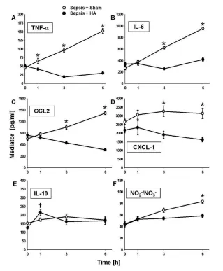
Figure 1.Effects of HA on circulating levels of inflammatory mediators. Postcannulation baseline values (that is, 24 h after implantation of an E. coli–impregnated fibrin clot and hence established sepsis) for all cytokines were not significantly different between the two experimental groups. Circulating TNF-α(A), IL-6 (B), CCL2 (C) and CXCL-1 (D) were signifi-cantly lower in the HA group versus the sham group (*p < 0.05 versus sham group; †p <
0.05 versus HA at 0 h). Circulating NO2–/NO3–after 6 h of treatment was significantly lower
(F) (*p < 0.001 versus sham; †p < 0.05 versus HA at 0 h). Note: Some error bars are not
as follows. All the tubing in the extracor-poreal circulation was flushed with he-parinized saline (5 units/mL). The extra-corporeal circuit was started from the femoral vein and returned back to the ip-silateral internal jugular vein by action of a mechanic minipump (Fisher Scientific). For HA, a 1.5-g CytoSorb cartridge (Cytosorbents) with a dead space of 0.3–0.4 mL was placed in the circuit of animals randomized to the HA group, whereas no cartridge was used in the sham group. Blood flow was maintained by the minipump at a rate of 0.8–1.0 mL/ min and regulated by a stopcock at the precartridge side. Both the size of the de-vice and the blood flow rate were scaled to those used for humans (approximately a 300-g device on the basis of body weight with a blood flow of 100–150 mL/ min on the basis of comparable blood volume). Blood samples (0.4 mL) were collected from the internal jugular vein at 0, 1, 3 and 6 h (that is, 24–30 h of total experimental sepsis). After blood with-drawal, the catheters were flushed with an equal volume of heparinized saline (5 U/mL). After 6 h, the treatment was stopped and an intraperitoneal fluid sample was collected by injecting 10 mL sterile isotonic saline into the peritoneum via an 18-gauge needle. The abdomen was gently manipulated and the peri-toneal fluid was aspirated for cytokine assay (n = 6, HA or sham) and total bac-terial count (n = 8, HA or sham). The rats were then euthanized by administering an overdose of pentobarbital sodium fol-lowed by cervical dislocation.
For the control animals, a separate group of animals (n = 9) were subjected to a laparotomy incision followed by implantation of a fibrin clot that lacks
E. coli. After 24 h after implantation of the fibrin clot, the animals were eutha-nized and blood samples were collected via a cardiac puncture.
Preparation and Dose Estimation of E. Coli–Impregnated Fibrin Clot
Bacteria were quantified using a spec-trophotometer (DU 530 UV/VIS; Beck-man Coulter, Brea, CA, USA) on the day
of bacterial fibrin clot implantation. A portion of this bacterial culture was added to a Lysogeny broth agar plate and incubated overnight at 37°C to obtain a count of implanted E. coliin a given rat. We determined that rats that received a fibrin clot containing 0.8 ×10⁸to 1.2 × 10⁸CFUs/clot did not show signs of sep-ticemia 24 h after implantation, but rats that received an inoculum of 1 ×10⁸to 2.0 ×10⁸CFUs/clot had a mortality rate of ~40–45% after 48 h of clot implantation (data not shown). An E. coliinoculum of 1.5 ×10⁸to 2.0 ×10⁸induced lethargy, hy-pothermia, reluctance to feed, tachycardia and tachypnea in rats, with a mortality rate of ~20–25% at 24 h (data not shown). Rats that received inocula higher than 2.0 ×10⁸CFUs/clot had a mortality rate of 80% during the first 24 h of implanta-tion (data not shown). Thus, we chose an
E. coliinoculum ranging from 1.5 ×10⁸to 2.0 ×10⁸CFUs/clot for the present study.
Blood Collection and Cytokine, NO2–/NO
3–and Aspartate Transaminase (AST)/Alanine Aminotransferase (ALT) Analysis
Blood (0.4 mL) was withdrawn from HA-treated and sham rats from the inter-nal jugular vein line at time 0 (24 h after implantation of the E. coli–impregnated fibrin clot) and 1, 3 and 6 h later. Plasma samples (n = 8 per group) were pre-pared and stored at –80°C until analysis.
Plasma cytokines were measured with a multiplex bead immunoassay system (Luminex; Millipore, Billerica, MA, USA). The cytokine assays included granulocyte/ macrophage colony-stimulating factor (GM/CSF), keratinocyte-derived
chemokine (GRO/KC, CINC-1, CXCL-1), interferon (IFN)-γ, IL-1α, IL-1β, IL-2, IL-4, IL-5, IL-6, IL-10, IL-12 p70, IL-18, monocyte chemoattractant protein-1 (MCP-1, CCL2) and TNF-α. Plasma NO2–/NO3–was measured by the nitrate reductase method using a commercially available kit (Cayman Chemical, Ann Arbor, MI, USA). AST and ALT were measured using a commercially available kit (Fujifilm, Asaka-shi, Satama, Japan; distributed by Heska Corporation, Love-land, CO, USA) according to the manu-facturer’s instructions.
Peritoneal Fluid Collection and Analyses
To determine baseline levels of peri-toneal cytokines and bacterial count at 0 h, an E. coli–impregnated fibrin clot was implanted in 12 separate rats that were not included in the total number of rats used for the two major experimental groups (HA and sham). After 30 h of sep-sis (immediately after euthanasia), 10 mL sterile isotonic saline was injected into the peritoneum. The peritoneal irrigation fluid was recovered after a laparotomy for cytokine assay (n = 6 per group) and
bacterial count (n = 8 per group) (11). A portion of the peritoneal fluid was streaked on an agar plate and incubated overnight at 37°C. After 24 h incubation, bacterial colonies were quantified as de-scribed above. The remaining peritoneal fluid was then stored at –80°C for cyto -kine assay (see above).
Statistical Analyses
All data are expressed as mean ± stan-dard error of the mean. Statistical analy-sis was performed by one-way analyanaly-sis of variance on ranks followed by the Holm-Sidak test, using SigmaPlot 11 soft-ware (Systat Softsoft-ware, San Jose, CA, USA), with p< 0.05 considered signifi-cant. A power calculation was performed
and suggested that n = 8 rats was a co-hort size sufficient to observe statistically significant differences in inflammatory mediators between treatment groups. Principal component analysis (PCA) was carried out essentially as described re-cently (12) using MATLAB software 7.6.0 (MathWorks, Natik, MA, USA).
All supplementary materials are available online at www.molmed.org.
RESULTS
Overview
In total, 21 animals were implanted with E. coli–impregnated fibrin clots; of these, 5 animals died after 24 h of the
fib-rin clot implantation (23.8% mortality), but before randomization to HA or sham circuit groups. Accordingly, these rats were excluded from further analysis. The remaining 16 animals were randomized to HA (n = 8) or sham (n = 8). The levels of circulating inflammatory mediators in the control group 24 h after implantation of a fibrin clot lacking E. colicompared with circulating inflammatory mediators of the sham group are shown in Supple-mentary Figure S1.
Effects of HA on Circulating Levels of Inflammatory Mediators and Markers of Liver Damage
We initially sought to determine if HA altered the balance of inflammatory
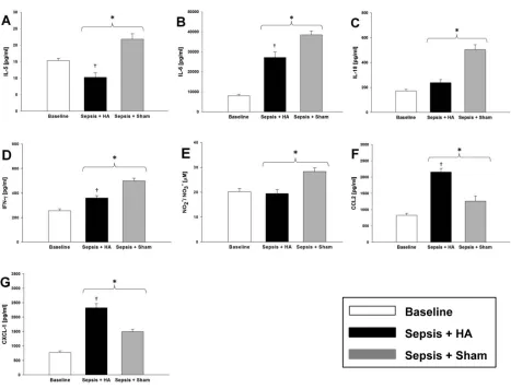
me-Figure 3.Effects of HA on peritoneal inflammatory mediators and bacteria. Rats were subjected to sepsis by the implantation of a fibrin clot impregnated with E. coli followed by sham or HA treatment, and peritoneal fluid was obtained as descibed in the Materials and Methods. Peri-toneal IL-5 (A), IL-6 (B), IL-18 (C), IFN-γ(D) and NO2–/NO
3–(E) were significantly lower (n = 6 rats; *p < 0.05 versus sham group; †p < 0.05 versus
diators in the setting of septic peritonitis; accordingly, we examined the effect of HA on circulating cytokines. The differences in circulating plasma cytokines for animals treated with HA or sham (n = 8 per group) are shown in Figure 1. Postcannulation baseline values (that is, 24 h after implan-tation of an E. coli–impregnated fibrin clot and hence established sepsis) for all cy-tokines were not significantly different between the two experimental groups. However, the concentrations of TNF-α (Figure 1A), IL-6 (Figure 1B), CCL2 (Fig-ure 1C) and CXCL-1 (Fig(Fig-ure 1D) were significantly lower in the HA group after
treatment compared with the sham group. Moreover, after only 1 h of the treatment, the concentrations of TNF-α (see Figure 1A), IL-6 (see Figure 1B) and CCL2 (see Figure 1C) were signifi-cantly lower in the HA group. At 3 and 6 h, TNF-α(see Figure 1A), IL-6 (see Figure 1B), CCL2 (see Figure 1C) and CXCL-1 (see Figure 1D) were still lower in the HA group compared with sham. Interestingly, circulating IL-10 levels were significantly higher (p = 0.002) after 1 h of treatment in the HA group com-pared with baseline, whereas the levels of IL-10 in sham animals were not
(Fig-ure 1E). However, there was no statisti-cally significant difference in IL-10 levels between HA and sham at any time point. The concentration of circulating NO2–/ NO3–at 6 h of treatment was signifi-cantly lower in the HA group when com-pared with the sham group (Figure 1F). The levels of circulating IFN-γ, IL-1β, IL-12 p70, IL-5, IL-18, GM/CSF and IL-1αwere unchanged over time in both sham and HA animals, whereas levels of circulating IL-4 and IL-2 were undetect-able in both groups (Supplementary Fig-ure S2). Circulating levels of AST were significantly lower (p = 0.041) in the HA
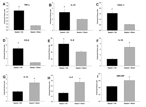
Figure 4.Ratios of local (peritoneal) mediators to systemic concentrations at 6 h for all the mediators that were measured in both sites. Rats were subjected to sepsis by the implantation of a fibrin clot impregnated with E. coli followed by sham or HA treatment, and plasma as well as peritoneal fluid was obtained as described in the Materials and Methods. Animals in the HA group had significantly higher lo-calized TNF-α(*p = 0.0010 versus sham) (A), IL-10 (*p = 0.040 versus sham) (B), CXCL-1 (*p = 0.001 versus sham) (C), CCL2 (*p = 0.001 ver-sus sham) (D) and IL-6 (*p = 0.029 verver-sus sham) (E). The sham group had significantly higher ratios of IL-18 (*p = 0.001 verver-sus HA) (F), IL-1β
group (186.25 ± 18.98 units/liter [U/L]) versus the sham group (252 ± 22.42 U/L) at 6 h after treatment. However, there was no statistically significant difference (p = 0.075) in plasma ALT between treat-ment groups (117.5 ± 3.65 U/L in HA versus 138.7 ± 10.42 U/L in sham group).
PCA Suggests Different Drivers of Inflammation in HA Versus Sham
We next used PCA in an attempt to identify the subsets of circulating media-tors that are most strongly indicative of membership in the HA or sham treat-ment group and that thereby might be considered principal drivers of the in-flammatory response pattern seen in each group. PCA suggested that the cir-culating inflammatory response in the sham group was primarily driven by IL-6 and TNF-α(Figure 2A), whereas the sepsis response in the HA group was pri-marily driven by TNF-α, CXCL-1, IL-10 and CCL2 (Figure 2B). This analysis sug-gests that, in this animal model (and in the time range studied), TNF-α_and IL-6 are principal drivers of sepsis and that HA modifies this process via CXCL-1,
IL-10 and CCL2 (and with a concomi-tantly reduced role for IL-6).
Effects of HA on Peritoneal
Inflammatory Mediators and Bacteria We next hypothesized that removal of mediators from the plasma by HA would not lead to reduced local inflammatory mediator concentrations and that, by in-creasing the ratio of local to systemic concentrations, we would observe im-proved bacterial clearance (13–17). We tested these hypotheses by examining the peritoneal fluid levels of inflamma-tory mediators (n = 6 per group) as well as changes in peritoneal bacterial counts (n = 8 per group). Figure 3 shows the dif-ference in concentrations of peritoneal cytokines in both HA and sham groups at 6 h (that is, 30 h after implantation of an E. coli–impregnated fibrin clot) versus the baseline at 0 h (24 h after clot implan-tation). The peritoneal fluid concentrations of IL5 (Figure 3A), IL6 (Fig -ure 3B), IL-18 (Fig-ure 3C) and IFN-γ (Figure 3D) and the stable NO reaction products NO2–/NO
3–(Figure 3E) were significantly lower in the HA group when compared with the sham group. Interestingly, CCL2 (Figure 3F) and CXCL-1 (Figure 3G) concentrations in the peritoneal fluid were significantly higher in the HA group when compared with the sham group. Peritoneal IL-2 (Supple-mentary Figure S3A) and GM-CSF (Sup-plementary Figure S3B) were elevated in the HA animals at 6 versus 0 h, but no statistically significant differences were observed between HA and sham.
The PCA of plasma inflammatory me-diators suggested that HA was associ-ated with effects on TNF-α, CXCL-1, IL-10 and CCL2. Because a healthy in-flammatory response remains localized, whereas a hallmark of detrimental in-flammation in sepsis manifests in the systemic spillover of inflammatory medi-ators (18), we hypothesized that HA may act in some fashion to maintain localized levels of these principal mediators and reduce their systemic spillover. To test this hypothesis, we calculated the ratios of local (peritoneal) mediators to
sys-temic concentrations at 6 h for the all me-diators that were detectable in both sites. This analysis demonstrated that animals in the HA group had significantly higher localized TNF-α(p = 0.0010; Figure 4A), IL-10 (p = 0.040; Figure 4B), CXCL-1 (p = 0.001; Figure 4C), CCL2 (p = 0.001; Fig -ure 4D) and IL-6 (p = 0.029; Figure 4E). In contrast, the sham group had signifi-cantly higher ratios of IL-18 (p = 0.001; Figure 4F), IL-1β(p = 0.003; Figure 4G), IL-5 (p = 0.001; Figure 4H) and GM-CSF (p = 0.005; Figure 4I). There was no sig-nificant difference in the peritoneal/ plasma ratios of IL-1αand IFN-γ (Sup-plementary Figure S4).
We next hypothesized that the altered local inflammatory response after HA treatment would result in reduced local bacterial burden. Figure 5 shows the peritoneal bacterial count in both HA and sham groups at 6 h versus the base-line at 0 h. As can be seen in Figure 5, peritoneal bacterial counts were signifi-cantly lower in the HA group when com-pared with the sham group.
DISCUSSION
The inflammatory response is com-partmentalized both structurally and across multiple scales of organization (19–22). We recently hypothesized that acute inflammation proceeds at a given “nested” level or scale until positive feedback exceeds a “tipping point.” Below this tipping point, inflammation is contained and manageable; when this threshold is crossed, inflammation be-comes disordered and dysfunction prop-agates systemically (23,24). Thus, circu-lating inflammatory mediators, including cytokines and free radical reaction prod-ucts such as NO2–/NO
3–, play an impor-tant role in the pathophysiology of sepsis (1). In patients with established sepsis, both proinflammatory and antiinflamma-tory mediators coexist in the circulation in markedly increased amounts (25–30), whereas paradoxically, the capacity to combat infection or to mount appropri-ate inflammatory responses is blunted (31). On the basis of these observations, we and others hypothesized that
re-Figure 5.Peritoneal bacterial count in HA and sham groups at 6 h versus the base-line at 0 h. Rats were subjected to sepsis by the implantation of a fibrin clot im-pregnated with E. coli followed by sham or HA treatment, and peritoneal fluid was obtained as described in the Materials and Methods. Peritoneal bacterial counts were significantly lower (n = 8 rats; *P < 0.001 versus sham) in the HA group (7 ×
108± 3.0
×107CFUs/mL) when compared
with the sham group (9.4 ×10⁸± 5.3
×107
moval of inflammatory mediators would be beneficial in sepsis. Indeed, HA has demonstrated efficacy in animal models (6,8) and is currently in clinical trials for the treatment of sepsis (clinicaltrials.gov, NCT00559130).
The primary goal of the present study was to begin to test the hypothesis that some of the benefit of HA could be ascribed to reprogramming compartmentalized and dysregulated inflammation in sepsis, by defining the effects of HA on the nature, intensity and compartmentalization of inflammatory mediators. This experimental model is associated with a clinically realistic mor-tality of 25–30%. Our results show that HA with the CytoSorb polymer can effectively remove proinflammatory cyto -kines (TNF-αand IL-6) as well as chemokines (CCL2 and CXCL-1) from the circulation, while also resulting in lower IL-5, IL-6, IL-18, IFN-γand NO2–/NO3–(along with higher levels of CXCL-1 and CCL2) in the peritoneal fluid after 6 h of HA.
Except for circulating IL-10 and IL-1β, which did not show any statistical signif-icance in HA versus sham treatment, the present results are consistent with other studies on the effects of HA on cytokine removal, both in ex vivoand rat cecal lig-ation and perforlig-ation models. The likely explanation is that, since molecular cap-ture with the HA device depends on con-centration, molecules with lower concen-trations will be captured with lower efficacy compared with those with higher concentrations. Various experi-mental models of sepsis have differences in cytokine expression, and this particu-lar model is associated with a relatively low expression of IL-10. Similar to the rat cecal ligation and perforation model, our septic peritonitis model is one of true in-fection, thereby approximating human sepsis. We also note that HA treatment did affect the ratio of plasma to peri-toneal IL-10, suggesting that compart-mental effects of this cytokine may be more important than systemic effects. Importantly, the mortality rate in our an-imal model that was seen at 20–24 h after
introducing the E. coli–impregnated fib-rin clot and immediately before estab-lishing the extracorporeal circuit (ap-proximately 25–30%) is similar to mortality rates seen in the human inten-sive care unit (ICU) patients with sepsis (32). However, it is important to note that these patients receive antibiotic and fluid therapy, unlike in our animal model. Moreover, the timing of mortality is different between our animal model and the clinical setting. We also note that HA treatment was initiated 24 h after the implantation of an E. coli–impregnated fibrin clot and continued until 30 h postimplantation, reflecting a clinically realistic timing of therapy. One advan-tage of the animal model used in the present study over cecal ligation and perforation is the ability to quantify both input and final numbers of bacteria, and we were thus able to demonstrate a sig-nificant reduction in peritoneal bacteria in HA versus sham.
To better understand the effects of HA on inflammation in our rat model, we sought to leverage the insights gained from data-driven analyses into quasi-mechanistic insights regarding the dy-namics of inflammation after sepsis. We therefore used PCA to identify the sub-sets of mediators that are most strongly indicative of HA or sham and that thereby might be considered principal drivers of the inflammatory response pattern seen in each group. Importantly, PCA is on the basis of time-dependent changes in variance (33); therefore, we hypothesized that this analysis would yield insights into the dynamic responses of the various inflammatory mediators. We recently demonstrated the utility of this method, along with other data-driven analyses, in suggesting the princi-pal drivers of trauma/hemorrhagic shock in mice (12). In the present study, PCA suggested that IL-6 and TNF-αare the principal circulating drivers of sepsis (as seen in the sepsis + sham experimen-tal group) and that HA modifies this pro-cess via CXCL-1, IL-10 and CCL2 (with a reduced contribution of IL-6). We con-firmed this computationally derived
hy-pothesis, in part, in that the peritoneal-to-plasma ratios of TNF-α, IL-10, CXCL-1 and CCL2 were higher in the HA group. However, the peritoneal-to-plasma ratio for IL-6 was also higher in the HA group compared with sham; the reason and po-tential role for elevated local IL-6 is at this point unclear, but these findings sug-gest that HA does more than simply re-move cytokines. Rather, this treatment appears to lead to inflammatory repro-gramming and recompartmentalization. This effect is also consistent with the re-sults of a recent study from our group, where we demonstrated reduced organ injury and improved survival with HA in CLP sepsis, even when we scaled the intervention back to a level at which typ-ical circulating inflammatory mediators (for example, TNF-αand IL-6) were not affected (34).
Although at first glance it would ap-pear that the simplest explanation for re-duced inflammatory mediators in the plasma is linked to the reduced bacterial counts in the peritoneum, it is not obvi-ous how this would have happened when the HA therapy (which to our knowledge has no direct antibiotic effect) was administered to the systemic circula-tion rather than to the peritoneal space. We hypothesize that HA helps to main-tain relevant inflammatory mediators in the peritoneal space and thereby reduces the number of bacteria. Presumably, as bacterial numbers decrease in the peri-toneum, the forward-feedback proinflam-matory loop driven by peritoneal bacteria results in further decreases in circulating inflammatory mediators. Although this hypothesis is consistent with our data, al-ternative explanations such as sequestra-tion of inflammatory mediators and bac-teria to other organs/compartments are also consistent with these data. In addi-tion, it is possible that, whereas liver damage was ameliorated by HA, that other measures of organ pathophysiology (for example, hemodynamic parameters) were not improved. Further studies will be required to examine these issues.
the hypothesis that reprogramming and recompartmentalization of induced inflammation resulted in im-proved bacterial clearance and reduced liver injury. We interpret these results as being consistent, since better leukocyte homing to the nidus of infection would have decreased bacterial counts and re-duced remote liver damage caused by activated leukocytes. Moreover, tissue samples taken from the rats’ kidney, liver and lung from either the HA or sham groups for hematoxylin and eosin stain-ing suggested worse pathology in the sham- versus HA-treated rats. However, these trends did not reach statistical sig-nificance in semiquantitative analysis (data not shown). Thus, our findings fur-ther the hypothesis that immune modu-lation induced by blood purification results in better localization of the in-flammatory response in severe sepsis and provide proof of concept for a new paradigm in the treatment of sepsis (35). This hypothesis is not limited to HA but may apply to other forms of blood purification including highvolume hemo -filtration and plasma exchange. For ex-ample, Yekebas et al.(36) demonstrated that a combination of hemofiltration and adsorption (accomplished with frequent filter changes) resulted in marked im-provement of immune function in ani-mals with experimental pancreatitis. However, we note that most studies of blood purification have failed to examine immune responses.
CONCLUSION
We recognize that there are several limitations in our model. For example, the animals did not receive supplemental oxygen, fluid resuscitation, vasopressors or other therapies. Antibiotics were not administered, since they may alter the in-flammatory response and may well have obscured any “signal.” We would also point out that many patients suffering from sepsis in the ICU acquire infections with organisms that are resistant to an-tibiotics or inadequate source control (37). Because the animals were eutha-nized 30 h after the initiation of septic
peritonitis, we cannot comment on the effects of HA on short- or long-term sur-vival. The animal model involved sepsis induced by a single strain of E. coli, and our results may have been different in the setting of polymicrobial infection (al-though prior studies in the setting of cecal ligation and perforation [8,34] sug-gest that HA is effective in the setting of realistic, polymicrobial infections). An-other limitation of our study is that, while we observed reduced bacterial counts in the peritoneal fluid of HA rats, the possibility remains that these bacteria migrated to distal organs rather than being killed. We believe that this is an unlikely possibility, since the reduced peritoneal bacterial counts were ob-served in tandem with reduced plasma inflammatory mediators and markers of liver damage, as well as with reduced histological damage in HA rats com-pared with sham animals.
Another important limitation to the in-terpretation of our data is the fact that it is likely that many noncytokine, low-mo-lecular-weight proteins adsorb to the HA beads, proteins that may affect inflam-mation and/or liver dysfunction via al-ternative mechanisms. A further limita-tion of our study is that the number of mediators, particularly antiinflammatory mediators, was restricted by the neces-sary use of rats for these experiments, an experimental consideration that limited the number of analytes we could mea-sure using commercially available, rat-specific Luminex™ beadsets. Similarly, we note that pathogen-associated (for ex-ample, LPS) and damage-associated (for example, HMGB-1) pattern molecules are also important in the pathophysiology of sepsis and were not evaluated in this study. Finally, we note that in silico meth-ods alone cannot validate associations and that more interventions (as well as true mechanistic computational model-ing) are needed to demonstrate specific causes and effects. We note that tools such as PCA serves only to suggest the primary drivers in a certain experimental condition by ranking the top inflamma-tory mediators that contributed most in
that specific experimental condition. PCA does this by measuring the variabil-ity among multiple inflammatory media-tors with respect to evolution of these mediators over time (12,24,33,38). Thus, by suggesting the components that con-tributed most, we hypothesized that PCA would raise specific hypotheses and direct our future work to focus on dem-onstrating these associations.
In conclusion, we suggest that HA that uses a large surface-area polymer ap-pears to reduce, relocalize and repro-gram sepsis-induced acute inflammation, while simultaneously reducing infectious burden and liver damage. Future studies are needed to fully define the effects of this emerging sepsis therapy.
ACKNOWLEDGMENTS
This work was supported by National Institutes of Health Grants
R01-HL080926, R33-HL089082 and P50-GM53789, as well as Department of De-fense contract W81XWH-08-2-0032. The authors would like to thank Dr. Timothy Oury (University of Pittsburgh, Depart-ment of Pathology) for assistance with histopathology analysis.
DISCLOSURE
JA Kellum is a paid consultant for CytoSorbents.
REFERENCES
1. Abraham E, Singer M. (2007) Mechanisms of sep-sis-induced organ dysfunction. Crit. Care Med.
35:2408–16.
2. Kellum JA, et al.(2007) Understanding the in-flammatory cytokine response in pneumonia and sepsis: results of the Genetic and Inflammatory Markers of Sepsis (GenIMS) Study. Arch. Intern. Med. 167:1655–63.
3. Namas R, et al.(2012) Sepsis: something old, something new, and a systems view. J. Crit. Care.
27:314.e1–11.
4. Matsuda K, Hirasawa H, Oda S, Shiga H, Nakan-ishi K. (2001) Current topics on cytokine removal technologies. Ther. Apher. 5:306–14.
5. Kellum JA, Venkataraman R. (2002) Blood purifi-cation in sepsis: an idea whose time has come?
Crit. Care Med. 30:1387–8.
short-term survival in lethal endotoxemia. Crit. Care Med. 32:801–5.
7. Venkataraman R, Subramanian S, Kellum JA. (2003) Clinical review: extracorporeal blood pu-rification in severe sepsis. Crit. Care. 7:139–45. 8. Peng ZY, Carter MJ, Kellum JA. (2008) Effects of
hemoadsorption on cytokine removal and short-term survival in septic rats. Crit. Care Med.
36:1573–7.
9. Schroder M, von LE, Hubbuch J. (2006) Direct quantification of intraparticle protein diffusion in chromatographic media. J. Phys. Chem. B.
110:1429–36.
10. Ahrenholz DH, Simmons RL. (1980) Fibrin in peri-tonitis. I. Beneficial and adverse effects of fibrin in experimental E. coliperitonitis. Surgery. 88:41–7. 11. Hau T, Simmons RL. (1980) Chemotactic
sub-stances in the treatment of experimental in-traperitoneal infections. Ann. Surg. 192:625–8. 12. Mi Q, et al.(2011) A dynamic view of trauma/
hemorrhage-induced inflammation in mice: principal drivers and networks. PLoS One.
6:e19424.
13. Secher T, et al.(2009) Crucial role of TNF recep-tors 1 and 2 in the control of polymicrobial sep-sis. J. Immunol. 182:7855–64.
14. Alves-Filho JC, et al.(2009) Regulation of chemo -kine receptor by toll-like receptor 2 is critical to neutrophil migration and resistance to polymi-crobial sepsis. Proc. Natl. Acad. Sci. U. S. A.
106:4018–23.
15. Spite M, et al.(2009) Resolvin D2 is a potent reg-ulator of leukocytes and controls microbial sep-sis. Nature. 461:1287–91.
16. Monserrat J, et al.(2009) Clinical relevance of the severe abnormalities of the T cell compartment in septic shock patients. Crit. Care. 13:R26. 17. Call DR, et al.(2001) Ratio of local to systemic
chemokine concentrations regulates neutrophil recruitment. Am. J. Pathol. 158:715–21.
18. Bone RC. (1996) Immunologic dissonance: a con-tinuing evolution in our understanding of the systemic inflammatory response syndrome (SIRS) and the multiple organ dysfunction syn-drome (MODS). Ann. Intern. Med. 125:680–7. 19. Boujoukos AJ, Martich GD, Supinski E,
Suffre-dini AF. (1993) Compartmentalization of the acute cytokine response in humans after intra-venous endotoxin administration. J. Appl. Physiol.
74:3027–33.
20. Schein M, Wittmann DH, Holzheimer R, Condon RE. (1996) Hypothesis: compartmentalization of cytokines in intraabdominal infection [review].
Surgery. 119:694–700.
21. Molina PE, Bagby GJ, Stahls P. (2001) Hemor-rhage alters neuroendocrine, hemodynamic, and compartment-specific TNF responses to LPS.
Shock. 16:459–65.
22. Cavaillon JM, Annane D. (2006) Compartmental-ization of the inflammatory response in sepsis and SIRS. J. Endotoxin Res. 12:151–70. 23. An G, Nieman G, Vodovotz Y. (2012)
Computa-tional and systems biology in trauma and sepsis:
current state and future perspective. Int. J. Burn Trauma. 1–10.
24. An G, Nieman G, Vodovotz Y. (2012) Toward computational identification of multiscale “tip-ping points” in acute inflammation and multiple organ failure. Ann. Biomed. Eng.40:2414–24. 25. Goldie AS, et al.(1995) Natural cytokine
antago-nists and endogenous antiendotoxin core anti-bodies in sepsis syndrome. The Sepsis Interven-tion Group. JAMA. 274:172–7.
26. van der Poll T, de Waal MR, Coyle SM, Lowry SF. (1997) Antiinflammatory cytokine responses during clinical sepsis and experimental endotox-emia: sequential measurements of plasma solu-ble interleukin (IL)-1 receptor type II, IL-10, and IL-13. J. Infect. Dis. 175:118–22.
27. Boomer JS, et al.(2011) Immunosuppression in patients who die of sepsis and multiple organ failure. JAMA. 306:2594–605.
28. Novotny AR, et al.(2012) Mixed antagonist re-sponse and sepsis severity-dependent dysbal-ance of pro- and anti-inflammatory responses at the onset of postoperative sepsis. Immunobiology.
217:616–21.
29. Osuchowski MF, Welch K, Siddiqui J, Remick DG. (2006) Circulating cytokine/inhibitor pro-files reshape the understanding of the SIRS/ CARS continuum in sepsis and predict mortality.
J. Immunol. 177:1967–74.
30. Tamayo E, et al.(2011) Pro- and anti-inflamma-tory responses are regulated simultaneously from the first moments of septic shock. Eur. Cy-tokine Netw. 22:82–7.
31. Pinsky MR. (2001) Sepsis: a pro- and inflammatory disequilibrium syndrome. Contrib. Nephrol.(132):354–66.
32. Angus DC, et al.(2001) Epidemiology of severe sepsis in the United States: analysis of incidence, outcome, and associated costs of care. Crit. Care Med. 29:1303–10.
33. Janes KA, Yaffe MB. (2006) Data-driven model-ling of signal-transduction networks. Nat. Rev. Mol. Cell. Biol. 7:820–8.
34. Peng ZY, et al.(2012) Acute removal of common sepsis mediators does not explain the effects of extracorporeal blood purification in experimental sepsis. Kidney Int. 81:363–9.
35. Peng Z, et al.(2010) Blood purification in sepsis: a new paradigm. Contrib. Nephrol. 165:322–8. 36. Yekebas EF, et al.(2001) Attenuation of
related immunoparalysis by continuous venous hemofiltration in experimental porcine pancreatitis. Crit. Care Med. 29:1423–30. 37. Kollef MH, Fraser VJ. (2001) Antibiotic resistance
in the intensive care unit. Ann. Intern. Med.
134:298–314.