RESEARCH
Allopatric integrations selectively
change host transcriptomes, leading to varied
expression efficiencies of exotic genes
in
Myxococcus xanthus
Li‑Ping Zhu, Xin‑Jing Yue, Kui Han, Zhi‑Feng Li, Lian‑Shuai Zheng, Xiu‑Nan Yi, Hai‑Long Wang, You‑Ming Zhang
and Yue‑Zhong Li
*Abstract
Background: Exotic genes, especially clustered multiple‑genes for a complex pathway, are normally integrated into chromosome for heterologous expression. The influences of insertion sites on heterologous expression and allotropic expressions of exotic genes on host remain mostly unclear.
Results: We compared the integration and expression efficiencies of single and multiple exotic genes that were inserted into Myxococcus xanthus genome by transposition and attB‑site‑directed recombination. While the site‑ directed integration had a rather stable chloramphenicol acetyl transferase (CAT) activity, the transposition produced varied CAT enzyme activities. We attempted to integrate the 56‑kb gene cluster for the biosynthesis of antitumor polyketides epothilones into M. xanthus genome by site‑direction but failed, which was determined to be due to the insertion size limitation at the attB site. The transposition technique produced many recombinants with varied production capabilities of epothilones, which, however, were not paralleled to the transcriptional characteristics of the local sites where the genes were integrated. Comparative transcriptomics analysis demonstrated that the allopatric integrations caused selective changes of host transcriptomes, leading to varied expressions of epothilone genes in different mutants.
Conclusions: With the increase of insertion fragment size, transposition is a more practicable integration method for the expression of exotic genes. Allopatric integrations selectively change host transcriptomes, which lead to varied expression efficiencies of exotic genes.
Keywords: Transposition, Site‑directed integration, Chloramphenicol acetyl transferase, Epothilone biosynthetic gene cluster, Expression efficiency, Transcriptome, Myxococcus xanthus
© 2015 Zhu et al. This article is distributed under the terms of the Creative Commons Attribution 4.0 International License (http:// creativecommons.org/licenses/by/4.0/), which permits unrestricted use, distribution, and reproduction in any medium, provided you give appropriate credit to the original author(s) and the source, provide a link to the Creative Commons license, and indicate if changes were made. The Creative Commons Public Domain Dedication waiver (http://creativecommons.org/publicdomain/ zero/1.0/) applies to the data made available in this article, unless otherwise stated.
Background
Heterologous expression is a routine laboratory tech-nique to obtain massive quantities of interesting prod-ucts, not only single recombinant proteins but also metabolites from complex biosynthetic pathways, such as microbial secondary metabolites. Efficient expres-sion and high yields of exotic genes in acclimatized
hosts are not only important for increasing the yields of desired products, but also useful in the discovery of novel compounds, for example, the products of those cryptic secondary metabolic pathways [1, 2]. Heterolo-gous expressions of single gene products can normally be achieved in high efficiency and yields in Escherichia coli after codon optimization, promoter selection, co-expres-sion with chaperon protein genes, and/or cultivation improvement [1–3]. However, heterologous expressions of metabolites from pathways containing cluster-organ-ized multiple-genes confront much more difficulties and
Open Access
*Correspondence: [email protected]
limits [4]. Researchers have to establish suitable transfer systems for large-sized gene clusters, maintain integrity of well-organized genes during transfer, trigger efficient expressions of multiple genes, provide sufficient supplies of substrates for the biosynthesis, facilitate secretion to eliminate products-feedback inhibition on biosynthesis or toxic effects on host, and reduce negative effects of local metabolisms on the desired expressions. Up to now, efficient expression of a complicated biosynthetic path-way in heterologous host is still a challenge.
Because of large size, multiple genes of complicated biosynthetic pathways are often integrated into chro-mosome for heterologous expression. For example, antitumor compounds epothilones were originally iso-lated from myxobacterial Sorangium cellulosum cul-tures [5]. The 56-kb biosynthetic gene cluster has been successfully introduced into different hosts, including
Streptomyces coelicolor [6, 7], S. venezuelae [8], E. coli
[9], Pseudomonas putida [10] and other
myxobacte-rium Myxococcus xanthus [10–12]. While the yields of epothilones in M. xanthus might be up to 160 μ
g/L-titer [11], the yields in those distantly related hosts were lower than 1 μg/L, or undetectable [9, 10], probably due
to incompatibility of the exotic DNA and/or metabolites. Thus, although distantly related hosts may have mer-its in genetic performances, growth and fermentation, closely related species turn to be preferable for heterolo-gous expression of the products from large gene clusters. However, the productions of epothilones are also greatly varied in those engineered Myxococcus strains, even with almost identical genetic backgrounds [10, 11], suggest-ing internal uncertainty for the expression of multiple exotic genes. For example, it is yet unclear whether and how insertion sites influence the expression efficiency of exotic genes and whether and how allotropic expressions influence on host cells.
Transposition is able to bring exotic genes into host chromosome randomly. Recently, transposon technique has been developed for heterologous expression of large-sized gene clusters [10], which provides an approach for analysis of the influences of integration patterns. In this study, we compared expression efficiencies of the chlo-ramphenicol acetyl transferase gene that was introduced into chromosome via transposition insertion or site-spe-cific insertion. We then constructed vectors containing the entire 56-kb epothilone gene cluster, stitched using a modified recombination strategy, for one-step introduc-tion into M. xanthus cells. We assayed effects of integra-tion sites on the yields of epothilones and transcriptome changes in different mutants. Our results indicated that allopatric integrations selectively change host transcrip-tomes, leading to varied expression efficiencies of exotic genes in M. xanthus.
Results and discussion
Site‑specific and transposition insertions of cat gene in M.
xanthus
There are usually two ways to introduce exotic genes into M. xanthus genome. One is arbitrary insertion via transposon such as miniHimar1, a plasmid derived from the mariner element Himar1 [13, 14], while the other is site-specific recombination, normally at the chromo-somal attB site via Mx8 att, developed from myxophage Mx8 DNA [15, 16]. To test whether insertion pattern influences heterologous expression, we introduced the chloramphenicol acetyl transferase (cat) gene into the genome of M. xanthus DZ2 via either transposition (Tp) or site-directed insertion (Mx8). Previous studies related to heterologous expressions of the epothilone biosyn-thetic gene cluster normally employed an exotic pro-moter like aphII, the kanamycin promoter in pKK-aphII [17], which is frequently used in M. xanthus [18, 19]. We once reported that the 843-bp epoP promoter for the bio-synthetic gene cluster of epothilones from S. cellulosum So0157-2 exhibited much higher activities than aphII in
E. coli [17]. Thus, four plasmids containing the
respec-tive promoter and integration elements, i.e. pTp-epoP, pMx8-epoP, pTp-aph and pMx8-aph, were constructed (Additional file 1: Figure S1), which were further elec-troporated into the M. xanthus DZ2 strain, respectively.
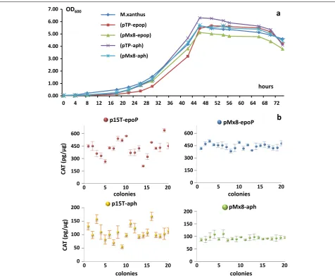
Compared with the wild type strain, the cat expres-sion had small effects on the growth of recombinant cells (Figure 1a). We picked-up 20 colonies randomly from each of the four recombinant categories to assay their chloramphenicol acetylation activities. The site-directed recombination produced rather stable CAT activities, i.e. approximately 450 and 100 pg/μg in the pMx8-epoP
and pMx8-aph transformants, respectively. However, the pTp-epoP and pTp-aph strains showed highly varied CAT activities, ranging from 209.31 to 638.49 pg/μg in
the pTp-epoP transformants or from 55.59 to 168.24 pg/
μg in the pTp-aph transformants (Figure 1b).
Further-more, it is clear to see that the M. xanthus transformants from the pTp-epoP or pMx8-epoP plasmids exhibited several times higher CAT activities than those from the corresponding pTp-aph or pMx8-aph plasmids. The epoP promoter is also more efficient than the aphII promoter
in M. xanthus. Accordingly, the original epoP promoter
was used for further heterologous expressions of the epothilone biosynthetic genes.
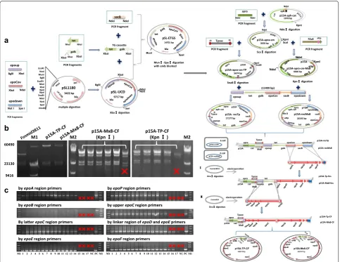
Construction of plasmids containing the entire epothilone biosynthetic gene cluster
integration in M. xanthus chromosome. The epothilone genes were from the Cosmid10 and Fosmid3B11 plas-mids, which separately contained a 38.5-kb fragment from epoA to the front part of epoD and a 34.4-kb fragment from the rear part of epoC to the downstream fragment of epoF. The two fragments overlapped a 6.5-kb region.
To simplify recombination and to avoid false-junction, we stitched the two fragments into a complete epothilone gene cluster using a one-stop-way strategy. The homolo-gous arms included an upstream arm, a downstream arm and a shared middle arm. The shared middle arm was designed to stitch the two large fragments, permitting the two rounds of recombination happening in no particular order for integration of the epothilone gene cluster frag-ments into plasmid. The three homologous arms were
constructed into a single vector, and the stitching was car-ried out in E. coli GB05-red by means of the red-ET recom-bination system (diagrammed in Figure 2a). The correct colonies were screened by the negative marker galk and sacB, and the junction regions were verified by restriction digestion (Figure 2b). After sequencing verification, we got the correct recombinant plasmid p15A-Tp-CF (68.8 kb).
We also constructed a recombinant plasmid contain-ing the Mx8 att sequence (Figure 2a, b) for site-specific recombination.
Integration of the epothilone biosynthetic genes
into M. xanthus chromosome
The plasmids were subject to one-step integration into
M. xanthus DZ2 chromosome, and the recombinant
0.00 1.00 2.00 3.00 4.00 5.00 6.00 7.00
0 4 8 12 16 20 24 28 32 36 40 44 48 52 56 60 64 68 72
M.xanthus (pTP-epop) (pMx8-epop) (pTP-aph) (pMx8-aph)
hours
OD600
a
CA
T (p
g/
μg
)
0 150 300 450 600
0 5 10 15 20
p15T-epoP
colonies
b
0 150 300 450 600
0 5 10 15 20
pMx8-epoP
colonies
0 50 100 150 200
0 5 10 15 20
p15T-aph
colonies
0 50 100 150 200
0 5 10 15 20
pMx8-aph
colonies
CA
T (p
g/
μg
)
Figure 1 Growth and CAT activities of M. xanthus transformants integrated from the plasmids pTP‑epoP, pMx8‑epop, pTP‑aph or pMx8‑aph. a The
colonies were screened by selection markers Apra and Cm. After 6–8 days of incubation at 30°C, we obtained 21 colonies from the CYE plate plus the Apra and Cm antibiotics in a single transformation performance. PCR amplification determined that 14 of the 21 antibiotics-resistant colonies were correct transformants, while the other seven contained partial or none of epothilone genes. A further transformation performance of p15A-Tp-CF in M. xanthus DK1622 strain yielded 30 correct transformants from 59 antibiotics-resistant colonies. We
also performed transformations in SW504, a difA mutant
of M. xanthus DK1622 [22, 23], from which 4 correct
transformants were obtained. Figure 2c demonstrates some PCR amplification results. Our results showed that the transposition technique was highly efficient to nail large exotic genetic fragments into the chromosome of different M. xanthus strains.
Electroporation of the p15A-Mx8-CF plasmid with
M. xanthus DZ2 strain obtained six tiny colonies on the
selection plate, which, however, were turned out to be
a
BglII XbaI epoup epoCov XbaI NdeI epodownNot I Spe I
pSL1180 3422 bp ori bla SpeI NotI NdeI SnaBIHpaI KpnI XbaI NheI EcoRVBglII MunINcoI EcoRI HindIII pSL-UCD 9717 bp NdeI NotI SpeI BglII XbaI bla galk tet XbaI XbaI tet NheI NheI galk XbaINheI XbaI
PCR fragments PCR fragments TG cassee sacB NdeI NdeI PCR fragment pSL-ETGS SpeI MunI bla tet 14701 bp muple digeson Xba digeson
Mun -Spe digeson with ends blunted
p15A-aph-cat 1978 bp KpnI NdeI aph apra NdeI NdeI p15A-apra-cm
3656 bp KpnIScaI
NdeI
Tpase
IR IR Mx8 attp
p15A-apra-cm-TP 5674 bp KpnI SnaBI p15A IR IR Sca digeson Nde digeson SnaB digeson PCR fragment PCR fragment PCR fragment p15A-apra-cm-Mx8 6646 bp KpnI NdeI NdeI p15A ap Kpn digeson (11499 bp)
epoup tet galk epoCov sacB epodown
p15A- recTp 17177 bp p15A epoCov galk tet epoup IR TpaseIR aph-catapra sacB p15A-recMx8 18145 bp p15Aepodown sacB epoCov galk tet Mx 8 ap aph-catapra
b
E.coli p15A-recTP p15A apra cat Tpase epoup tet galk epoCov sacB epodown p15A apra cat Tpase p15A-recTp electroporaon electroporaon Dra digeson Fosmid3B11 Dra digeson Cosmid10 p15A apra cat Tpase epoup tet galk p15A-Tp-fos p15A-Tp-CF p15A-Mx8-fos p15A-recMx8 p15A-Mx8-CF Mx8 Mx8 Mx8 E.coli p15A-recMx8 p15A-TP-CF 68815bp epoA epoP epoB epoC epoD epoEepoF orfAorfB p15A-Mx8-CF 69761bp epoP epoB epoC epoD epoEepoForfAorfB epoA
60490
23130
9416
M1 M2 p15A-Mx8-CF(Kpn ) p15A-TP-CF(Kpn ) M2
by epoAregion primers by epoPregion primers
by epoBregion primers by upper epoCregion primers
By laer epoCregion primers by linker region of epoDand epoEprimers
by epoEregion primers by epoFregion primers
c
M1 1 2 3 4 5 6 7 8 9 10 11 12 13 14 15 16 17 NC PC M1 M1 1 2 3 4 5 6 7 8 9 10 11 12 13 14 15 16 17 NC PC M1
Figure 2 Construction of plasmids containing the epothilone biosynthetic gene cluster and their expressions in M. xanthus. a Diagram of the plas‑
spontaneous mutants without any epothilone genes. We performed transformation of the plasmid in different M.
xanthus strains, which also failed in obtaining any
cor-rect transformant. To testify whether the failure was due to the size limitation for integration at the Mx8 att site, we constructed smaller plasmids of 42.0-kb p15A-Mx8-fos, derived from the recombination of the linear fosmid3B11 with p15A-recMx8, and 18.1-kb p15A-recMx8. The trans-formation performance with these two plasmids under the same conditions produced 4 and 47 correct colonies
in M. xanthus DZ2, respectively. We thus concluded that
the recombination efficiency of the Mx8 att site decreased sharply with the increase of the integration fragment sizes.
Epothilone production abilities and integration sites in the transposition transformants
We assayed the production abilities of epothilones in 48 correct recombinant transformants derived from
differ-ent M. xanthus strains, i.e. DZ2, DK1622 or SW504. The
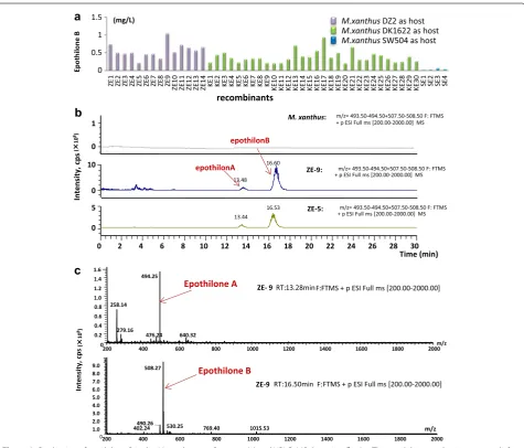
strains were cultivated in the CYE medium for 5 days of incubation, and the production of epothilones was char-acterized by high performance liquid chromatography-mass spectrometry (HPLC–MS) techniques. The results showed that all these recombinants were able to produce epothilones A and B; whereas no epothilone was detect-able in the original M. xanthus strains (Figure 3a; detailed information of the yields in different recombinants is
0 0.5 1 1.5 ZE 1 ZE 2 ZE 3 ZE 4 ZE 5 ZE 6 ZE 7 ZE 8 ZE 9
ZE10 ZE11 ZE12 ZE13 ZE14 KE
1 KE 2 KE 3 KE 4 KE 5 KE 6 KE 7 KE 8 KE 9
KE10 KE11 KE12 KE13 KE14 KE15 KE16 KE17 KE18 KE19 KE20 KE21 KE22 KE23 KE24 KE25 KE26 KE27 KE28 KE29 KE30 SE
1 SE 2 SE 3 SE 4 Epothilone B recombinants
M.xanthusDZ2 as host M.xanthusDK1622 as host M.xanthusSW504 as host
a
b
13.44 0
0 2 4 6 8 10 12 14 16 18 20 22 24 26 28 30
Time (min) 0 16.53 0 16.60 13.48 5 10
1 M. xanthus: + p ESI Full ms [200.00-2000.00] MSm/z= 493.50-494.50+507.50-508.50 F: FTMS
m/z= 493.50-494.50+507.50-508.50 F: FTMS + p ESI Full ms [200.00-2000.00] MS
ZE-9:
m/z= 493.50-494.50+507.50-508.50 F: FTMS + p ESI Full ms [200.00-2000.00] MS
ZE-5: Intensity, cp s ( 10 8) epothilonA epothilonB
ZE- 9RT:13.28minF:FTMS + p ESI Full ms [200.00-2000.00]
200 400 600 800 1000 1200 1400 1600 1800 2000m/z
0 0.2 0.4 0.6 0.8 1.0 1.2 1.4 Intensity, cp s 494.25 258.14 476.24 640.32 279.16 1.6 Epothilone A
ZE-9 RT:16.50min F:FTMS + p ESI Full ms [200.00-2000.00]
200 400 600 800 1000 1200 1400 1600 1800 2000
m/z 0 1.0 2.0 3.0 4.0 5.0 6.0 7.0 8.0 9.0 508.27 490.26 530.25 769.40 1015.53 402.24 Epothilone B ( 10 8) (mg/L)
c
Figure 3 Production of epothilone B in the M. xanthus transformants (a) and HPLC–MS detection (b, c). a The epothilone productions, assayed after
7‑days fermentation. ZE‑, KE‑ and SE‑ series strains are termed for those recombinants from M. xanthus DZ2, DK1622 and SW504 strains, respectively.
provided in Additional file 2: Table S1; Figure 3b, c dem-onstrate the HPLC–MS detection of epothilones pro-duction). Similar to that of the CAT enzyme activities in transposition transformants, the yields of epothilones varied significantly in these recombinants. For exam-ple, in the M. xanthus DK1622 host, the yields in CYE medium ranged from 0.045 to 0.17 mg/L of epothilone A and 0.22–1.05 mg/L of epothilone B. Among the three employed M. xanthus hosts, the yield ranges were similar in the DZ2 and DK1622 mutants, but much low in the SW504 mutants. The production abilities of epothilones
in M. xanthus transposition recombinants from DZ2 or
DK1622 were rather similar to that in those wild type epothilone-producing S. cellulosum strains [24]. How-ever, in contrast to the 2:1 ratio of epothilones A to B or almost single A without B in S. cellulosum strains [24], the ratios of epothilones A–B produced in these M. xan-thus producers were all approximately 1:5 (Additional file 2: Table S1). Compared to those previously reported epothilone-producing M. xanthus recombinants (approx-imately 0.1 mg/L or less) [10, 11], some of the transposi-tion transformants gave much higher productransposi-tions under unimproved fermentation conditions.
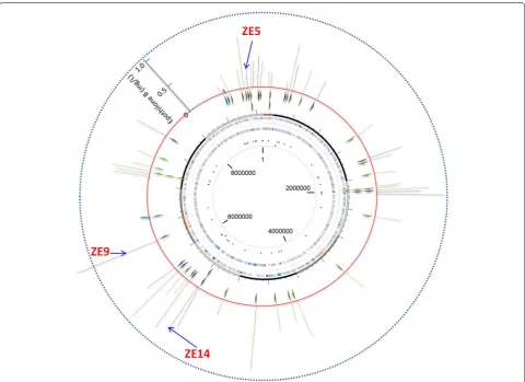
We mapped the integration sites of the epothilone genes in genome using the genome walking technique. Sequencing revealed that the epothilone genes were located at 41 different sites in the 48 recombinants, of which six were repeatedly inserted (Figure 4; details are provided in Additional file 2: Table S1). The epothilone genes were all in the same direction, and these integra-tion sites scattered along the Myxococcus chromosome, seemly with no bias. The recombinants with the same integration sites also had similar yields of epothilones, which was consistent with that of the site-directed inser-tion of CAT gene at the attB site (Figure 1b). However, no clear correlation was observed between the production abilities of epothilones and the insertion sites in genome (Figure 4).
Transcriptomics variation in epothilone‑producing transposition transformants
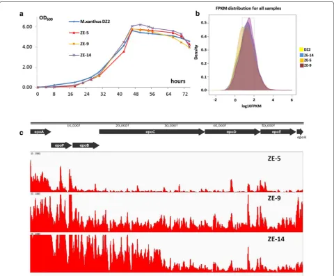
The above results suggested that the production differ-ences probably resulted from the infludiffer-ences of inser-tion sites on the expressions of the epothilone genes. In order to investigate how allopatric integrations influ-enced the yields of epothilones, we assayed the transcrip-tomes in three recombinants, as well as wild type strain DZ2. The three recombinant strains, i.e. ZE-5 (inserted at MXAN_7320), ZE-9 (inserted at MXAN_5011), and ZE-14 (inserted at MXAN_4403), were varied of their yields of epothilones in the CYE medium (0.22 ± 0.08, 1.05 ± 0.21 and 0.66 ± 0.09 mg/L epothilone B, respec-tively; Additional file 2: Table S1). To confirm the total
biosynthetic genes were at the same place in each strain, we performed PCR amplification of the junction regions between the seven modules of the gene cluster. The results showed that all the modules were neighbored in each of these three recombinants (Additional file 3: Fig-ure S2). Growth assays indicated that the three strains had similar growth curves as the wild type strain (Fig-ure 5a). The RNA materials were extracted from the cells, which were collected when XAD-16 resin was harvested for the assay of epothilone production. After confirma-tion of the extracts free of DNA, we sequenced transcrip-tomes using strand specific RNA-seq technique (Illumina Miseq 2000 technique). The sequencing obtained 18.3, 21.3, 18.5 and 20.8 million reads for DZ2, ZE5, ZE9 and ZE14, respectively. Totally, 97.4–98.2% of reads were mapped to genome, and the distribution had good corre-lation with the gene number in each strand. We checked the expression levels of the insertion-mutated genes in the wild type strain DZ2 (Additional file 2: Table S1), which, however, showed no correlations with the produc-tion abilities in the transposiproduc-tion recombinants.
Comparative transcriptomes showed similar distribu-tions of expression density of the total genes in the four recombinants (Figure 5b). The expression patterns of the epothilone genes varied significantly in the three recom-binants (Figure 5c), but parallel to their production abili-ties. The expression levels of the epothilone biosynthetic genes in ZE-5 were low, especially the latter part of this big gene cluster. In contrast, the epothilone genes expres-sions in ZE-9, which had approximately five times of the epothilone production in ZE-5, were rather high in dif-ferent regions in the gene cluster. In the ZE-14 strain, although expressions of the front region were much high, the hinder genes had sharply decreased expressions, which probably led to the medium level of the epothilone production.
Compared to the M. xanthus DZ2 strain, many
Myx-ococcus-own genes were up- or down-regulated
(hypothetical protein) and MXAN_4372 (DNA-binding protein) were significantly down regulated. It is noted that the locations of these up- or down- regulated genes normally had no relationship with the insertion sites
in Myxococcus genome. These transcriptome changes
indicated that allopatric integration of exotic genes in genome had some specific effects on the transcriptome in host, which probably resulted in distinct expressions of the exotic genes.
Conclusions and outlook
In contrary to potential size-limitation of the inserted fragments at directed sites, demonstrated using the attB site in M. xanthus, transposition attempts to position large-sized genetic materials randomly into genome, thus provides much more opportunities for the integration of exotic genes. Such random transposition integration is blind, and the introduced exotic genes may inactivate or disturb functions of host genes, but also yields varied
expression levels of exotic genes. The expression differ-ences of allopatrically integrated genes did not follow the local expression characteristics, but rather the results of some global changes of the host transcriptome. We might be able to improve the expressions of the exotic genes further by genome engineering based on the transcrip-tome changes. In addition, the expressions of epothilone genes were rather different in different cluster regions– increasing the expression of low-expressed genes will be also useful for the yield improvement.
Methods
Bacterial strains, plasmids and culture conditions
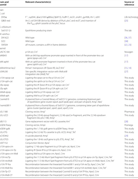
Bacterial strains and plasmids used in this study are listed in Table 1. Escherichia coli DH5α was used for
rou-tine transformations and sub-cloning. E. coli GB05-red, a derivative of DH10B [25], was used in recombination performance. E. coli strains were grown routinely in Luria Broth (LB) medium (10 g/L peptone, 10 g/L yeast extract
Figure 4 Distribution of the integration sites of the epothilone genes (the small triangles inside the red ring; all inserted clockwise) and the produc‑
and 5 g/L NaCl; pH7.2), and Myxococcus xanthus strains were grown in CYE medium [10 g/L casitone, 5 g/L yeast extract, 10 mM 3-(N-morpholino) propanesulfonic acid (MOPS; pH 7.6) and 4 mM MgSO4] [26]. The growth temperatures were 37°C for E. coli and 30°C for M. xan-thus, respectively. For selection of constructed plasmids or transformants, different antibiotics, i.e. ampicillin [Amp], 100 μg/mL; kanamycin [Km], 40 μg/mL;
chlo-ramphenicol [Cm], 10 μg/mL; or apramycin [Apra],
25 μg/mL, were added in LB or CYE media.
Construction of CAT reporter vectors
We amplified the 836-bp replicon p15A from pACYC184 using the primer pair of P15A. The chloramphenicol
acetyl transferase (cat) gene was initiated by a pro-moter of either the epothilone operon propro-moter (epop) or the kanamycin promoter (aphII). These two primers are recognizable in E. coli and M. xanthus [10, 11, 17]. The 1.6-kb NdeI-KpnI fragment epop-cat was ampli-fied from the pKK843 plasmid [17] using the CM-EPOP primer pair, while the 1.1-kb NdeI-KpnI fragment
aphII-cat was amplified from pKK-aphII [17] using the
CM-APH primer pair. The epop-cat and aphII-cat were ligated to the p15A replicon, generating the vectors of p15A-epop-cat and p15A-aph-cat, respectively. The 2.0-kb transposon element IR-Tpase-IR was amplified from plasmid pMiniHimar-lacZ [13] using the TPASE prime pair, while the 3.0-kb site-specific recombination
Figure 5 Transcriptomes of M. xanthus DZ2 and three recombinants with insertions of epothilone biosynthetic genes. a The growth curves of the
Table 1 Bacterial strains and plasmids used in this study Strain and
plasmid Relevant characteristic(s) Source or reference
Strain E. coli
DH5α F−, supE44, ΔlacU169 (ϕ80lacZΔM15), hsdR17, recA1, endA1, gyrA96, thi‑1, relA1 Life technologies
GB05‑red An E. coli DH10B derive by deletion of fhuA, ybcC and recET, and insertion of the PBAD‑gbaA cassette at the ybcC locus
[25]
S. cellulosum
So0157‑2 Epothilone producing strain This lab
M. xanthus
DK1622 Wild type [36, 37]
DZ2 Wild type [38]
SW504 dif mutant, contains a difA in frame deletion [22, 23]
Plasmid
pACYC184 p15A ori; Cmr Novagen
pKK843 With an 843‑bp epothione promoter epop inserted in front of the promoter‑less cat
gene (epop‑cat); Cmr [17]
pKK‑aphII With an aphII promoter fragment inserted in front of the promoter‑less cat
gene (aphII‑cat); Cmr [17]
pMiniHimar‑lacZ Himar1 transposon (IR‑Tpase‑IR), lacZ; Kmr [13, 14]
pSWU30 Site specific integration vector with Mx8 attB
integration site (Mx8); Tetr [27]
p15A‑epop‑cat Ligating the epop‑cat to the p15A ori; Cmr This study
p15A‑aph‑cat Ligating the aphII‑cat to the p15A ori; Cmr This study
pTp‑epop Ligating the IR‑Tpase‑IR to p15A‑epop‑cat; Cmr This study
pTp‑aph Ligating the IR‑Tpase‑IR to p15A‑aph‑cat; Cmr This study
pMx8‑epop Ligating Mx8 to p15A‑epop‑cat; Cmr This study
pMx8‑aph Ligating Mx8 to p15A‑aph‑cat; Cmr This study
Cosmid10 A plasmid from a Cosmid library of So0157‑2 genome, containing foremost part
of epothilone gene cluster (epoA, epoP, epoB, epoC and part of epoD); Ampr, Neor This study
Fosmid3B11 A plasmid from a fosmid library of So0157‑2 genome, containing latter part of epothilone
gene cluster (partial epoC, epoD, epoE, epoF); Cmr This study
pSL1180 Cloning vector; Ampr Pharmacia
pSL‑UCD Ligating the 2.0‑kb epoup fragment, 2.2‑kb epoCov fragment, and the 2.2‑kb epodown
fragment into pSL1180; Ampr This study
pBJ113 Gene replacement vector with KG cassette; Kmr [28]
pGEM‑Teasy Cloning vector, Ampr Promega
pGEM‑galk Ligating the 1.7‑kb galk gene to pGEM‑Teasy; Ampr This study
pSL‑ETG Ligating the 3.2‑kb TG cassette to pSL‑UCD; Ampr, Tetr This study
pCVD442 A suicide plasmid; Ampr [29]
pSL‑ETGS Ligating the 1.8‑kb sacB gene to pSL‑ETG; Ampr, Tetr This study
pSET152 Conjunction Vector; Aprar This study
p15A‑apra‑cm Ligating 1.7‑kb apra fragment to p15A‑aph‑cat; Aprar, Cmr This study
p15A‑apra‑cm‑Tp Ligating IR‑Tpase‑IR to p15A‑apra‑cm; Aprar, Cmr This study
p15A‑apra‑cm‑Mx8 Ligating Mx8 to p15A‑apra‑cm; Aprar, Cmr This study
p15A‑recTp Ligating the 11.5‑kb MunI‑SpeI fragment from pSL‑ETGS to p15A‑apra‑cm‑Tp; Aprar, Cmr, Tetr This study
p15A‑recMx8 Ligating the 11.5‑kb MunI‑SpeI fragment from pSL‑ETGS to p15A‑apra‑cm‑Mx8; Aprar, Cmr, Tetr This study
p15A‑TP‑fos Recombination between the linearized Fosmid3B11 and p15A‑recTp; Aprar, Cmr, Tetr This study
p15A‑Mx8‑fos Recombination between the linearized Fosmid3B11 and p15A‑recMx8; Aprar, Cmr, Tetr This study
p15A‑Tp‑CF Recombination between the linearized Cosmid10 and p15A‑TP‑fos; Aprar, Cmr This study
element Mx8 was generated from the template pSWU30 [27] using the MX8 primer pair. Finally, the plasmids pTp-epop (or pTp-aph) and epop (or pMx8-aph) were constructed by ligating the IR-Tpase-IR and Mx8 to p15A-epop-cat (or p15A-aph-cat), respectively, which had been digested with NdeI; and the DNA ends were blunted using the T4 DNA polymerase. The PCR primer pairs used in this study are summarized in Addi-tional file 5: Table S3.
Stitching and recombination of epothilone gene cluster
We constructed cosmid and fosmid libraries of
Soran-gium cellulosum So0157-2 genome. Southern blotting
hybridization with epothilone gene probes revealed clones containing the epothilone genes. The harbored epothilone genes fragments in the positive clones were determined by the end sequencing. We selected Cos-mid10 and Fosmid3B11 for our construction. After completely sequencing, Cosmid10 contained a fragment of complete epoA to epoC and part of epoD, while Fos-mid3B11 contained part of epoC till the downstream sequence of epoF.
To stitch the two epothilone-genes fragments in Cosmid10 and Fosmid3B11, transitional recombinant vectors p15A- recT and p15A-recM were constructed as follows. First, the 2.0-kb BglII-XbaI epoup fragment (upstream of epoA), 2.2-kbXbaI-NdeI epoCov frag-ment (overlapping the epoC sequence), and the 2.2-kb NotI-SpeI epodown fragment (downstream of epoF) were amplified from the genomic DNA of S. cellulo-sum So0157-2 using the primer pairs of UP, C-LAP and DOWN, respectively. The three PCR products, used as the homologous arms for recombination, were ligated serially into the corresponding restriction sites of pSL1180, generating pSL-UCD. Then, the 1.7-kb galk gene, containing XbaI and NheI sites at the 5′ end and XbaI site at the 3′ end, was amplified from pBJ113 [28] using the GALK primer pair, which was further ligated into the XbaI site of pGEM-Teasy vector, gen-erating pGEM-galk. The 1.5-kb NheI tet fragment (tetracycline gene), amplified from pSWU30 using the TET primer pair, was ligated into the NheI site of the galk fragment in pGEM-galk. The 3.2-kb tet-galk fragment (TG cassette), used for positive and negative selection, was cleaved with Xba I, and ligated into the XbaI site of pSL-UCD, generating pSL-ETG. The plas-mid pSL-ETG was cleaved with NdeI and ligated with the 1.8-kb Nde I sacB gene, which was amplified from pCVD442 [29] using the SACB primer pair, producing pSL-ETGS.
In another route, a 1.7-kb apra fragment (apramycin gene), amplified from pSET152 [30] using the APRA primer pair, was cloned into NdeI site of p15A-aph-cat to
create p15A-apra-cm. Then, the PCR fragment IR-Tpase -IR or Mx8 was ligated into p15A-apra-cm, which there-after was cleaved at the 3′ end of the cat gene by ScaI, yielding the plasmid cm-Tp and p15A-apra-cm-Mx8, respectively. Finally, the 11.5-kb MunI-SpeI fragment from pSL-ETGS with the blunt ends was cloned into the p15A-apra-cm-Tp, which had been cleaved with SnaBI with a blunt end, to create the purposed vector p15A- recTp. The plasmid was cloned into p15A-apra-cm-Mx8, which was further cleaved with KpnI, form-ing the blunt ends, to create the other purposed vector p15A-recMx8.
The vectors p15A-recTp and p15A-recMx8 was subsequently engineered for recombining with the epothilone gene cluster from Cosmid10 and Fos-mid3B11. First, both Cosmid10 and Fosmid3B11 were cleaved with DraI, removing the irrelevant sequences and exposing the terminal sequences with epothilone genes, which contained the identical homologous arms epoup, epoCov and epodown. Then, the linearized Fosmid3B11 was electroporated into the E. coli strain GB05-red harboring p15A-recTp or p15A-recMx8 for the first round of recombination and the transformants with the objective recombinant TP-fos or p15A-Mx8-fos were selected on the LB plate containing Apra and Cm with extra 10% sucrose for negative selection. Similarly, in the second round the linearized Cosmid10 was electroporated into E. coli GB05-red harboring the plasmid from the first round and the transformants were selected on the LB plate containing extra 1% 2-deoxygalactose (DOG). Ultimately, the engineered recombinant plasmids p15A-Tp-CF and p15A-Mx8-CF containing the integrated epothilone gene cluster were constructed. All the above PCR products were ampli-fied using the pfu DNA polymerase to ensure high fidelity and sequenced to confirm their identities. A diagrammatic sketch for the construction is provided in Figure 2.
Electro‑transformation of M. xanthus
The constructed expression plasmids were introduced into M. xanthus cells by electroporation. Briefly, M.
xanthus cells from 50-mL overnight cultures (OD600
was about 0.6) were collected and washed thrice with ice-cold water for preparing competent cells. 1.5 mL of the culture was centrifuged and the precipitate was resuspended with 50 μL of ice-cold water in tube,
mixed with 3 μg of DNA, and electroporated at a
was added to 2 mL 0.5% soft agar and the mixture was spread on 1.5% CYE selection agar plates. Resistant colonies that appeared after 6 days of incubation were checked by colony PCR with the following primers: primer pairs of CM-APH and CM-EPOP were used to check the integration of cat gene; Epothilone-specific primers, EA, EP, EB, EC1, EC2, EE and EF, with a 51% coverage of the whole gene cluster, were designed to detect the regions located in ORFs epoA, epoP, epoB, epoC, epoE or epoF, respectively, to verify the integra-tion of the whole biosynthetic gene cluster in the M.
xanthus chromosome.
Measurement of chloramphenicol acetyl transferase (CAT)
activity in M. xanthus
To measure the chloramphenicol acetyl transferase (CAT) activity, the bacterial pellets were harvested by centrifugation at 8,000×g for 5 min after the cultures reached the mid-exponential phase (36 h of incubation; OD600 was about 3.0). The pellet was resuspended and washed twice with distilled water and then resuspended in 1 mL of lysis buffer. The crude bacterial extracts were prepared by sonication, and the supernatant was col-lected by centrifugation at 12,000×g for 2 min, and used for characterizing chloramphenicol acetyl transferase (cat) gene expression activity with the CAT ELISA kit, as previously described [17].
Epothilone extraction and detection
M. xanthus strains containing the complete epothilone
gene cluster were grown overnight in 100 mL CYE medium supplemented with Apra and Cm antibiotics. The cultures were inoculated at a ratio of 2:100 into CYE medium containing 2% of the XAD-16 resin for the absorption of epothilone products. After 7 days rotation at the 250 rpm speed and 30°C, the mixtures of cells and resin were harvested by centrifugation and extracted with two volume of methanol by shaking at room temperature overnight [31]. After centrifuged for 20 min, the supernatant was moved into a rotary evap-orator to remove the solvent. The residue was further re-dissolved in 1 mL of methanol, and an aliquot of 20 μL of the sample was injected into a Finnigan HPLC
system interfaced with a Finnigan MSQ classic quadru-pole mass spectrometer (Thermo Finnigan, USA). The analysis was carried out on a Shim-pack MRC-ODS RP column (4.6 mm × 250 mm, 4.60 μm; Shimadzu, Japan)
at a temperature of 28°C with a mobile phase of 60% methanol (HPLC grade) and 40% buffer (0.2% acetate acid/18 MΩ Millipore water) at a flow rate of 1.0 mL/
min. The MS analysis was performed under the fol-lowing conditions: ESI-positive, probe temperature of 450°C, cone voltage of 75 V, full scan mass range from
200 to 2,000 m/z at 2 Hz scan speed, and SIM scan at 494 [M + H]+ for epothilone A and 508 [M + H]+ for
epothilone B [32]. Epothilones were identified by com-parison to the retention time, 249 nm of UV spectra and the MS2 pattern of the authentic reference stand-ard sample containing epothilones A and B, which were obtained from the culture of S. cellulosum So0157-2 as reported previously [31, 33]. The production levels of epothilones were averaged from three independent cul-tivations and extractions.
Determination of the integration sites
A Genome Walking kit (TaKaRa, Japan) was used to verify the insertion sites of the gene integration. Three specific primers, Cm1, Cm2 and Cm3, designed in this work (Additional file 5: Table S3) were employed with the random primer AP1, which is provided by the kit, in three PCR rounds, to amplify a product containing partial cat region, the IR sequence of the transposon and the flanking unknown genome region of M. xan-thus, respectively. Genomic DNA of the transformants with the epothilone gene cluster was extracted and used as the template in the first round PCR using the primer pair AP1/Cm1; the diluted product from the first PCR round was orderly used as the template for the second round, using the primer pair AP1/Cm2; and the prod-uct from the second round was diluted as the template for the third PCR round, using the AP1/Cm3 primer pair. The PCR systems and procedures were performed according to the kit protocol. The PCR products were detected by electrophoresis, and the single DNA bands with appropriate size from the third PCR round were withdrawn by the DNA Extraction kit (Promega, USA) and sequenced after subcloning to the pGEM-Teasy vector.
Transcriptomics analysis
In this study, we exploit the ssRNA-seq method to iden-tify the transcriptional template strands of M. xanthus DZ2 and three transformants ZE-5, ZE-9 and ZE-14 at a whole genome level using the Illumina-platform high throughput sequencing. First of all, the total RNA of the
M. xanthus strains were extracted according to the
200 ng of M. xanthus polyA+ RNA by heating at 98°C
for 40 min in 0.2 mM sodium citrate, pH 6.4 (Ambion, USA). The fragmented RNA was concentrated to 5 μL,
mixed with 3 μg random hexamers, incubated at 70°C
for 10 min, and then cooled on ice. The RNA mixtures were further added with 4 μL of 5× first-strand buffer,
2 μL of 100 mM DTT, 1 μL of 10 mM dNTPs, 4 μg of
actinomycin D (USB), 200 U SuperScript III, and 20 U SUPERase-In (Ambion, USA), incubated at room tem-perature for 10 min followed by 1 h at 55°C to synthe-size the first-strand cDNA. First-strand cDNA was cleaned up by extraction twice with phenol: chloroform: isoamyl alcohol (25:24:1), followed by ethanol precipita-tion with 0.1 volumes 5 M ammonia acetate to remove dNTPs and re-suspension in 104 μL ddH2O.
Second-strand cDNA was synthesized by adding 4 μL 5×
first-strand buffer, 2 μL 100 mM DTT, 4 μL 10 mM dNTPs
with dTTP replaced by dUTP (Sigma-Aldrich, USA), 30 μL 5× second strand buffer, 40 U Escherichia coli
DNA polymerase, 10 U E. coli DNA ligase, 2 U E. coli RNase H and incubating at 16°C for 2 h. A paired-end library for Illumina sequencing was prepared accord-ing to the instructions provided with the followaccord-ing modifications. First, five times less adapter mixture was ligated to the cDNAs. Second, 1 U USER (New Eng-land Biolabs, USA) was incubated with 180- to 480-bp size-selected, adapter-ligated cDNA at 37°C for 15 min followed by 5 min at 95°C before PCR. Third, PCR was performed with Phusion High-Fidelity DNA Poly-merase with GC buffer (New England Biolabs, USA) and 2 M betaine (Sigma, USA). Fourth, PCR prim-ers were removed using 1.8× volume of AMPure PCR Purification kit (BeckmanCoulter Genomics, USA). Transcriptome sequencing was performed at the BGI Corporation. Reagents were all from Invitrogen (Carls-bad, USA) except as noted.
To assay the growth curves, M. xanthus colonies were first cultured overnight in CYE medium. Then, the cul-tures were transferred at a ratio of about 2:100 into 5 mL fresh CYE medium with a start OD600 of 0.15, and assayed of the OD600 values periodically.
The reference genome (M. xanthus DK1622) and the gene annotation was retrieved from GenBank (accession no. NC_008095.1). After removing the reads containing sequencing adapters and low-quality reads (reads con-taining Ns >10%), the remaining 90 bp clean reads with high quality were aligned with the reference genome using Bowtie software [35]. Then the RPKM method was used to normalize the transcript level, which was expressed as the number of reads per kilobase of exon region per million mapped reads (RPKM). Go annotation of the genes was performed using Blast2GO software and visualized by WEGO software.
Authors’ contributions
LPZ, YZL designed the experiments. LPZ, XJY, KH analyzed and interpreted the data. LPZ, XJY, ZFL, LSZ, XNY, HLW performed the experiments. LPZ, YZL wrote the manuscript. YMZ, YZL supervised the research. All authors read and approved the final manuscript.
Acknowledgements
This work was financially supported by the National Science Foundation of China (NSFC) Key Program [No. 31130004] and National High‑tech R&D Program of China (863 Program) [No. 2012AA02A701]. We thank Chun‑ yan Lin and Guang‑rong Zhao from Tianjin University for exchanged communications.
Compliance with ethical guidelines
Competing interests
The authors declare that they have no competing interests.
Received: 17 May 2015 Accepted: 7 July 2015
References
1. Rosano GL, Ceccarelli EA (2014) Recombinant protein expression in
Escherichia coli: advances and challenges. Front Microbiol. 5, article 172 2. Ross AC, Gulland LE, Dorrestein PC, Moore BS (2015) Targeted capture
and heterologous expression of the Pseudoalteromonas alterochromide gene cluster in Escherichia coli represents a promising natural product exploratory platform. ACS Synth Biol. doi:10.1021/sb500280q
3. Wang Y, Li YZ (2014) Cultivation to improve in vivo solubility of overex‑ pressed arginine deiminases in Escherichia coli and the enzyme character‑ istics. BMC Biotechnol 14:53. doi:10.1186/1472‑6750‑14‑53
4. Ongley SE, Bian X, Neilan BA, Müller R (2013) Recent advances in the heterologous expression of microbial natural product biosynthetic path‑ ways. Nat Prod Rep 30:1121–1138
5. Gerth K, Bedorf N, Höfle G, Irschik H, Reichenbach H (1996) Epothilons A and B: antifungal and cytotoxic compounds from Sorangium cellulosum
(Myxobacteria). Production, physico‑chemical and biological properties. J Antibiot (Tokyo) 49:560–563
6. Tang L, Shah S, Chung L, Carney J, Katz L, Khosla C et al (2000) Cloning and heterologous expression of the epothilone gene cluster. Science 287:640–642
Additional files
Additional file 1: Figure S1. Diagram for the construction of the CAT reporter gene vectors. The epop and aphII promoters were linked to p15A
to generate p15‑epop‑cat and p15A‑aph‑cat, respectively. Then, the IR-Tpase-IR or Mx8 attp element was subcloned to each of the two plasmids above, respectively, to build the four final plasmids pTp‑epoP, pMx8‑ epoP, pTp‑aph and pMx8‑aph, which were initiated by aphII and epoP, respectively. The plasmids were electroporated into M. xanthus to assay the promoter activities.
Additional file 2:Table S1. The insertion sites of the epothilone genes and the production of epothilones in different transposition recombinants.
Additional file 3: Figure S2. PCR amplification of the junction regions between the seven epothilone‑modules. Genome templates for each PCR sample are as follows: 1, M. xanthus DZ2; 2, M. xanthus ZE5; 3, M. xanthus
ZE9; 4, M. xanthus ZE14; 5, S. cellulosum So0157‑2; 6, distilled water; M, DNA Marker DL2000 plus II.
Additional file 4: Table S2. Significantly up‑ and down‑regulated
Myxococcus genes in different recombinants.
7. Zhang L, Zhao GP, Ding XM (2011) Tandem assembly of the epothilone biosynthetic gene cluster by in vitro site‑specific recombination. Sci Rep Uk. doi:10.1038/srep00141
8. Park SR, Park JW, Jung WS, Han AR, Ban YH, Kim EJ et al (2008) Heterolo‑ gous production of epothilones B and D in Streptomyces venezuelae. Appl Microbiol Biotechnol 81:109–117
9. Mutka SC, Carney JR, Liu Y, Kennedy J (2006) Heterologous production of epothilone C and D in Escherichia coli. Biochemistry 45:1321–1330 10. Fu J, Wenzel S, Perlova O, Wang J, Gross F, Tang Z et al (2008) Efficient
transfer of two large secondary metabolite pathway gene clusters into heterologous hosts by transposition. Nucleic Acids Res 36(17):e113 11. Julien B, Shah S (2002) Heterologous expression of epothilone biosyn‑
thetic genes in Myxococcus xanthus. Antimicrob Agents Chemother 46:2772–2778
12. Tang L, Chung L, Carney JR, Starks CM, Licari P, Katz L (2005) Generation of new epothilones by genetic engineering of a polyketide synthase in
Myxococcus xanthus. J Antibiot (Tokyo) 58:178–184
13. Bouhenni R, Gehrke A, Saffarini D (2005) Identification of genes involved in cytochrome c biogenesis in Shewanella oneidensis, using a modified mariner transposon. Appl Environ Microbiol 71:4935–4937
14. Zhang CY, Cai K, Liu H, Zhang Y, Pan HW, Wang B et al (2007) New locus important for Myxococcus social motility and development. J Bacteriol 189:7937–7941
15. Tojo N, Sanmiya K, Sugawara H, Inouye S, Komano T (1996) Integration of bacteriophage Mx8 into the Myxococcus xanthus chromosome causes a structural alteration at the C‑terminal region of the IntP protein. J Bacte‑ riol 178:4004–4011
16. Viswanathan P, Murphy K, Julien B, Garza AG, Kroos L (2007) Regulation of dev, an operon that includes genes essential for Myxococcus xanthus
development and CRISPR‑associated genes and repeats. J Bacteriol 189:3738–3750
17. Zhu LP, Li ZF, Sun X, Li SG, Li YZ (2013) Characteristics and activity analysis of epothilone operon promoters from Sorangium cellulosum strains in
Escherichia coli. Appl Microbiol Biotechnol 97:6857–6866
18. Hao T, Biran D, Velicer GJ, Kroos L (2002) Identification of the Omega4514 regulatory region, a developmental promoter of Myxococcus xanthus that is transcribed in vitro by the major vegetative RNA polymerase. J Bacteriol 184:3348–3359
19. Zhang C, Cai K, Pan H, Liu H, Li Y (2010) Construction and application of plasmid pZCY11 for analyzing gene functions and expressions in Myxo-coccus. Wei Sheng Wu Xue Bao. 50:29–35
20. Julien B, Shah S, Ziermann R, Goldman R, Katz L, Khosla C (2000) Isolation and characterization of the epothilone biosynthetic gene cluster from
Sorangium cellulosum. Gene 249:153–160
21. Molnar I, Schupp T, Ono M, Zirkle RE, Milnamow M, Nowak‑Thompson B et al (2000) The biosynthetic gene cluster for the microtubule‑stabilizing agents epothilones A and B from Sorangium cellulosum So ce90. Chem Biol 7:97–109
22. Yang Z, Geng Y, Xu D, Kaplan HB, Shi W (1998) A new set of chemotaxis homologues is essential for Myxococcus xanthus social motility. Mol Microbiol 30:1123–1130
23. Wang J, Hu W, Lux R, He X, Li Y, Shi W (2011) Natural transformation of
Myxococcus xanthus. J Bacteriol 193:2122–2132
24. Li PF, Li SG, Li ZF, Zhao L, Wang T, Pan HW et al (2013) Co‑cultivation of
Sorangium cellulosum strains affects cellular growth and biosynthesis of secondary metabolite epothilones. FEMS Microbiol Ecol 85:358–368 25. Fu J, Bian X, Hu S, Wang H, Huang F, Seibert PM et al (2012) Full‑length
RecE enhances linear‑linear homologous recombination and facilitates direct cloning for bioprospecting. Nat Biotechnol 30:440–446 26. Goldman B, Bhat S, Shimkets LJ (2007) Genome evolution and the emer‑
gence of fruiting body development in Myxococcus xanthus. PLoS One 2:e1329
27. Wu SS, Wu J, Kaiser D (1997) The Myxococcus xanthus pilT locus is required for social gliding motility although pili are still produced. Mol Microbiol 23:109–121
28. Julien B, Kaiser AD, Garza A (2000) Spatial control of cell differentiation in
Myxococcus xanthus. Proc Natl Acad Sci U S A. 97:9098–9103
29. Xia ZJ, Wang J, Hu W, Liu H, Gao XZ, Wu ZH et al (2008) Improving conju‑ gation efficacy of Sorangium cellulosum by the addition of dual selection antibiotics. J Ind Microbiol Biotechnol 35:1157–1163
30. Wilkinson CJ, Hughes‑Thomas ZA, Martin CJ, Bohm I, Mironenko T, Dea‑ con M et al (2002) Increasing the efficiency of heterologous promoters in actinomycetes. J Mol Microbiol Biotechnol 4:417–426
31. Gong GL, Sun X, Liu XL, Hu W, Cao WR, Liu H et al (2007) Mutation and a high‑throughput screening method for improving the production of Epothilones of Sorangium. J Ind Microbiol Biotechnol 34:615–623 32. Li SG, Zhao L, Han K, Li PF, Li ZF, Hu W et al (2014) Diversity of epothilone
producers among Sorangium strains in producer‑positive soil habitats. Microb Biotechnol 7:130–141
33. Perlova O, Fu J, Kuhlmann S, Krug D, Stewart AF, Zhang Y et al (2006) Reconstitution of the myxothiazol biosynthetic gene cluster by Red/ET recombination and heterologous expression in Myxococcus xanthus. Appl Environ Microbiol 72:7485–7494
34. Parkhomchuk D, Borodina T, Amstislavskiy V, Banaru M, Hallen L, Krobitsch S et al (2009) Transcriptome analysis by strand‑specific sequencing of complementary DNA. Nucleic Acids Res 37:e123
35. Langmead B, Salzberg SL (2012) Fast gapped‑read alignment with Bowtie 2. Nat Methods 9:357–359
36. Kaiser D (1979) Social gliding is correlated with the presence of pili in
Myxococcus xanthus. Proc Natl Acad Sci USA 76:5952–5956
37. Wang Y, Zhang WY, Zhang Z, Li J, Li ZF, Tan ZG et al (2013) Mechanisms involved in the functional divergence of duplicated GroEL chaperonins in
Myxococcus xanthus DK1622. PLoS Genet 9:e1003306
38. Müller S, Willett JW, Bahr SM, Darnell CL, Hummels KR, Dong CK et al. (2013) Draft genome sequence of Myxococcus xanthus wild‑type strain DZ2, a model organism for predation and development. Genome Announc 1: e00217–13
Submit your next manuscript to BioMed Central and take full advantage of:
• Convenient online submission
• Thorough peer review
• No space constraints or color figure charges
• Immediate publication on acceptance
• Inclusion in PubMed, CAS, Scopus and Google Scholar
• Research which is freely available for redistribution