Mieszko Więckiewicz
1, A–F, Klaus W. Boening
2, A–F, Gert Richter
2, A–F,
Włodzimierz Więckiewicz
3, C–FThe Use of Light-Cured Resin as an Alternative Method
of Occlusal Splints Manufacturing –
In Vitro
Study
1 Division of Dental Materials, Faculty of Dentistry, Wroclaw Medical University, Poland
2 Department of Prosthetic Dentistry, Faculty of Medicine, Dresden University of Technology, Germany 3 Department of Prosthetic Dentistry, Faculty of Dentistry, Wroclaw Medical University, Poland
A – research concept and design; B – collection and/or assembly of data; C – data analysis and interpretation;
D – writing the article; E – critical revision of the article; F – final approval of article; G – other
Abstract
Background. Temporomandibular disorders are very common nowadays. One of the methods to treat these prob-lems is occlusal splint therapy. Modern materials should be introduced to this treatment.
Objectives. The aim of this paper was to evaluate the properties of light-activated urethane dimethacrylate and the quality of the bonds it creates with thermoforming foils.
Material and Methods. Thermoforming foils were covered with light-cured resin. A bond was formed between the materials using an adhesive. A coating lacquer was used on the resinas a final preparatory step. Three laboratory tests were run: dye penetrant inspection, a Vickers microhardness test and a linear polymerization shrinkage test. The materials were layered and then cured with a polymerizing lamp emitting light of a wavelength of 400 Nm, according to the manufacturer’s instructions. All the occlusal splints were fitted to upper dental arch. The devices had been made in an articulator on specially prepared gypsum models. The results were analyzed statistically using a one-sided binomial test, Spearman’s rank-order correlation coefficient and the Friedman ANOVA (p = 0.05).
Results. In the dye penetrant inspection, only one sample out of sixty showed the effects of color penetration to the adhesive connection. The dye only penetrated the layer of lacquer coating the resin. The average value of the Vickers microhardness test with a load of F = 50 g applied to the material surface for 30 s was HV0.05 = 7.43 N/mm2.
The average linear shrinkage of the resin observed after polymerization was 1.175%.
Conclusions. Light-cured resin and an adhesive connection between the resin and thermoforming foil do not show susceptibility even to strong dye. The maximum polymerization shrinkage occurs immediately after curing. The light-cured resin that was tested seems to be a good alternative method for occlusal splints manufacturing (Adv Clin Exp Med 2014, 23, 6, 977–985).
Key words: occlusal splints, light-cured resin, dye penetrant inspection, Vickers microhardness, polymerization shrinkage.
Adv Clin Exp Med 2014, 23, 6, 977–985 ISSN 1899–5276
ORIGINAL PAPERS
© Copyright by Wroclaw Medical University
According to De Kanter et al., theincidence of temporomandibular disorders ranges from 6% to 93% [1]. Various studies confirm that women suf-fer from temporomandibular disorders more often than men [2, 3]. Those disorders may be related to soft tissues, temporomandibular joints, stomato-gnathic system muscles or component parts of the nervous system.
One of the most common methods to treat temporomandibular disorders is occlusal splint therapy [4–8]. The first written report referring to that type of treatment was published by Karolyi in 1901 [9]. Since that time many different oral ap-pliances for treating temporomandibular disor-ders have been introduced, with constantly chang-ing shapes and concepts.
Heat-cured polymethyl methacrylate is a ma-terial that has been commonly used in manufac-turing occlusal splints. It was introduced by Wal-ter Wright in 1937 as a maWal-terial to manufacture denture bases [10]. It has many useful properties, such as adequate hardness, a low shrinkage level, chemical stability, resistance to abrasion, easy han-dling and processing. These features make it an ex-cellent material for use in treating functional dis-orders of the stomatognathic system. It should be mentioned that heat-cured methacrylate is cheaper than light-cured resin. This makes it more accessi-ble and improves the cost effectiveness of the treat-ment. However, despite its good properties, it’s not a perfect material. The main problem is that some people have a hypersensitivity to the monomer that appears in acrylate materials, which can affect both dental technicians and patients, causing skin aller-gies and respiratory alleraller-gies [11]. Acrylate materi-als materi-also have strong, pungent odor that disappears right after polymerization.
Advances in science have permitted research-ers to start investigating materials that would pro-vide better and more biocompatible clinical so-lutions. Good examples of such materials are light-cured resins and thermoformable materials, widely used in modern dentistry, e.g. for forming occlusal splints used in treating temporomandibu-lar disorders, occlusal interferences and in orthog-nathic surgery [12–15].
The aim of this study was the in vitro evalua-tion of light-cured urethane dimethacrylate Light-don Splint resin (Dreve Dentamid Gmbh, Unna, Germany) used in combination with a thermo-formable foil to construct occlusal splints. The ma-terials were tested for dye penetration between the two materials, microhardness and linear polymer-ization shrinkage.
Material and Methods
Light-cured Lightdon Splint resin was ap-plied on 1 mm thick Erkodur thermoformable foil (Erkodent Gmbh, Pfalzgrafenweiler, Germa-ny). The thermoforming process was used to mold them in a Erkoform 3D vacuum unit (Erkodent Gmbh, Pfalzgrafenweiler, Germany). The chemi-cal composition of Lightdon Splint resin is tetra-hydrofurfuryl-2-methacrylat (25–50%), polyure-thaneacrylate (10–25%), poly-i-buthylmethacrylat (2.5–10%) and photo initiator (0.5–0.7%) [16]. Lightdon Bonding adhesive (Dreve Dentamid Gmbh, Unna, Germany) was used to bind the resin to the thermoformable foils, allowing the occlusal surface to be prepared with accurate canine guid-ance. After polymerization, finishing with milling burs and polishing the last surface, the manufactur-er advises covmanufactur-ering the resin surface with Plaquit lacquer (Dreve Dentamid Gmbh) to protect the resin from deposition of dental plaque [17].
The light-curing lamp used in the study (Indi-vido Light Box, Voco Gmbh, Cuxhaven, Germa-ny) had a power of 36 W and emitted light of a 400 Nm wavelength. The exposure times for particular components were as follows:
– Lightdon Bonding adhesive: 1 min; – Lightdon Splint resin: 5 min; – Plaquit lacquer: 4 min.
The materials were applied and cured accord-ing to the manufacturers’ recommendations.
All the occlusal splints were attached to upper dental arche (Fig. 1). The appliances had been pre-pared by the same person on gypsum models in a Protar 3 articulator (KaVo Dental Gmbh, Biber-ach, Germany). The final wet polishing was done in a Rotopol 22 polisher (Struers, Rodovre, Den-mark) using waterproof silicon carbide paper disks (Struers) with decreasing grain sizes (FEPA 320, 2400 and 400).
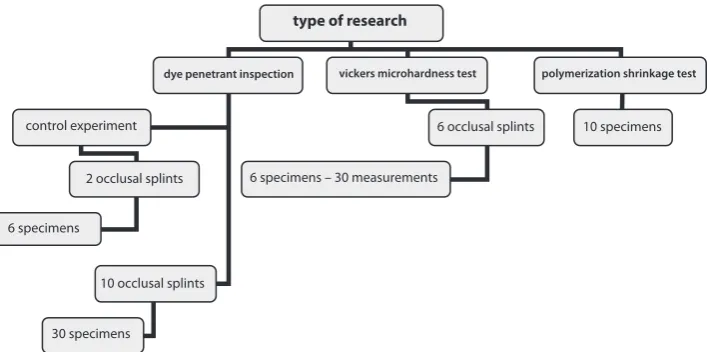
Fig. 1. The flowchart presents the number of occlusal splints and specimens used in the study
type of research
dye penetrant inspection vickers microhardness test polymerization shrinkage test
control experiment
2 occlusal splints
10 occlusal splints
6 occlusal splints 10 specimens
6 specimens
30 specimens
Three laboratory tests were designed and run: dye penetrant inspection, a Vickers microhardness test and a linear polymerization shrinkage test.
Dye Penetrant Inspection
This test was designed to test the susceptibili-ty to moisture penetration of the light-cured resin, lacquer and the bonding material between the res-in and the thermoformable foils. To achieve this, 10 similar occlusal splints were made. The materi-als were layered and exposed to light according to manufacturers’ recommendations. Then each ap-pliance was divided into 3 parts by cutting it be-tween the maxillary canine and the first premolar on both left and right sides (Fig. 2). After cutting, the new surfaces were polished, coated with Pla-quit lacquer and exposed to light to prevent any dye penetration from this side. A total of 30 such specimens were prepared. The prepared specimens were subjected to thermocycling (2500 cycles in distilled water) using a Festo FPC 101 Step Con-troller (Festo AG & Co. KG, Esslingen, Germany). Each cycle comprised 27 s of immersion time at temperatures of 5˚C ± 2˚C and 55˚C ± 2˚C, and 15 s for taking the samples to another bath.
The specimens were then immersed for 24 h in a 2% solution of methylene blue at 37˚C. After that time, the samples were rinsed with water and al-lowed to dry. Subsequently, each preparation was cut transversely in the middle and polished. The penetration of the dye was evaluated on the surface that was formed after the cut from the vestibular
and lingual sides. The samples were examined un-der a Leica MZ12 microscope (Meyer Instruments, Houston, Texas, USA) at a magnification of ×32.
As a control, another 6 specimens, not coat-ed with Plaquit lacquer, were preparcoat-ed, but in this case the Lightdon Bonding adhesive was replaced with an alginate isolator. Further procedures were the same as described above.
Vickers Microhardness Test
The part of the study was designed to evaluate the microhardness of the resin as an essential fea-ture of materials used in the prosthetic rehabilita-tion of patients with greater chewing forces. The Vickers method uses a square-based diamond pyr-amid (called an indenter) with an apical angle of 136˚ between the surfaces to evaluate the hardness. The indenter is pressed into the surface of the test piece using a prescribed force (F) and time. The measurement is usually performed at an ambi-ent temperature between 10˚C do 35˚C [18]. The Vickers hardness (HV) number is then determined by the ratio F/A where F is the force applied to the diamond and A is the surface area of the resulting indentation, according to following equation:
HV = 0.102 × F/A = 0.1891 F/d2 [N/mm2]
where
HV: Vickers hardness, F: load in Newtons [N], 0.1891: Vickers constant,
d = d1 + d2/2: the average value of the imprint
diagonal [mm],
d1, d2: diagonal values [mm].
The test was carried out 24 h after polymeriza-tion. The load used in this study (F = 50 g) was ap-plied to the surface for 30 s. To achieve this, 6 iden-tical occlusal splints were prepared with profiled canine guidance. All the materials were layered and then exposed to UV light according to manufactur-ers’ recommendations. Then each appliance was cut at the level of the canine guidance and 5 mm further, in the direction of the first premolar tooth (Fig. 3). Subsequently, gypsum cylinders were prepared and the specimens were localized inside them, to pro-vide stability during the test (Fig. 4). All the speci-mens were then subjected to wet polishing. A Neo-phot 32 microscope (Carl Zeiss, Jenna, Germany), was used to carry out the measurements. Five im-prints were made on each of 6 prepared cross-sec-tional specimens (30 measurements in all). The in-denter was then pressed, starting from the apex point of canine guidance and moving towards the Erkodur plate in 0.45 mm intervals. The diagonals of the imprints were measured according to the norm EN ISO 6507-1:2005 [18].
Polymerization Shrinkage Test
The aim of this test was to assess the amount of linear polymerization shrinkage of the light- -cured resin at specified time intervals and in specified storage conditions. For this purpose, 10 specimens were prepared in the same silicon ma-trix. The samples were made of light-cured res-in and had the dental arch shape. Three stares-inless steel pins were implanted in each sample, forming a shape similar to a triangle (with sides approxi-mately 40 mm in length). The triangle sides were marked A, B and C (Fig. 5). A ZKM 05-250 D mea-suring unit (Carl Zeiss, Jena, Germany) was used to measure the 3 sides of each triangle. Subsequently,
every line segment (A, B and C) of each sample was measured 4 times: before polymerization; 1 h after light curing; 24 h after polymerization; and finally after being placed in a 37˚C water bath (H&H De-vice Technology, Dresden, Germany)for 24 h. The polymerization time for each sample was 4 min. Differences in the length changes of line segments were examined between:
I: the length measured before polymerization and the length measured 1 h after polymerization (0–1 h);
II: the length measured 1 h after polymeriza-tion and the length measured 24 h after polymer-ization (1–24 h);
III: the length measured after placing the mate-rial in the water bath for 24 h and the length mea-sured 24 h after polymerization (H20-24 h).
Statistical Analysis
The statistical analysis was performed using STATISTICA software (version 10, StatSoft Inc., Tulsa, Oklahoma, USA). For all the tests, p = 0.05 was treated as statically significant.
The results of dye penetrant inspection were analyzed statistically using separate one-sided bi-nomial tests for the vestibular and lingual sides of each sample. The chance value of dye penetration in the observations was 0.5.
The average value of Vickers microhardness was calculated, as well as standard deviation. The results were then analyzed statistically using Spear-man’s rank-order correlation coefficient.
All differences and length changes of all 3 sides (A, B and C) in the polymerization shrinkage test
Fig. 3. An example of an occlusal splint prepared for the Vickers microhardness test. The cutting lines were marked in black. The distance between cuts is 5 mm, and was marked with a green line
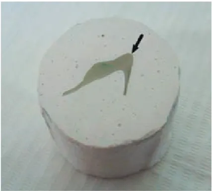
Fig. 4. A gypsum cylinder with specimen localized inside it. The black arrow indicates the location where the measurement was started for the Vickers micro-hardness test, and its direction
were analyzed under different experimental con-ditions, separately for each line segment, using the nonparametric Friedman ANOVA. For param-eters that showed statistically significant differ-ences, post-hoc tests were performed. The average value of length changes was calculated, as well as standard deviation.
Results
Dye Penetrant Inspection
For the vestibular surface, there was no dye penetration into the adhesive connection between the resin and the thermoformable foil in any of 30 measurements (Fig. 6). The one-sided binomi-al test proved that this result differed substantibinomi-ally from the assumed value of color penetration prob-ability [P (p=q=0.5) [[Y = 0]; p < 0.0001]. The lack of
color penetration is statistically significant. There was only one instance of color penetration from the lingual side (Fig. 7). The one-sided binomial test proved that this result differed substantially from the assumed value of color penetration prob-ability [P (p=q=0.5) [Y ≤ 1]; p < 0.0001]. The low
inci-dence of dye penetration is statistically significant. In the control experiment, dye penetration oc-curred in all 6 measurements (on both the vestibu-lar and lingual surfaces). Dye eventually penetrated the space between the resin and the thermoform-able foil. In these tests the Lightdon Bonding adhe-sive was replaced with an alginate isolator (Fig. 8).
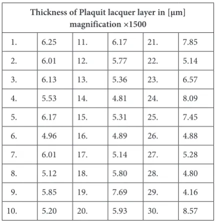
There was no dye penetration into the bulk of the light-cured resin; however, dye penetrated across the entire width of the lacquer coating the resin. The mean value of the lacquer thickness was
measured using a Philips XL 30 ESEM scanning electron microscope (FEI, Hillsboro, Oregon, USA). Thirty randomly selected measurements were performed with a magnification of ×1500 (Table 1). After the data analysis had been done, the average thickness of the lacquer layer was cal-culated as 5.975 µm.
Vickers Microhardness Test
Thirty measurements were done to calculate the average value of Vickers microhardness (Ta-ble 2). The average value of Vickers microhard-ness obtained in the study was HV0.05 = 7.43 N/mm2
(SD = 0.84). Spearman’s rank-order correlation co-efficient was computed separately for each specimen to assess the relationship between Vickers micro-hardness and the depth of the light-cured resin layer on Erkodur foil. There was no correlation between the 2 variables in any of the samples (Table 3).
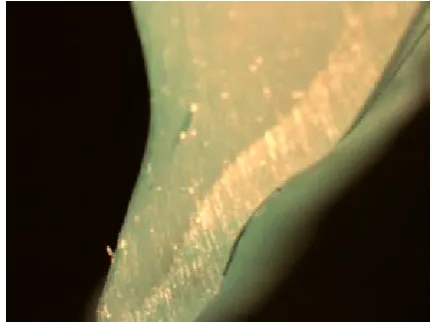
Fig. 6. The absence of dye penetration into the adhe-sive connection between the tested resin and the ther-moformable foil in one of the samples (lingual side surface, magnification ×32)
Fig. 7. Visible minor dye penetration into the adhe-sive connection between the light-cured resin and the thermoformable foil (lingual side surface, magnifica-tion ×50)
Polymerization Shrinkage Test
Line Segment A
The differences observed in the 3 length changes of line segment A were statistically signif-icant [Friedmann ANOVA (n = 10) chi2 = 15.80;
df = 2; p = 0.0004]. The post-hoc analysis showed that in line segment A the length changes de-scribed above as set I (before polymerization and 1 h after polymerization) were significantly great-er than the changes described as set II [Friedman
post-hoc > 1.6; p < 0.001] and those described as set III [Friedman post-hoc > 1.07; p < 0.05]. The length differences in sets II and III did not differ significantly.
Line Segment B
The differences observed in the 3 length changes of line segment B were statistically signif-icant [Friedman ANOVA (n = 10), chi2 = 18.20;
df = 2; p = 0.0001]. The post-hoc analysis proved that in line segment B the length changes in set I were significantly greater than the changes in set II [Friedman post-hoc > 1.86; p < 0.0001] and in set III [Friedman post-hoc > 1.07; p < 0.05]. The length differences in sets II and III did not differ significantly.
Line Segment C
The differences observed in the 3 lengths of line segment C were statistically significant [Friedman ANOVA (n = 10), chi2 = 15.80; df = 2; p = 0.0004].
The post-hoc analysis proved that in line segment C the length changes in set I were significant-ly greater than the changes in set II [Friedman
post-hoc > 1.6; p < 0.001]] and in set III [Friedman
post-hoc > 1.07; p < 0.05]. The length differences in sets II and III did not differ significantly.
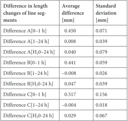
The study showed that polymerization shrink-age cause length changes in particular segments. The average values of the differences in length changes of line segments A, B and C (with stan-dard deviation) are shown in Table 4. The aver-age linear shrinkaver-age after polymerization was ap-prox. 1.175%.
Discussion
Dye Penetrant Inspection
Making occlusal splints from 2 different ma-terials carries the risk of lack of a tight bond be-tween the layers. This situation can lead to prob-lems with microorganisms, discoloration and the separation of the components of the appliance. Knowing the shrinkage of methacrylate resins, it
Table 1. Plaquit lacquer thicknessobtained among 30 randomly selected locations on different samples
Thickness of Plaquit lacquer layer in [µm] magnification ×1500
1. 6.25 11. 6.17 21. 7.85 2. 6.01 12. 5.77 22. 5.14 3. 6.13 13. 5.36 23. 6.57 4. 5.53 14. 4.81 24. 8.09 5. 6.17 15. 5.31 25. 7.45 6. 4.96 16. 4.89 26. 4.88 7. 6.01 17. 5.14 27. 5.28 8. 5.12 18. 5.80 28. 4.80 9. 5.85 19. 7.69 29. 4.16 10. 5.20 20. 5.93 30. 8.57
Table 2. Vickers microhardness values obtained in the study (HV 0.05)
Specimen
number Number of Vickers microhardnes measurement[N/mm2]
I II III IV V
1. 7.6 7.3 7.43 7.22 7.7 2. 8.85 8.42 8.27 8.47 8.07
3. 8.42 7.74 8.63 8.63 8.58
4. 6.4 6.72 7.7 7.48 7.22
5. 6.26 6.5 7.0 6.83 6.75
6. 7.06 6.47 6.4 6.5 6.13
Table 3. Spearman’s rank-order correlation coefficients between Vickers microhardness and the depth of the light-cured resin layer on Erkodur foil in all the analyzed specimens
Specimen
number Number of measurements rs p
1. 5 0.1 0.87
2. 5 –0.7 0.19
3. 5 0.6 0.32
4. 5 0.6 0.28
5. 5 0.6 0.28
seems appropriate to use thermocycling to reflect the clinical situation before samples are subjected to dye [19]. Out of 60 measurements done from the vestibular and lingual sides, only one instance of dye penetration to the adhesive connection was observed. This one case is not statistically signifi-cant. There was no dye penetration into the deep-er structure of the resin; dye only penetrated the lacquer coating layer. However, due to its small average thickness of 5.975 µm, this technique of making occlusal appliances can still be regarded as resistant to coloring agents. The additional test us-ing an isolatus-ing layer confirmed the methodologi-cal validity of the test performed. Research by Arias et al. and other authors has shown that the proce-dures followed in the current study are appropriate for evaluating the penetration of the dye into the adhesive connection [20–23]. Omitting the lacquer coating is not recommended, but it should be em-phasized that it is not absolutely necessary.
Vickers Microhardness Test
In the light of contemporary knowledge it seems reasonable to use the Vickers method to measure the microhardness of thin, hard coatings. Sharp and well-formed imprints were obtained us-ing this procedure, which ensured the accuracy of the results. The study showed that changes in the Vickers microhardness with the increasing depth of the test material are not statistically significant. It can be concluded that the layer of light-cured resin is fully polymerized throughout its entire thickness. When compared to the results pub-lished by Danesh et al., in which the microhardness
of different available light-cured and chemically hardened resins was measured [24], the average microhardness value obtained in the current study is lower. It should be noted that in both studies the same prescribed values of load and pressing time were applied. The average values of Vickers micro-hardness given by Chuenarrom et al. for both den-tin and tooth enamel are much higher than the val-ue of microhardness obtained for Lightdon Splint resin [25]. In fact, from the analysis of these da-ta it can be concluded that the tested material has a lower hardness level than other available resins.
Polymerization Shrinkage Test
Polymerization shrinkage is one of the big-gest and most important problems associated with the use of light-cured resins, and it occurs in the hardening process of every polymer. In occlusal splint therapy and restorative dentistry the phe-nomenon has considerable significance. The main task of occlusal appliances is to provide occlusal stability. When it is necessary to make an occlu-sal splint, either a direct method (in the patient’s mouth) or an indirect method (in an articulator) can be used. Thanks to light-cured resins, more time can be spent forming and properly shaping the occlusal appliance. Even if the model of the splint is close to perfect, a high level of polymer-ization shrinkage could ruin the work and disrupt the vertical dimensions as well as the occlusal con-tacts. A research study carried out by Cattani-Lor-ente et al. showed that the linear shrinkage of pop-ular composite resins used to restore hard dental tissues ranged from 1.54% to 2.11% [26].In anoth-er study, Arora et al. presented the linear polymanoth-er- polymer-ization shrinkage of 4 available acrylate materials polymerized at high temperature as ranging from 0.37% to 1.18% [27]. In the current study the lin-ear polymerization shrinkage of Lightdon Splint resin was calculated as approximately 1.175%; this is a reasonably low degree of shrinkage, which definitely makes clinical work easier. However, it must be emphasized that the length changes of line segments A, B and C in set I (the length measured before polymerization and the length measured 1 h after polymerization) were statistically more significant than the changes in sets II and III. In their research, Inoue et al. showed that the type of light, its intensity and time exposure have a signif-icant influence on polymerization shrinkage [28]. It is therefore very important to follow the polym-erization instructions specified by the manufac-turer. The influence of time and environment on the material were taken under consideration dur-ing the tests conducted in the current study. It is commonly known that one of the properties that
Table 4. Average values and standard deviation of length differences of line segments A, B and C obtained during the polymerization shrinkage test under changing envi-ronmental conditions and time
Difference in length changes of line seg-ments
Average difference [mm]
Standard deviation [mm]
Difference A[0–1 h] 0.450 0.071 Difference A[1–24 h] 0.008 0.039 Difference A[H20–24 h] 0.040 0.079
Difference B[0–1 h] 0.441 0.059 Difference B[1–24 h] –0.008 0.026 Difference B[H20-24 h] 0.047 0.039
characterizes polymers is their ability to absorb water from the environment; the organic matrix and inorganic filler content are responsible for that process. The absorption of water leads to the deg-radation of polymer bonds due to hydrolysis [29], which means destabilization of the occlusion may occur due to increasing expansion of the material. In their study, Danesh et al. proved that in light-cured resins the absorption of water is much high-er than among the chemically-hardened polymhigh-ers used in the manufacture of occlusal splints [30]. These considerations led the authors of the cur-rent study to investigate whether the absorption of water from the environment would have a sig-nificant influence on the behavior of the polymer. The test did not reveal any statistically significant changes in the lengths of line segments A, B or C in sets II and III. This result indicates that nei-ther the passage of time after polymerization nor
the humidity of the environment have any impli-cations in terms of increasing the volume of the tested resin. Clinically, this means there would be no destabilization of occlusion.
Within the limitations of this in vitro study it was concluded that neither the light-cured res-in nor the adhesive connection between the resres-in and the thermoformable plate showed susceptibil-ity to dye penetration. The application of Lightdon Bonding improves the contact tightness between the tested resin and thermoformable foil. Coating the resin with lacquer is not absolutely necessary. The microhardness of the resin after polymeriza-tion is consistent. The maximum polymerizapolymeriza-tion shrinkage occurs immediately after curing. A moist environment and the passage of time do not desta-bilize the resin structure after polymerization. The light-cured resin tested may be an alternative ma-terial for use in occlusal splints manufacturing.
References
[1] de Kanter RJ, Truin GJ, Burgersdijk RC, Van‘t Hof MA, Battistuzzi PG, Kalsbeek H, Kayser AF: Prevalence in the Dutch adult population and a meta-analysis of signs and symptoms of temporomandibular disorder. J Dent Res 1993, 72, 11, 1509–1518.
[2] Poveda Roda R, Bagán JV, Díaz Fernández JM, Hernández Bazán S, Jiménez Soriano Y: Review of temporo-mandibular joint pathology. Part I: Classification, epidemiology and risk factors. Med Oral Patol Oral Cir Buca 2007, 12, 4, 292–298.
[3] LeResche L: Epidemiology of temporomandibular disorders: implications for the investigation of etiologic factors. Crit Rev Oral Biol Med 1997, 8, 3, 291–305.
[4] Okeson JP: Long-term treatment of disk-interference disorders of the temporomandibular joint with anterior repositioning occlusal splints. J Prosthet Dent 1988, 60, 5, 611–616.
[5] Ramfjord SP, Ash MM: Reflections on the Michigan occlusal splint. J Oral Rehabil 1994, 21, 5, 491–500.
[6] Dao TT, Lavigne GJ: Oral Splints: the Crutches for Temporomandibular Disorders and Bruxism? Crit Rev Oral Biol Med 1998, 9, 3, 345–361.
[7] Fayed MM, El-Mangoury NH, El-Bokle DN, Belal AI: Occlusal splint therapy and magnetic resonance imaging. World J Orthod 2004, 5, 2, 133–140.
[8] Noriyuki N, Masahiko F, Tomohiro I, Kazunobu K, Toshihiko M: Effects of jaw clenching while wearing an occlu-sal splint on awareness of tiredness, bite force, and EEG power spectrum. J Prosthot Res 2009, 53, 3, 120–125.
[9] Karolyi M: Beobachtungen über Pyorrhea Alveolaris. Oesterr-Ung Vierteljahrschr Zahnheilk 1901, 17, 279–283.
[10] Tandon R, Gupta S, Kumar Agarwal S: Denture base materials: From past to future. Indian J Dent Sci 2010, 2, 2, 33–39.
[11] Seppäläinen AM, Rajaniemi R: Local neurotoxicity of methyl-meth-acrylate among dental technicians. Am J Ind Med 1984, 5, 6, 471–477.
[12] Magdaleno F, Ginestal E: Side effects of stabilization occlusal splints: a report of three cases and literature review. Cranio 2010, 28, 2, 128–135.
[13] dos Santos Jr J, Gurklis M: Immediate fabrication of occlusal bitesplints using visible light-cured material. Compend Contin Educ 1994, 15, 2, 228–232.
[14] DaneshG, LippoldC, JoosU, MeyerU: Technical and clinical assessment of the use of a new material-based splint in orthognathic surgery. Int J Oral Maxillofac Surg 2006, 35, 9, 796–799.
[15] Terai H, Shimahara M: Intermaxillary fixation using thermoforming plate. J Oral Maxillofac Surg 2002, 60, 9, 1092–1094.
[16] Lightdon Splint. Technical Data Sheet. Unna, Germany 2010.
[17] Lightdon Splint. Safety Data Sheet. Unna, Germany 2007.
[18] European Norm EN ISO 6507-1, 2005.
[19] Pazinatto FB, Campos BB, Costa LC, Atta MT: Effect of the number of thermocycles on microleakage of resin composite restorations. Pesqui Odontol Bras 2003, 17, 4, 337–341.
[20] Arias VG, Campos IT, Pimenta LAF: Microleakage Study of Three Adhesive Systems. Braz Dent J 2004, 15, 3, 194–198.
[22] Al-Boni R, Raja OM: Microleakage evaluation of silorane based composite versus methacrylate based composite. J Conserv Dent 2010, 13, 3, 152–155.
[23] Koyuturk AE, Kusgoz A, Ulker M, Yesilyurt C: Effects of mechanical and thermal aging on microleakage of dif-ferent fissure sealants. Dent Mater J 2008, 27, 6, 795–801.
[24] DaneshG, LippoldC, Ziebura T, Reinhardt KJ, Schafer E, Ehmer U:In vitro investigations on suitability of light- -cured resins for interocclusal splints: part II: Surface Hardness. J Orofac Orthop 2006, 67, 2, 138–147.
[25] Chuenarrom C, Benjakul P, Daosodsai P: Effect of indentation load and time on Knoop and Vickers Microharness Tests for enamel and dentin. Mater Res 2009, 12, 4, 473–476.
[26] Cattani-Lorente M, Godin Ch, Bouillaguet S, Meyer JM: Linear polymerization shrinkage of new restorative composite resins. Eur Cells Mater 2003, 5, 1, 40–41.
[27] Arora S, Khindaria SK, Garg S, Mittal S: Comparative evaluation of linear dimensional changes of four com-mercially available heat cure acrylic resins. Contemp Clin Dent 2011, 2, 3, 182–187.
[28] Inoue K, Howashi G, Kanetou T, Masumi S, Ueno O, Fujii K: Effect of light intensity on linear shrinkage of photo-activated composite resins during setting. J Oral Rehabil 2005, 32, 1, 22–27.
[29] Toledano M, Osorio R, Osorio E, Fuentes V, Prati C, Garcia-Godoy F: Sorption and solubility of resin-based restorative dental materials. J Dent 2003, 31, 1, 43–50.
[30] Danesh G, Lippold C, Mischke KL, Varzideh B, Reinhardt KJ, Dammaschke T, Schäfer E: Polymerization char-acteristics of light- and auto-curing resins for individual splints. Dent Mater 2006, 22, 5, 426–433.
Address for correspondence:
Mieszko Więckiewicz
Division of Dental Materials, Faculty of Dentistry Wroclaw Medical University
Krakowska 26 50-425 Wrocław Poland
Tel.: +48 660 47 87 59
E-mail: [email protected]
Conflict of interest: None declared