International Journal of Nanomedicine
Nanostructured self-assembling peptides
as a defined extracellular matrix for long-term
functional maintenance of primary hepatocytes
in a bioartificial liver modular device
Shibashish Giri1
Ulf-Dietrich Braumann2,3 Priya Giri1,3
Ali Acikgöz1,4 Patrick Scheibe3,5 Karen Nieber6 Augustinus Bader1 1Department of Cell Techniques
and Applied Stem Cell Biology, Center for Biotechnology and Biomedicine (BBZ), 2Institute for
Medical Informatics, Statistics, and Epidemiology (IMISE), University of Leipzig, Leipzig, Germany;
3Interdisciplinary Center for
Bioinformatics (IZBI), University of Leipzig, Leipzig, Germany;
4Klinikum St Georg, Leipzig, Germany; 5Translational Center for Regenerative
Medicine (TRM Leipzig), 6Department
of Pharmacology for Natural Sciences, Institute of Pharmacy, University of Leipzig, Leipzig, Germany
Correspondence: Shibashish Giri Department of Cell Techniques and Applied Stem Cell Biology, Center for Biotechnology and Biomedicine, University of Leipzig, Deutscher Platz 5, D-04103 Leipzig, Germany
Tel +49 34 1973 1353 Fax +49 34 1973 1329
Abstract: Much effort has been directed towards the optimization of the capture of in vivo hepa-tocytes from their microenvironment. Some methods of capture include an ex vivo cellular model in a bioreactor based liver module, a micropatterned module, a microfluidic 3D chip, coated plates, and other innovative approaches for the functional maintenance of primary hepatocytes. However, none of the above methods meet US Food and Drug Administration (FDA) guidelines, which recommend and encourage that the duration of a toxicity assay of a drug should be a minimum of 14 days, to a maximum of 90 days for a general toxicity assay. Existing innovative reports have used undefined extracellular matrices like matrigel, rigid collagen, or serum supplementations, which are often problematic, unacceptable in preclinical and clinical applications, and can even interfere with experimental outcomes. We have overcome these challenges by using integrated nanostructured self-assembling peptides and a special combination of growth factors and cytokines to establish a proof of concept to mimic the in vivo hepatocyte microenvironment pattern in vitro for predicting the in vivo drug hepatotoxicity in a scalable bioartificial liver module. Hepatocyte functionality (albumin, urea) was measured at days 10, 30, 60, and 90 and we observed stable albumin secretion and urea function throughout the culture period. In parallel, drug metaboliz-ing enzyme biomarkers such as ethoxyresorufin-O-deethylase, the methylthiazol tetrazolium test, and the lactate dehydrogenase test were carried out at days 10, 30, 60, and 90. We noticed excellent mitochondrial status and membrane stability at 90 days of culture. Since alpha gluta-thione S-transferase (GST) is highly sensitive and a specific marker of hepatocyte injury, we observed significantly low alpha GST levels on all measured days (10, 30, 60, and 90). Finally, we performed the image analysis of mitochondria-cultured hepatocytes at day 90 in different biophysical parameters using confocal microscopy. We applied an automatic algorithm-based method for 3D visualization to show the classic representation of the mitochondrial distribution in double hepatocytes. An automated morphological measurement was conducted on the mito-chondrial distribution in the cultured hepatocytes. Our proof of concept of a scalable bioartificial liver modular device meets FDA guidelines and may function as an alternative model of animal experimentation for pharmacological and toxicological studies involving drug metabolism, enzyme induction, transplantation, viral hepatitis, hepatocyte regeneration, and can also be used in other existing bioreactor modules for long-term culture for up to 90 days or more.
Keywords: image analysis, 3D visualization, bioreactor, FDA guidelines, primary hepato-cytes, hepatotoxicity
Introduction
Despite the tremendous efforts in hepatic tissue engineering,1 there is a lack of defined
long-term functional maintenance of primary hepatocytes. Much encouraging effort
Dove
press
O R I G I N A L R E S E A R C h
open access to scientific and medical research
Open Access Full Text Article
International Journal of Nanomedicine downloaded from https://www.dovepress.com/ by 118.70.13.36 on 23-Aug-2020
For personal use only.
Number of times this article has been viewed
This article was published in the following Dove Press journal: International Journal of Nanomedicine
has been made to optimize the capturing of the in vivo hepa-tocytes microenvironment for functional maintenance of primary hepatocytes in a micropatterned co-culture module,2
bioreactor-based liver module,3–5 microfluidic 3D chip,6 and
other innovative approaches.7–9 The most encouraging reports
relied on undefined extracellular matrices like Matrigel, rigid collagen, or serum supplementations, which are often problematic and unacceptable in preclinical and clinical applications. Moreover, animal-derived extracellular matri-ces can give rise to immunological conditions and zoonosis. The paracrine fashion secretion pattern of several soluble factors from feeder cells for primary cells cultures10 is more
suitable, but it is much more difficult to test the effect of the exogenous factors on the functional maintenance of primary hepatocytes. However, most experimental approaches aimed at the long-term functional maintenance of primary hepato-cytes in a culture from 1 week to several weeks (42 days) have been reported by various investigators,2–4 including us.11–13
However, to the best of our knowledge, there is no report for the long-term functional maintenance of primary hepatocytes up to 90 days within a defined hepatic microenvironment to provide a better strategy as an alternative to in vivo animal experimentation. US Food and Drug Administration (FDA) rules require the survival potential of primary hepatocytes of a minimum of 14 days and a maximum of 90 days for in vivo toxicological experiments.14 We have designed
nano-structured self-assembling peptides as a defined extracellular matrix (ECM) for the long-term functional maintenance of primary hepatocytes in a defined bioartificial liver modular device. Our bioartificial liver modular device in its ex vivo form aims to mimic the in vivo liver for the next genera-tion of ex vivo drug screening and many other liver related experiments. Our bioartificial liver device faithfully mimics the in vivo liver15 in various ways11,17 and was previously
evaluated in a novel porcine hepatectomy model16 and viral
infection.18 The present proof of concept study of the
bio-artificial liver modular device could replace the millions of animals that are currently sacrificed in preclinical testing and could open up a new vista for the production of safer pharmaceuticals. The integration of nanostructured self-assembling peptides into a bioartificial liver modular device to generate 3D cell interactions with a special combination of cytokines and growth factors is the focus of this study. Fundamentally, in vivo hepatocytes interact with their com-plex surrounding environment through exposure to a network of cytokines, chemokines, and growth factors19 to perform
and regulate hepatic cellular functions. The ECM is one of the key components of the hepatic microenvironment and
serves as a reservoir for several soluble factors that cells can use for various functions. In vivo hepatocytes interact with soluble signals via the ECM that controls the diffusion and activity of soluble factors that interact with the surrounding environment. Since the in vivo ECM is in the nanorange, to create such a nanoscale ECM for hepatocytes culture is challenging. Furthermore, cells acquire complex 3D geom-etries for intricate interactions with adjacent cells, cytokines, and growth factors.20 The nanoscale ECM can surround each
hepatocyte cell (10–20 µM) in 3D rather than the partial attachment observed in the conventional 2D scaffold. Such a nanoscale scaffold allows each hepatocyte cell to have its own position in direct contact and to interact with the network of cytokines, chemokines, and growth factors in the cultiva-tion media. The long-term maintenance of the hepatocyte function and optimization of the culture conditions under a defined hepatic microenvironment is one of the most signifi-cant challenges in developing an effective bioartificial liver model in addition to the drug discovery process. Current trends of in vitro hepatic toxicological models do not reflect the in vivo hepatic microenvironments. According to a 2006 survey report of pharmaceutical companies, hepatotoxicity was ranked first in terms of adverse events and it remains the most common reason for the restriction or withdrawal of a drug from the market by the FDA. Although there are many reasons underlying drug-induced hepatotoxicity, one of the most important is the limitation of existing in vitro cellular hepatocyte models. Short-term culture very often suffers from rapid loss of hepatic cellular activity due to 2D interaction, unlike the in vivo milieu. Consequently, it is rather difficult to use these short-term models for long-term experiments involving drug metabolism, virus infection, carcinogenesis, and other such studies.
To integrate a defined and highly biocompatible sub-strate into a bioartificial liver system that could support the long-term maintenance of hepatocyte function is one of the prominent challenges in developing effective bioartificial liver support as well as drug discovery. We have used a self-assembling peptide as a defined substrate or ECM for ex vivo long-term functional maintenance of primary hepa-tocytes under 3D defined culture conditions in a bioartificial modular device. These self-assembling peptides comprise a RAD motif, which is a 16-residue peptide composed of alternating hydrophilic arginine, hydrophobic alanine, and hydrophilic aspartic acid (RADARADARADARADA). The most common well-known motif cell adhesion sequence is RGD (arginine-glycine-aspartate). It is a tri-peptide and its sequence can be found in proteins of the ECM or cell Dovepress
Giri et al
International Journal of Nanomedicine downloaded from https://www.dovepress.com/ by 118.70.13.36 on 23-Aug-2020
adhesion proteins. The RGD peptide motif was discovered to be a major element in the recognition system for essential cell adhesion.21 Although the RGD peptide motif is the most
common element to use for cell adhesion and culture, only some RGD-containing proteins can support cell adhesion22
despite the large number of RGD-containing proteins. There-fore, in vitro cultured cells may not come into contact with these specific RGD-containing proteins during cell adhesion and expansion and will eventually be lost. This is because the RGD sequences are not freely available for in vitro cultured cells during cell adhesion and expansion. Another disad-vantage of these conventional RGD-containing extracellular matrices is that sometimes the RGD motif itself may not be compatible with integrin binding.23 Therefore, here we
inte-grated self-assembling peptides that contain the RAD motif as a defined substrate for cell adhesion as they are more com-patible for cell attachment three-dimensionally. Interestingly, the RAD motif in self-assembling peptides mimics the known cell adhesion properties of the RGD motif.24 Previously, the
RAD motif self-assembling peptide had been used for in vitro 3D culture for a wide range of cells. We have reported 3D scaffolding and signaling in bioartificial liver modules for up to 35 days under exposure of various growth factors, cytokines, and hormones, but here we have optimized and standardized a novel in vitro 3D defined culture system in a bioarticial liver (BAL) module up to 90 days of culture to fulfill the FDA guidelines for general toxicological studies. To the best of our knowledge, we are the first group to use self-assembling peptides for ex vivo long-term functional maintenance of primary hepatocytes in a clinically relevant bioartificial liver module.
Hepatocyte functionality (albumin, urea) was measured at days 10, 30, 60, and 90. We evaluated the potential for hepatotoxicity by analyzing drug metabolizing enzyme bio-markers such as ethoxyresorufin-O-deethylase, the methylthi-azol tetrmethylthi-azolium (MTT) test, and the lactate dehydrogenase (LDH) test at days 10, 30, 60, and 90. Liver failure is linked by the common pathway of mitochondrial failure.25 Thus, we
aimed to perform image analysis of mitochondria of single or double hepatocyte cells. Currently, there are few methods26
to quantify the mitochondrial image, but we have shown a 3D visualization, which revealed a classic representation of the mitochondrial distribution around the liver cell. The idea of recreating a 3D defined hepatic microenvironment has become very challenging owing to the establishment of the structural and functional complexity of in vivo hepatocytes in an ex vivo bioartificial liver module. This optimized 3D defined culture system in bioartificial liver modules could
be useful for physiological and pathophysiological states of the liver, pharmacological and toxicological screening of drug candidates, and generation of clinical grade hepa-tocyte cells.
Materials and methods
Cell culture
Hepatocytes were isolated from male Sprague-Dawley rats (weighing 200–250 g) by the two-step collagenase perfusion method, as previously described.27 Pure hepatocytes were
obtained by Percoll gradient separation. Cell viability was assessed by trypan blue exclusion and hepatocytes with a viability of greater than 85%–90% were used. Hepatocytes were cultured in Williams’ E medium supplemented with L-glutamine 2 mM, penicillin 100 U/mL, streptomycin 100 µg/mL, dexamethasone 1 µM, insulin 0.2 U/mL, and glucagon 4 ng/mL without fetal bovine serum. The cells were seeded at a density of 2.5 × 10⁵ cells/cm2 in a RAD
peptides coated bioreactor under a defined combination of growth factors and cytokines (Williams’ E medium +
Activin A + WNT 3a + HGF + FGF-4 + EGF + Oncostatin M + FGF-10 + BMP-4 + Dex + RA all from Sigma-Aldrich Chemie Gmbh [Munich, Germany] to mimic the in vivo hepatic microenvironment)28 and were incubated at 37°C in a
humidified atmosphere containing 5% CO2 and 20% O2 (v/v). The fundamental design of our flat membrane bioreactor11
and the minibioreactor13 is similar. Funtional maintenance of
primary hepatocytes was conducted three times.
Self-assembling peptides coated procedure in bioartificial liver module
The self-assembling peptide was discovered from a segment in a yeast protein by Zuotin29,30 and is now commercially
available under the name PuraMatrix™ (BD Biosciences, San Jose, CA, USA). The viscosity of the self-assembling peptides stock solution of our aliquots (1.5 mL microtube) decreased by vortexing for 30 minutes in a bath sonicator. If air bubbles were present, the aliquots were centrifuged at high speed for a few seconds. Then, a 0.5% solution of self-assembling peptides was prepared by diluting it with sterile water. In order to create the nanoscaffold in the six-well bioreactor, 1.2 mL of 0.5% (v/v) self-assembling peptides (300 µL for a 24-well bioreactor, 1 mL for a six-well bioreac-tor, and 75 µL for a 96-well format bioreactor) was uniformly distributed over each well and then 2.4 mL of Williams’ Medium E was added very carefully to each well of the six-well bioreactor. To promote gelation, we put the bioreactor in an incubator for 1 hour. After the nanostructure hydrogel was Dovepress Nanostructured self-assembling peptides and maintenance of hepatocytes
International Journal of Nanomedicine downloaded from https://www.dovepress.com/ by 118.70.13.36 on 23-Aug-2020
assembled, we carefully changed the medium using a wide top micropipette. Aspirator use was avoided because of the risk of destroying the nanostructured hydrogel. The medium was changed (300 µL per well for 24 wells) twice over a period of 1 hour to equilibrate the gel at physiological pH and finally the bioreactor was put in an incubator overnight with the medium. After that we seeded the desired cell density. The culture medium (250 µL) was replaced with fresh medium every 78 hours, and the supernatant was stored at −20°C for LDH, albumin, and urea.
RNA isolation
Total RNA was isolated from the cells using the RNeasy Mini Kit (Qiagen, Hilden, Germany) according to the manufac-turer’s instructions. RNA concentration was quantified and their purity was determined by standard spectrophotometric methods. RNA samples were stored at −80°C.
cDNA synthesis and reverse transcription (RT) polymerase chain reaction (PCR)
Total RNA was isolated from the cultured cells using an RNeasy MinElute Cleanup Kit (Qiagen) and then 8 µg of the RNA was reverse transcribed into cDNA from cultured rat hepatocytes, prepared using the Fastlane Cell cDNA kit (Qiagen) according to the manufacturer’s instructions. Quantitative real-time PCR was performed using SYBR Green PCR Master Mix (Applied Biosystems, Carlsbad, California). Primers for rat albumin and CYPA31 and GAPDH genes were selected (rat CYP3A1-specific primer set: forward, 5′ GCCATCACGGACACAGAAATA 3′; reverse, 5′ GAACGTGGGTGACAGTAAGGCT 3′; rat
albumin forward, 5′ ATACACCCAGAAAGCACCTC
3′; reverse, 5′ CACGAATTGTGCGAATGTCAC 3′; rat housekeeping gene forward 5′ CAG TTC CAC CCA CCT CAG AT 3′ and reverse 5′ TTT TGG GCT CCT TCA GAG TG 3′). Primer (Sigma-Aldrich) concentrations were optimized before use. SYBR Green Master Mix (1×) was used with 1 µM of forward and reverse primers in a total volume of 12 µL that also included 1 µL of cDNA. All PCR reactions were performed in duplicate. PCR amplification was performed as follows: 95°C for 10 minutes, 40 cycles of 95°C for 15 seconds, 60°C for 10 seconds, and 68°C for 1 minute on a Mastercycler Realplex (Eppendorf, Hamburg, Germany). The PCR products were then analyzed using the electrophoresis of 2% agarose gels stained with ethidium bromide for visualization. The housekeeping gene (GAPDH) was used as an internal standard to determine the relative levels of albumin, albumin and CYP3A1 gene expression.
Biochemical assays
Albumin and urea were measured by enzyme-linked immu-nosorbent assay, as previously described.27 LDH activity was
determined in a colorimetric enzymatic assay (Cytotoxicity Detection Kit, Cat No 1644793; Roche, Mannheim, Germany). For induction of CYP3A1 expression, dexam-ethasone (as a 10-mM solution in dimethyl sulfoxide; final concentration: 10 µM) was added to the replacing medium on various days: day 10, day 30, day 60, and day 90. Cell viabil-ity was then assessed by 3-(4,5-dimethylthiazol-2-yl)-2,5-diphenyltetrazolium bromide MTT assay (Sigma-Aldrich). The protocol of MTT assay ethoxyresorufin-O-deethylase is given in our previous report.31α-glutathione S-transferase
(GST) was measured in the culture supernatant as a marker for hepatic damage by a commercially available enzyme immunoassay method (Biotrin Rat Alpha GST EIA; Biotrin, Dublin, Ireland).
Statistical analysis
All experimental results are shown as mean ± standard error, and analyzed for significance using Wilcoxon’s test for non-paired examination. P-values of less than 0.05 were judged to be statistically significant.
Image analysis of confocal microscopy of mitochondria
We analyzed mitochondrial structural status by confocal microscopy by using MitoTracker (Life Technologies), a dye used for fluorescent mitochondrial markers. This was simply incubated in submicromolar concentrations (dilution with culture medium: 1:1000) of the MitoTracker probe in cultured hepatocytes and directly observed with the fluorescent microscope. In general, MitoTracker probes are cell-permeant mitochondrion-selective dyes that diffuse passively across the plasma membrane and accumulate in active mitochondria and stain mitochondria with a bright red, fluorescein-like fluorescence.
Visualization and quantification of mitochondrial distribution
Mitochondria play a central part in cellular survival32,33
and apoptotic death. With time, liver cells malfunction and die because of a lack of energy. A central factor in liver cell survival is the decay of the mitochondria in cells. The survival/apoptosis of cells is accompanied by an increase/ decrease in the numbers of mitochondria per cell. So, from the experimentalist point of view, it is important to know the mitochondrial distribution around the liver cell nucleus. Dovepress
Giri et al
International Journal of Nanomedicine downloaded from https://www.dovepress.com/ by 118.70.13.36 on 23-Aug-2020
When cells come from in vivo to in vitro, they require an in vivo-like microenvironment. In long-term culture, cells are unable to get the appropriate in vivo-like microenvironment. Under this stress condition, mitochondria are unable to survive and they swell and finally burst. Currently there are few methods to quantify the mitochondria, but apart from these methods, we have explained 3D visualization which shows a classic representation of mitochondrial distribu-tion around liver cells. Experimental outcomes have been observed using confocal microscopy and microscopy of cultured hepatocytes has been stained using MitoTrackers. High quality fluorescence images were captured for the subsequent quantitative analysis of mitochondria using the computer algebra system Mathematica (v 8; Wolfram Research, Inc, Champaign, IL, USA), which offers a variety of image processing functionalities.
Data-set and an image processing pipeline
In this section we describe the data set (Figure 1), followed by the image processing pipeline according to Figure 2. Three-dimensional volume data sets of mitochondria in rat liver cells expressing MitoTrackers were acquired by the digitization of liver cell cultures using confocal microscopy with a 10 × objective. Respective 2D image stacks have dimensions of 1024 × 1024 × n voxels, and n is adapted according to cell extent. Each 2D image in a stack is in 24 bit color depth which is converted into scalar 8 bit for processing.
We have noticed some artifacts in the middle of the nucleus region of a 2D image. There is some a priori knowl-edge about an acceptable minimum mitochondrial site, and apart from this, the selected 2D slide has been taken from a 3D stack and the artifacts shadow comes from the other slide.
3D visualization results
The ability of a confocal microscope to create sharp opti-cal sections makes it possible to build 3D renderings of the specimen. Data collected from a series of optical sections imaged at short and constant intervals along the optical axis are used to create some 3D visualization. Results are shown in Figure 1A and B and are realized as shaded 3D surface rendering. In the data, however, despite reflecting a 3D topology, the cell appears to be implausibly squeezed, so that the visible mitochondria enclosing the nucleus (not stained) occur along a ring instead of a sphere. That is why for further quantitative assessments we decided to stick to selected single images taken centrally from the volume
data set. For this work we rather take the 3D visualizations of the location of mitchondria around nucleous.
Image processing pipeline 2D
Confocal laser scanning microscopy (CLSM) image data (Figure 2A), has a certain degree of noise due to image capture conditions; eg, due to the statistical characteristic of light, scattering, etc. In order to reduce this effect we applied a smoothing operation implemented as an edge preserving total variation filtering,34 assuming Poisson noise. The results
are shown in Figure 2B. Due to a particularly high variabil-ity of fluorescence intensvariabil-ity and contrast, for mitochondria segmentation, we used an iterative active-contour level-set segmentation in its variant proposed by Chan and Vese,35 as
shown in Figure 2C. For further processing we required a number of cell masks according to the respective number of cells. We applied a dilation operation,36 ie, a Minkowski-sum
of binary image and a circular area structuring element, to have the mitochondria segments merged and to get some connected segments according to the number of cells. Connected component labeling37 was accomplished to allow
for small element (artifact) removal. Overall, cell masks were then computed according to the connected segment’s convex hulls,38 as shown in Figure 2D. Further, we needed
to take the difference of the masks and label region shown in Figure 2D, then needed to do another connected compo-nent labeling to sort out small noncentral rests (Figure 2E). We then needed to compute the convex hulls of the large inner regions to get the inner masks of cell nuclei; output is shown in Figure 2F. Further, we subtracted the nuclei masks from the outer masks to obtain representative ring-shaped mitochondrial distribution regions, as shown in Figure 2G. Since we were interested in the mitochondrial distribution around the nuclei, we focused on the characteristics of the margin thickness. The thickness measurement was taken along the course of a closed circular line as was obtained by skeletonization39 of the ring, while distances were
com-puted using Euclidean distance transform39 applied on the
ring (Figure 2H).
Results
Morphological analysis of long-term
culture
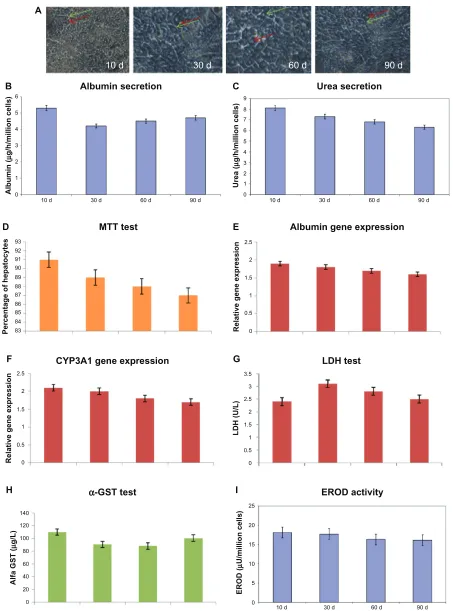
Cultured hepatocytes were continuously observed under phase-contrast microscopy at day 10, day 30, day 60, and day 90. All hepatocytes showed well-defined cell membranes, nuclei and nucleoli, bile canaliculi, and distinct cytoplasm (Figure 3A). Hepatocytes cultured in the bioartificial liver Dovepress Nanostructured self-assembling peptides and maintenance of hepatocytes
International Journal of Nanomedicine downloaded from https://www.dovepress.com/ by 118.70.13.36 on 23-Aug-2020
module adopted an in vivo like polygonal and established extensive cell-to-cell contracts with natural organization.
Stability of albumin and urea secretion at various days
The albumin secretions were 26.4 ± 5.1 mg/mL, 24.2 ± 6.3 mg/mL, 21.8 ± 3.3 mg/mL, and 25.4 ± 1.3 mg/mL at day 10, day 30, day 60, and day 90, respectively. These results showed stable albumin secretion throughout the culture period (Figure 3B). The urea secretions were 46.4 ± 2.1 mg/mL, 39.2 ± 2.3 mg/mL, 34.8 ± 8.3 mg/mL, and 40.4 ± 2.3 mg/mL at day 10, day 30, day 60, and day 90,
respectively (Figure 3C). These results showed stable albu-min and urea secretion throughout the culture period. So, under the appropriate 3D hepatic culture conditions, cultured hepatocytes might recover from the stress of the isolation procedure from the in vivo liver. If one can count per cell, the present showed that the albumin and urea performance per cell was almost similar to that in the in vivo liver.
hepatocyte cell number at day 10, day 30, day 60, and day 90
Cell death was assessed by MTT assay in primary hepato-cytes and it was found that there is no significant cell death 0
20
40
x [µM]
y [µM] z [µM]
60
0
20 40
60
0 2 4 6 A
0
20
40
x [µM]
y [µM] z [µM]
60
80
0 20
40 60
80
0 2 4 6 B
Figure 1 Mitochondriadata sets depicted in 3D visualization using shaded surface rendering.
Note: Asupplementary 3D movie is available to view: http://youtu.be/Alc_72FM9ho.
Dovepress
Giri et al
International Journal of Nanomedicine downloaded from https://www.dovepress.com/ by 118.70.13.36 on 23-Aug-2020
on these days. Mitochondria metabolism such as the reduc-tion of a tetrazolium salt (MTT) was used here. Although primary hepatocytes did not proliferate significantly during long-term maintenance, they remained excellent and viable up to 90 days, as demonstrated by the MTT assay. There was no significant difference in cell viability between day 30, day 60, and day 90 (Figure 3D).
The number of mitochondria is higher at the end day of the culture period (day 90)
Since mitochondria is the main target of cellular stress, apop-tosis, and ageing, in these conditions the shape of the mito-chondria changes and becomes swollen. The mitomito-chondria
of primary hepatocytes cultured in the 3D hepatic defined microenvironment in the bioartificial liver module were very well shaped and uniformly distributed throughout the cytoplasm, which was confirmed by confocal microscopy at day 90 (Figure 4). It was interesting to find a high number of mitochondria present in culture hepatocytes at day 90 which was noticed by confocal microscopy using MitoTracker red. The mitochondria were found throughout the cytoplasm.
Gene expression of albumin and CYP3A1
RT-PCR analysis revealed that the albumin and CYP3A1 gene expression was also stable throughout the culture period (Figure 3E and F). The mRNAs of albumin and CYP3A1
A
Confocal laser scanning microscopy image (mitochondria staining)
•
B
Total-variation filtering
•
C
Binarization
•
Dilation
•
Labeling
•
D
Artifact removal
•
Separation
•
Convex hulls (cell masks)
•
E
Subtraction: cell masks – dilated mitochondria regions
•
F
Nuclei region isolation
•
Convex hull of cell nuclei regions
•
G
Subtraction of convex hull (D and F)
•
Ring-shaped mitochondria regions
•
H
Euclidean distance mapping
•
Skeletonization of mitochondria regions
•
Labeling
•
Figure 2 Proposedimage processing pipeline towards 2D mitochondrial distribution assessment. (A) Confocal laser scanning microscopy image (mitochondria staining); (B) total-variation filtering; (C) binarization, dilation, labeling; (D) artifact removal, separation, convex hulls (cell masks); (E) cell masks minus mitochondria regions, labeling; (F) nuclei region isolation, convex hull of cell nuclei regions; (G) subtraction of convex hulls (D and F), ring-shaped mitochondria regions; (H) Euclidean distance mapping, skeletonization of mitochondria regions.
Notes: The single steps basically do not require any specific interaction; for example, the number of cells can be implicitly determined by means of size considerations referring to the label images. What the pipeline eventually provides is some chain of mitochondria margin measurements obtained along the skeleton positions.
Dovepress Nanostructured self-assembling peptides and maintenance of hepatocytes
International Journal of Nanomedicine downloaded from https://www.dovepress.com/ by 118.70.13.36 on 23-Aug-2020
10 d 30 d 60 d 90 d
0
10 d 30 d 60 d 90 d
Albumin
(
µ
g/h/million cells)
1 2 3 4 5 6
Albumin secretion
83
Percentage of hepatocytes
84 85 87 86 88 89 90 91 92 93
Relative gene expression 0 0.5 1 1.5 2 2.5
MTT test Albumin gene expression
0
10 d 30 d 60 d 90 d
Urea
(
µ
g/h/million cells)
2 1 3 4 5 7
6 8 9
Urea secretion A
B C
D E
Figure 3 (A)Morphological analysis (scale bar 100 µM) at various days. Green arrows indicate bile duct and red arrows indicate nuclei. (B) Albumin secretion. (C) Urea secretion. (D) MTT test. (E) Gene expression of albumin. (F) Gene expression of CYP3A1. These gene expressions are normalized against the Housekeeping gene (GAPDH) as an internal standard. (G) LDh test; (H) α-GST test at various days (day 10, day 30, day 60, and day 90). (I) EROD activity.
Notes: Resultsare presented as the mean ± SD from three independent experiments. P, 0.05 was considered to be statistically significant.
Abbreviations: α-GST,alpha glutathione S-transferase; EROD, ethoxyresorufin-O-deethylase; LDH, lactate dehydrogenase; MTT, methyl thiazol tetrazolium; SD, standard deviation.
Relative gene expression 0 0.5 1 1.5 2 2.5
Alfa GST
(
µ
g/L)
0 20 40 60 80 140
120
100
EROD
(
µ
U/million cells
)
0 5 10 25
20
15
LDH (U/L)
0 0.5 1 1.5 2 2.5 3 3.5
CYP3A1 gene expression LDH test
α-GST test EROD activity
10 d 30 d 60 d 90 d
F G
H I
Dovepress
Giri et al
International Journal of Nanomedicine downloaded from https://www.dovepress.com/ by 118.70.13.36 on 23-Aug-2020
were detected at day 10, day 30, day 60, and day 90 which was also significantly high. The albumin mRNA and CYP3A1 were significantly expressed in the hepatocytes cultured in the bioartificial liver modules for up to 90 days. We confirmed that the defined hepatic microenvironment was highly important to maintain such hepatic gene expression throughout the period.
Stable hepatocyte membrane stability up to 90 days
The LDH test is frequently used to check for tissue dam-age as elevated levels of LDH may indicate liver damdam-age. The LDH release of cells is an index of their viability. It is a sign of the integrity of the plasma membrane since the LDH activity outside the cell is measured. The LDH releases were 2.4 ± 1.1 U/L, 4.2 ± 6.3 U/L, 2.8 ± 3.3 U/L, and 2.4 ± 1.3 U/L at day 10, day 30, day 60, and day 90, respectively (Figure 3G). These findings demonstrate that this present study has major advantages over the conventional in vitro culture regarding the cellular integrity.
Negligible amount of α-GST throughout culture period
The release of α-GST either from in vitro hepatocytes culture or in vivo liver is a more sensitive parameter than the release of conventionally used liver enzymes (aspartate aminotransferase, alanine aminotransferase, and LDH) in the assessment of early hepatocellular damage. α-GST is a cytosolic enzyme mostly located in hepatocytes with a uni-form distribution in the liver. Accumulating evidence from numerous clinical and experimental studies has shown that
α-GST is an early and sensitive biomarker for hepatocyte injury. Herein, we showed the α-GST was 110 ± 1.5 µg/L, 90 ± 6.8 µg/L, 78 ± 4.3 µg/L, and 100.4 ± 7.3 µg/L at day 10, day 30, day 60, and day 90, respectively (Figure 3H).
This highly sensitive method also supports that this 3D culture system in the bioartificial module provides ideal conditions for long-term functional maintenance.
Image analysis results
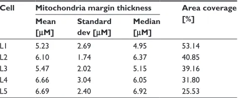
We have analyzed the mitochondria margins using the image processing pipeline in 2D, as was explained above. The results for the specimen with two cells, already depicted in Figure 1A and B, are given in Figure 4. Curves show the course of the mitochondria margin thickness drawn as a function of the normalized perimeter (obtained in counter-clockwise direction). Since cells appear arbitrarily rotated, curves were periodically shifted so that their minimum is placed at the zero position. Another example with three cells is given in Figure 5. Further characteristic numbers derived from the curves are given in Table 1 for all specimens; ie, mean, standard deviation, and median. Additionally, a simple measure named “area coverage,” addressing an approxima-tion of the mitochondria density, is given (see table capapproxima-tion for details).
Discussion
To the best of our knowledge, this is the first report for the long-term functional maintenance of primary hepatocytes up to 90 days under a defined 3D hepatic microenvironment in a bioartificial liver module device that mimics the state of the art liver. We optimized our bioartificial liver module model for long-term culture for up to at least 90 days to meet the FDA guidelines,14 which recommend that the duration of a
toxicity assay of a drug should be a minimum of 14 days, to a maximum of 90 days for a general toxicity assay. Repeated drug dose toxicity testing in animals for 28 and 90 days is used to evaluate chronic toxic effects to find out the observed primary toxic effect on various organ systems, including Norm perimeter
0
0.0 0.2 0.4 0.6 0.8 1.0
Margin
[
µ
M]
2 4 6 8 10 12
Figure 4 The first mitochondria margin measurement example plots describe the course of the determined mitochondria margin’s thickness (counterclockwise cycle). The cyan curve represents the upper right cell (L1)’s mitochondria margin, and the magenta curve represents that of the lower left cell (L2). Curves are plotted so that their global minimum is placed at the abscissa’s origin.
Dovepress Nanostructured self-assembling peptides and maintenance of hepatocytes
International Journal of Nanomedicine downloaded from https://www.dovepress.com/ by 118.70.13.36 on 23-Aug-2020
the liver, from minimal dose to maximum dose tolerance. Drug screening data obtained from unnatural conventional 2D systems is often confusing and matrigel, collagen based systems are often associated with unphysiological substances, while animal models are expensive, time consuming, and present ethical dilemmas. Herein, we reported a challeng-ing, alternative, novel, non-animal approach in a bioartificial liver modular platform to replace animal testing for assessing the chronic toxic effects of drug candidates to find out the observed primary toxic effects before further trials to the later clinical stage. The 3D defined hepatocyte culture system pres-ents the great challenge of maintaining hepatocyte cells for the extended period of time of up to 90 days that is required to assess the chronic toxic drug screening effects. The pres-ent proof of concept experimpres-ent generated great challenges with a new strategy (self-assembling peptides, growth factors, cytokines, enhanced oxygenation, and 3D signaling) for the long-term maintenance of primary hepatocytes in a bioar-tificial liver module using a defined microenvironment. We hope that this 3D interaction of growth factors and cytokines in nanostructured self-assembling peptides in a bioartificial liver modular device might add significant value to other existing toxicology screening devices as well as to the phar-maceutical industrial methodology, enabling more accurate toxicological assays and increasing the predictive accuracy during drug candidate screening. This 3D signaling can be useful in other existing micropatterned co-culture modules,2
bioreactor based liver modules,3–5 microfluidic 3D chips,6 and
other innovative approaches. It is a formidable challenge to establish an in vitro hepatocyte cellular model to allow the long-term functional maintenance of primary hepatocytes up to 90 days, which can be useful for basic studies of hepa-tocytes physiology, drug metabolism, enzyme induction, transplantation, viral hepatitis, hepatocyte regeneration, and
other purposes. After 80 years of reliance on animal testing that gives unforeseen and unrealistic results, it is time to explore the modular device to replace animals and provide relevant outcomes for safer drugs.
This nanostructured self-assembling peptide was origi-nally found in Zuotin.41,42 It contains alternating hydrophobic
and hydrophilic residues which are characterized by a stable sheet structure that undergoes self-assembly into nanofibers similar to those in other biological protein self-assembly.29
The surface of an in vivo hepatocyte cell is composed of thousands of membrane proteins, cell receptors, different lipids, proteins, and carbohydrates. All of these complex forms are arranged in 3D and perform cellular physiological functions. Every growth factor or cytokine has its specific membrane receptors out of thousands of membrane receptors. Here we utilized a nanostructured self-assembling peptide to create such a 3D microenvironment in an ex vivo model in a bioartificial liver modular device so that the exposed growth factors or cytokines were able to freely search out their receptors on the membrane of hepatocytes
three-Table 1 Characteristic numbers obtained from the mitochondria margin measurements (see also Figure 4)
Cell Mitochondria margin thickness Area coverage [%]
Mean [μM]
Standard dev [μM]
Median [μM]
L1 5.23 2.69 4.95 53.14
L2 6.10 1.74 6.37 40.85
L3 5.47 2.02 5.15 39.16
L4 6.66 3.04 6.05 31.80
L5 6.69 2.40 6.92 25.53
Notes: Mean, standard deviation, and median were determined from the curves depicted in Figure 5. The area coverage refers to the respective portion of segmented mitochondria; eg, Figure 4C, covered by the determined ring-shaped mitochondria regions, eg, Figure 4G. It can be considered as an indirect measure for mitochondria packing density.
Norm perimeter
0
0.0 0.2 0.4 0.6 0.8 1.0
Margin
[
µ
M]
2 4 6 8 10 12
Figure 5 Thesecond mitochondria margin measurement example.
Notes: Theplots illustrate the margin thicknesses (counterclockwise cycle). The red curve represents the lower left cell (L3)’s mitochondria margin, the green curve represents that of the upper cell (L4), and the blue curve represents that of the lower right cell (L5). Curves are plotted so that their global minimum is placed at the abscissa’s origin.
Dovepress
Giri et al
International Journal of Nanomedicine downloaded from https://www.dovepress.com/ by 118.70.13.36 on 23-Aug-2020
dimensionally, and to recapitulate in vivo milieu rather than two-dimensionally in existing conventional in vitro models. Growth factors and cytokines may not be able find their appropriate membrane receptors in flattened 2D culture, so are unable to recapitulate in an in vivo microenvironment.43,44
Therefore, when hepatocytes are removed from their in vivo microenvironment and isolated hepatocytes are cultured in 2D conditions, they will lose their hepatospecific functions quickly. This is because these hepatocytes become more flattened. The use of short peptides is always more advan-tageous than naturally derived proteins for cell adhesion. This is because short peptides provide a lot less variables than naturally derived proteins.46 Further, self-assembling
peptide scaffolds facilitate the slow and sustained release of active cytokines that are extremely important for long-term culture.44 We used a self-assembling peptide nanoscaffold
built from a peptide that was discovered from a segment of the yeast protein Zuotin.28,29 Zuotin is a member of a new
class of peptides that preferentially bind to the left-handed Z-DNA binding protein in Saccharomyces cerevisiae.28 The
world’s most important yeast, S. cerevisiae, has been a very useful fungus in baking and brewing since ancient times. Furthermore, S. cerevisiae yeast is completely harmless for healthy people.
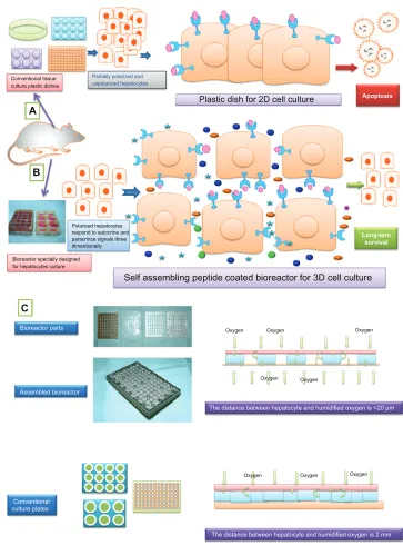
When hepatocytes are isolated from the in vivo liver and put in ex vivo conditions, hepatocytes search in the in vivo microenvironment during the initial vitro culture conditions. Generally, if hepatocytes do not get into the in vivo milieu, then finally the in vitro hepatocytes try to search for the neighbor cell to communicate for the exchange of many molecules to survive and function. Therefore, cultured hepa-tocytes get more elongated and stretched. Herein, we provide a hypothetical diagram (Figure 6) for how hepatocytes suffer and behave in unnatural conventional 2D culture conditions and lose hepatospecific functions and die quickly. The whole cell membrane of the seeded hepatocyte cells are partially polarized to interact with unrealistic stimulation by growth factors, cytokines, nutrients, and signals of the surrounding environment. This is because one side of the hepatocyte cell body will be in direct contact with the rigid ECM which cre-ates unnatural cell interaction with the surrounding ECM. Half or less of the hepatocyte membrane receptor is avail-able to interact for exposure with growth factors, cytokines, nutrients, and signals in the 2D system. Thus, cells in a 2D conventional culture are partially polarized which can seri-ously impair cellular communication, the transport of oxygen and nutrients, the removal of wastes, and cellular metabolism. In contrast, in a 3D nanorange microenvironment using the
nanostructured self-assembling peptides reported here, all functional motifs on the nanostructured self-assembling peptides encircle the whole hepatocyte cell body in all dimensions where all growth factors, cytokines, nutrients, and signals can interact three-dimensionally just like in an in vivo hepatic microenvironment. In the 3D interaction these cytokines or growth factors freely search out their receptors three-dimensionally, rather than two-dimensionally as in existing conventional in vitro models. These types of defined, nanostructured, self-assembling peptides could be highly valuable for both the development of the defined 3D culture system as well as effective bioartificial liver configuration.
Different bioreactor configurations have been developed to obtain a BAL device but none of them reported the long-term survivability and functionality of primary hepatocytes. However, the long-term functional maintenance of primary hepatocytes of up 90 days requires an appropriate in vivo microenvironment. This has not been developed so far because the scaffold and cells do not match the complete profile of the in vivo microenvironment. The actual microen-vironment is comprised of a collection of cells, the ECM/ scaffold, cytokines, and growth factors, which form the basis of normal tissue architecture and function. A large number of research studies have shown that using collagen to fix hepa-tocytes in the manner of sandwich configuration could create a matrix environment close to that seen in vivo and reported encouraging hepatic function up to a few weeks. Although the double layer of collagen sandwich configuration cellular model has been widely accepted for various hepatic studies, it is still a monolayer attachment culture. The 3D signaling in an in vitro cellular model with special reference to three-dimensional interactions of growth factors, cytokines, and hormones either in a conventional monolayer or sandwich configuration is inherently asymmetric and does not reflect an authentic in vivo environment.
Virtually, in native liver tissue, hepatocytes reply in a 3D hepatic environment with a nanorange ECM to provide adequate oxygen and solution factor transport. Further, the liver lobule is a functional unit of a whole liver that consists of hepatocytes that are arranged into hepatic cords separated by the sinusoidal space called the space of Disse (10–15 µM). The fundamental concept of in vivo hepatocytes is that each hepatocyte has direct contact with the space of Disse for the uptake of nutrients, growth factors, cytokines, hormones, oxygen, and other things. So, surrounding the in vivo hepatocyte is the nanorange environment. Thus, the substrate or ECM used during in vitro culture should be smaller than the hepatocytes cells, so that the scaffold can Dovepress Nanostructured self-assembling peptides and maintenance of hepatocytes
International Journal of Nanomedicine downloaded from https://www.dovepress.com/ by 118.70.13.36 on 23-Aug-2020
bind three-dimensionally. The average size of the hepatocytes is 10–20 µM. It is widely believed that when the size of the ECM is larger than the cells, the cells cannot be surrounded by the biomaterial scaffold. Most conventional biomaterials used for hepatocyte cell cultures are in the microscale range, where upon attachment the cells still exist in a 2D topography,
which is very common in conventional cell cultures in a bioartificial liver device and culture plates. Great difficulty is encountered if the cells cannot be attached in an in vivo 3D topology where the signaling, as well as the diffusion, is inherently asymmetric in traditional 2D culture and is the cur-rent main limitation for long-term in vitro culture. Therefore,
Conventional tissue culture plastic dishes
Polarized hepatocytes respond to autocrine and paracrince signals three dimentionally
Bioreactor specially designed for hepatocytes culture
Oxygen Oxygen
Oxygen Oxygen
Oxygen Oxygen Oxygen Oxygen Self assembling peptide coated bioreactor for 3D cell culture
Plastic dish for 2D cell culture
Partially polarized and unpolarized hepatocytes
Apoptosis
Long-tern survival
The distance between hepatocyte and humidified oxygen is <20 µm
The distance between hepatocyte and humidified oxygen is 2 mm Bioreactor parts
Assembled bioreactor
Conventional culture plates
A
B
C
Figure 6 Ahypothetical diagram showing how primary hepatocyte suffers in conventional 2D culture (A) in comparison to 3D culture (B) with special reference cytokines and growth factor interaction in exposure media during in vitro culture. (C) The difference of oxygenation in bioreactor and conventional culture plates with special reference to the distance between cultured hepatocyte and humidified oxygen.
Notes: Partially polarized or unpolarized hepatocytes of conventional tissue culture plastic dishes do not facilitate autocrine and paracrine soluble signals from their surrounding environments and finally lead to apoptosis. But under 3D culture, hepatocytes are polarized and able to respond to both paracrine and autocrine soluble signals three dimensionally and maintain their functional property on a long-term basis. 2D conventional culture is partially polarized which can seriously impair cellular communication, the transport of oxygen and nutrients, the removal of wastes, and cellular metabolism. By contrast, in a 3D nanorange microenvironment, using the nanostructured self-assembling peptides reported here, all functional motifs on the nanostructured self-assembling peptides encircle the whole hepatocyte cell body in all dimensions where all growth factors, cytokines, nutrients, and signals can interact three-dimensionally, just like in an in vivo hepatic microenvironment.
Dovepress
Giri et al
International Journal of Nanomedicine downloaded from https://www.dovepress.com/ by 118.70.13.36 on 23-Aug-2020
a nanostructured self-assembling peptide is essential to create an authentic 3D microenvironment by holding hepatocytes of all dimensions.
Oxygen supplies during primary hepatocyte culture are a critical issue since conventional cultures are often under oxygen-deficient culture conditions and are thus forced into anaerobic metabolic states. Fundamentally, the oxygen con-sumption does not only depend on hepatocellular uptake rates but is also limited by culture medium thickness as well as ambient oxygen concentration. However, despite this oxygen supply limitation, hepatocytes generally tolerate hypoxia due to their extraordinary capacity to satisfy energy requirements by anaerobic glycolysis. This hypoxia situation leads to an inefficient utilization of glucose since the conversion of glucose to lactate leads to the generation of 2 mol ATP/mol glucose compared to 38 mol ATP/mol glucose during oxida-tive phosphorylation.47 Enhanced oxygenation is important
for the in vitro liver cellular microenvironment5 and the BAL
model.48 Around half a century ago, Stevens reported that
in vitro liver cells obtain 4% of their oxygen requirement and degenerate rapidly.49 The oxygen supply in an in vivo liver is
2000 nmol/mL, but in vitro hepatocyte cells get an oxygen supply of less than 200 nmol/mL,50 which is a significant
current limitation and neglected area, particularly in primary hepatocyte culture. In our construction, our bioreactor offers a maximal oxygen supply of 90 µmol per 1.77 cm2 via direct
delivery of oxygen to the cells from the bottom of the device.13
Our bioreactor also allows direct contact of every individual liver cell with the oxygen supply, for enhanced oxygenation that is closer to the in vivo liver.5,11,12
While the available image data was obtained from CLSM, basically providing the image series for our image analysis, we have restricted ourselves to 2D image-based assessments on single images taken centrally from the stacks. The reason was two-fold: first the cells considerably adhere to the ground, leading to heavy distortions so that the cells appear to be strongly flattened. Thus, even dedicated efforts to analyze the 3D morphology (with respect to the mitochondria in this work) remains of limited use (Figure 1). While the mitochon-dria should be expected to be distributed on a spherical hull, what we see is some degenerate torus-like spatial distribution, an obvious phenomenon which undermines a reasonable 3D morphometry. The second reason in part is a consequence of the first. Due to the distortions, single mitochondria pieces appear even closer to each other than under physiological con-ditions. This effect, however, worsens the conditions for the following image processing and analysis, since mitochondria pieces in particular appear to be densely packed. In turn, this
also affects any segmentation capabilities; ie, the separated depiction of mitochondria pieces. However, there is another reason why a strict 3D analysis does not appear worthwhile at this point: position dependent blurring of the noisy images, an effect, which is inherent to CLSM, requiring effective image restoration, is always a challenge. It can basically be tackled using a variety of image deconvolution techniques.51 This
effort, however, was not undertaken here, due to the strong cell distortions discussed above. Interestingly,52 we did not
apply image deconvolution, but in spite of this outline, a 3D image analysis processing chain ended up with a couple of 3D based morphometric numbers with respect to single mitochondria pieces. With respect to the blurring and artifacts in our image data, a similar single mitochondria assessment approach did not appear feasible. High quality fluorescence images were captured for the subsequent quantitative analy-sis of mitochondria using the Mathematica software, which offers a variety of image processing functionality. It has been found that in liver cells, total mitochondrial volume is distrib-uted into many mitochondrial elements. Also, it shows that a healthy cell in a bioreactor has a very good level of mito-chondrial volume around the nucleus. Previously, we have investigated the automatic algorithms-based image analysis of various biomedical images that enable the quantitative analysis and 3D visualization of medical images of numerous modalities such as microscopy histological structure.
The main highlights of the present study are that this optimized 3D defined culture system in bioartificial liver modules could be useful for drug metabolism, enzyme induction, transplantation, viral hepatitis, and hepatocyte regeneration. This proof of concept may be convenient for other existing bioartificial devices and hepatotoxicity assessments. Nanostructured self-assembling peptides are an excellent substrate for 3D culture and long-term culture. 2D/3D visualization of mitochondria revealed healthy shapes and distributions at 90 days of culture. This bioartificial liver module might be an alternative to animal experimentation. This proof of concept may meet the regulatory guidelines as well as pharmaceutical good manufacturing practice guide-lines as all conditions are absolutely defined.
Instead, we decided to do the mitochondria growth analy-ses that refer to representative single images taken from the image stacks. Applying our image processing pipeline in 2D detailed above, we could obtain a new comprehensive description of determined mitochondria margins, mainly using distance transforms and skeletons. The method was exemplified in this work and needs to be assessed in a comparative study.
Dovepress Nanostructured self-assembling peptides and maintenance of hepatocytes
International Journal of Nanomedicine downloaded from https://www.dovepress.com/ by 118.70.13.36 on 23-Aug-2020
Conclusion
We have developed a defined, cost effective, efficacious, and scalable ex vivo bioartificial modular device for performing long-term functional maintenance of primary hepatocytes to meet the regulatory guidelines and provide alternative ani-mal experimentation with special reference to chronic dose screening of drug candidates that can also reduce the number of animals used in in vivo testing. The long-term functional maintenance of primary hepatocytes in this clinically relevant modular device opens up a new avenue not only for rapid drug screening testing from sub-acute to chronic exposure but also has clinical implications for the bioartificial liver support system. This concept may be useful for other existing bioar-tificial liver systems to make them more efficient, especially for the long-term functional maintenance of housed primary hepatocytes in these systems. Herein, we proved that using nanostructured, self-assembling peptides and 3D interaction of growth factors and cytokines, it is possible to mirror the in vivo liver microenvironment. This 3D interaction is very effective to recapitulate the physiological functions of in vivo hepatocytes after the isolation of hepatocytes from the in vivo liver. Pharmaceutical companies have been under high pressure to develop safer drugs because a number of approved drugs entering the market have failed in the last decade. Many pharmaceutical and biotechnology companies are now searching for effective strategies to improve the preclinical stage to recognize unsuccessful drug candidates early in the drug discovery process. The pharmaceutical and biotechnology industries are increasingly seeking an ex vivo cellular model that mimics the state of the art liver which could replace the use of animals in drug screening tests in the early stage of drug candidate selection. A modular novel bioreactor-based bioartificial liver support system was designed and constructed using self-assembling peptides and a special combination of growth factors, cytokines, and hormones in order to simplify the tedious operation of artificial liver treatment and to improve the applicability of the system in the bioartificial liver system and in the in vitro hepatic model to produce safer drugs. Additionally, we have shown the 3D visualization of well-shaped mitochondria of single/double hepatocyte cells at day 90, since mitochondria is directly proportional to the survivability of hepatocytes. With the proof of concept, in the future, it is hoped that several basic hepatocyte physiological disease-screening models, such as those that are induced pluripotent stem cells-based, can be integrated into a modular device platform to realize a complete in vivo liver for different genetic background populations. This modular optimized device has great
potential to predict in vivo toxicity and is very much suitable to measure metabolism and the toxicity of drugs with a very good correlation to the in vivo situation. It will minimize the gap between the in vivo situation and in vitro situation. This proof of concept study in a bioartificial liver modular device could replace the millions of animals that are currently sacrificed in preclinical testing and open up a new vista for new, safer pharmaceutical products.
Acknowledgments
The authors wish to thank Frank Struck for supplying the primary hepatocytes. Special thanks also go to Ingo Schäfer for his confocal microscopy. The funding for this project was provided by the Medicine Faculty of the University of Leipzig, TRM Leipzig, and IZBI Leipzig, Leipzig, Germany.
Disclosure
The authors report no conflicts of interest in this work.
References
1. Domansky K, Inman W, Serdy J, Dash A, Lim MH, Griffith LG.
Perfused multiwell plate for 3D liver tissue engineering. Lab Chip. 2010;
10(1):51–58.
2. Khetani SR, Bhatia SN. Microscale culture of human liver cells for
drug development. Nat Biotechnol. 2008;26(1):120–126.
3. Zeilinger K, Schreiter T, Darnell M, et al. Scaling down of a clinical three-dimensional perfusion multicompartment hollow fiber liver bioreactor developed for extracorporeal liver support to an analytical
scale device useful for hepatic pharmacological in vitro studies. Tissue
Eng Part C Methods. 2011;17(5):549–556.
4. Tostões RM, Leite SB, Miranda JP, et al. Perfusion of 3D encapsulated hepatocytes – a synergistic effect enhancing long-term functionality in
bioreactors. Biotechnol Bioeng. 2011;108(1):41–49.
5. Kidambi S, Yarmush RS, Novik E, Chao P, Yarmush ML, Nahmias Y. Oxygen-mediated enhancement of primary hepatocyte metabolism,
functional polarization, gene expression, and drug clearance. Proc Natl
Acad Sci U S A. 2009;106(37):15714–15719.
6. Toh YC, Lim TC, Tai D, Xiao G, van Noort D, Yu H. A microfluidic
3D hepatocyte chip for drug toxicity testing. Lab Chip. 2009;9(14):
2026–2035.
7. van Midwoud PM, Merema MT, Verpoorte E, Groothuis GM. A microfluidic approach for in vitro assessment of interorgan
interac-tions in drug metabolism using intestinal and liver slices. Lab Chip.
2010;10(20):2778–2786.
8. Ho CT, Lin RZ, Chang WY, Chang HY, Liu CH. Rapid heterogeneous liver-cell on-chip patterning via the enhanced field-induced
dielectro-phoresis trap. Lab Chip. 2006;6(6):724–734.
9. Zhang C, Zhao Z, Abdul Rahim NA, van Noort D, Yu H. Towards a human-on-chip: culturing multiple cell types on a chip with
compartmentalized microenvironments. Lab Chip. 2009;9(22):
3185–3189.
10. Tompa A, Langenbach R. Promoting effect of feeder cells in
main-tenance of adult rat hepatocytes. Acta Morphol Acad Sci Hung.
1980;28(4):393–405.
11. Maringka M, Giri S, Bader A. Preclinical characterization of primary porcine hepatocytes in a clinically relevant flat membrane bioreactor.
Biomaterials. 2010;31(1):156–172.
Dovepress
Giri et al
International Journal of Nanomedicine downloaded from https://www.dovepress.com/ by 118.70.13.36 on 23-Aug-2020
International Journal of Nanomedicine
Publish your work in this journal
Submit your manuscript here: http://www.dovepress.com/international-journal-of-nanomedicine-journal
The International Journal of Nanomedicine is an international, peer-reviewed journal focusing on the application of nanotechnology in diagnostics, therapeutics, and drug delivery systems throughout the biomedical field. This journal is indexed on PubMed Central, MedLine, CAS, SciSearch®, Current Contents®/Clinical Medicine,
Journal Citation Reports/Science Edition, EMBase, Scopus and the Elsevier Bibliographic databases. The manuscript management system is completely online and includes a very quick and fair peer-review system, which is all easy to use. Visit http://www.dovepress.com/ testimonials.php to read real quotes from published authors. 12. Giri S, Acikgöz A, Pathak P, et al. Three dimensional cultures of rat
liver cells using a natural self-assembling nanoscaffold in a clinically
relevant bioreactor for bioartificial liver construction. J Cell Physiol.
2012;227(1):313–327.
13. Schmitmeier S, Langsch A, Jasmund I, Bader A. Development and characterization of a small-scale bioreactor based on a bioartificial hepatic culture model for predictive pharmacological in vitro screenings.
Biotechnol Bioeng. 2006;95(6):1198–1206.
14. FDA (Food and Drug Administration), Center for Drug Evaluation and Research. August, 2005. Guidance for Industry, Impurities in Drug Prod-uct. Revision I, http://www.fda.gov/Cder/Guidance/6423dftrev1.html.
15. Strain AJ, Neuberger JM. A bioartificial liver–state of the art. Science.
2002;295(5557):1005–1009.
16. Giri S, Bader A. Improved preclinical safety assessment using micro-BAL devices: the potential impact on human discovery and drug
attrition. Drug Discov Today. 2011;16(9–10):382–397.
17. Frühauf NR, Oldhafer KJ, Höltje M, et al. A bioartificial liver support system using primary hepatocytes: a preclinical study in a new porcine
hepatectomy model. Surgery. 2004;136(1):47–56.
18. Frühauf JH, Mertsching H, Giri S, Frühauf NR, Bader A. Porcine endogenous retrovirus released by a bioartificial liver infects primary
human cells. Liver Int. 2009;29(10):1553–1561.
19. Si-Tayeb K, Lemaigre FP, Duncan SA. Organogenesis and development
of the liver. Dev Cell. 2010;18(2):175–189.
20. Pampaloni F, Reynaud EG, Stelzer EH. The third dimension bridges
the gap between cell culture and live tissue. Nat Rev Mol Cell Biol.
2007;8(10):839–845.
21. Pierschbacher MD, Ruoslahti E. Cell attachment activity of fibronec-tin can be duplicated by small synthetic fragments of the molecule.
Nature. 1984;309(5963):30–33.
22. D’Souza SE, Ginsberg MH, Plow EF. Arginyl-glycyl-aspartic acid (RGD):
a cell adhesion motif. Trends Biochem Sci. 1991;16(7):246–250.
23. Ruoslahti E. RGD and other recognition sequences for integrins. Annu
Rev Cell Dev Biol. 1996;12:697–715.
24. Prieto AL, Edelman GM, Crossin KL. Multiple integrins mediate cell
attachment to cytotactin/tenascin. Proc Natl Acad Sci U S A. 1993;
90(21):10154–10158.
25. Schafer DF, Sorrell MF. Power failure, liver failure. N Engl J Med.
1997;336(16):1173–1174.
26. Mitra K, Wunder C, Roysam B, Lin G, Lippincott-Schwartz J. A hyperfused mitochondrial state achieved at G1-S regulates cyclin E
buildup and entry into S phase. Proc Natl Acad Sci U S A. 2009;
106(29):11960–11965.
27. Bader A, Fruhauf N, Tiedge M, et al. Enhanced oxygen delivery reverses anaerobic metabolic states in prolonged sandwich rat hepatocyte
culture. Exp Cell Res. 1999;246(1):221–232.
28. Taub R. Liver regeneration: from myth to mechanism. Nat Rev Mol
Cell Biol. 2004;5(10):836–847.
29. Zhang S, Lockshin C, Herbert A, Winter E, Rich A. Zuotin, a
puta-tive Z-DNA binding protein in Saccharomyces cerevisiae. EMBO J.
1992;11(10):3787–3796.
30. Zhang S. Fabrication of novel biomaterials through molecular
self-assembly. Nat Biotechnol. 2003;21(10):1171–1178.
31. Giri S, Acikgöz A, Pathak P, et al. Three dimensional cultures of rat liver cells using a natural self-assembling nanoscaffold in a clinically
relevant bioreactor for bioartificial liver construction. J Cell Physiol.
2012;227(1):313–327.
32. Gomes LC, Di Benedetto G, Scorrano L. During autophagy mitochon-dria elongate, are spared from degradation and sustain cell viability.
Nat Cell Biol. 2011;13(5):589–598.
33. Blackstone C, Chang CR. Mitochondria unite to survive. Nat Cell Biol.
2011;13(5):521–522.
34. Chan TF, Osher S, Shen J. The digital TV filter and nonlinear denoising.
IEEE Trans Image Process. 2001;10(2):231–241.
35. Chan TF, Vese LA. Active contours without edges. IEEE Trans Image
Process. 2001;10(2):266–277.
36. Gonzalez RC, Woods RE. Dilation. In: Digital Image Processing, 3rd ed.
Upper Saddle River: Prentice Hall; 2008. 633.
37. Wu K, Otoo E, Suzuki K. Optimizing two-pass connected-component
labeling algorithms. Pattern Anal Appl. 2009;12(2):117–135.
38. Bentley JL, Clarkson KL, Levine DB. Fast linear expected-time
algorithms for computing maxima and convex hulls. Algorithmica.
1993;9(2):168–183.
39. Saito T, Toriwaki JI. New algorithms for Euclidean distance
transforma-tion on an n-dimensional digitized picture with applicatransforma-tions. Pattern
Recognit. 1994;27(11):1551–1565.
40. Rosenfeld A. A characterization of parallel thinning algorithms.
Information and Control. 1975;29(3):286–291.
41. Zhang S. Beyond the Petri dish. Nat Biotechnol. 2004;22(2):151–152.
42. Zhang S, Holmes T, Lockshin C, Rich A. Spontaneous assembly of a self-complementary oligopeptide to form a stable macroscopic
membrane. Proc Natl Acad Sci U S A. 1993;90(8):3334–3338.
43. Pampaloni F, Reynaud EG, Stelzer EH. The third dimension bridges
the gap between cell culture and live tissue. Nat Rev Mol Cell Biol.
2007;8(10):839–845.
44. Bezbradica JS, Medzhitov R. Integration of cytokine and heterologous
receptor signaling pathways. Nat Immunol. 2009;10(4):333–339.
45. Gelain F, Unsworth LD, Zhang S. Slow and sustained release of active
cytokines from self- assembling peptide scaffolds. J Control Release.
2010;145(3):231–239.
46. Mager MD, LaPointe V, Stevens MM. Exploring and exploiting
chem-istry at the cell surface. Nat Chem. 2011;3(8):582–589.
47. Jensen MD, Wallach DF, Sherwood P. Diffusion in tissue culture
on gas-permeable and impermeable supports. J Theor Biol. 1976;
56(2):443–458.
48. Poyck PP, Mareels G, Hoekstra R, et al. Enhanced oxygen availability
improves liver-specific functions of the AMC bioartificial liver. Artif
Organs. 2008;32(2):116–126.
49. Stevens KM. Oxygen requirements for liver cells in vitro. Nature.
1965;206(980):199.
50. Nahmias Y, Kramvis Y, Barbe L, Casali M, Berthiaume F, Yarmush ML. A novel formulation of oxygen-carrying matrix enhances
liver-specific function of cultured hepatocytes. FASEB J. 2006;20(14):
2531–2533.
51. Sarder P, Nehorai A. Deconvolution Methods for 3-D fluorescence
microscopy images. IEEE Signal Process Mag. 2006;23(3):32–45.
52. Dwarakapurm S, Roysam B, Lin G, Mitra K. Quantitative analysis
of mitochondrial tubulation using 3D imaging. Research Thrust R2
Presentations. 2007;Paper 35. Available from: http://iris.lib.neu.edu/
censsis_r2_pres/35/. Accessed January 01, 2007.
53. Huang B, Jones SA, Brandenburg B, Zhuang X. Whole-cell 3D STORM reveals interactions between cellular structures with nanometer-scale
resolution. Nat Methods. 2008;5(12):1047–1052.
Dovepress
Dove
press
Nanostructured self-assembling peptides and maintenance of hepatocytes
International Journal of Nanomedicine downloaded from https://www.dovepress.com/ by 118.70.13.36 on 23-Aug-2020